malaya - studentsrepo.um.edu.mystudentsrepo.um.edu.my/7816/5/fatemeh.pdf · matlamat kajian ini...
TRANSCRIPT
EEG ANALYSIS OF OPIOID DEPENDENTS
DURING METHADONE TREATMENT
FATEMEH MOLAEI VANEGHI
RESEARCH PROJECT SUBMITTED IN PARTIAL FULFILLMENT FOR THE
DEGREE OF MASTER OF ENGINEERING (BIOMEDICAL)
FACULTY OF ENGINEERING
UNIVERSITY MALAYA
KUALA LUMPUR
2011
Univers
ity of
Mala
ya

UNIVERSITI MALAYA ORIGINAL LITERARY WORK DECLARATION
Name of Candidate: (I.C/Passport No:)
Registration/Matric No:
Name of Degree: Master of XXXXXXXXX Title of Project Paper/Research Report/Dissertation/Thesis (“this Work”):
Title
Field of Study:
I do solemnly and sincerely declare that:
(1) I am the sole author/writer of this Work; (2) This Work is original; (3) Any use of any work in which copyright exists was done by way of fair dealing and for
permitted purposes and any excerpt or extract from, or reference to or reproduction of any copyright work has been disclosed expressly and sufficiently and the title of the Work and its authorship have been acknowledged in this Work;
(4) I do not have any actual knowledge nor do I ought reasonably to know that the making of this work constitutes an infringement of any copyright work;
(5) I hereby assign all and every rights in the copyright to this Work to the University of Malaya (“UM”), who henceforth shall be owner of the copyright in this Work and that any reproduction or use in any form or by any means whatsoever is prohibited without the written consent of UM having been first had and obtained;
(6) I am fully aware that if in the course of making this Work I have infringed any copyright whether intentionally or otherwise, I may be subject to legal action or any other action as may be determined by UM.
Candidate’s Signature Date
Subscribed and solemnly declared before,
Witness’s Signature Date
Name:
Designation:
Univers
ity of
Mala
ya

i
ACKNOWLEDGEMENTS
My sincere appreciation goes to my supervisor, Dr. Fatimah Ibrahim who helped me
understand the meaning of enthusiasm and hard work. I am very grateful to her for raising
my interest in neuroscience and brain research and also for revising my paper patiently.
I also want to express my warmest thanks to Dr. Ng Siew Cheok who provides me
with his wide knowledge and experience in EEG signal processing.
Last but not least, very special and humble thanks goes to Dr. Rusdi who provides
us with the facilities required for conducting the experiments and giving his support in
every step of running this study.
Univers
ity of
Mala
ya
ii
ABSTRAK
Matlamat kajian ini adalah untuk mempelajari tentas perubahan struktur otak pada
penagih heroin setelah penggunaan awal metadon dan menentukan peranan metodon dalam
menormalkan kecelaruan psikofisiologik berkaitan kebergantungan kepada opioid. Untuk
mengkaji kemampuan metadon dalam memulihkan semula struktur fungsi otak daripada
penagih opioid, komposisi daripada ayunan elektroensefalografi (EEG) jalur frekuensi lebar
(0.5Hz – 60Hz) dikaji. Analisis komponen merdeka (ICA) daripada isyarat EEG kulit
kepala digunakan untuk mengesan kawasan kortikal dan aktiviti spektro-temporal yang
didapati pada kebergantungan opioid dan pengubatan metodon. Hasil kajian ini
menunjukkan bahawa, semasa keadaan kebergantungan opioid, aktiviti sumber otak yang
responsif terhadap opioid terpumpun pada medial prefrontal cortex (mPFC) dan luasan
sistem limbik, dan aktiviti-aktiviti ini nyata sekali menurun setelah penggunaan metadon.
Analisis spektrum daripada akitiviti kortikal menunjukkan peningkatan akitiviti alfa, beta,
dan gama semasa kebergantungan opioid, manakala sesaat selepas penggunaan metodon,
berlaineudaripede aktiviti gama, aktifiti alfa dan beta menurun.
Univers
ity of
Mala
ya
iii
ABSTRACT
This study aims to explore the structural brain changes in heroin abusers after the
first administration of methadone and therefore examine the role of methadone in
normalizing the psychophysiological impairments associated with the state of opioid
dependency. The ability of methadone to restore the normal cortical functioning in opioid
dependents the composition of electroencephalographic (EEG) oscillations within a broad
frequency band (0.5Hz – 60Hz) was explored. The Independent Component Analysis (ICA)
was used to identify the cortical regions and their relevant spectro-temporal activities
involved in opioid dependency and methadone therapy based on the information content of
the scalp EEG signal. It has been shown that within the state of opioid dependency the
majority of brain activities responsive to opioids are located within the medial prefrontal
cortex (mPFC) and the extended limbic system, and these activities reduced significantly
after methadone administration. Spectral activities of the brain within alpha, beta, and
gamma frequencies increased during opioid dependency while, unlike the gamma spectrum,
the alpha and beta spectral activities underwent a decline early after the onset of methadone
administration.
Univers
ity of
Mala
ya

iv
TABLE OF CONTENTS
ACKNOWLEDGEMENTS………………………………………………..............i
ABSTRAK…………………………………………………………………............ii
ABSTRACT…………………………………………………………………….....iii
TABLE OF CONTENTS…………………………………………………............iv
LIST OF TABLES………………………………………………………………...vi
LIST OF FIGURES……………………………………………………………….vii
CHAPTER I: INTRODUCTION…………………………………………..............1
1.1 Overview…………………………………………………………...….1
1.2 Problem Statement……………………………………………………..1
1.3 Objectives……………………………………………………………...2
1.4 Scope of Study………………………………………………………....2
1.5 Significance of Study…………………………………………………..2
1.6 Outline of the Report…………………………………………………..3
CHAPTER II: LITERATURE REVIEW……………………………………….....4
2.1 Introduction……………………………………………………………...4
2.2 Physiology of the Brain………………………………………………….4
2.3 Opioid Dependency……………………………………………………..5
2.3.1 Neurobiology…………………………………………………..5
2.3.2 Neuropsychology………………………………………...........7
2.4 EEG Analysis……………………………………………………............7
2.5 Methadone Treatment……………………………………………...........8
2.6 Independent Component Analysis (ICA)………………………………..9
Univers
ity of
Mala
ya
v
CHAPTER III: METHODOLOGY……………………………………………….10
3.1 Introduction……………………………………………………………..10
3.2 Subjects………………………………………………………………....10
3.3 Trial Design…………………………………………………………….10
3.4 Data Acquisition (EEG Recordings)…………………………………...12
3.5 Data Preprocessing……………….…………………………….............12
3.5.1 Duration of EEG Signal…………………………………........12
3.5.2 Artifact Removal……………………………………………..13
3.5.3 Filtering the Data……………………………………………..15
3.6 Analysis of Independent Components………………………………….15
3.7 Component Source Modeling…………………………………………..16
3.8 Localization of Component Equivalent Dipoles……………………….16
3.9 Dipoles Clustering……………………………………………………...17
3.10 Spectral Analysis……………………………………………………..18
CHAPTER IV: RESULTS AND DISCUSSION…………………………………19
4.1 Introduction………………………………………………….…………19
4.2 Cortical Localization of Component Equivalent Dipoles……………..19
4.3 Spectral Analysis………………………………………………………22
4.4 Statistical Analysis……………………………………………………..23
CHAPTER V. CONCLUSION AND FUTURE WORK………………………….25
5.1 Introduction…………………………………………………………….25
5.2 Conclusion……………………………………………………………...25
5.2 Recommendation for Future Study…………………………………….26
REFERENCES………………………………………………………………….....27
Univers
ity of
Mala
ya

vi
List of Tables
Table 2.1: Cortical regions and their functions………………………………………5
Table 3.1: Algorithms of artifact removal……………………………………….....14
Univers
ity of
Mala
ya
vii
List of Figures
Figure 2.1: Physiology of the brain………………………………………………….4
Figure 3.1: Overview of the methodology………………………………………….10
Figure 4.1: Independent components clustering……………………………………20
Figure 4.2: Cortical localization of dipoles………………………………………...21
Figure 4.3: Clusters spectrum………………………………………………………22
Figure 4.4: Comparison of clusters spectrum………………………………………23
Figure 4.5: Statistical analysis of relative power values……………………………24
Univers
ity of
Mala
ya
1
1 CHAPTER I: INTRODUCTION
1.1 Overview
Opioid dependency is a chronic brain syndrome associated with
psychophysiological disorders. Although the possibility of relapsing back to state of opioid
dependency is quite high particularly in early abstinence, neuropsychophysiological deficits
during the state of opioid dependency or after opioid cessation are barely investigated. On
the other hand, structural cortical alterations and their correlation with the onset age and
duration of opioid abuse, remain unknown.
Methadone is known as a long term acting medication for opioid dependency. While
oxycodone, morphine, heroin or other types of addictive drugs have a short term effect on
the body and brain, methadone has long lasting effects. Like other opioids, methadone can
cause dependence but since it has steadier effects on the Mu receptors, it produces less
tolerance and withdrawal symptoms. Methadone treatment can also normalize the
disruption of the hormones in opioid dependents. Effects of methadone on mediating the
symptoms of opioid abuse are not limited to the physiological treatment. It also provides
psychological medication like facilitating the behavioral therapy (White, 2007).
1.2 Problem Statement
Despite of a large number of advantages associated with methadone as an influential
therapy for opioid dependency, still the efficiency of this treatment is highly dependent
upon the prescription condition. Since methadone is considered as a long term medication
with addictive effects, the risk of getting back opioid dependency is very high if the patient
cannot meet the required period of the treatment while the methadone itself can cause
dependency. Yet the optimal dosage of methadone administration has not been discovered.
Univers
ity of
Mala
ya
2
Moreover the psychophysiological side effects of methadone therapy need to be considered.
All of these facts about the methadone therapy make it of great importance to investigate
different psychophysiological aspects of this treatment to identify the optimal therapy.
1.3 Objectives
The aim of this study is to investigate the efficiency of methadone treatment in the
state of opioid dependency.
1.4 Scope of study
The main scopes of conducting this study include:
Providing a novel and highly efficient method to process the
electroencephalographic (EEG) signal while being able to accurately track cortical
regions of the EEG.
Spectral analysis of neurophysiological alteration through the state of opioid
dependency and methadone-based treatment to investigate the effects of opioids and
methadone on the brain structure.
Evaluating the efficiency of methadone treatment in medication of opioid
dependency.
1.5 Significance of Study
The effectiveness of this spatial filtering lies within the application of the
information content of EEG signal itself rather than a predefined set of cortical locations to
separate the scalp data into brain and artifact sources (Makeig et al., 2002). Another major
advantage of applying ICA for EEG source localization is that the locally coherent cortical
activities which constitute one particular EEG source will be joint together making a single
independent component (IC) which includes projection of those activities to all of the scalp
Univers
ity of
Mala
ya

3
electrodes, whereas the unrelated EEG source activities will be eliminated from this IC and
contributed to other ICs (Onton & Makeig, 2005).
1.6 Outline of the Report
This project is categorized in five chapters each investigating one aspect of the
study. Statement of the problem, objectives of the study along with the scopes and
significance of conducting the experiment are covered through the first chapter. Chapter II
presents the wide range literature related to EEG analysis, opioid dependency and
methadone medication while it tries to evaluate the most significant finding regarding the
objectives of the present study. Chapter III deals with the theoretical and practical aspects
of the methodology used in this study, and how it can be applied to obtain the most reliable
results. Chapter IV represents the result of implementing the study and how they can be
interpreted regarding the effects of methadone therapy in opioid abusers. Lastly, chapter V
draws a conclusion on the efficiency of methadone medication based on the results of the
previous chapter and also recommends the future work in this area.
Univers
ity of
Mala
ya

4
2 Chapter II: REVIEW OF RELATED LITERATURE
2.1 Introduction
This chapter covers the literature regarding the neurophysiological and
neuropsychological aspects of drug dependency, methadone treatment, and the
methodological strategies of temporal and spectral analysis of the EEG signal focusing on
the Independent Component Analysis (ICA) to investigate opioid dependent EEG signal.
2.2 Physiology of Brain
The following table gives an overview of the brain structures and their functions
while their basic physiology is demonstrated in Figure 2.1 as follows.
Figure 2.1 Basic Physiology of Brain (Swanson, 2011).
Univers
ity of
Mala
ya
5
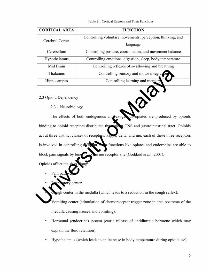
Table 2.1 Cortical Regions and Their Functions
CORTICAL AREA FUNCTION
Cerebral Cortex Controlling voluntary movements, perception, thinking, and
language
Cerebellum Controlling posture, coordination, and movement balance
Hypothalamus Controlling emotions, digestion, sleep, body temperature
Mid Brain Controlling reflexes of swallowing and breathing
Thalamus Controlling sensory and motor integration
Hippocampas Controlling learning and memory
2.3 Opioid Dependency
2.3.1 Neurobiology
The effects of both endogenous and exogenous opiates are produced by opioids
binding to opioid receptors distributed throughout CNS and gastrointestinal tract. Opioids
act at three distinct classes of receptors: kappa, delta, and mu, each of these three receptors
is involved in controlling different brain functions like opiates and endorphins are able to
block pain signals by binding to the mu receptor site (Goddard et al., 2001).
Opioids affect the sites along:
• Pain pathways.
• Respiratory center.
• Cough center in the medulla (which leads to a reduction in the cough reflex).
• Vomiting center (stimulation of chemoreceptor trigger zone in area postrema of the
medulla causing nausea and vomiting).
• Hormonal (endocrine) system (cause release of antidiuretic hormone which may
explain the fluid retention).
• Hypothalamus (which leads to an increase in body temperature during opioid use).
Univers
ity of
Mala
ya
6
• Immune system (there may be a degree of immunosuppression due to these drugs
and this can lead to increased susceptibility to infections in vulnerable patients.
Whether this is a problem with prescribed doses is unclear).
The opioid system is connected with most neurotransmitter networks in the body.
The interaction between the opioids and the dopaminergic system appears to be involved in
addiction, tolerance, and withdrawal symptoms (Grace, 2000). The relevant interaction
appears to occur along the mesolimbic projection, particularly in the ventral tegmental area
(VTA) and nucleus accumbens (NAc) (Walwyn et al., 2010). The limbic system controls
emotions. Opiates change the limbic system to produce increased feelings of pleasure,
relaxation and contentment (Kreek, 1992). The brainstem controls things your body does
automatically, like breathing or coughing. Opiates can act on the brainstem to stop
coughing or slow breathing. The spinal cord transmits pain signals from the body. By
acting here, opiates block pain messages and allow people to bear even serious injuries.
Opiates act not only on the central structures of the reward circuit (the VTA and the
NA), but also on other structures that are naturally modulated by endorphins including the
amygdale, locus coeruleus, arcuate nucleus, periaqueductal grey matter which also
influence dopamine levels, though indirectly, and thalamus which would explain their
analgesic effect (Kosten, 1990).
The brain pleasure centers affected by drugs:
• Ventral Tegmental Area (VTA) which is located in the midbrain and contains the
dopaminergic neurons that innervate the limbic system and the prefrontal cortex.
• Nucleus Accumbens (NA) or Ventral Striatum which is located in the septal region,
innervated by the ventral tegmental area, and serves as an interface between the
limbic system and the motor system.
Univers
ity of
Mala
ya
7
• Prefrontal Cortex whose role in the processes of attention and motivation is well
established (Robinson & Berridge, 2000). The following figure demonstrates these
three regions within the cortex.
2.3.2 Neuropsychological changes
Neuropsychological research reported short term disfunction in visual and verbal
memory, attention, working memory and concentration through the state of opioid
dependency. Even a general intellectual decline has been shown while intoxicated or very
recently detoxified (Khantzian et al., 1984; Prosser et al., 2006). It has been shown that
after rapid detoxification heroin abusers who had shown a deficit in attention, working and
episodic memory, and verbal fluency during abuse did not differ from controls after one to
two weeks of abstinence. It seems that opioid abuse induces partially transient alterations of
cognitions. On the other hand, after long term abstinence a consistent deficit in executive
functioning, especially in impulse control has been found (Davis et al., 2002; Fu et al.,
2008; Mintzer et al., 2005). The prefrontal cortex is involved in cognitive functions such as
planning, anticipation and establishment of goals, organization and motivation of behavior,
defined as executive functions (Fellows, 2007). The functional imaging studies of
substance abusers also point to those frontal pathways related to cognition (Ghodse &
Galea, 2010; Volkow et al., 2002).
2.4 EEG Studies
The signal recorded at each scalp electrode is the sum of activities originating in
nearly all cortical sources and artifact source domains from movements, muscles, eyes,
electrodes, and the electrical environment. So far most of the EEG studies are based on the
analysis of electrical potential time series receiving at each scalp electrode, which reveals
little information regarding the type, number and spatial distribution of the brain sources
Univers
ity of
Mala
ya
8
generating them. So EEG recordings are usually considered to have low spatial resolution.
Furthermore, different patterns of cortical folding across individuals may cause the spatially
equivalent brain source areas to have different orientations, relative amplitude, and
distances across most of the cortex, making significant differences in their projections to
the scalp electrodes. Thus, comparing EEG signals of the subjects across equivalent scalp
electrodes may not be accurate.
2.5 Methadone Treatment
Methadone is considered to be the standard agonist substitution therapy for heroin
abusers. Recent findings in clinical neuroscience reveal the importance of methadone to
decrease craving for opioids (Greenwald et al., 2007) while it can also prime craving and
opioid cue response (Curran et al., 2001; Gerra et al., 2003; Shaham et al., 1996). Although
many studies have already supported the efficiency of methadone treatment as a medication
for opioid abuse (Ball & Ross, 1991; Simpson et al., 1997). Still some issues remain.
Methadone prevents symptoms of withdrawal through affecting the same opioid receptors
as opioids. While it is associated with physical dependency, it causes less psychological
dependency as compared with other opioids like heroin since it does not give the same
sense of euphoria.
Methadone treatment can affect brain cognitive function; as compared to abstinent
subjects methadone subjects have shown considerably poorer performance in learning tests
and also immediate recall (Gritz et al., 1975). It has been reported that that patients
underwent methadone treatment have shown some impairments in attention tests and
psychomotor speed (Mintzer & Stitzer, 2002). Furthermore a significant alteration in levels
of cerebral phospholipid metabolite (Silveri et al., 2004)and also higher production of
interleukins (IL-6) (Zajocov et al., 2004) was observed in methadone treatment subjects.
Univers
ity of
Mala
ya
9
It has been shown that while a long period (minimum of three months) of abstinence
may significantly or even completely normalize the EEG of opioid-dependent
patients(Bauer, 2001; Costa & Bauer, 1997; Gekht et al., 2003; Shufman et al., 1996)
methadone treatment can recover the temporal structure of EEG oscillations in the opioid
abusers (with slight slowing down of EEG dynamics) (Gritz, Shiffman, et al., 1975)
comparable to the healthy subjects. The electroencephalographic signal of opioid
dependents during withdrawal and opioid abuse is reorganized significantly in terms of
EEG oscillations and their temporal activity (Fingelkurts, Kivisaari, Autti, Borisov,
Puuskari, Jokela, & Kahkonen, 2006) Methadone can restore these structural changes
which extensively distribute over the cortex back to the normal level of healthy subjects
(Fingelkurts et al., 2007).
2.6 Independent Component Analysis (ICA)
As an alternative approach to simultaneously monitoring field activities of the brain
sources, independent component analysis (ICA) (Bell & Sejnowski, 1995; Cardoso &
Laheld, 1996)was applied. Each subject scalp EEG data was separated into the active
cortical and artifact sources based on the physiologically and statistically reasonable
assumption of the ICA that these cortical activities should be nearly temporally independent
of each other (Makeig et al., 1996; Makeig et al., 1999).
Univers
ity of
Mala
ya
10
3 CHAPTER III: METHODOLOGY
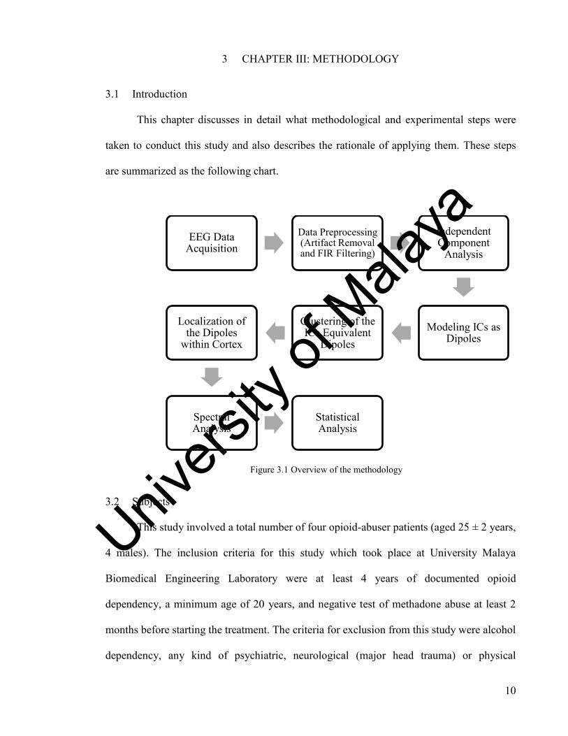
3.1 Introduction
This chapter discusses in detail what methodological and experimental steps were
taken to conduct this study and also describes the rationale of applying them. These steps
are summarized as the following chart.
Figure 3.1 Overview of the methodology
3.2 Subjects
This study involved a total number of four opioid-abuser patients (aged 25 ± 2 years,
4 males). The inclusion criteria for this study which took place at University Malaya
Biomedical Engineering Laboratory were at least 4 years of documented opioid
dependency, a minimum age of 20 years, and negative test of methadone abuse at least 2
months before starting the treatment. The criteria for exclusion from this study were alcohol
dependency, any kind of psychiatric, neurological (major head trauma) or physical
EEG Data Acquisition
Data Preprocessing (Artifact Removal and FIR Filtering)
Independent Component
Analysis
Modeling ICs as Dipoles
Clustering of the ICs Equivalent
Dipoles
Localization of the Dipoles
within Cortex
Spectral Analysis
Statistical Analysis
Univers
ity of
Mala
ya

11
(vascular pathology) disease which can have adverse effects on the routine therapy, and any
records of poly-substance abuse. All of the patients have been abusing heroin for at least 6
years and none of them had used methadone before. None of them reported any episodic
(irregular) use of alcohol, benzodiazepines, cannabis, and amphetamines while heroin and
street buprenorphine were reported as the only drugs taken daily (regularly) for the past few
years (at least 3 years). The study was approved by the Ethics Committees of University
Malaya Hospital and all of the patients submitted their consent form before being enrolled
in the study.
3.3. Trial Design
On the day of admission, each subject underwent breath screening, blood test and
urine samples to verify a). Heroin abuse within 12 hours prior to the EEG recording, b).
Abstinence of methadone (this allowed us to examine the exact instantaneous effect of
methadone), and c). Absence of any withdrawal symptoms (subjects required to be on their
daily dosage to avoid these symptoms). Methadone was used in dosage of 90-120 mg
(depending on the subject) in the morning.
Once the instruments were calibrated and the scalp electrodes located on the scalp,
the subject was placed in a comfortable situation in a dimmed electromagnetically shielded
recording room and the whole procedure was explained. Subjects were instructed to look
straightly forward (even while their eyes were closed) and have a very comfortable position
without any movement. These considerations reduce artifacts originating from eye
movement and muscle tensions. Subjects underwent 10 minutes of EEG recording before
and a few minutes after taking methadone, 5 minutes with their eyes opened and 5 minutes
closed. Subject behavior through the experiment was monitored using a TV camera.
Univers
ity of
Mala
ya
12
3.4. Data Acquisition (EEG Recording)
EEG recording carried out in an electrically and magnetically shielded recording
room in the BIOMEDICAL Laboratory of the UNIVERSITY MALAYA Engineering
Faculty. EEG activities were captured using 32-channel EEG data acquisition equipment
within a frequency band of 0.5Hz to 85Hz (256Hz sampling rate). We applied the standard
international 10/20 electrode system with the Cz electrode as the reference. Before data
collection, for each subject we monitored the impedance of all the electrodes to verify its
value to be under 5 kΩ. Based on the physiologically plausible facts regarding the cortical
areas which are most affected by drugs, and also to reduce the complexity of the analysis
we analyzed EEGs from 19 (F7/8 .Fz . F3/4 . T7/8 . C5/6 . C3/4 . Cz . P7/8 . P3/4 .Pz .O1/2)
electrodes widely distributed across the scalp allowing us to accurately capture the brain
signals.
3.5. Data Pre-Processing
A clean decomposition of EEG signal by ICA which can yield highly accurate and
reliable results is dependent upon two main factors; first the number of time points in each
EEG channel data and second applying the universal rule of GIGO “garbage in, garbage
out” to ICA.
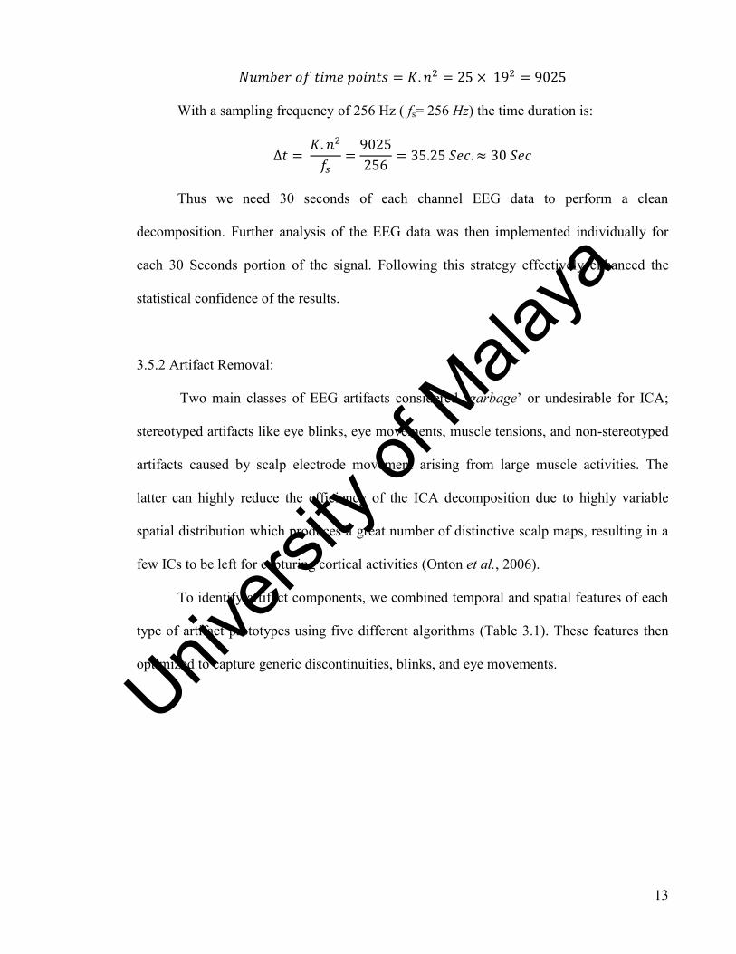
3.5.1 Duration of EEG Signal
Number of Time Points: Assuming to have n channels of EEG data, the number of
time points which are required for a clean decomposition is related to the number of
channels squared (Onton & Makeig, 2006). Running so many trials and errors, we
considered the best value for K to be 25. While the number of channels was 19 and K being
equal to 25 the sufficient number of data points needed for a clean decomposition would
be:
Univers
ity of
Mala
ya
13
With a sampling frequency of 256 Hz ( fs= 256 Hz) the time duration is:
Thus we need 30 seconds of each channel EEG data to perform a clean
decomposition. Further analysis of the EEG data was then implemented individually for
each 30 Seconds portion of the signal. Following this strategy effectively enhanced the
statistical confidence of the results.
3.5.2 Artifact Removal:
Two main classes of EEG artifacts considered „garbage‟ or undesirable for ICA;
stereotyped artifacts like eye blinks, eye movements, muscle tensions, and non-stereotyped
artifacts caused by scalp electrode movement arising from large muscle activities. The
latter can highly reduce the efficiency of the ICA decomposition due to highly variable
spatial distribution which produces a great number of distinctive scalp maps, resulting in a
few ICs to be left for capturing cortical activities (Onton et al., 2006).
To identify artifact components, we combined temporal and spatial features of each
type of artifact prototypes using five different algorithms (Table 3.1). These features then
optimized to capture generic discontinuities, blinks, and eye movements.
Univers
ity of
Mala
ya

14
Table 3.1 Algorithms of Artifact Removals and Their Index of Sensitivity
ARTIFACT REMOVAL ALGORITHM SENSITIVITY OF THE APPLIEDALGORITHM
Eye Blink
Spatial Average Difference (SAD) Higher amplitude in frontal areas
Temporal Kurtosis (TK) Outliers in the amplitude distribution typical of
blinks
Horizontal
Eye
Movement
Spatial Eye Difference (SED) Large amplitudes in frontal channels near the eyes
typical of horizontal eye movements
Maximum Epoch variance (MEV) Slower fluctuations typical of vertical eye
movements
Vertical Eye
Movement
Spatial Average Difference (SAD) Higher amplitude in frontal areas
Maximum Epoch variance (MEV) Slower fluctuations typical of vertical eye
movements
Generic
Discontinuities
Generic Discontinuities Spatial
Feature (GDSF) Local spatial discontinuities
Maximum Epoch variance (MEV) Slower fluctuations typical of vertical eye
movements
Pruned ICs completely corrected from the artifact effects then back projected
together by ICA (Makeig et al., 1997) to make the clean brain activities ready for further
analysis.
EEG streams, fully corrected from the influence of artifacts, contained a continuous
5-min signal (with eyes closed) for each patient and control subject. Due to the technical
requirements of the tools which were later used to process the data, EEGs from 20
electrodes (F7/8, Fz, F3/4, T3/4, C5/6, Cz, C3/4, T5/6, Pz, P3/4, Oz, O1/2) were analyzed
with a converted sampling rate of 128 Hz.
Prior to the spectral analysis, each EEG signal was band pass filtered within the 0.5-30 Hz
frequency range. This frequency range was chosen because approximately 98% of spectral
power lies within it (Collura et al., 2008).
Univers
ity of
Mala
ya

15
3.5.3 Filtering
Considering the fact that the natural brain activities are represented by different
types of simultaneous oscillations (Basar et al., 2000), spectral filtering and isolating EEG
data into different frequency bands would mask the natural composition of cortical
activities (Fingelkurts, Kivisaari, Autti, Borisov, Puuskari, Jokela, & Kähkönen, 2006)
Temporal activity of each independent component is the resultant of instantaneous and thus
broad band filtering of EEG scalp data, therefore it represents the synchronous segment of
activity within a single or two connected cortical patches. This fact also supports the
drawbacks of applying spectral filtering to the EEG data prior to the main analysis
(Fingelkurts & Kahkonen, 2005).
Therefore we considered a broad frequency band (0.5Hz – 45Hz) for our analysis
knowing that also 98% of the whole power of the EEG signal lies within this spectrum
(Fingelkurts et al., 2000) which makes it easier to capture and monitor the activities of
brain sources. This broad band-pass filtering enhanced the efficiency of ICA decomposition
as well (Delorme & Makeig, 2004)since it efficiently minimized the appearance of 50Hz or
60Hz linear trends. We applied EEGLAB toolbox of the MATLAB software to implement
the Finite Impulse Response (FIR) Butterworth band-pass filtering of the data. The
advantages of this non-linear filtering algorithm arise from its flat band passing which is
excellent in simulating the band pass of an ideal filter.
3.6 Independent Component Analysis (ICA)
As the next step, we performed ICA decomposition using infomax ICA algorithm
(provided in EEGLAB Toolbox of MATLAB) which requires a few matrix inversion and
therefore runs very fast. The main advantage of this algorithm is that it can detect not only
supergaussian (peaky) sources of activities but also subgaussian distributions.
Univers
ity of
Mala
ya

16
This feature of infomax algorithm is of great importance while detecting artifacts
(Lee et al., 1999).Experimental trials and errors revealed that for our analysis applying the
infomax ICA yields cleaner decompositions.
3.7 Components Source Modeling
As we have previously seen in this study, low spatial resolution of the EEG scalp
signals makes it so difficult to indicate the location of even the most intensive underlying
sources in the cortex. Therefore, we directly applied an inverse modeling of cortical source
locations from distribution of EEG potentials on the scalp. But this approach might be quite
intractable since almost the entire EEG scalp signals are sum of the activities of multiple
cortical and non-cortical sources. ICA has paved the way to solve this problem by applying
inverse modeling of ICs scalp maps rather than their projection to each of the scalp
electrodes.
3.8 Modeling Components Equivalent Dipoles
The most realistic approach to this inverse modeling tries to match each ICs scalp
map with the closest projection of a single equivalent dipole(Scherg & Von Cramon,
1985).We used DIPFIT plug-in of EEGLAB toolbox to localize the equivalent dipoles of
ICs. We applied the standard four shell (Skin, Skull, CSF, and Cortex) spherical head
model to simulate the brain. To make the coordinates of the dipoles returned by DIPFIT,
meaningful channel were co-registered to the surface of the selected head model. After fine
fitting of the dipoles, for each subject we were able to find single equivalent dipole models
for 20 ICs whose projection patterns across scalp significantly (with no more than 15%
residual variance (R.V.) between the scalp projection of the dipole and the actual IC scalp
Univers
ity of
Mala
ya

17
map) match the observed IC map. To have a highly accurate modeling if an IC scalp map
didn‟t adequately modeled by a single equivalent dipole we would exclude it from the
analysis. Performing further analysis requires adequate identification of physiologically
plausible components originating from the cortical sources. A workable criterion to
distinguish between cortical and non-cortical ICs is that the cortical independent
component should match adequately to a single (or bilateral) equivalent dipole(s) (R.V. <
15%) which localized inside the volume of the head. Modeling ICs to the best-fitting
equivalent dipoles not only provide localization of cortical activities to a certain brain
region; but also paves the way to evaluate and visualize the spatial uniformity of clusters
which include functionally comparable or equivalent IC activities across subjects.
3.9. Components Clustering
To identify and correlate equivalent ICs across subjects we applied a clustering
approach. We analyzed and compared independent components from different subjects and
found ICs which were functionally or spatially equivalent, these equivalent components
then were grouped together as one cluster. In other words, ICs representing the same
functional activities were spatially modeled as equivalent dipoles in one single cluster.
Clustering independent components according to their equivalent dipoles 3D locations we
were able to analyze brain activities in almost all cortical areas or at least in cortical regions
having adequate densities of dipoles across subjects. We used K-means distance-based
algorithm to run the clustering of ICs equivalent dipoles. Physiological differences of
cortical structures in location and orientation of gyrus, sulci, and also the brain shape may
reduce the reliability and confidence in the results achieved by comparing cortical areas of
ICs equivalent dipoles across subjects. Therefore, to normalize 3D locations of equivalent
Univers
ity of
Mala
ya
18
dipoles across subjects, we performed a spatial normalization of each subject head shape to
a standard four shell head model learned from 3D locations of each subject scalp
electrodes. To this end, we performed a true comparison of dipoles 3D locations across the
subjects.
3.10 Spectral Analysis
Although clustering of independent components across subjects to find the most
significant functional or spatial correlation among different cortical activities is itself a
statistical approach, to put more confidence in our analysis regarding the source
localization of the cortical activities involved in states of opioid dependency and methadone
treatment, we also compared broad band (0.5Hz-120Hz) relative power across three
clusters of ICs in alpha, beta, and gamma frequencies.
Univers
ity of
Mala
ya
19
4 CHAPTER IV: RESULTS AND INTERPRETATIONS
4.1 Introduction
This chapter provides the results of EEG source localizations along with the spectral
and statistical analysis of the results. We applied ICA for localizing the EEG sources within
the cortex while these cortical sources were modeled as equivalent dipoles of the
independent components (ICs). Spectral analysis of the clustered dipoles was implemented
in alpha, beta, and gamma frequencies. Finally we applied t-test for statistical analysis of
the results.
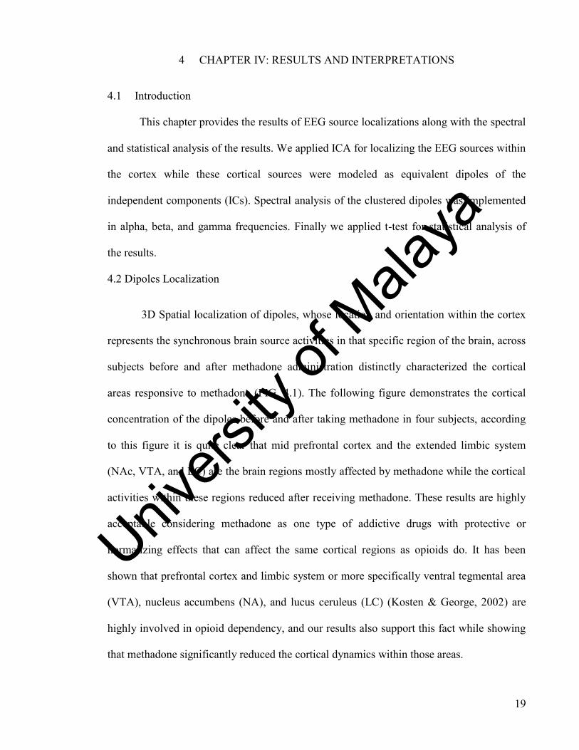
4.2 Dipoles Localization
3D Spatial localization of dipoles, whose location and orientation within the cortex
represents the synchronous brain source activities in that specific region of the brain, across
subjects before and after methadone administration distinctly characterized the cortical
areas responsive to methadone (FIG. 4.1). The following figure demonstrates the cortical
concentration of the dipoles before and after taking methadone in four subjects, according
to this figure it is quite clear that mid prefrontal cortex and the extended limbic system
(NAc, VTA, and LC) are the brain regions mostly affected by methadone while the cortical
activities within these regions reduced after receiving methadone. These results are highly
acceptable considering methadone as one type of addictive drugs with protective or
normalizing effects that can affect the same cortical regions as opioids do. It has been
shown that prefrontal cortex and limbic system or more specifically ventral tegmental area
(VTA), nucleus accumbens (NA), and lucus ceruleus (LC) (Kosten & George, 2002) are
highly involved in opioid dependency, and our results also support this fact while showing
that methadone significantly reduced the cortical dynamics within those areas.
Univers
ity of
Mala
ya
20
Figure 4.1 Clustering ICs from 4 subjects based on their functional consistency and
common spatial projection of their equivalent dipoles
Univers
ity of
Mala
ya
21
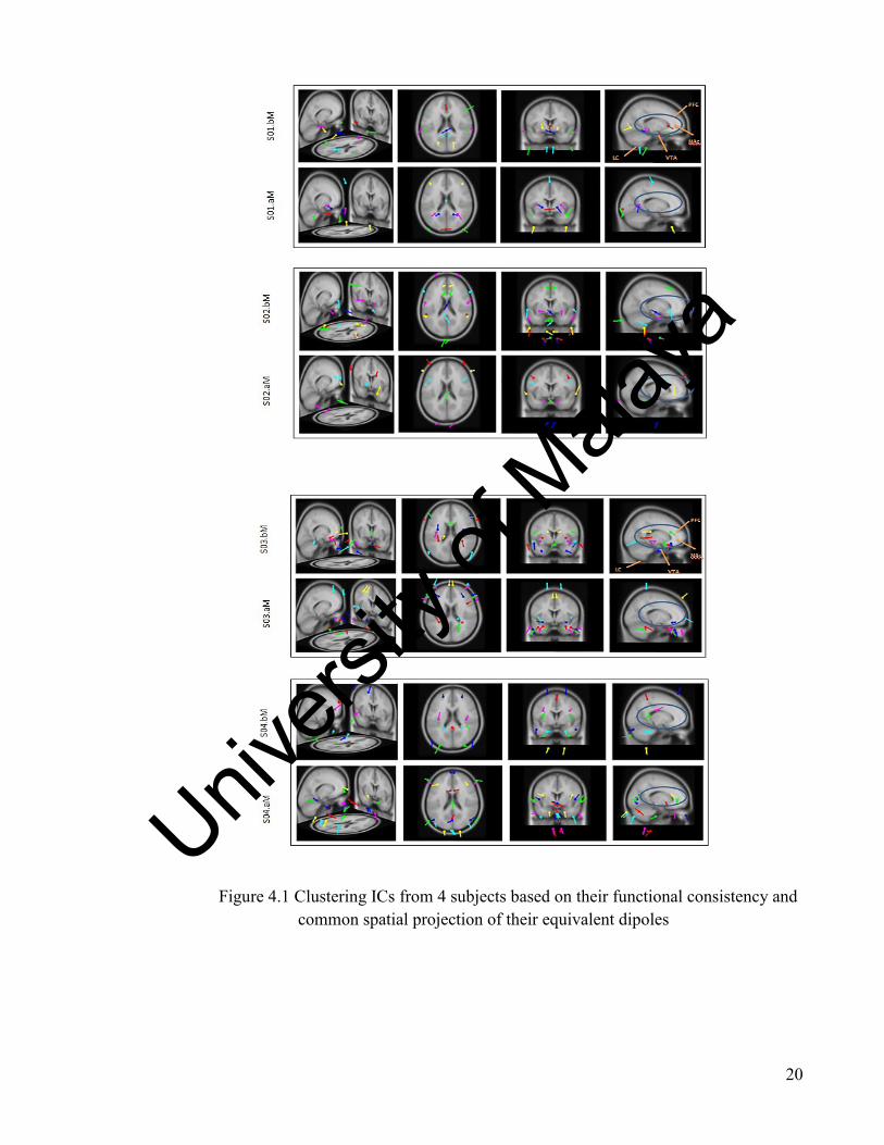
We identified 3 homogenous clusters of 20 ICs whose equivalent dipoles accurately
(R.V. <15) project to the scalp map of those ICs (Figure 4.2). Spatial 3D localization of
these dipoles within 3 clusters precisely demonstrated the concentration of the locally
coherent cortical activities in medial prefrontal cortex (mPFC) and extended limbic system
through the state of opioid dependency and methadone-based treatment in all 4 subjects.
Since the clustering algorithm which was used in our analysis (K-Means) efficiently finds
the functionally and spatially coherent activities across the cortex, therefore it provides
more accurate and plausible results as compared to the analysis of single subjects in the
previous section. So it is quite clear that the concentration of coherent cortical activities
across the mid prefrontal cortex and extended limbic system (VAT, NAc) is more distinct
across the clusters rather than the subjects. These findings also support the efficiency and
precision of our clustering algorithm.
Figure 4.2 . Localization of the most functionally and spatially coherent cortical activities
across the clusters and their corresponding spectrum
Univers
ity of
Mala
ya
22
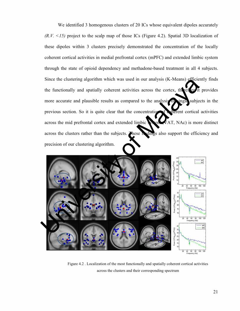
4.3 Spectral Analysis
To put more confidence in our results, we perform spectral analysis across the
clusters in the state of opioid dependency (before receiving methadone therapy) and
methadone treatment. The results (Figure 4.3) revealed a significant increase in the brain
source activities within gamma spectrum while the cortical activities of the alpha and beta
rhythms decreased after early onset of methadone treatment. These findinge were of great
consistency across all three clusters while they highly agree with the results of statistical
analysis as well.
Figure 4.3 Clusters spectrum
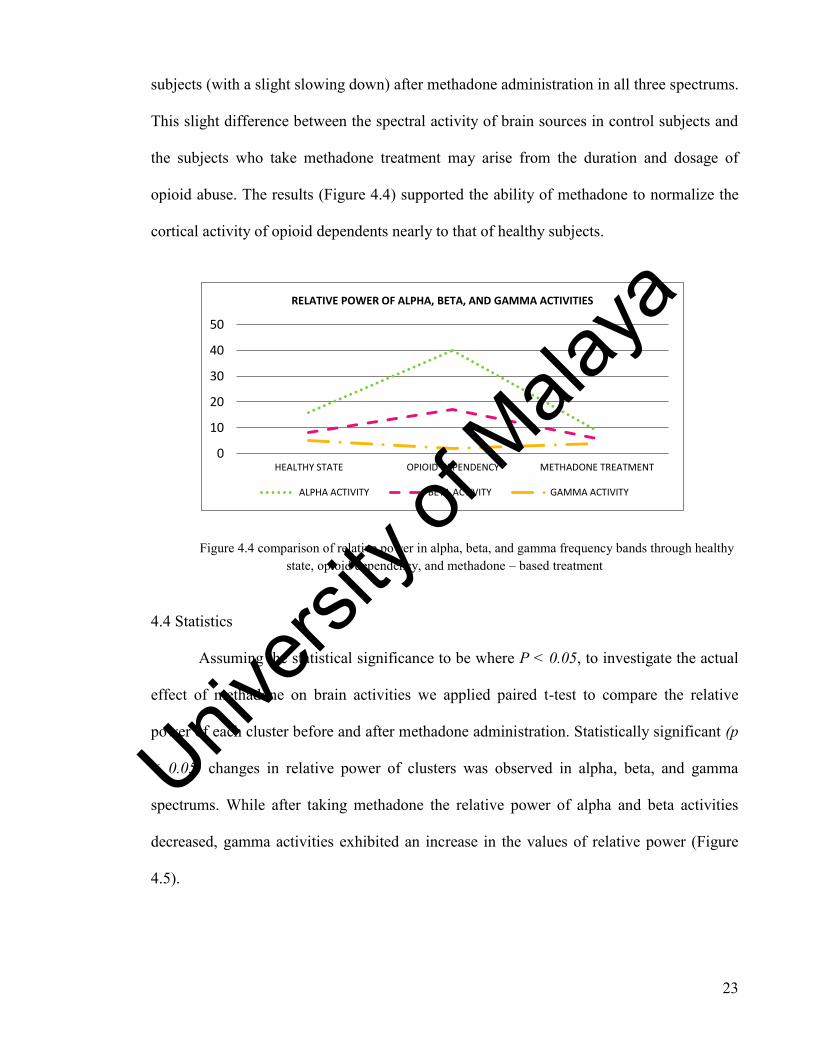
Finally we compared the results of spectral analysis within the healthy state, opioid
dependency and methadone treatment to investigate the effectiveness of methadone to
restore the normal structural functioning of the brain comparable to the healthy subjects.
This comparative approach revealed that through the state of opioid dependency and
methadone treatment spectral activity of cortical sources has changed considerably in alpha,
beta, and gamma frequency bands. These spectral activities underwent an upward trend
through the state of opioid dependency and then dropped almost to the level of control
Univers
ity of
Mala
ya
23
subjects (with a slight slowing down) after methadone administration in all three spectrums.
This slight difference between the spectral activity of brain sources in control subjects and
the subjects who take methadone treatment may arise from the duration and dosage of
opioid abuse. The results (Figure 4.4) supported the ability of methadone to normalize the
cortical activity of opioid dependents nearly to that of healthy subjects.
Figure 4.4 comparison of relative power in alpha, beta, and gamma frequency bands through healthy
state, opioid dependency, and methadone – based treatment
4.4 Statistics
Assuming the statistical significance to be where P < 0.05, to investigate the actual
effect of methadone on brain activities we applied paired t-test to compare the relative
power of each cluster before and after methadone administration. Statistically significant (p
< 0.05) changes in relative power of clusters was observed in alpha, beta, and gamma
spectrums. While after taking methadone the relative power of alpha and beta activities
decreased, gamma activities exhibited an increase in the values of relative power (Figure
4.5).
0
10
20
30
40
50
HEALTHY STATE OPIOID DEPENDENCY METHADONE TREATMENT
RELATIVE POWER OF ALPHA, BETA, AND GAMMA ACTIVITIES
ALPHA ACTIVITY BETA ACTIVITY GAMMA ACTIVITY
Univers
ity of
Mala
ya

24
Figure 4.5 .Statistical Analysis of Relative Power Values within Alpha, Beta, and Gamma
Frequency Bands Before and After Methadone Administration
FREQUENCY
BANDS
BEFORE TAKING METHADONE AFTER TAKING METHADONE
Mean S.D. P (< 0.05) Mean S.D. P (< 0.05)
ALPHA 25.1489 5.75 0.0612 23.7522 6.14 0.0612
BETA 3.2848 0.9505 < 0.000 2.5712 0.9788 < 0.000
GAMMA 5.7877 1.36 0.002 6.9657 1.4 0.002 Univers
ity of
Mala
ya
25
5 Chapter V: CONCLUSION AND FUTURE WORK
5.1 Introduction
In this chapter the contribution of the results to optimal methadone treatment will be
discussed.
5.2 Conclusion
Considering the allostatic state of opioid dependency defined as a chronic deviation
from the normal structural functioning of the brain, the main finding of this study supports
the important role of methadone to restore the normal cortical activities in opioid
dependents comparable to healthy subjects (FIG. 4.4). Reorganization of the brain temporal
activity within the state of opioid dependency which is contributed to the disorganization
syndrome in drug dependency and reward regulation (Fingelkurts, Ermolaev, et al., 2006;
Haig et al., 2000) reveals the significant role of psychotropic drugs to normalize the
temporal structure of the brain.
Source localization of the cortical activities provides a potent approach to accurately
investigate the actual cortical sources involved in opioid dependency which in return makes
it easier to identify the most effective therapy for opioid abuse. Most of the research
contributing to the temporal and spatial analysis of brain dynamics, apply advanced image
processing techniques like MRI or CT, from this perspective the significance of this study
derives from the efficient methodology used for brain source localization based on
independent component analysis (ICA). This study is the first of its kind since it represent a
completely novel approach to characterize the capability of methadone treatment in
normalizing the cortical functions of opioid dependents.
Univers
ity of
Mala
ya

26
Despite of the effectiveness of methadone treatment which has been proven in this
study, these physiological medications still need to be conjugated with the psychological
treatment knowing that opioid addiction is a psychophysiological disorder.
5.3 Future Work
Further analysis is required to estimate the optimal dosage of methadone which is
highly dependent on brain dynamics of individuals and also the duration of treatment. Since
investigation of the actual cortical activities is highly complex, there may be many
contributing factors while analyzing the structural brain dynamics, therefore a plausible
examination of all the contributing factors to the opioid addiction may require more sample
data.
Univers
ity of
Mala
ya
27
REFERENCES
Ball, J. C., & Ross, A. (1991). The effectiveness of methadone maintenance treatment:
Patients, programs, services, and outcome: Springer-Verlag Publishing.
Basar, E., Basar-Eroglu, C., Karakas, S., & Schurmann, M. (2000). Brain oscillations in
perception and memory. International journal of psychophysiology, 35(2-3), 95-
124.
Bauer, L. O. (2001). CNS recovery from cocaine, cocaine and alcohol, or opioid
dependence: a P300 study. Clinical neurophysiology, 112(8), 1508-1515.
Bell, A. J., & Sejnowski, T. J. (1995). An information-maximization approach to blind
separation and blind deconvolution. Neural computation, 7(6), 1129-1159.
Cardoso, J. F., & Laheld, B. H. (1996). Equivariant adaptive source separation. Signal
Processing, IEEE Transactions on, 44(12), 3017-3030.
Collura, T. F., Thatcher, R. W., & Lambos, W. A. (2008). EEG biofeedback training using
live z-scores and a normative database. Introduction to quantitative EEG and
neurofeedback: advanced theory and applications, 103.
Costa, L., & Bauer, L. (1997). Quantitative electroencephalographic differences associated
with alcohol, cocaine, heroin and dual-substance dependence. Drug and alcohol
dependence, 46(1-2), 87-93.
Curran, H. V., Kleckham, J., Bearn, J., Strang, J., & Wanigaratne, S. (2001). Additional
methadone increases craving for heroin: a double-blind, placebo-controlled study of
chronic opiate users receiving methadone substitution treatment.
Psychopharmacology, 154(2), 153-160.
Davis, P., Liddiard, H., & McMillan, T. (2002). Neuropsychological deficits and opiate
abuse. Drug and alcohol dependence, 67(1), 105-108.
Delorme, A., & Makeig, S. (2004). EEGLAB: an open source toolbox for analysis of
single-trial EEG dynamics including independent component analysis. Journal of
neuroscience methods, 134(1), 9-21.
Fellows, L. K. (2007). The role of orbitofrontal cortex in decision making. Annals of the
New York Academy of Sciences, 1121(1), 421-430.
Fingelkurts, A. A., Ermolaev, V. A., & Kaplan, A. Y. (2006). Stability, reliability and
consistency of the compositions of brain oscillations. International journal of
psychophysiology, 59(2), 116-126.
Fingelkurts, A. A., Grin, E., Ermolaev, V., & Kaplan, A. Y. (2000). Adaptive classification
of EEG spectral patterns: the comparison between healthy subjects and patients with
different brain pathologies. Vestnik Moskovskogo Universiteta (Bulletin of Moscow
State University). Series: Biology, 4, 3-11.
Fingelkurts, A. A., & Kahkonen, S. (2005). New perspectives in pharmaco-
electroencephalography. Progress in Neuro-Psychopharmacology and Biological
Psychiatry, 29(2), 193-199.
Fingelkurts, A. A., Kahkonen, S., Kivisaari, R., Borisov, S., Puuskari, V., Jokela, O., et al.
(2007). Composition of EEG oscillations and their temporal characteristics:
Methadone treatment. International journal of psychophysiology, 64(2), 130-140.
Fingelkurts, A. A., Kivisaari, R., Autti, T., Borisov, S., Puuskari, V., Jokela, O., et al.
(2006). Reorganization of the composition of brain oscillations and their temporal
characteristics in opioid dependent patients. Progress in Neuro-
Psychopharmacology and Biological Psychiatry, 30(8), 1453-1465.
Fingelkurts, A. A., Kivisaari, R., Autti, T., Borisov, S., Puuskari, V., Jokela, O., et al.
(2006). Increased local and decreased remote functional connectivity at EEG alpha
Univers
ity of
Mala
ya

28
and beta frequency bands in opioid-dependent patients. Psychopharmacology,
188(1), 42-52.
Fu, L., Bi, G., Zou, Z., Wang, Y., Ye, E., Ma, L., et al. (2008). Impaired response inhibition
function in abstinent heroin dependents: an fMRI study. Neuroscience letters,
438(3), 322-326.
Gekht, A., Polunina, A., Briun, E., & Davydov, D. (2003). Brain bioelectrical activities in
heroin addicts during early abstinence period]. Zhurnal nevrologii i psikhiatrii imeni
SS Korsakova/Ministerstvo zdravookhraneniia i meditsinskoĭ promyshlennosti
Rossiĭskoĭ Federatsii, Vserossiĭskoe obshchestvo nevrologov [i] Vserossiĭskoe
obshchestvo psikhiatrov, 103(5), 53.
Gerra, G., Ferri, M., Polidori, E., Santoro, G., Zaimovic, A., & Sternieri, E. (2003). Long-
term methadone maintenance effectiveness: psychosocial and pharmacological
variables. Journal of Substance Abuse Treatment, 25(1), 1-8.
Ghodse, A., & Galea, S. (2010). Opioid analgesics and narcotic antagonists. Side Effects of
Drugs Annual, 32, 183-224.
Goddard, A. W., Mason, G. F., Almai, A., Rothman, D. L., Behar, K. L., Petroff, O. A. C.,
et al. (2001). Reductions in occipital cortex GABA levels in panic disorder detected
with 1h-magnetic resonance spectroscopy. Archives of general psychiatry, 58(6),
556.
Grace, A. A. (2000). The tonic/phasic model of dopamine system regulation and its
implications for understanding alcohol and psychostimulant craving. Addiction,
95(8s2), 119-128.
Greenwald, M., Johanson, C. E., Bueller, J., Chang, Y., Moody, D. E., Kilbourn, M., et al.
(2007). Buprenorphine duration of action: mu-opioid receptor availability and
pharmacokinetic and behavioral indices. Biological psychiatry, 61(1), 101-110.
Gritz, E. R., Shiffman, S. M., Jarvik, M. E., Haber, J., Dymond, A. M., Coger, R., et al.
(1975). Physiological and psychological effects of methadone in man. Archives of
general psychiatry, 32(2), 237.
Haig, A. R., Gordon, E., De Pascalis, V., Meares, R. A., Bahramali, H., & Harris, A.
(2000). Gamma activity in schizophrenia: evidence of impaired network binding?
Clinical neurophysiology, 111(8), 1461-1468.
Khantzian, E. J., Gawin, F., Kleber, H. D., & Riordan, C. E. (1984). Methylphenidate
(Ritalin) treatment of cocaine dependence--A preliminary report. Journal of
Substance Abuse Treatment, 1(2), 107-112.
Kosten, T. R. (1990). Neurobiology of abused drugs: opioids and stimulants. Journal of
Nervous and Mental Disease.
Kosten, T. R., & George, T. P. (2002). The neurobiology of opioid dependence:
implications for treatment. Science &# x0026; Practice Perspectives, 1(1), 13.
Kreek, M. J. (1992). Rationale for maintenance pharmacotherapy of opiate dependence.
Lee, T. W., Girolami, M., & Sejnowski, T. J. (1999). Independent component analysis
using an extended infomax algorithm for mixed subgaussian and supergaussian
sources. Neural computation, 11(2), 417-441.
Makeig, S., Bell, A. J., Jung, T. P., & Sejnowski, T. J. (1996). Independent component
analysis of electroencephalographic data. Advances in neural information
processing systems, 145-151.
Makeig, S., Jung, T. P., Bell, A. J., Ghahremani, D., & Sejnowski, T. J. (1997). Blind
separation of auditory event-related brain responses into independent components.
Proceedings of the National Academy of Sciences, 94(20), 10979.
Univers
ity of
Mala
ya

29
Makeig, S., Westerfield, M., Jung, T. P., Covington, J., Townsend, J., Sejnowski, T. J., et
al. (1999). Functionally independent components of the late positive event-related
potential during visual spatial attention. The Journal of neuroscience, 19(7), 2665.
Makeig, S., Westerfield, M., Jung, T. P., Enghoff, S., Townsend, J., Courchesne, E., et al.
(2002). Dynamic brain sources of visual evoked responses. Science, 295(5555),
690.
Mintzer, M. Z., Copersino, M. L., & Stitzer, M. L. (2005). Opioid abuse and cognitive
performance. Drug and alcohol dependence, 78(2), 225-230.
Mintzer, M. Z., & Stitzer, M. L. (2002). Cognitive impairment in methadone maintenance
patients. Drug and alcohol dependence, 67(1), 41-51.
Onton, J., & Makeig, S. (2005). Independent component analysis (ICA) source locations
vary according to task demands. Org. Hum. Brain Mapp., Abstracts.
Onton, J., & Makeig, S. (2006). Information-based modeling of event-related brain
dynamics. Progress in Brain Research, 159, 99-120.
Onton, J., Westerfield, M., Townsend, J., & Makeig, S. (2006). Imaging human EEG
dynamics using independent component analysis. Neuroscience & Biobehavioral
Reviews, 30(6), 808-822.
Prosser, J., Cohen, L. J., Steinfeld, M., Eisenberg, D., London, E. D., & Galynker, I. I.
(2006). Neuropsychological functioning in opiate-dependent subjects receiving and
following methadone maintenance treatment. Drug and alcohol dependence, 84(3),
240-247.
Robinson, T. E., & Berridge, K. C. (2000). The psychology and neurobiology of addiction:
an incentive–sensitization view. Addiction, 95(8s2), 91-117.
Scherg, M., & Von Cramon, D. (1985). Two bilateral sources of the late AEP as identified
by a spatio-temporal dipole model. Electroencephalography and Clinical
Neurophysiology/Evoked Potentials Section, 62(1), 32-44.
Shaham, Y., Rajabi, H., & Stewart, J. (1996). Relapse to heroin-seeking in rats under
opioid maintenance: the effects of stress, heroin priming, and withdrawal. The
Journal of neuroscience, 16(5), 1957-1963.
Shufman, E., Perl, E., Cohen, M., Dickman, M., Gandaku, D., Adler, D., et al. (1996).
Electro-encephalography spectral analysis of heroin addicts compared with
abstainers and normal controls. The Israel journal of psychiatry and related
sciences, 33(3), 196.
Silveri, M. M., Pollack, M. H., Diaz, C. I., Nassar, L. E., Mendelson, J. H., Yurgelun-Todd,
D. A., et al. (2004). Cerebral phosphorus metabolite and transverse relaxation time
abnormalities in heroin-dependent subjects at onset of methadone maintenance
treatment. Psychiatry Research: Neuroimaging, 131(3), 217-226.
Simpson, D. D., Joe, G. W., & Brown, B. S. (1997). Treatment retention and follow-up
outcomes in the Drug Abuse Treatment Outcome Study (DATOS). Psychology of
Addictive behaviors, 11(4), 294.
Swanson, L. W. (2001). Brain architecture:Understanding the basic plan:Oxford Univ Pr.
Volkow, N. D., Fowler, J. S., Wang, G. J., & Goldstein, R. Z. (2002). Role of dopamine,
the frontal cortex and memory circuits in drug addiction: insight from imaging
studies. Neurobiology of Learning and Memory, 78(3), 610-624.
Walwyn, W. M., Miotto, K. A., & Evans, C. J. (2010). Opioid pharmaceuticals and
addiction: The issues, and research directions seeking solutions. Drug and alcohol
dependence, 108(3), 156-165.
White, W. L. (2007). Counselor Magazine's Addiction Professional's Reference Guide.
Univers
ity of
Mala
ya

30
Zajocov, A., Wilczek, H., & Hol, V. (2004). Short Communication The Alterations of
Immunological Reactivity in Heroin Addicts and Their Normalization in Patients
Maintained on Methadone. Folia Biologica (Praha), 50, 24-28.
Univers
ity of
Mala
ya