shamah mcb 93
TRANSCRIPT
MOLECULAR AND CELLULAR BIOLOGY, Dec. 1993, p. 7203-72120270-7306/93/127203-10$02.00/0Copyright © 1993, American Society for Microbiology
Vol. 13, No. 12
Dominant-Negative Mutants of Platelet-Derived Growth FactorRevert the Transformed Phenotype of
Human Astrocytoma CellsSTEVEN M. SHAMAH,1 CHARLES D. STILES,`* AND ABHIJIT GUHA1'2
Department ofMicrobiology and Molecular Genetics, Harvard Medical School, and Dana-FarberCancer Institute, Boston, Massachusetts 02115,1 and Division ofNeurosurgery and
Surgical Oncology, University of Toronto, Toronto, Ontario, Canada2
Received 10 November 1992/Returned for modification 25 January 1993/Accepted 26 August 1993
Malignant astrocytoma is the most common primary human brain tumor. Most astrocytomas express acombination of platelet-derived growth factor (PDGF) and PDGF receptor which could close an autocrine loop.It is not known whether these autocrine loops contribute to the transformed phenotype of astrocytoma cells orare incidental to that phenotype. Here we show that dominant-negative mutants of the PDGF ligand break theautocrine loop and revert the phenotype of BALB/c 3T3 cells transformed by the PDGF-A or PDGF-B (c-sis)gene. Then, we show that these mutants are selective in that they do not alter the phenotype of 3T3 cellstransformed by an activated Ha-ras or v-src gene or by simian virus 40. Finally, we show that these mutantsrevert the transformed phenotype of two independent human astrocytoma cell lines. They have no effect on thegrowth of human medulloblastoma, bladder carcinoma, or colon carcinoma cell lines. These observations areconsistent with the view that PDGF autocrine loops contribute to the transformed phenotype of at least somehuman astrocytomas.
The platelet-derived growth factors (PDGFs) are com-posed of two subunits, PDGF-A and PDGF-B (c-Sis), whichassociate through disulfide bonds to form PDGF-AA, PDGF-AB, and PDGF-BB (8, 15, 18, 35). Two PDGF receptorsubunits, a and A, associate noncovalently to form func-tional homodimers or heterodimers which differ in the abilityto interact with the three isoforms of PDGF (19, 20, 26, 42).PDGF-a receptor subunits can affiliate with either an A or Bchain of the PDGF ligand. PDGF-13 receptor subunits recog-nize PDGF-B chains preferentially. Thus, a:a homodimersfunction as the universal PDGF receptor, responsive to allthree isoforms of PDGF. The 1:13 receptor homodimersrespond preferentially to PDGF-BB, and a:3 receptor het-erodimers have an intermediate response profile (19, 21, 26,42).PDGF autocrine loops are established when PDGF ligands
and responsive PDGF receptor subunits are expressed in thesame cell. In tissue culture, these autocrine loops can bothinitiate and sustain a transformed phenotype (4, 11, 23, 28).In human neoplastic disease, PDGF autocrine loops havebeen linked to sarcoma, lung carcinoma, and malignantastrocytoma (1, 22, 45). The link is especially pervasive inmalignant astrocytoma. Although the PDGF ligand andreceptor genes are autonomous and independently regu-lated, the majority of human astrocytomas and astrocytomacell lines express a combination of these genes that could, inprinciple, close a PDGF autocrine loop and drive the unreg-ulated mitotic process in these tumors (14, 22, 39). Still, therole of PDGF autocrine loops in human astrocytoma hasremained unclear. At one extreme, these loops may becompletely incidental to the disease. As an intermediateview, the closure of PDGF autocrine loops may be an earlyinitiating event which becomes irrelevant to the malignantphenotype as the tumor progresses and acquires additional
* Corresponding author.
genetic lesions. Finally, it is possible that PDGF autocrineloops contribute to the transformed phenotype of astrocy-toma cells.To document the contribution of such loops to malignant
growth, it is necessary to disrupt them and demonstrate thatnormal growth regulation is restored. A bulk of evidencesuggests that although v-sis can activate intracellular formsof PDGF-1 receptor (10, 28), these receptors must belocalized to the cell surface for v-sis to induce mitogenicsignalling and autocrine transformation (10, 16, 32). Accord-ingly, reagents that antagonize the interaction betweenPDGF and its receptor at the cell surface, such as suraminand PDGF-neutralizing antibodies, have been found to re-duce the mitogenicity and revert the phenotype of v-sis-transformed cells (5, 11, 32). However, other groups havedemonstrated mitogenically active intracellular forms ofv-sis and maintain that intracellular PDGF autocrine loopsinvolving PDGF-13 receptors can initiate cellular transforma-tion (3, 4, 33). Thus, PDGF autocrine loops might be morecomprehensively targeted for disruption by interfering withPDGF ligand formation within the cell interior.
In previous studies, we constructed two mutations withina PDGF-A cDNA clone which exhibit dominant-negativeproperties (36). In COS cell cotransfection studies, onemutant, 1308, suppresses biologic activity of PDGF-A and-B subunits by forming unstable heterodimers. A secondmutant, 1317, forms stable but inactive heterodimers withPDGF-A subunits. Heterodimers between 1317 and PDGF-Bcan also form. However, these heterodimers are functional,thus restricting the dominant-negative action of 1317 toPDGF-A. It occurred to us that these mutants might becapable of disrupting autocrine loops involving the PDGFsand their receptors. In principle, these mutants could act atthe very onset of autocrine loop closure by suppressing theformation of PDGF ligand within the Golgi apparatus. In thisstudy, we tested the mutants initially in a defined context,using PDGF-transformed BALB/c 3T3 cells, and then in
7203
7204 SHAMAH ET AL.
A B
-29kDa
X PI)DGF-A/pMT2 (5XCS);- PDGF A/pMT2 ('5ZPPP)* pMT2 (5MBCS)* pMT2 ('9%PPP)
-T
0
EU
-1
.c-SS/pMT2(5.BCS)c-SIS/pMT2 (5XPPP)pMT2 (5XBCS)
- pMT2 (5%PPP)
DAYS I)AYS
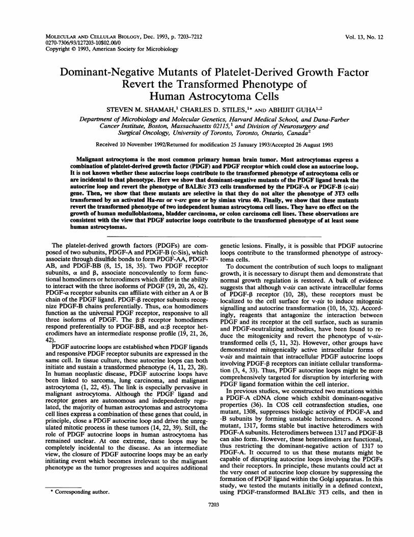
FIG. 1. Growth of PDGF-transformed cells in the presence or absence of serum. (A) Immunoprecipitations of metabolically [35S]cysteine-labeled PDGF-A-transformed CM with a PDGF-AA-specific antibody demonstrated PDGF-AA homodimers (-32 kDa) which could becompeted for with 1 ptg of unlabeled, recombinant PDGF-AA. The growth of PDGF-A-transformed cells in 5% BCS versus 5% PPP was
measured at the indicated days following plating at 2,000 cells per cm2. (B) The CL of metabolically labeled c-sis-transformed cells wasimmunoprecipitated with a PDGF-BB-specific antibody, revealing two bands (-30 and -24 kDa) that were specifically competed for with 1pg of unlabeled, recombinant PDGF-BB. The growth of c-sis-transformed cells in BCS versus PPP was determined as in panel A.
several human astrocytoma cell lines. We show that PDGFdominant-negative mutants revert the phenotype of PDGF-transformed cells and at least some human astrocytomas.These data are consistent with the notion that PDGF auto-crine loops are an active factor in the transformed phenotypeof these tumors.
MATERIALS AND METHODS
cDNA plasmid constructs. Full-length mouse PDGF-A (37)and human c-sis (a kind gift from the laboratory of D.Bowen-Pope, University of Washington, Seattle) cDNAswere cloned into the EcoRI site of the pMT2 expressionvector (27) and expressed under the control of the adenovi-rus major late promoter. The 1308 and 1317 PDGF dominant-negative mutants were cloned into the HindIII site of thepLNCX vector (38) and expressed under the control of thecytomegalovirus immediate-early promoter. The neomycinresistance gene is expressed by pLNCX; resistance to hy-gromycin B was conferred to cells by cotransfection withpY3 (7).
Cell transfections. Transfection of BALB/c 3T3 cells wasperformed by a calcium phosphate precipitation technique.Approximately 18 h prior to transfection, 106 cells per dishwere plated on 100-mm-diameter tissue culture dishes inDulbecco's modified Eagle medium (DMEM; GIBCO) sup-plemented with 10% bovine calf serum (BCS; HyClone). Amixture of DNA (20 pug of pMT2 constructs with 1 pug ofpY3, or 20 jxg of pLNCX constructs) in 0.5 ml of 0.125 MCaCl2 was added dropwise to 0.5 ml of 2x HBS buffer (280mM NaCl, 1.5 mM Na2HPO4. 7H20, 50 mM N-2-hydroxy-ethylpiperazine-N'-2-ethanesulfonic acid [HEPES]) with
constant aeration and incubated for 30 min at room temper-ature (RT) to facilitate the formation of a calcium phosphateprecipitate. The precipitate was added to cells withoutmedium for 20 min at RT, and then the cells were incubatedfor 4 h with 4 ml of 10% BCS at 370C. After the precipitatewas removed, the cells were shocked with 15% glycerol-1 xHBS for 3 min, rinsed twice with phosphate-buffered saline(PBS), and grown in 10% BCS. Clonal lines were isolated inthe third to fourth weeks of drug selection with 200 pg ofhygromycin B (Calbiochem) per ml and/or 500 ,ug of neomy-cin sulfate (GIBCO) per ml. Four-millimeter squares oftrypsin-soaked Whatman 3MM filter paper were applieddirectly to PBS-rinsed cell colonies, incubated for 3 min at370C, and transferred to six-well tissue culture dishes (Fal-con) for subsequent clonal expansion.For the astrocytoma cell lines, transfections were per-
formed with Lipofectin reagent (GIBCO/BRL) as follows.Eighteen hours prior to transfection, 106 cells were plated onfibronectin-coated (1 Pg/cm2 for 1 h; Collaborative Biomed-ical Products) 100-mm-diameter tissue culture dishes. Lipo-fectin was added to DMEM at a concentration of 16 1.l/mland then mixed with an equal volume of DMEM containingDNA (pLNCX constructs) for a final concentration of 81.d/ml. Cells were rinsed twice with PBS and incubated withthe Lipofectin-DNA mixture (4 ml per dish) at 370C for 5 h,at which time an equal volume of 10% BCS was added.Drug-resistant cultures were selected in 500 pug of neomycinsulfate per ml.
Metabolic labeling and immunoprecipitation. Primaryclones were first screened for expression of PDGF-A or c-sismRNA by Northern (RNA) analysis (data not shown). One
X 7.E- E-ra
Q.A.
< -o~a
CL X~9 - - L)n6CL a
a CL
u
C UL U --
--29gkDa
0
er
E
To"I
wX
MOL. CELL. BIOL.
7,..W
A
:i
PDGF DOMINANT-NEGATIVE MUTANTS REVERT ASTROCYTOMAS 7205
A100000
C.)-J-J50
50000
* PDGF-A+ VECTOR* PDGF-A +1317(12)* PDGF-A+ 1317(13)
PDGF-A +1308(15)X PDGF-A + 1308(18)
WT 3T3'
Lo0 ~ .- .
B
0
CD
-1
0
150000
10000
50000 -
0o i .
2
c-SIS+ VECTc-SIS+1317(c-SIS+ 1317(:
El c-SIS+ 1308(!c-1SS+ 1308(WT 3T3*
2
8
8
4
DAYS
'OR1)'3)9)11)
4
DAYS
CM
COlii0j
2 4 8
DAYS
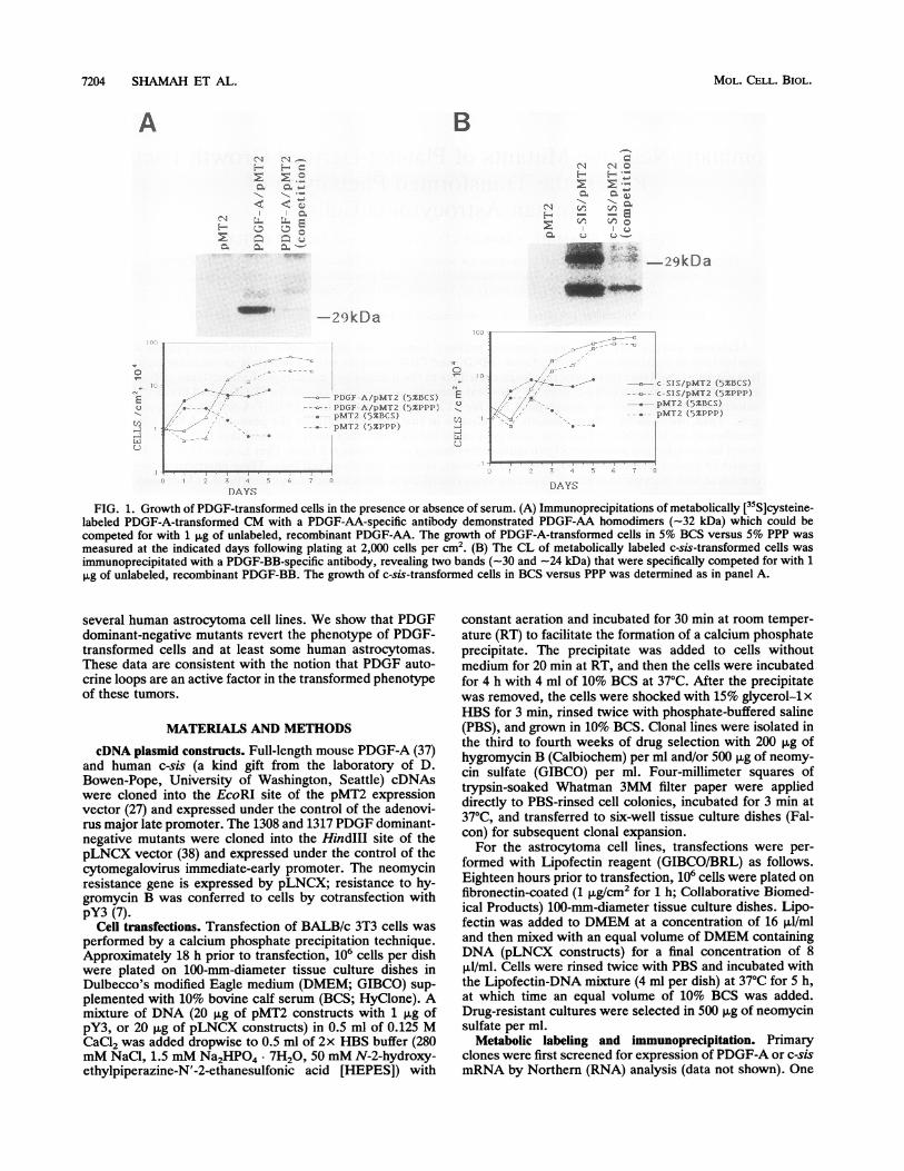
FIG. 2. Growth suppression of PDGF-A and PDGF-B (c-sis)-transformed cells by dominant-negative mutants. The indicated celllines were plated in duplicate wells at 2,000 cells percm2 and grown
in 5% PPP. Cells were harvested and counted at the indicated days.(A) PDGF-A-transformed cells with or without 1317 or 1308; (B)c-sis-transformed cells with or without 1317 or 1308; (C) Ha-ras-transformed cells with or without 1317 or 1308. Numbers in paren-
theses represent different clonal cell lines. Each value (with theexception of Ha-ras values) represents the average standard error
of the mean of two independent growth curve experiments. WT,wild type.
subline expressing abundant PDGF-A mRNA and anotherexpressing abundant c-sis mRNA were metabolically labeledto verify PDGF protein expression. Cells were grown to
confluence on five 100-mm-diameter tissue culture dishes(Falcon), rinsed twice with cysteine-free DMEM, and incu-
bated with 250puCi of[355]cysteine (NEN) in 1 ml ofcysteine-free DMEM for 4 h at 370C with intermittentrocking. After the conditioned medium (CM) was harvested,the cells were rinsed twice with PBS and lysed with 1 ml ofradioimmunoprecipitation assay (RIPA) buffer (150 mMNaCl, 50 mM Tris [pH 8], 1% Nonidet P-40, 10 mMdeoxycholic acid, 0.1% sodium dodecyl sulfate [SDS]) con-taining 1 mM phenylmethylsulfonyl fluoride (PMSF). TheCM and cell lysates (CL) were concentrated in AmiconCentriprep-10 concentrators (10,000-molecular-weight cut-off) and then clarified by centrifugation before immunopre-cipitation.
Immunoprecipitations were performed with a polyclonalmouse PDGF-AA-specific antibody (1:100; kindly providedby Chiayeng Wang) and a monoclonal human PDGF-BB-specific antibody (1:400; Upstate Biotechnology Inc. [UBI]).Antibodies were added directly to CL, or with RIPA-PMSFto CM, and incubated for 3 h at 40C with constant rocking.For competition experiments, 1pug of unlabeled, recombi-nant PDGF-AA (UBI) or PDGF-BB (UBI) was added tothe CL or CM prior to addition of antibodies. ProteinA-Sepharose CL-4B resin (Pharmacia) was added for 1 h at40C and subsequently rinsed twice with RIPA-PMSF. Thesamples were reconstituted in nonreducing Laemmli samplebuffer, boiled for 5 min, and analyzed by SDS-polyacryl-amide gel (14% acrylamide) electrophoresis (PAGE).Growth in serum and PPP. The PDGF-A-transformed,
PDGF-B (c-sis)-transformed, and pMT2-transfected primarycell lines were plated in 10% BCS at a density of 2,000cells/cm2 into 24-well plates. After 24 h, the medium waschanged to either 5% BCS or 5% platelet-poor plasma (PPP),and new medium added every third day. Duplicate wellswere trypsinized and counted with a Coulter Counter(Coulter Electronics, Inc.), and the average number of cellsper square centimeter was recorded daily for 8 days. Cellstransfected with the dominant-negative mutants were simi-larly plated into six-well dishes also at a starting density of2,000 cells per cm2.Colony formation assay. Cells were transfected with the
dominant-negative mutants as described above. Followingthe transfection, cells were grown in 10% BCS for 24 h andthen split 1:2 into either 5% BCS or 5% PPP with neomycinsulfate (500,ug/ml) and hygromycin B (200,ug/ml). Fortransfection of astrocytoma cells, the medium was changedto 5% PPP and 500,ug of neomycin sulfate per ml 48 h aftertransfection for simultaneous selection of drug resistanceand growth in PDGF-deprived medium. After approximately3 weeks, drug-resistant colonies were photographed at lOxmagnification or fixed with 10% formalin and stained with0.625% crystal violet for analysis.Anchorage-independent growth. Sterilized 1.8% agar in
water was mixed with equal volumes of 2x DMEM-20%BCS, layered on the bottom of 60-mm-diameter culturedishes (Falcon), and allowed to gel at RT. A methocellulosesuspension with lx DMEM-10% BCS was prepared aspreviously described (40). A suspension of 105 cells in 4 ml ofmethocellulose-BCS was layered on top of the agarose bed.Fresh methocellulose-BCS (3 ml) was added weekly.Western immunoblotting. Cells were grown to confluence
in 150-mm-diameter dishes and incubated in 5% PPP for 48h. As controls, BALB/c 3T3 cells were incubated for 15 minin the absence or presence of 30 ng of PDGF-AA orPDGF-BB (UBI) per ml. Cellular protein was isolated at 4°Cwith 0.5 ml of lysis buffer (10 mM NaP buffer [pH 7.2], 150mM NaCl, 1% Nonidet P-40, 10% glycerol, 0.2% NaF,0.44% NaPPJ) containing 1 mM sodium orthovanadate, 5 ,ug
mmmmn=aa_
MMMMME3EW"
VOL. 13, 1993
7206 SHAMAH ET AL.
A LIL... A B
(i*. * A pILNCX
l_1317p1 NCX
c Balb/c3T3 SRC
pLNCX
*.42: * S.
1317pLNCX
1308pLNCX
1308pL.NCX
1317pLNCX
1308pLNCX
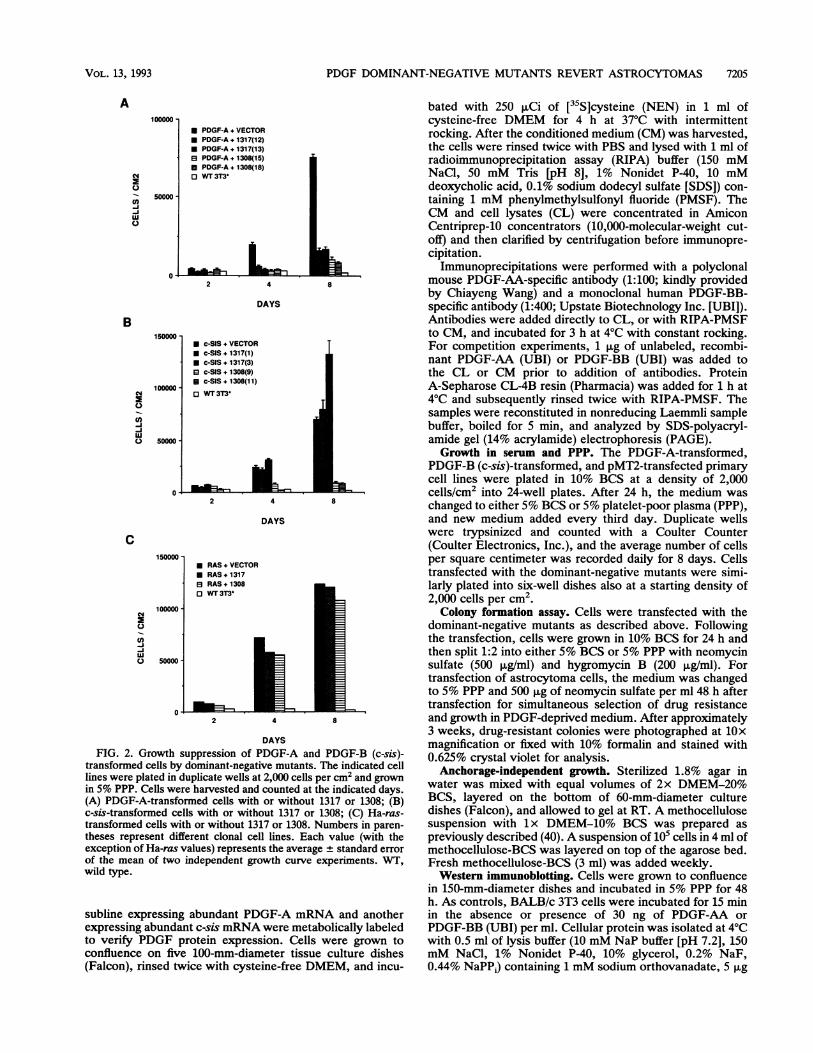
FIG. 3. Inhibition of colony formation by dominant-negative mutants. PDGF-A-transformed (A), PDGF-B (c-sis)-transformed (B), normal(neomycin resistant) (C), src-transformed (D), or SV40-transformed (E) BALB/c 3T3 cells were transfected with the dominant-negativemutant constructs as described in Materials and Methods and grown in 5% PPP with hygromycin B and neomycin drug selection. Afterapproximately 3 weeks, cells were fixed and stained as described in the text and evaluated for the number and size of cell colonies.
of aprotinin per ml, and 1 mM PMSF. The CL weremicrocentrifuged to remove cellular debris. The proteincontent was quantified (Bio-Rad protein analysis), and theCL were stored at -700C.For anti-PDGF-a receptor blots, PDGF-A-transformed
(-300 jig) or wild-type (-150 ,ug) 3T3 CL were resuspendedin (lx) reducing Laemmli buffer, boiled for 5 min, andseparated on SDS-7.5% polyacrylamide gels. The gels weretransblotted to nitrocellulose and blocked at 370C with 2%gelatin in Tris-buffered saline (TBS; 10mM Tris [pH 8], 0.9%NaCl) for 1 h. A polyclonal anti-PDGF-a receptor antibody(diluted 1:100 in TBS containing 0.1% Tween 20 [TBSTJ;kindly provided by Chiayeng Wang) or a monoclonal an-tiphosphotyrosine antibody (diluted 1:5,000; kindly providedby Thomas Roberts) was applied overnight at RT. Foranti-PDGF-P receptor blots, c-sis-transformed (-3,000 ,ug)or wild-type (-30 jig) 3T3 CL were first enriched forPDGF-P receptors by immunoprecipitation (as outlinedabove) with a polyclonal anti-PDGF-3 receptor antibody(1:100; UBI) prior to Western blotting. The immunoprecip-itates were resuspended in reducing Laemmli buffer, boiledfor 5 min, and separated on SDS-7.5% polyacrylamide gels.The gels were transblotted to Immobilon-P (Millipore) andblocked at RT with 5% bovine serum albumin in PBS for 2 h.Anti-PDGF-P receptor antibody or antiphosphotyrosine an-tibody (both diluted 1:5,000 in blocking buffer) was appliedfor 1.5 h at RT. After a rinse with TBST, the secondaryanti-rabbit or anti-mouse alkaline phosphatase-conjugatedantibody (1:7,000; Promega) was applied for 1 h at RT inTBST. Alkaline phosphatase reactions were carried out withthe addition of 66 AL of nitroblue tetrazolium and 33 Al of5-bromo-4-chloro-3-indolyl phosphate to 10 ml of developingbuffer (100 mM Tris [pH 9.5], 100 mM NaCl, 5 mM MgCI2).
CL used in all immunoprecipitations and immunoblots werenormalized on the basis of total protein content.Nude mice. Pooled colonies of U343 astrocytoma cells
transfected with the 1308 dominant-negative mutant or thecontrol expression vector (pLNCX) were trypsinized andresuspended in DMEM. By using a tuberculin syringe and a27-gauge hypodermic needle, 10 cells (0.3 ml) were injectedinto the subcutaneous interscapular tissue of 4- to 6-week-old male Swiss nude mice (Taconic). The tumor diameterwas recorded for four animals per experimental set on aweekly basis starting at 3 weeks postinjection. The meantumor diameter ± standard error of the mean was calculated,and the statistical significance at 10 weeks was determinedby Student's t test (STAT-SAK; G. E. Dallal).
RESULTS
Transformation of BALB/c 3T3 cells with PDGF-A orPDGF-B (c-sis) enhances cell growth in PDGF-deficient me-dium. PDGF-A or PDGF-B (c-sis) cDNAs were subclonedinto the pMT2 expression vector and cotransfected with thehygromycin B resistance plasmid, pY3, into BALB/c 3T3fibroblasts. Two hygromycin B-resistant sublines expressinghigh levels of PDGF-A or -B mRNA (data not shown) wereselected for further characterization. One cell line producesPDGF-AA homodimer and releases it into the cell culturemedium (Fig. 1A). Another produces PDGF-BB homodimer,the bulk of which remains cell associated, as noted by others(31) (Fig. 1B). Both of the PDGF-producing sublines werereleased from their growth dependence on exogenous PDGF(Fig. 1). This release was demonstrated by comparing cellgrowth in medium supplemented with PPP with growth inmedium supplemented with BCS. As the product of unclot-
MOL. CELL. BIOL.
C ;. S]
.I.IA.
PDGF DOMINANT-NEGATIVE MUTANTS REVERT ASTROCYTOMAS 7
c-ASIS
* 2L'.'s'
c-SIS/ 1308 PD(
i.r.,'A
PDGF-A
PDGF A/1317
!
-
GF-A/ 1308
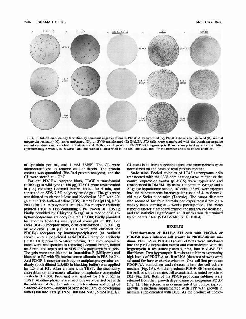
FIG. 4. Inhibition of anchorage-independent growth by domi-nant-negative mutants. PDGF-A and c-sis-transformed cells with orwithout 1317 or 1308 expression were analyzed for anchorage-independent growth. Cells (105) were plated in a methocellulose-BCS suspension and allowed to grow for 3 to 4 weeks, with freshmethocellulose-BCS added weekly. The suppressive effect of thedominant-negative mutants was measured as the ability to preventthe growth of colonies in methocellulose.
ted blood, PPP contains little, if any, PDGF (8, 44). Cellstransformed with either PDGF-A or PDGF-B (c-sis) grewnearly as well in PPP- or BCS-supplemented medium. Bycontrast, a control culture of BALB/c 3T3 cells, transfectedwith vector alone, showed superior growth in the BCS-supplemented medium. In addition to reducing the growthdifferential between PPP- and BCS-supplemented media,transformation with either PDGF-A or PDGF-B (c-sis) in-creased the final cell density at confluence (Fig. 1).PDGF dominant-negative mutants restore serum depen-
dence of 3T3 cells transformed by PDGFs. The PDGF-A andPDGF-B (c-sis)-transformed 3T3 cell sublines were sub-jected to secondary transfections with expression vectorsencoding either of two PDGF dominant-negative mutants,1317 and 1308. Two subclones from each transfection groupwhich expressed high levels of 1317 or 1308 mRNA (data notshown) were selected for further study. First, we tested thesecondary transfectants for growth in PPP-supplementedmedium. Expression of either mutant suppressed the growthof PDGF-A-transformed sublines (by approximately 70%)compared with a control PDGF-A-transformed sublinewhich received only the pLNCX vector during secondarytransfection (Fig. 2A). The PDGF-B (c-sis)-transformed sub-lines expressing 1308 were likewise growth inhibited (by
A PDGF-AVWT 3T3 TRANSFORMED
LL
0C-
- co2n0k
- 200 kDa
Ab: Ptyr I -4-+
Ab: PDGF -Rec
B
- 200 kDa
W4T INTNSFORMED
C- SI SWVT 3T3 TRANSFORMAED
LL
0C-
coOT2-200 kDa
Ab: Ptyr -4-
200 kDaAb: PDGF -Rec l * -40O
FIG. 5. Dominant-negative mutants disrupt PDGF autocrineloops. PDGF autocrine loops were analyzed by measuring the levelof constitutive receptor tyrosine phosphorylation in cells preincu-bated with 5% PPP for 48 h at 370C. (A) Cell lysates of wild-type(WT) 3T3 cells (untreated or PDGF treated) or PDGF-A-trans-formed cells (untreated or transfected with 1317 or 1308) wereresolved by SDS-PAGE, transferred to nitrocellulose, and analyzedby Western immunoblotting with antiphosphotyrosine (Ptyr; top)and anti-PDGF-a receptor (a-Rec; bottom) antibodies (Ab). ThePDGF treatment for the wild-type 3T3 cells was with 30 ng ofPDGF-AA per ml for 15 min at 370C. (B) CL of wild-type 3T3 cells(untreated or PDGF treated) or PDGF-B (c-sis)-transformed cells(untreated or transfected with 1308) was immunoprecipitated withan anti-PDGF-P receptor (P-Rec) antibody to enrich for receptorsprior to Western immunoblotting with antiphosphotyrosine (top)and anti-PDGF-j receptor (bottom) antibodies. For the experimentsin panel B, PDGF treatment for the wild-type 3T3 cells was with 30ng of PDGF-BB per ml for 15 min at 370C. To compensate forreceptor downregulation, we had to load unequal quantities of CL inthe various lanes. For the experiments in panel A, we used -150 pugof CL from BALB/c 3T3 cells and -300 jig of CL from PDGF-A-transformed cells. For the experiments in panel B, the differentialwas even greater: -30 ,ug of CL from BALB/c 3T3 cells and -3,000pig of CL from PDGF-B (c-sis)-transformed cells.
approximately 90%) compared with the control PDGF-B(c-sis)-transformed subline (Fig. 2B). The 1317 mutant failedto suppress the growth of c-sis-transformed cells and, in fact,facilitated growth, consistent with our previous findingswhich suggest that the 1317:PDGF-B heterodimer retainsfunctional activity (36). The inhibition of growth by 1308 and1317 is specific to PDGF-transformed cells, as they wereunable to inhibit the growth of Ha-ras-transformed BALB/c3T3 cells (Fig. 2C).A colony formation assay for the action ofPDGF dominant-
negative mutants. To eliminate the possibility of clonalselection bias and to rapidly screen other cell lines, we useda colony formation assay for growth in PDGF-free medium.The dominant-negative mutant expression vectors weretransfected into 3T3 cells which had been transformed with
VOL. 13, 1993 7207
A I
., . f I IA- A, .
.4t .-A
7208 SHAMAH ET AL.
PDGF-A, PDGF-B (c-sis), or two unrelated oncogenicagents, v-src and simian virus 40 (SV40). Transfected cellswere selected for neomycin resistance in PPP-supplementedmedium. Under these conditions, only cells which haveescaped the requirement for exogenous PDGF yield macro-scopically visible colonies (2, 48). As expected, wild-typeBALB/c 3T3 cells failed to form colonies in PPP-supple-mented medium, whereas cells transformed by PDGF-A,PDGF-B (c-sis), v-src, or SV40 did form colonies (Fig. 3).Transfection with either 1317 or 1308 reduced the numberand average size of PDGF-A-transformed cell colonies (Fig.3A). Only the 1308 mutant suppressed colony formation ofcells transformed by PDGF-B (c-sis) (Fig. 3B). The 1308 and1317 mutants neither suppressed nor enhanced colony for-mation of v-src- and SV40-transformed cells (Fig. 3D and E).As noted in growth rate measurements (Fig. 2) and in accordwith previous transfection studies on COS cell cultures (36),1317 actually enhanced the number and average size ofcolonies of cells transformed with PDGF-B (c-sis).
Dominant-negative mutants inhibit anchorage-independentgrowth of PDGF-transformed 3T3 cells. One hallmark oftransformed cell lines is the ability to develop colonies in softagar or methocellulose suspension. The PDGF-B (c-sis)-transformed cells developed large colonies (approximately50 cells or more) growing in a methocellulose suspensionwithin 14 days (Fig. 4). Expression of 1308 in these cellsinhibited growth in methocellulose. In agreement with re-sults in two different growth assays (Fig. 2 and 3), expressionof 1317 actually enhanced anchorage-independent growth ofthe PDGF-B (c-sis)-transformed cells. The PDGF-A-trans-formed 3T3 cells also developed colonies in methocellulosesuspension, albeit smaller than the ones generated byPDGF-B (c-sis)-transformed cells (Fig. 4). Expression ofeither 1308 or 1317 in PDGF-A-transformed cells inhibitedanchorage-independent growth, in accord with our growthrate measurements and colony formation assays (Fig. 2 and3).
Disruption of PDGF autocrine loops by the dominant-negative mutants. The tyrosine phosphorylation status ofPDGF receptors was examined by Western immunoblottingwith an antiphosphotyrosine antibody. When normal 3T3cells were cultured in PPP-supplemented medium, the PDGFreceptors were not phosphorylated unless challenged withexogenous PDGF. In the same medium, receptors wereconstitutively tyrosine phosphorylated in cells transformedwith PDGF-A or PDGF-B (c-sis) (Fig. 5). Expression of the1308 mutant eliminated this constitutive tyrosine phosphor-ylation in either PDGF-A- or PDGF-B (c-sis)-transformedcells (Fig. 5). In keeping with its biologic and biochemicalaction spectrum, expression of 1317 eliminated the constitu-tive phosphotyrosine signal in cells transformed by PDGF-A(Fig. 5A). Western immunoblotting with anti-PDGF-a andanti-PDGF-1 receptor antibodies showed that loss of theconstitutive tyrosine phosphate signal on PDGF receptorssignal did not reflect a loss of PDGF receptor protein.Indeed, total receptor protein was more abundant followingtransfection with dominant-negative mutants. This apparentupregulation was especially evident in the PDGF-B (c-sis)-transformed cells following transfection with 1308 (Fig. SB).Taken together, these data indicate that PDGF autocrine
loops are closed in 3T3 cells that express either PDGF-A orPDGF-B and that our dominant-negative mutants disruptthese loops. It should be noted, however, that acquisitionand reversion of the transformed phenotype do not correlatein a linear way with the abundance of tyrosine-phosphory-lated PDGF receptor. The total abundance of PDGF recep-
A VECTOR 1308
U343
U87
B
EJ
SW48()
DaOY
HeLica
FIG. 6. PDGF dominant-negative mutant inhibits astrocytomacell growth. Astrocytoma cell lines U87 and U343 were transfectedwith 1308 and analyzed for colony formation in 5% PPP. Cells (106per dish) were transfected with 20 pug of 1308/pLNCX (1308) or 20±g of pLNCX (vector) and grown in 5% PPP supplemented with 5008Lg of neomycin sulfate per ml. Drug-resistant colonies were stainedin 0.05% cresyl violet approximately 3 weeks after transfection.
tor protein in our PDGF-B (c-sis)-transformed 3T3 cells isless than 1% of that of control 3T3 cells (see the legend toFig. 5). Although receptors are upregulated when PDGFautocrine loops are broken by our dominant-negative mu-tants, they do not return to the levels seen in wild-type 3T3cells (see Discussion).
Inhibition of astrocytoma cell growth by the 1308 dominant-negative mutant. Of the two mutants that we characterized,only 1308 was active on both PDGF-A- and PDGF-B (c-sis)-transformed 3T3 cells. For this reason, we expressed 1308 intwo independent astrocytoma cell lines, U87 MG (U87) andU343 MGa Cl 2:6 (U343). These cell lines were previouslycharacterized and found to express both PDGF-A andPDGF-B together with PDGF-3 receptors; U343 expresseshigher levels of the ligands, whereas U87 expresses morereceptors (39). As judged from these expression patterns,
MOL. CELL. BIOL.
PDGF DOMINANT-NEGATIVE MUTANTS REVERT ASTROCYTOMAS 7209
FIG. 7. Morphological changes in astrocytoma cells induced by a PDGF dominant-negative mutant. U343 astrocytoma cells transfectedwith the control expression vector (A) or with 1308 (B) were pooled and plated at equal density (5 x 10⁵ cells per dish) in 5% PPP andphotographed on the same day at 10x magnification.
both cell lines are candidates for possessing PDGF autocrineloops. Using the colony formation assay described above(Fig. 3), we found that both U87 and U343 cells formedcolonies in PPP-supplemented medium. Thus, as predicted,these cells do not require exogenous PDGF for growth (Fig.6A). When either astrocytoma line was transfected with1308, colony formation in PPP-supplemented medium wasmarkedly inhibited (Fig. 6A).As controls for specificity, we studied the response of one
human cell line derived from a different type of brain tumor(the medulloblastoma cell line DaOY) and two human tumorcell lines that express an activated ras gene (the EJ bladdercarcinoma line and the SW480 colon carcinoma). None ofthese cell lines were affected by 1308 (Fig. 6B), althoughthey expressed equivalent amounts of 1308 mRNA (data not
shown). We were somewhat surprised to find that 1308suppresses colony formation in HeLa cells, a human cervicalcarcinoma cell line. However, Western blot analysis (datanot shown) indicates that our HeLa cell stocks expressPDGF-P receptors. For this reason and others (see Discus-sion), it is possible that the response of HeLa cells to 1308reflects the presence of a PDGF autocrine loop in a hithertounexpected context.
Morphological changes in astrocytoma cells. The U343astrocytoma cell line forms multilayered, refractile fociwhen grown in culture (Fig. 7A). Expression of the 1308mutant in these cells resulted in distinct morphologicalchanges. Both the number and size of foci that developedwhen grown in PPP-supplemented medium were reducedrelative to control cells transfected with vector alone (Fig.
VOL. 13, 1993
7210 SHAMAH ET AL.
12 -2- VECTOR1308
EE
C-"uJ
aC 4 -A +_0
0 6 10
TIME (weeks)
FIG. 8. A PDGF dominant-negative mutant reduces tumorige-nicity of astrocytomas in nude mice. U343 astrocytoma cells trans-fected with 1308 or the control expression vector were pooled andinjected subcutaneously into four nude mice per experimental set at107 cells per animal. The data represent mean tumor diameters +standard errors of the means. The tumor diameter of cells express-ing 1308 is 70% smaller than that of control cells at 10 weeks (t = 2.6,P < 0.05).
7B). Microscopic analysis also supports the view that 1308does not function as a toxin. We never observed cell debris,cytoplasmic vacuoles, or other visual indicators of a toxicresponse to 1308 expression (Fig. 7B). Cells expressing thedominant-negative mutant remained capable of sustainedgrowth in serum and PPP-supplemented medium, albeit atrates slower than those of control cells. Similar morpholog-ical changes were observed for U87 cells expressing the 1308dominant-negative mutant (data not shown).
Decreased tumorigenicity in nude mice of astrocytoma cellsexpressing 1308. U343 cells expressing 1308 were injectedinto nude mice as a measure of the tumorigenicity of thesecells. Each of four mice injected in the interscapular regionwith the control U343 cells developed moderate-size tumorsthat persisted through 10 weeks (Fig. 8). In contrast, U343cells expressing the 1308 dominant-negative mutant inducedsmall tumor formation in only two of four injected mice. At10 weeks, the mean diameter of tumors formed by U343 cellswas reduced by 70% in cells expressing 1308.
DISCUSSION
Malignant astrocytomas are the most common primaryintracranial tumors. In the United States, these tumorsaccount for approximately 15,000 deaths per year and 4 to5% of all cancer-related deaths (34). Several lines of evi-dence suggest that PDGF may contribute to the growth ofthese tumors. The majority of astrocytoma cell lines testedexpress PDGFs and PDGF receptor subunits which couldpotentially close autocrine loops and lead to stimulation ofthe tumor cells (39). Operative specimens from high-gradeastrocytomas express greater amounts of both PDGF-A andPDGF-B than do lower-grade astrocytomas or nonneoplasticglial tissue (14, 22). These high-grade astrocytomas alsoexpress PDGF-a receptors, implicating autocrine-mediatedgrowth (12, 14, 22, 30). It is not clear, however, that PDGFautocrine loops contribute to the malignant growth of thesehuman brain tumors. High levels of expression of othergrowth factors and/or growth factor receptors have beendemonstrated in astrocytomas (9, 17, 46, 47). Moreover,these tumors almost certainly accumulate other kinds of
genetic lesions in the process of becoming a clinicallydetectable tumor mass (6, 13, 25, 43).
In this study, we used dominant-negative mutants of thePDGF ligand to address the role of PDGF autocrine loops inhuman astrocytoma. Starting with well-characterized 3T3cell lines, we found that our mutants break PDGF autocrineloops, as indicated by Western blot analysis of receptorphosphorylation status. The mutants revert the transformedphenotype of 3T3 cells that display these loops. The selec-tive action of the mutants on 3T3 cells transformed byPDGF-A or PDGF-B (c-sis) is in accord with the biochemicalproperties of the mutant proteins (36). One of the mutants,1308, reverts the transformed phenotype of astrocytoma celllines which express both PDGF ligands and 3 receptorsubunits.To interpret these results, it is important to show that
growth responses to PDGF dominant-negative mutants areconfined to cells that exhibit PDGF autocrine loops. Neithermutant affects the phenotype of 3T3 cells transformed byactivated ras, v-src, or SV40, agents that function down-stream of PDGF receptor activation. Two human carcinomacell lines possessing activated ras oncogenes and a humanmedulloblastoma cell line were also unaffected by expres-sion of the 1308 mutant. We would not have expected 1308 toinhibit growth of the two carcinoma lines because ras-transformed 3T3 cells were not affected. Thus, these unre-sponsive human lines represent an important positive con-trol for species specificity. Growth inhibition of the twoastrocytoma lines does not reflect a generalized nonspecific,toxic response to expression of a mutated mouse proteinwithin human cells. We were surprised to see that expres-sion of 1308 inhibits growth of the HeLa cell line, which wasderived decades ago from a cervical carcinoma (29). We areunaware of any data linking HeLa cells or cervical carci-noma to PDGF autocrine loops. However, Western blotanalysis shows that the HeLa cell stocks in our laboratoryexpress PDGF-13 receptors (data not shown). Others haveshown that the endometrial cells which line the porcineuterus express PDGF-f receptors in vivo (41). Moreover,PDGF autocrine loops have been implicated as a growthcontrol mechanism for placental cytotrophoblasts that haveinvaded the maternal endometrium (24). Further studies areneeded to determine whether the HeLa cell response to 1308is a nonspecific artifact or, alternatively, whether the mutanthas revealed a PDGF autocrine loop in an interesting andhitherto unexpected context.Our Western blot analysis of PDGF-transformed 3T3 cells
raises other interesting questions for future study. As shownin Fig. 5, the total abundance of constitutively phosphory-lated (and presumably active) PDGF-P receptor in PDGF-B(c-sis)-transformed 3T3 cells is less than 1% of that detectedin normal 3T3 controls exposed to moderate concentrationsof exogenous PDGF. The low amplitude of the phosphoty-rosine signal reflects the fact that the total abundance ofPDGF-P receptor protein in the transformed 3T3 cells is lessthan 1% of wild-type levels. Even when the autocrine loop isbroken by expression of 1308, the upregulated receptorabundance is no more than 10% of that of wild-type 3T3cells. These quantitative findings raise several interestingquestions. The mitogenic signals delivered by autocrine-activated PDGF-P receptors appear to be disproportionatelyeffective, and it would be interesting to know why. It wouldalso be interesting to know whether the loss of PDGFreceptors in PDGF-transformed cells is adaptive. Sincesignals from autocrine-activated receptors appear to beextraordinarily potent, one might imagine that, in excess,
MOL. CELL. BIOL.
PDGF DOMINANT-NEGATIVE MUTANTS REVERT ASTROCYTOMAS 7211
they could be toxic. Further studies are required to addressthese issues.
In summary, our studies on 3T3 cells show that dominant-negative mutants of a PDGF ligand disrupt autocrine loopswhich can form in the cell interior. These mutants revert thephenotype of PDGF-transformed 3T3 cells and do not per-turb the growth of cells transformed by other agents. Byextension, our studies on human tumor-derived cell lines areconsistent with the view that PDGF autocrine loops contrib-ute to the unregulated growth of some astrocytomas. Abroader survey of human astrocytoma cell lines is needed todetermine whether the responses to PDGF dominant-nega-tive mutants exhibited by the U343 and U87 lines are typicalor atypical. Additional studies could also determine whethergrowth responses to PDGF dominant-negative mutants havediagnostic, prognostic, or therapeutic utility.
ACKNOWLEDGMENTS
We thank Chiayeng Wang for providing the anti-PDGF-A andanti-PDGF-a receptor antibodies and Brian Druker and ThomasRoberts for providing the antiphosphotyrosine antibody and helpfuldiscussions. The EJ and SW480 cell lines were kindly provided byGeoffrey Cooper. We also thank Anthony Pawson (LunenfeldResearch Institute, Mount Sinai Hospital, Toronto, Canada) forallowing us to do the nude mouse work in his laboratory.
This research was supported by grant GM 31489 from the Na-tional Institutes of Health. A. Guha is supported by a clinicalscientist award from the Medical Research Council of Canada.
REFERENCES1. Antoniades, H. N., T. Galanopoulos, J. Neville-Golden, and C. J.
O'Hara. 1991. Malignant epithelial cells in primary human lungcarcinomas coexpress in vivo platelet-derived growth factor(PDGF) and PDGF receptor mRNA and their protein products.Proc. Natl. Acad. Sci. USA 89:3942-3946.
2. Armelin, H. A., M. C. S. Armelin, K. Kelly, T. Stewart, P.Leder, B. H. Cochran, and C. D. Stiles. 1984. Functional role forc-myc in mitogenic response to platelet-derived growth factor.Nature (London) 310:655-660.
3. Bejcek, B., D. Y. Li, and T. F. Deuel. 1989. Transformation byv-sis occurs by an internal autoactivation mechanism. Science245:1496-1498.
4. Bejcek, B. E., R. M. Hoffman, D. Lipps, D. Y. Li, C. A. Mitchell,P. W. Majerus, and T. F. Deuel. 1992. The v-sis oncogeneproduct but not platelet-derived growth factor (PDGF) A ho-modimers activate PDGF a and P receptors intracellularly andinitiate cellular transformation. J. Biol. Chem. 267:3289-3293.
5. Betsholtz, C., A. Johnsson, C.-H. Heldin, and B. Westermark.1986. Efficient reversion of simian sarcoma virus-transformationand inhibition of growth factor-induced mitogenesis by suramin.Proc. Natl. Acad. Sci. USA 83:6440-6444.
6. Bigner, S. H., J. Mark, P. C. Burger, M. S. Mahaley, D. E.Bullard, L. H. Muhlbaier, and D. D. Bigner. 1988. Specificchromosomal abnormalities in malignant human gliomas. Can-cer Res. 48:405-411.
7. Blochlinger, K., and H. Digglemann. 1984. Hygromycin B phos-photransferase as a selectable marker for DNA transfer exper-iments with higher eucaryotic cells. Mol. Cell. Biol. 4:2929-2931.
8. Bowen-Pope, D. F., C. E. Hart, and R A. Seifert. 1989. Sera andconditioned media contain different isoforms of platelet-derivedgrowth factor (PDGF) which bind to different classes of PDGFreceptor. J. Biol. Chem. 264:2502-2508.
9. Ekstrand, A. J., C. D. James, W. K. Cavanee, B. Seliger, R. F.Pettersson, and V. P. Collins. 1991. Genes for epidermal growthfactor receptor, transforming growth factor a, and epidermalgrowth factor and their expression in human gliomas. CancerRes. 51:2164-2172.
10. Fleming, T., T. Matsui, C. Molloy, K. Robbins, and S. Aaronson.1989. Autocrine mechanism for v-sis transformation requires
cell surface localization of internally activated growth factorreceptors. Proc. Natl. Acad. Sci. USA 86:8063-8067.
11. Fleming, T. P., T. Matsui, M. A. Heidran, C. J. Molloy, J.Artrip, and S. A. Aaronson. 1992. Demonstration of an activatedplatelet-derived growth factor autocrine pathway and its role inhuman tumor cell proliferation in vitro. Oncogene 7:1355-1359.
12. Fleming, T. P., A. Saxena, W. C. Clark, J. T. Robertson, E. H.Oldfield, S. A. Aaronson, and I. U. Ali. 1992. Amplificationand/or overexpression of platelet-derived growth factor recep-tors and epidermal growth factor receptor in human glial tu-mors. Cancer Res. 52:4550-4553.
13. Fults, D., D. Brockmeyer, M. W. Tullous, C. A. Pedone, andR. M. Cawthon. 1992. p53 mutations an loss of heterozygosityon chromosome 17 and 10 during astrocytoma progression.Cancer Res. 52:674-679.
14. Guha, A., K Dashner, P. M. Black, J. Wagner, and C. D. Stiles.Unpublished data.
15. Hammacher, A., U. Hellman, A. Johnsson, A. Ostman, K.Gunnarsson, B. Westermark, and C.-H. Heldin. 1988. A majorpart of PDGF purified from human platelets is a heterodimer ofone A and one B chain. J. Biol. Chem. 263:16493-16498.
16. Hannink, M., and D. J. Donoghue. 1988. Autocrine stimulationby the v-sis gene product requires a ligand-receptor interactionat the cell surface. J. Cell Biol. 107:287-298.
17. Harsh, G. R., M. L. Rosenblum, and L. T. Williams. 1989.Oncogene related growth factors and growth factor receptors inhuman malignant glioma derived cell lines. J. Neuro-Onc.7:47-56.
18. Hart, C. E., M. Bailey, D. A. Curtis, S. Osbourn, E. Raines, R.Ross, and J. W. Forstrom. 1990. Purification of PDGF-AB andPDGF-BB from human platelet extracts and identification of allthree PDGF dimers in human platelets. Biochemistry 29:166-172.
19. Heideran, M. A., J. H. Pierce, J.-C. Yu, D. Lombardi, J. E.Artrip, T. P. Fleming, A. Thomason, and S. A. Aaronson. 1991.Role of a1x receptor heterodimer formation in 1P platelet-derivedgrowth factor (PDGF) receptor activation by PDGF-AB. J. Biol.Chem. 266:20232-20237.
20. Heldin, C.-H., G. Backstrom, A. Ostman, A. Hammacher, L.Ronnstrand, K. Rubin, M. Nister, and B. Westermark. 1988.Binding of different dimeric forms of PDGF to human fibro-blasts: evidence for two separate receptor types. EMBO J.7:1387-1393.
21. Heldin, C.-H., A. Ernlund, C. Rorsman, and L. Ronnstrand.1989. Dimerization of B-type platelet-derived growth factorreceptors occurs after ligand binding and is closely associatedwith receptor kinase activation. J. Biol. Chem. 264:8905-8912.
22. Hermanson, M., K. Funa, M. Hartman, L. Claesson-Welsh,C.-H. Heldin, B. Westermark, and M. Nist6r. 1992. Platelet-derived growth factor and its receptors in human glioma tissue:expression of messenger RNA and protein suggests the pres-ence of autocrine and paracrine loops. Cancer Res. 52:3213-3219.
23. Hermanson, M., M. Nister, C. Betsholotz, C.-H. Heldin, B.Westermark, and K. Funa. 1988. Endothelial cell hyperplasia inhuman glioblastoma: coexpression of mRNA for platelet-de-rived growth factor (PDGF) B chain and PDGF receptor sug-gests autocrine growth stimulation. Proc. Natl. Acad. Sci. USA85:7748-7752.
24. Holmgren, L., L. Claesson-Welsh, C.-H. Heldin, and R. Ohlsson.1992. The expression of PDGF a- and 1-receptors in subpopu-lations of PDGF-producing cells implicates autocrine stimula-tory loops in the control of proliferation in cytotrophoblasts thathave invaded the maternal endometrium. Growth Factors6:219-231.
25. James, C. D., E. Carlbom, J. P. Dumanski, M. Hansen, M.Norderskjold, V. P. Collins, and W. K. Cavanee. 1988. Clonalgenomic alterations in glioma malignancy stages. Cancer Res.48:5546-5551.
26. Kanakaraj, P., S. Raj, S. Khan, and S. Bishayee. 1991. Ligand-induced interaction between a- and 1-type platelet-derivedgrowth factor (PDGF) receptors: role of receptor heterodimersin kinase activation. Biochemistry 30:1761-1767.
VOL. 13, 1993
7212 SHAMAH ET AL.
27. Kaufman, R. J., M. V. Davies, V. K. Pathak, and J. W. B.Hershey. 1989. The phosphorylation state of eucaryotic initia-tion factor 2 alters translational efficiency of specific mRNAs.Mol. Cell. Biol. 9:946-958.
28. Keating, M. T., and L. T. Williams. 1988. Autocrine stimulationof intracellular PDGF receptors in v-sis transformed cells.Science 239:914-916.
29. Key, G. O., W. D. Coffman, and M. T. Kubicek. 1952. Tissueculture studies of the proliferative capacity of cervical carci-noma and normal epithelium. Cancer Res. 12:264-265.
30. Kumabe, T., Y. Sohma, T. Kayama, T. Yoshimoto, and T.Yamamoto. 1992. Amplification of a platelet-derived growthfactor receptor gene lacking an exon coding for a portion of theextracellular region in a primary brain tumor of glial origin.Oncogene 7:627-633.
31. LaRochelle, W. J., M. May-Siroff, K. C. Robbins, and S. A.Aaronson. 1991. A novel mechanism regulating growth factorassociation with the cell surface: identification of a PDGFretention domain. Genes Dev. 5:1191-1199.
32. Lee, B. A., and D. J. Donoghue. 1992. Intracellular retention ofmembrane-anchored v-sis protein abrogates autocrine signaltransduction. J. Cell Biol. 118:1057-1070.
33. Lokeshwar, V. B., S. S. Huang, and J. S. Huang. 1990. Intra-cellular turnover, novel secretion, and mitogenically activeintracellular forms of v-sis gene product in simian sarcoma
virus-transformed cells. Implications for intracellular loop auto-crine transformation. J. Biol. Chem. 265:1665-1675.
34. Mahaley, M. S., C. Mettlin, N. Natarajan, E. R. Laws, and B. B.Peace. 1989. National survey of patterns of care for brain tumorpatients. J. Neurosurg. 71:826-836.
35. Matsui, T., J. H. Pierce, T. P. Fleming, J. S. Greenberger, W. J.LaRochelle, M. Ruggiero, and S. A. Aaronson. 1989. Indepen-dent expression of human a or 1P platelet-derived growth factorreceptor cDNAs in a naive hematopoietic cell leads to func-tional coupling with mitogenic and chemotactic signalling path-ways. Proc. Natl. Acad. Sci. USA 86:8314-8318.
36. Mercola, M., P. L. Deininger, S. M. Shamah, J. Porter, C.Wang, and C. D. Stiles. 1990. Dominant-negative mutants of a
platelet-derived growth factor gene. Genes Dev. 4:2333-2341.37. Mercola, M., C. Wang, J. Kelly, C. Brownlee, J. Jackson-
Grusby, C. D. Stiles, and D. Bowen-Pope. 1990. Selectiveexpression of PDGF-A and its receptor during early mouseembryogenesis. Dev. Biol. 138:114-122.
38. Miller, A. D., and G. J. Rosman. 1989. Improved retroviralvectors for gene transfer and expression. BioTechniques 7:980-990.
39. Nistir, M., L. Claesson-Welsh, A. Eriksson, C.-H. Heldin, and B.Westermark. 1991. Differential expression of platelet derivedgrowth factor receptors in human malignant glioma cell lines. J.Biol. Chem. 266:16755-16763.
40. Risser, R., and R. Pollack. 1974. A non-selective analysis ofSV40 transformation of mouse 3T3 cells. Virology 59:471-489.
41. Ronnstrand, L., M. P. Beckmann, B. Faulders, A. Ostman, B.Ek, and C.-H. Heldin. 1987. Purification of the receptor forplatelet-derived growth factor from porcine uterus. J. Biol.Chem. 262:2929-2932.
42. Seifert, R. A., C. E. Harts, P. E. Phillips, J. W. Forstrom, R.Ross, M. J. Murrays, and D. F. Bowen-Pope. 1989. Two differentsubunits associate to create isoform-specific PDGF receptors. J.Biol. Chem. 264:8771-8778.
43. Sidransky, D., T. Mikkelsen, K. Schwechheimer, M. L. Rosen-blum, W. Cavanee, and B. Vogelstein. 1992. Clonal expansion ofp53 mutant cells is associated with brain tumor progression.Nature (London) 355:846-847.
44. Singh, J. P., M. A. Chaikin, and C. D. Stiles. 1982. Phylogeneticanalysis of platelet-derived growth factor by radio-receptorassay. J. Cell Biol. 95:667-671.
45. Smits, A., K. Funa, F. S. Vassbotn, M. Beausang-Linder, F. afEkenstam, C.-H. Heldin, B. Westermark, and M. Nister. 1992.Expression of platelet-derived growth factor and its receptors inproliferative disorders of fibroblastic origin. Am. J. Pathol.140:639-648.
46. Takahashi, J. A., H. Mori, M. Fukumoto, K. Igarashi, M. Jaye,Y. Oda, H. Kikuchi, and M. Hatanaka. 1990. Gene expression offibroblast growth factors in human gliomas and meningiomas:demonstration of cellular source of basic fibroblast growthfactor mRNA and peptide in tumor tissues. Proc. Natl. Acad.Sci. USA 87:5710-5714.
47. Wong, A. J., S. H. Bigner, D. D. Bigner, K. W. Kinzler, S. R.Hamilton, and B. Vogelstein. 1987. Increased expression of theepidermal growth factor receptor gene in malignant gliomas isinvariably associated with gene amplification. Proc. Natl. Acad.Sci. USA 84:6889-6903.
48. Zhan, X. I., and M. Goldfarb. 1986. Growth factor requirementof oncogene-transformed NIH 3T3 and BALB/c 3T3 cells cul-tured in defined media. Mol. Cell. Biol. 6:3541-3544.
MOL. CELL. BiOL.