zirconia reinforced hydroxyapatite ...eprints.usm.my/10412/1/zirconia_reinforced...zirconia...
TRANSCRIPT
ZIRCONIA REINFORCED HYDROXYAPATITE BIOCOMPOSITE FOR STRENGTH AND
TOUGHNESS IMPROVEMENT
FIRMANDIKA HARDA
UNIVERSITI SAINS MALAYSIA
2008
Saya isytiharkan bahawa kandungan yang dibentangkan di dalam tesis ini adalah
hasil kerja saya sendiri dan telah dijalankan di Universiti Sains Malaysia kecuali
dimaklumkan sebaliknya. Tesis ini juga tidak pernah disertakan untuk ijazah yang
lain sebelum ini.
Disaksikan Oleh: Tandatangan Calon Tandatangan Penyelia/Dekan Nama Calon: Firmandika Harda
ZIRCONIA REINFORCED HYDROXYAPATITE BIOCOMPOSITE FOR STRENGTH AND TOUGHNESS IMPROVEMENT
by
FIRMANDIKA HARDA
Thesis submitted in fulfillment of the requirements
for the degree of Master of Science
July 2008
ii
ACKNOWLEDGEMENTS
I cherish this chance to show my sincere gratefulness to my supervisor, Prof.
Ahmad Fauzi Mohd. Noor and co-supervisor, Dr. Hasmaliza Mohamad for their
support, guidance and inspiration to complete this research project at USM. I also
would like to acknowledge my advisor, Prof. Kunio Ishikawa at Department of
Biomaterials, Kyushu University, for his valuables comment and input throughout
the project. I wish to thank my advisor, Dr. Aditianto Ramelan in ITB, for his
constant support and advices.
I am grateful to JICA-AUN/SEED-Net program for financial support and the
opportunity to undertake this work. Thank you very much to AUN/SEED-Net Chief
Advisor, Prof. Kazuo Tsutsumi, Mr. Sakae Yamada, Ms. Kalayaporn, and also Mrs.
Irda, and Mrs. Norpisah from USM.
I would like to thank all lecturers, administrative and technical staffs in the
School of Materials and Mineral Resources Engineering, USM for their continual
assistance and supports during the two years it took to finish this work, especially
Mrs. Jamilah, Ms. Hakishah, Mr. Sahrul, Mrs Fong, Mr. Hasnur, Mr. Rashid, Mr.
Mokhtar, and Mr. Shahid for their invaluable assistance and technical support.
I want to express gratitude to all postgraduate students in School of Materials
and Mineral Resources Engineering USM and all my friends in USM, it was an
unforgettable moment having great companion during my study. All activities that
iii
we had together will be etched in my mind. Thanks to all my friends in PPI’s
Engineering, especially for Niki, Pak Teguh, Pak Sugeng, Bang Irvan, Pak Kusmono,
Kak Hamidah, Mba Yanti, Cahyo, Tedi and Pak Syafrudin for their support and
friendship.
Finally, I would like to take this opportunity to express my gratitude to my
family for their love, unfailing advice and support, especially my parents. Special
thanks to my beloved wife Widyastuti for her endless care, who have been a
continuous source of encouragement and support.
iv
TABLE OF CONTENTS
Page ACKNOWLEDGEMENTS ii
TABLE OF CONTENTS iv
LIST OF TABLES ix
LIST OF FIGURES x
LIST OF ABBREVIATION xiv
LIST OF PUBLICATIONS xvi
ABSTRAK xviii
ABSTRACT xix
CHAPTER 1 : INTRODUCTION
1.1 Background and Problem statement
1
1.2 Objectives of the Research
5
1.3 Project Overview
5
CHAPTER 2 : LITERATURE REVIEW
2.0 Introduction
7
2.1 Bone Tissue
8
2.1.1 Composition of Bone Tissue 8
2.1.2 Structure and Properties of Bone Tissue
10
2.2 Ceramics and Bioceramics Implants
13
2.2.1 Classification of Bioceramics 15
2.2.1.1 Bioinert ceramics 16
2.2.1.2 Bioresorbable ceramics 16
v
2.2.1.3 Bioactive ceramics 17
2.2.2 Applications of Bioceramics 18
2.3 Calcium Phosphate (CaP) Bioceramics
20
2.3.1 Hydroxyapatite 22
2.3.1.1 Hydroxyapatite as an implant materials 23
2.3.1.2 Mechanical properties and manufacturing techniques of hydroxyapatite
24
2.3.2 β-Tricalcium Phosphate (β-TCP) 28
2.3.3 Biphasic Calsium Phosphate 29
2.4 Ceramic Biocomposites
31
2.5 Zirconia
33
2.5.1 Stabilization of Zirconia
34
2.5.2 Mechanical Properties of Zirconia
37
2.6 Biological Performance Evaluation
38
CHAPTER 3 : MATERIALS AND METHODOLOGY
3.0 Introduction
41
3.1 Raw Materials
42
3.2 Experiment I (Y2O3-ZrO2/HAp Biocomposite)
43
3.2.1 Effect of Y2O3-ZrO2 Addition and Sintering Temperature 43
3.2.1.1 Batching and Powders Mixing
46
3.2.1.2 Drying and Sieving
46
3.2.1.3 Dry Pressing
46
3.2.1.4 Sintering
47
3.2.2 Effect of CaF2 Addition to Y2O3-ZrO2/HAp Biocomposite 48
3.3 Experiment II (CaO-ZrO2/HAp Biocomposite)
49

vi
3.3.1 Synthesis of Nanosized CaO-ZrO2 Powders 49
3.3.2 Effect of CaO-ZrO2 Addition and Sintering Temperature
52
3.4 Evaluation of Bioactivity In Vitro
52
3.4.1 Preparation of simulated body fluid
53
3.4.2 Immersion of samples in SBF
54
3.5 Characterization
54
3.5.1 X-Ray Diffraction (XRD) 54
3.5.2 Scanning Electron Microscope (SEM) 57
3.5.3 Transmission Electron Microscope (TEM) 58
3.5.4 Surface Area Determination 58
3.5.5 Particle Size Analysis 59
3.5.6 Density and Porosity Measurement 59
3.5.7 Linear Shrinkage Measurement 62
3.5.8 Hardness Test 63
3.5.9 Fracture Toughness Measurement 64
3.5.10 Flexural Strength Test 65
CHAPTER 4 : RESULTS AND DISCUSSION
4.0 Introduction 67
4.1 Characterization of Raw Materials 67
4.1.1 Particle size distribution 68
4.1.2 Morphological Analysis 69
4.1.3 Elemental Analysis 71
4.2 Experiment I (Y2O3-ZrO2/HAp Biocomposite) 72
4.2.1 Effect of Y2O3-ZrO2 addition and sintering Temperature 72
vii
4.2.1.1 XRD analysis 72
4.2.1.2 Elemental analysis 78
4.2.1.3 Evaluation of physical properties 80
4.2.1.4 Microstructural observation 84
4.2.1.5 Evaluation of mechanical properties 85
4.2.2 Effect of 2 wt% CaF2 addition to Y2O3-ZrO2/HAp
biocomposite
88
4.2.2.1 XRD analysis 89
4.2.2.2 Evaluation of physical properties 93
4.2.2.3 Microstructural observation 96
4.2.2.4 Evaluation of mechanical properties 97
4.2.3 Effect of varying the amount of CaF2 addition to Y2O3-
ZrO2/HAp biocomposite
99
4.2.3.1 XRD analysis 99
4.2.3.2 Evaluation of physical properties 103
4.2.3.3 Microstructural observation 107
4.2.3.4 Evaluation of mechanical properties 109
4.2.3.5 Evaluation of bioactivity in SBF 114
4.3 Experiment II (CaO-ZrO2/HAp Biocomposite) 117
4.3.1 Characterization of synthesized CaO-ZrO2 powder 118
4.3.2 Effect of CaO-ZrO2 addition 119
4.3.2.1 XRD analysis 119
4.3.2.2 Evaluation of physical properties 121
viii
4.3.2.3 Microstructural observation 124
4.3.2.4 Evaluation of mechanical properties
125
CHAPTER 5 : CONCLUSION AND RECOMMENDATION
5.1 Conclusion 128
5.2 Recommendation for Future Research
130
REFERENCES
131
APPENDICES
APPENDIX A ICDD Card
139
APPENDIX B Example of Calculation
141
APPENDIX C XRD Pattern 146
ix
LIST OF TABLES
Page
2.1 Composition of adult human and bovine cortical bone 9
2.2 Enamel and bone component of the human adult 10
2.3 Mechanical properties of bone tissues 12
2.4 Calcium phosphate ceramics and their applications 21
2.5 Comparative mechanical properties of dense HAp and human enamel 25
2.6 Characteristic of ZrO2 based ceramics 38
3.1 Summary of raw materials used for ZrO2/HAp biocomposite 42
3.2 Summary of materials used for CaO-ZrO2 synthesis 43
3.3 Summary of composition for the study on the effect of Y2O3-ZrO2
addition
44
3.4 Summary of variable parameters 44
3.5 Summary of composition for the study on the effect of CaF2 addition 48
3.6 Ion concentration of simulated body fluid and human blood plasma 52
3.7 Reagents for preparation of SBF (pH 7.25) 53
4.1 Relative percentages of HAp and β-TCP phases present in the Y2O3-
ZrO2/HAp samples
76
4.2 Ca/P ratio of Y2O3-ZrO2/HAp composite samples calculated from
EDX results
80
4.3 Relative percentages of HAp and β-TCP phases present in the Y2O3-
ZrO2/HAp samples with 2 wt% CaF2
91
4.4 Relative percentages of HAp and β-TCP phases present in 5ZH and
20ZH samples with various CaF2 addition
102
4.5 Relative percentages of HAp and β-TCP phases present in 5CaZH and
20CaZH after sintering at different temperatures
121
x
LIST OF FIGURES
Page
2.1 Longitudinal section of a human femur 11
2.2 Effect of age on strength of bone 12
2.3 Bioactivity spectrum for various bioceramics implants 15
2.4 Clinical uses of bioceramics 19
2.5 The atomic arrangement of calcium hydroxyapatite 23
2.6 Schematic illustration of zirconia polymorphs 34
2.7 Phase diagram for (a) CaO-ZrO2 system; (b) Y2O3-ZrO2 system 35
2.8 Representation of stress-induced transformation toughening process 36
2.9 SEM micrograph of (a) surface and (b) cross section of apatite layer
formed on glass ceramic A-W in SBF
39
3.1 Flow chart of Y2O3-ZrO2/HAp biocomposite sample preparation 45
3.2 Green bodies of Y2O3-ZrO2/HAp biocomposite 47
3.3 Flow chart of synthesis of CaO-ZrO2 49
3.4 Schematic illustration of the density measurement by Archimedes
method
60
3.5 Schematic illustration of ZrO2/HAp biocomposite samples 62
3.6 Schematic of indentation mark in Vickers microhardness
measurement
63
3.7 Schematic diagram of radial crack by indentation 64
3.8 Schematic diagram of a three point bending test 65
4.1 Particle size distribution curve for (a) HAp and (b) Y2O3-ZrO2
powder
68
4.2 SEM image of HAp powder at (a) 20 k and (b) 50 k magnifications 70
4.3 SEM image of Y2O3-ZrO2 powder at (a) 20 k and (b) 50 k
magnifications
70
4.4 SEM image and EDX spectrum of HAp powder 71
4.5 XRD patterns of Y2O3-ZrO2/HAp with various Y2O3-ZrO2 amount
sintered at (a) 1050oC, (b) 1150oC and (c) 1250oC
73
4.6 XRD patterns of (a) 5ZH and (b) 20ZH sintered at different
temperatures
77
xi
4.7 SEM image and EDX spectrum of 20ZH-1250 sample 79
4.8 Relative densities of Y2O3-ZrO2/HAp with various Y2O3-ZrO2
amount as a function of sintering temperature
81
4.9 Apparent porosities of Y2O3-ZrO2/HAp with various Y2O3-ZrO2
amount as a function of sintering temperature
82
4.10 Firing shrinkages of Y2O3-ZrO2/HAp with various Y2O3-ZrO2
amount as a function of sintering temperature at (a) length, (b)
width and (c) thickness direction
83
4.11 SEM images of (a) monolithic HAp, (b) 5ZH, (c) 10ZH and (d)
15ZH sintered at 1150oC for 5 h
85
4.12 Flexural strength (MOR) of Y2O3-ZrO2/HAp with various Y2O3-
ZrO2 amounts as a function of sintering temperature
86
4.13 Microhardness of Y2O3-ZrO2/HAp with various Y2O3-ZrO2
amounts as a function of sintering temperature.
87
4.14 Fracture toughness (KIC) of Y2O3-ZrO2/HAp with various Y2O3-
ZrO2 amounts as a function of sintering temperature
88
4.15 XRD patterns of Y2O3-ZrO2/HAp with various Y2O3-ZrO2 amount
added with 2 wt% CaF2 sintered at (a) 1150oC and (b) 1250oC
90
4.16 (a) Relative densities and (b) apparent porosities of Y2O3-ZrO2/HAp
with various Y2O3-ZrO2 amount added with 2 wt% CaF2 as a
function of sintering temperature.
94
4.17 Firing shrinkages of Y2O3-ZrO2/HAp with various Y2O3-ZrO2
amount added with 2 wt% CaF2 as a function of sintering
temperature at length direction.
95
4.18 SEM images of fractured surface of 20ZH composite (a) without
CaF2 and (b) with 2 wt% CaF2 after sintering at 1250oC for 5 h.
96
4.19 (a) Flexural strength, (b) Hardness and (c) Fracture toughness of
Y2O3-ZrO2/HAp with 2 wt% CaF2 addition as a function of
sintering temperature.
98
4.20 XRD patterns of (a) 5ZH and (b) 20ZH with various CaF2 addition
after sintering at 1250oC for 5 h.
100
xii
4.21 XRD patterns of 5ZH with 5 wt% CaF2 addition after sintering at
different temperatures.
101
4.22 (a) Relative densities and (b) apparent porosities of 5ZH with
various CaF2 additions as a function of sintering temperature.
104
4.23 (a) Relative densities and (b) apparent porosities of 20ZH with
various CaF2 additions as a function of sintering temperature.
105
4.24 Firing shrinkages of (a) 5ZH and (b) 20ZH with various CaF2
additions as a function of sintering temperature at length direction.
106
4.25 SEM images of 20ZH composite with (a) 1 wt% CaF2, (b) 2 wt%
CaF2, (c) 3 wt% CaF2 and (d) 5 wt% CaF2 after sintering at 1250oC
for 5 h.
107
4.26 SEM images of 5ZH composite with (a) 1 wt% CaF2, (b) 2 wt%
CaF2, (c) 3 wt% CaF2 and (d) 5 wt% CaF2 after sintering at 1250oC
for 5 h.
108
4.27 Fractured surface of 20ZH composite with 5wt% CaF2 after
sintering at 1250oC for 5 hours.
109
4.28 (a) Flexural strength, (b) Hardness and (c) Fracture toughness of
5ZH composite with various CaF2 amounts addition as a function of
sintering temperature.
111
4.29 (a) Flexural strength, (b) Hardness and (c) Fracture toughness of
20ZH composite with various CaF2 amounts addition as a function
of sintering temperature.
113
4.30 SEM images of the surfaces of (a) 5ZH-5CF and (b) 20ZH-5CF
before and after soaking in SBF for 7 days.
116
4.31 XRD pattern for CaO-ZrO2 powder synthesized by polymer
precursor method after calcinations at 700oC for 2h.
118
4.32 (a) SEM image and (b) TEM image of synthesized CaO-ZrO2
powder after calcinations at 700oC for 2 h.
119
xiii
4.33 XRD patterns of (a) 5CaZH and (b) 20CaZH sintered at different
temperatures.
120
4.34 (a) Relative densities and (b) apparent porosities of CaO-ZrO2/HAp
as a function of sintering temperature.
123
4.35 Firing shrinkages of CaO-ZrO2/HAp as a function of sintering
temperature at length direction.
124
4.36 SEM images of 5CaZH composite after sintering at (a) 1100oC, (b)
1150oC, (c) 1200oC and (d) 1250oC for 5 h.
125
4.37 (a) Flexural strength, (b) Hardness and (c) Fracture toughness of
5CaZH and 20CaZH composite compared with monolithic HAp as
a function of sintering temperature.
127
xiv
LIST OF ABBREVIATION
Al2O3 : Alumina
BCP : Biphasic Calcium Phosphate
BET : Brunauer, Emmet and Teller
c- ZrO2 : Cubic Zirconia
Ca : Calcium
CaF2 : Calcium Fluoride
CaO : Calcia
CaO-ZrO2 : Calcia stabilized zirconia
CaP : Calcium Phosphate
CDA : Calcium Deficient Apatite
CeO2 : Ceria
CIP : Cold Isostatic Pressing
EDX : Energy Dispersive X-ray Spectroscopy
FESEM : Field Emission Scanning Electron Microscope
FSZ : Fully Stabilized Zirconia
HAp : Hydroxyapatite
ICDD : International Centre for Diffraction Data
m- ZrO2 : Monoclinic Zirconia
MgO : Magnesia
MOR : Modulus of Rupture
MPa : Megapascal
OH : Hydroxide
P : Phosphorus
PO4 : Phosphate
PSZ : Partially Stabilized Zirconia
SBF : Simulated body Fluid
SEM : Scanning Electron Microscope
SiC : Silicon Carbide
t- ZrO2 : Tetragonal Zirconia
TEM : Transmission Electron Microscopy
xv
TTCP : Tetra-Calcium Phosphate
XRD : X-Ray Diffraction
Y2O3 : Yttria
Y2O3-ZrO2 : Yttria stabilized zirconia
ZrO2 : Zirconia
ZrO2/HAp : Zirconia reinforced hydroxyapatite biocomposite
β-TCP : Beta Tri-Calcium Phosphate
-TCP : Alpha Tri-calcium Phosphate
xvi
LIST OF PUBLICATIONS
1. Firmandika Harda, Ch’ng Lay Ean, Aditianto Ramelan, Kunio Ishikawa and
Ahmad Fauzi Mohd Noor. (2007) Physical and Mechanical Properties of ZrO2-
HAp Biocomposite Aided by CaF2. Proceeding. International Conference on
Engineering and Environment (ICEE) 2007. 10th-11th May 2007. Faculty of
Engineering, Prince of Songkla University, Thailand.
2. Firmandika Harda, Hasmaliza Mohamad, Aditianto Ramelan, Kunio Ishikawa
and Ahmad Fauzi Mohd Noor. (2007) Mechanical Properties Improvement of
Zirconia-Hydroxyapatite Bio-Composite with Addition of CaF2. Proceeding. 10th
AUN/SEED-Net/JICA Field-Wise Seminar on Engineering Materials, 5th-6th June
2007, School of Materials and Mineral Resources Engineering, Universiti Sains
Malaysia, Malaysia.
3. Firmandika Harda, Hasmaliza Mohamad, Aditianto Ramelan, Kunio Ishikawa and
Ahmad Fauzi Mohd Noor. (2007) Fracture Toughness Improvement of Zirconia-
Hydroxyapatite Biocomposite. Proceeding. International Conference On Sustainable
Materials (ICOSM) 2007. 9th-11th June 2007. School of Materials Engineering,
Universiti Malaysia Perlis, Malaysia.
4. Firmandika Harda, Niki Prastomo, Wong Gar Shen, Siew Fook Wai, Zainovia
Lockman, Hasmaliza Mohamad and Ahmad Fauzi Mohd Noor. (2007) Chemical
Synthesis of Nanocrystals Tetragonal Zirconia Powders. Proceeding. 12th Asian
Chemical Congress (12ACC). 23rd-25th August 2007, Institut Kimia Malaysia and
Federation of Asian Chemical Societies, Malaysia.
xvii
5. Firmandika Harda, Aditianto Ramelan, Kunio Ishikawa and Ahmad Fauzi Mohd
Noor. (2008) Investigation on Phase Stability of Zirconia/Biphasic Calcium
Phosphate (ZrO2/BCP) Biocomposite. Proceeding. International Conference on
X-Rays & Related Techniques in Research and Industry ICXRI 2008, 2nd-6th June
2008, X-Ray Application Malaysia Society (XAPP) and Universiti Malaysia
Sabah, Malaysia.
xviii
BIOKOMPOSIT HIDROKSIAPATIT DENGAN PENGUAT ZIRKONIA
UNTUK PENINGKATAN KEKUATAN DAN KELIATAN
ABSTRAK
Biokomposit zirkonia/hidroksiapatit (ZrO2/HAp) telah dihasilkan dengan
objektif untuk meningkatkan kekuatan patah dan keliatan patah bioseramik HAp
monilitik. Y2O3-ZrO2 komersil dan CaO-ZrO2 disintesis telah dipillih sebagai bahan
penguat di dalam matrik HAp. Kaedah pemprosesan seramik yang lazim,
termasuklah pencampuran, penekanan dan pensinteran, telah digunakan untuk
menghasilkan sampel ZrO2/HAp. Pada mulanya, HAp yang diperkuatkan dengan
nisbah berbeza ZrO2 komersil (5, 10, 15 dan 20% berat) telah dihasilkan
menggunakan penekan isostatik sejuk (CIP) dan kemudiannya disinter pada suhu
1050°C hingga 1250°C dalam persekitaran udara selama 5 jam. Keputusan
menunjukkan sifat fizikal dan mekanikal merosot dengan penambahan Y2O3-ZrO2
yang lebih tinggi. Nilai kekuatan patah dan keliatan patah adalah 73 MPa dan 0.85
MPa.m1/2 untuk sampel 5% berat Y2O3-ZrO2/HAP. Ini berpunca daripada penguraian
HAp ke β-TCP. Bagi meningkatkan keupayaan pensinteran dan kestabilan fasa
biokomposit ini, sedikit CaF2 telah ditambahkan. Penambahan CaF2 didapati mampu
mengekalkan fasa HAp dan menghadkan penukarannya kepada β-TCP. Kekuatan
patah dan keliatan patah optimum untuk 20% berat Y2O3-ZrO2 dengan 5% berat
CaF2 adalah 126 MPa dan 1.9 MPa.m1/2 manakala bagi 5% berat Y2O3-ZrO2 dengan
5% berat CaF2 nilainya adalah 151 MPa dan 1.4 MPa.m1/2. Pada masa sama, kaedah
penguraian polimer telah digunakan untuk mensintesis serbuk tetragonal CaO-ZrO2
bersaiz nano. Kajian awal menunjukkan penambahan CaO-ZrO2 mampu
meningkatkan kekuatan dan keliatan HAp berbanding penambahan Y2O3-ZrO2. Nilai
diperolehi adalah lebih tinggi berbanding sifat optimum HAp monolitik.
xix
ZIRCONIA REINFORCED HYDROXYAPATITE BIOCOMPOSITE FOR STRENGTH AND TOUGHNESS IMPROVEMENT
ABSTRACT
Zirconia/hydroxyapatite (ZrO2/HAp) biocomposites were fabricated with the
objective to improve the strength and fracture toughness of monolithic HAp
bioceramics. Commercial Y2O3-ZrO2 and synthesized CaO-ZrO2 were selected as the
reinforcement material for the HAp matrix. Conventional ceramic processing route
was used to prepare the samples of ZrO2/HAp, which involved mixing, compaction
and sintering. Initially HAp reinforced with different ratios of commercial Y2O3-
ZrO2 (5, 10, 15 and 20 wt%) were compacted by using cold isostatic press (CIP) and
subsequently sintered from 1050°C to 1250°C in air atmosphere for 5 hours. The
results show that the physical and mechanical properties had deteriorated with higher
amount of Y2O3-ZrO2. The value in fracture strength and toughness was 73 MPa and
0.85 MPa.m1/2 for the sample 5 wt% Y2O3-ZrO2/HAP. This was due to
decomposition of HAp to β-TCP. To improve sinterability and the phase stability of
these biocomposites, small amounts of CaF2 were added. Addition of CaF2 was able
to retain the HAp phase, limiting its transformation to β-TCP. The optimum flexural
strength and fracture toughness values for 20 wt% Y2O3-ZrO2 with 5 wt% CaF2
addition was 126 MPa and 1.9 MPa.m1/2, respectively while that for 5 wt% Y2O3-
ZrO2 and 5 wt% CaF2, the values were 151 MPa and 1.4 MPa.m1/2. At the same time,
a polymer precursor decomposition method was adopted to synthesize nanosized
tetragonal CaO-ZrO2. Preliminary study with the incorporation of synthesized CaO-
ZrO2 without CaF2 was found to be effective to improve the strength and toughness
of the HAp as compared with addition of Y2O3-ZrO2. The values obtained were
relatively higher than the optimum properties of monolithic HAp.
1
CHAPTER 1
INTRODUCTION
1.1 Background and Problem Statement
Hydroxyapatite (HAp), a very important bioceramic, has been studied and
used extensively in medical applications for repair or replacement of bone tissues in
human body system. With the chemical formula Ca10(PO4)6(OH)2, hydroxyapatite is
chemically similar to the mineral component of bone and teeth, and it will form an
artificial bone-like structure with the surrounding bone tissue when implanted
[Hench and Wilson, 1993]. The reason for using hydroxyapatite as a bone substitute
material is because natural bone is approximately 70% hydroxyapatite by weight and
50% hydroxyapatite by volume [Shackelford, 1999].
HAp is frequently used for reconstruction and replacement of damaged bone
or tooth zones in plastic and dental surgeries as well as in coatings on dental and
orthopedic implants [Muster, 1992]. Metals coated with hydroxyapatite have also
been introduced as artificial bones. The hydroxyapatite coating will help the
surrounding tissue to bond firmly with the implant while the metal provides the
strength for the artificial bone [Oonishi, 1991].
2
Hydroxyapatite is reported to have a calcium to phosphate ratio of 1.67:1, and
is relatively insoluble, thus suitable for prosthetic applications [Akao et. al., 1981]. It
has consistent bioactive properties and therefore is well suited as a calcium
phosphate coating for total joint arthoplasty and total knee arthoplasty. HAp has also
been shown to be biocompatible, nontoxic, and capable of bonding directly to bone,
thus allowing for true osteointegration [Søballe and Friedman, 1996; Epinette, 1999].
However, sintered hydroxyapatite material exhibits relatively poor
mechanical properties than natural bone. For instance, it has a low fracture toughness
value, low strength and high brittleness. The fracture toughness (KIc) of the HAp
ceramics does not exceed the value of about 1 MPa.m1/2 as compared with 2-12
MPa.m1/2 for human bone [Suchanek et. al., 1997; Qing-Liang et. al, 2004]. Because
of these disadvantages, HAp are not viable as heavy-loaded implants. Their medical
applications are limited to small unloaded implants, coatings and low loaded porous
implants.
For full utilization of bioactive HAp based implants, the improvement of
mechanical properties are necessary. One of the possible methods to improve these
properties can be by the incorporation of reinforcing phases into HAp, i.e. as a
composite. The addition of bioinert materials such as alumina (Al2O3) was found to
improve the mechanical properties of hydroxyapatite [Champion et. al., 1996;
Gautier et. al., 1996]. However, other researchers have found that adding Al2O3, an
amphoteric material, had encouraged reaction with the HAp, forming tricalcium
3
phosphate and calcium aluminate and thereby reducing the mechanical properties
[Ahmad Fauzi and Saud, 2004].
Other candidate material for the reinforcing phase for HAp is zirconia (ZrO2)
[Rapacz Kmita et. al., 2006]. Zirconia-based materials exhibit exceptional toughness
due to martensitic transformation of tetragonal (t) to monoclinic ZrO2 (m) (t → m).
Medical applications of ZrO2 implants confirmed their satisfactory biocompatibility,
although it cannot bond well to bone tissue. Nevertheless, it is also considered as a
bioinert material [Park and Bronzino, 2003].
Zirconia is a well-known polymorph that occurs in three forms: monoclinic
(m), tetragonal (t) and cubic (c). Pure zirconia is monoclinic at room temperature.
This phase is stable up to 1170°C. Above this temperature it transforms into
tetragonal and then into cubic phase above 2370°C. During cooling, a t → m
transformation takes place in a temperature range of about 100°C below 1070°C. The
phase transformation taking place while cooling is associated with a volume
expansion of approximately 3-4%. Stresses generated by the expansion originate
cracks in pure zirconia ceramics that, after sintering in the range 1500-1700°C, will
break catastrophically into pieces at room temperature [Porter and Heuer, 1977;
Piconi and Maccauro, 1999].
4
However, the addition of stabilizing oxides, like CaO, MgO, CeO2 or Y2O3,
to pure ZrO2 allows generation of multiphase materials typically referred as Partially
Stabilized Zirconia (PSZ), whose microstructure at room temperature generally
consists of cubic zirconia with tetragonal zirconia precipitates. The precipitates may
exist at grain boundaries or within the cubic matrix grains. The mechanical strength
of PSZ was improved by a homogeneous and fine distribution of the tetragonal phase
within the cubic matrix [Green et. al., 1989].
The tetragonal metastable precipitates finely dispersed within the cubic
matrix were able to be transformed into the monoclinic phase when the constraint
exerted on them by the matrix was relieved, i.e. by a crack advancing in the material.
In that case, the stress field associated with expansion due to the phase
transformation acts in opposition to the stress field that promotes the propagation of
the crack. Enhancement in toughness will be obtained, because the energy associated
with crack propagation is dissipated both in the t → m transformation and in
overcoming the compression stresses due to the volume expansion.
With this background, this research was conducted to investigate the
possibility of the improvement of HAp mechanical properties by using partially
stabilized zirconia as the reinforcing phases. The transformation toughening
mechanism in zirconia was expected to occur and thus improve the fracture
toughness of HAp. The stability of the phases also needs to be studied carefully to
maintain the biocompatibility of the system. Therefore, in this research, work was
5
focused to optimize the strength and fracture toughness of the reinforced
hydroxyapatite biocomposite.
1.2 Objective of the Research
The objective of this research is to develop hydroxyapatite biocomposite with
improved fracture toughness and strength with good physical properties and
biocompatibility. With this main objective, the following studies were conducted:
1. Preparation of ZrO2/HAp biocomposite bodies by conventional ceramic
processing technique and characterizing the physical and mechanical properties.
Commercial Y2O3-ZrO2 and synthesized CaO-ZrO2 were used as the reinforcing
material.
2. Investigation of the bioactivity of Y2O3-ZrO2/HAp biocomposite using simulated
body fluid (SBF).
1.3 Project Overview
In this study, the conventional ceramic processing technique which involved
wet mixing and dry pressing were selected. This technique was selected due to the
simple process, low production cost, equipment availability and also easily
adjustable parameters. Various parameters were investigated in preparing the
ZrO2/HAp biocomposite. The amount of ZrO2 addition was varied in order to study
the optimum composition of ZrO2/HAp biocomposite. The sintering process was
conducted in several different temperatures to investigate the most favorable
sintering temperature to achieve optimum properties of the ZrO2/HAp biocomposite
which are comparable with human bone properties. The effect of ZrO2 addition and
sintering temperature to the phase stability of the HAp was also studied. Two types
6
of zirconia, i.e. commercial Y2O3-ZrO2 and synthesized CaO-ZrO2, were used in this
study. The influences of both type of ZrO2 were analyzed in this study.
The prepared ZrO2/HAp biocomposite samples were characterized using X-
Ray diffraction (XRD) for phase identification and scanning electron microscope
(SEM) for topography and morphology analysis. The density and the shrinkage of
the samples were measured, and the mechanical properties of the composite were
determined by using Vickers hardness test and three point bending test.
7
CHAPTER 2
LITERATURE REVIEW
2.0 Introduction
Biomaterials are widely used to make devices to replace a part or a function
of the body in a safe and biocompatible manner. The development and applications
of biomaterials have been continually expanded since the first vanadium steel plates
were introduced in 1912 by W.D. Sherman to help in the fixation of bone fractures
[Park and Bronzino, 2003]. Over the years, various definitions of the term
biomaterials have been proposed. Black (1999) defined biomaterials as materials
intended to interface with biological systems to evaluate, treat or replace any tissues,
organ, or function of the body. Other definition have simply defined biomaterials as
synthetic materials used to make devices to replace part of a living system or to
function in intimate contact with living tissue [Park, 2000].
Hydroxyapatite (HAp) is one of biomaterials that have received large
attention due to its chemical composition which is similar to that of the mineral
constituent of bone [Shackelford, 1999]. Bioactive properties of HAp allow the
formation of tissue and bone-like layer on its surface hence encourages better
fixation of implants [Chen et. al., 2002]. Therefore, many studies are continuously
being carried out to investigate the properties of HAp and to expand its applications
in wider usage as orthopedic implants.
8
This review starts with an overview about the structure and properties of bone
tissue. Bioceramic materials will be described in more detail starting with its
classifications and applications. Then the properties of HAp and other calcium
phosphate materials are also the presented thoroughly in this chapter. This is
followed by a review on ceramic biocomposite concept and explanation on the role
of zirconia as a candidate for reinforcement phase for HAp. At the end, some
evaluation techniques on the biological performance of materials are briefly
explained.
2.1 Bone Tissue
Most bioceramics implants are in contact with bone, and therefore it is
necessary to understand the characteristic of bone. Bone is a living material
composed of cells and a blood supply covered by a strong, interwoven composite
structure [Hench and Wilson, 1993]. Each bone organ is made up of bone tissue with
cartilage tissue, other connective tissue and nerves. The major roles of bone are to
serve as a support for the body’s muscles, protection for some parts of the body, and
also act as a reserve of calcium for the body [Krajewski and Ravaglioli, 1991].
2.1.1 Composition of Bone Tissue
Bone tissue is a composite made of 60 to 70% mineral substances and 30 to
40% organic tissues. This ratio holds true although the properties of a bone varies
from point to point, and the ratio of the substances in the skeleton also differ from
one bone organ to another [Krajewski and Ravaglioli, 1991]. The composition of
bone depends on a large number of factors, e.g., species, age, sex, type of bone tissue
and the location from which the sample is taken [Katz, 2000]. However, in general
9
there are three major components of bone tissue, and they are [Hench and Wilson,
1993]:
1. Collagen, which is flexible and very tough
2. Biological apatites, bone mineral, which is the reinforcing phase of the
composite
3. Bone matrix or ground substance, which performs various cellular support
functions.
Collagen is a natural, polymeric protein and the most important structural
material in vertebratas. Collagen constitutes 36% of bone by weight, and the form of
collagen found in bone is termed Type I. It is the dominant form throughout the body
[Shackleford, 1999]. Collagen, which can be considered as the matrix of bone, is a
triple helix structure and each strand being made up sequences of amino acids
[Meyers et. al., 2008]. Some data for the composition of adult human and bovine
cortical bone are given in Table 2.1.
Table 2.1 Composition of adult human and bovine cortical bone [Katz, 2000] Species % H2O % Apatite % Dry weight collagen
Bovine 9.1 76.4 21.5
Human 7.3 67.2 21.2
Biological apatites, which are the mineral phases of bone tissue, are usually
referred to as calcium hydroxyapatite, although biological apatites are usually
calcium deficient and carbonate substituted [LeGeros and LeGeros, 1993; Landi et.
al., 2003]. Small amounts of magnesium, fluoride, carbonate, chloride and potassium
10
are also found in the mineral substance of bone. Table 2.2 shows the comparative
composition of human enamel and bone.
Table 2.2: Enamel and bone component of the human adult [LeGeros and LeGeros, 1993]
Constituent Enamel Bone
Calcium, Ca2+ 36.0 24.5
Phosphorus, P 17.7 11.5
Ca/P molar ratio 1.62 1.65
Sodium, Na+ 0.5 0.7
Potassium, K+ 0.08 0.03
Magnesium, Mg2+ 0.44 0.55
Carbonate, CO32- 3.2 5.8
Fluoride, F- 0.01 0.02
Chloride. Cl- 0.30 0.10
Total inorganic 97.0 65.0
Total organic 1.0 25.0
Absorbed H2O 1.5 9.7
2.1.2 Structure and Properties of Bone Tissue
The complexity of bone’s properties comes from the complexity of its
structure [Katz, 2000]. Therefore it is important to understand the bone structure in
order to analyze the properties. There are two principal types of bone, i.e. cancellous
bone and cortical bone. Cancellous bone, also called trabecular or spongy bone, is
less dense than cortical bone (compact bone). Cortical bone is found in long bones
(femur, tibia, fibula), while cancellous bone is found in the core of bones, in flat
bones and occurs across the ends of the long bones [Meyers et. al., 2008].
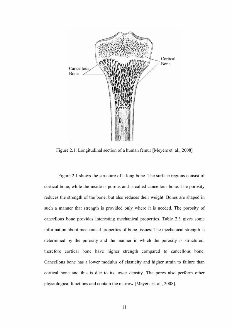
11
Figure 2.1: Longitudinal section of a human femur [Meyers et. al., 2008]
Figure 2.1 shows the structure of a long bone. The surface regions consist of
cortical bone, while the inside is porous and is called cancellous bone. The porosity
reduces the strength of the bone, but also reduces their weight. Bones are shaped in
such a manner that strength is provided only where it is needed. The porosity of
cancellous bone provides interesting mechanical properties. Table 2.3 gives some
information about mechanical properties of bone tissues. The mechanical strength is
determined by the porosity and the manner in which the porosity is structured,
therefore cortical bone have higher strength compared to cancellous bone.
Cancellous bone has a lower modulus of elasticity and higher strain to failure than
cortical bone and this is due to its lower density. The pores also perform other
physiological functions and contain the marrow [Meyers et. al., 2008].
12
Table 2.3: Mechanical properties of bone tissues [Hench and Wilson, 1993] Property Cortical Bone Cancellous Bone
Compressive Strength (MPa)
100-230 2-12
Flexural Strength (MPa)
50-150 10-20
Strain to failure
1-3 5-7
Young’s Modulus (GPa)
7-30 0.5-0.05
Fracture Toughness, KIC (MPa.m1/2)
2-12 N.A.
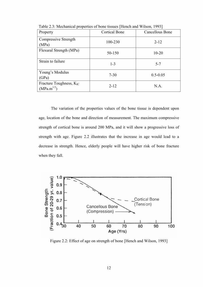
The variation of the properties values of the bone tissue is dependent upon
age, location of the bone and direction of measurement. The maximum compressive
strength of cortical bone is around 200 MPa, and it will show a progressive loss of
strength with age. Figure 2.2 illustrates that the increase in age would lead to a
decrease in strength. Hence, elderly people will have higher risk of bone fracture
when they fall.
Figure 2.2: Effect of age on strength of bone [Hench and Wilson, 1993]
13
2.2 Ceramics and Bioceramics Implants
Ceramic is defined as the art and science of making and using solid articles
that have as their essential component being inorganic nonmetallic materials
[Kingery et. al., 1976]. Ceramics are refractory, polycrystalline compounds, usually
inorganic, including silicates, metallic oxides, carbides, nitrides, and various
refractory hydrides and sulfides.
Unlike metals and polymers, ceramics are difficult to shear plastically due to
the ionic nature of the bonding and the minimum number of slip systems. Ceramics
are nonductile materials and are generally hard. Ceramics are very susceptible to
microcracks because, instead of undergoing plastic deformation, they will fracture
elastically on initiation of crack. At the crack tip the stress could be many times
higher than the stress in the material. Other characteristics of ceramic materials are
their high melting temperatures and low conductivity of electricity and heat. These
characteristic are due to the chemical bonding within the ceramics [Park and Lakes,
1992].
During the last couple of decades, ceramics has been use to improve the
quality of human life. Innovative techniques for fabricating ceramics have led to their
use as advance materials. Specially designed and fabricated ceramics for the repair
and reconstruction of diseased, damaged or worn out parts of the body are widely
developed. Ceramics used for the purpose to improve the quality of human life are
called bioceramics. Most clinical applications of bioceramics are related to the repair
of the skeletal tissues, composed of bones, joints and teeth, and to augment both hard
14
and soft tissues. Ceramic are also used to replace parts of the cardiovascular system,
especially heart valves [Hench and Wilson, 1993].
In order to be classified as a bioceramic, the ceramic material must meet or
exceed the following desired properties of implantable bioceramics [Bilotte, 2003],
that is they should be:
1. Nontoxic
2. Noncarsinogenic
3. Nonallergic
4. Noninflammatory
5. Biocompatible
6. Biofunctional for its lifetime in the host
Although bioceramics seem to be a suitable candidate for implant materials,
the natural problem in conventional ceramics are also the challenge that inhibits the
application of bioceramics. Primary drawbacks of bioceramics are their brittleness,
high Young modulus, and inferior workability. Brittleness becomes a significant
problem when bioceramics are used in positions with high stress loading. Stress
shielding is observed when bone tissue is bonded with materials with high Young’s
modulus, where the bone around the materials is resorbed [Ishikawa et. al., 2003].
Because the bioceramics workability is not good, it is difficult for the surgeon to
shape the bioceramics when necessary during the surgery [Shackelford, 1999].
15
2.2.1 Classification of Bioceramics
Bioceramics can be classified independently in terms of their behavior in the
body environment. Such a classification method is especially useful and relevant
since bioceramics are used in human body. This method classifies bioceramics as
relatively bioinert or nonabsorbable, bioactive or surface reactive, and biodegradable
or bioresorbable ceramics [Bilotte, 2003]. It is also critical that any bioceramics
implant avoid a toxic response that kills cells in the surrounding tissues and can
cause systemic damage to the patient [Hench and Wilson, 1993].
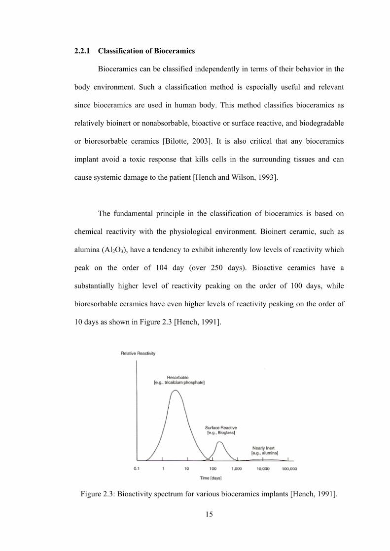
The fundamental principle in the classification of bioceramics is based on
chemical reactivity with the physiological environment. Bioinert ceramic, such as
alumina (Al2O3), have a tendency to exhibit inherently low levels of reactivity which
peak on the order of 104 day (over 250 days). Bioactive ceramics have a
substantially higher level of reactivity peaking on the order of 100 days, while
bioresorbable ceramics have even higher levels of reactivity peaking on the order of
10 days as shown in Figure 2.3 [Hench, 1991].
Figure 2.3: Bioactivity spectrum for various bioceramics implants [Hench, 1991].
16
2.2.1.1 Bioinert ceramics
Bioinert ceramics are defined as ceramics that are stable in human body and
do not show harmful response or bioactivity. Bioinert ceramics maintain their
physical and mechanical properties while in the host [Bilotte, 2003]. In general, the
living body recognizes artificial materials as foreign materials when the latter are
implanted in the living body and consequently the artificial materials are covered
with fibrous layer. The thickness of fibrous layer depends on the tissue compatibility
of the implanted materials. Biologically inactive, nearly inert ceramics (like alumina
and zirconia) will elicit thin fibrous capsule at their interface, while metals and most
polymers may cause a total encapsulation of the implant within the fibrous layer. For
an implant made of bioinert materials, a tight mechanical fit with the host tissue is
very important in order to prevent interfacial movement and subsequent clinical
failure [Hench and Wilson, 1993].
Example of relatively bioinert ceramics are dense and porous alumina,
zirconia ceramics, and a single-phase calcium aluminates. Bioinert ceramics are
usually applied as structural support implants, such as bone plates, bone screws, and
femoral heads. Examples of a non-structural support uses are ventilation tubes,
sterilization devices, and drug delivery devices [Bilotte, 2003].
2.2.1.2 Bioresorbable Ceramics
Bioresorbable or biodegradable ceramics degrades upon implantation in the
host. The resorbed material is replaced by endogeneous tissues. Instead of replacing
the tissues, the material encourages the regeneration of tissues to take their place, and
the degradation rate varies from material to material. Examples of bioresorbable
17
ceramics are aluminum calcium phosphate, coralline, plaster of paris, and beta-
tricalcium phosphate (β-TCP) [Bilotte, 2003].
There are a few issues or criteria that need to be taken into consideration in
order to successfully implant the bioresorbable ceramics. The constituent of the
materials need to be of a composition that can be broken down chemically by body
fluids. The degradation products must also be non-toxic chemical compounds that
can be easily disposed of without damaging the surrounding cells or harming the
host’s health. Moreover, the resorption rate of the materials must also match the
restoration rate of the tissues even as the material provides a sufficient mechanical
strength to support the host tissue while the regeneration of tissues takes place
[Hench and Wilson, 1993].
2.2.1.3 Bioactive ceramics
Bioactive ceramics are defined as the material that bond directly with bone
without having fibrous connective tissues between them. Upon implantation in the
host, bioactive ceramics form a strong bond with adjacent tissue. This characteristic
of direct bonding is extremely useful, especially when the biomaterial is used in an
area where the material will be bonded with bone.
In order for an implant to perform optimally, its properties, such as a
controlled rate of chemical reactivity, morphology and phase composition need to be
carefully engineered according to its function and rate of bonding to the host tissue.
A small change in composition can change the properties of the bioceramics from
nearly inert to bioresorbable to bioactive. In bioactive ceramics, unlike bioresorbable
18
ceramics, chemical reactions only occur at the surface while the rest of the implant
remains largely stable [Hench and Wilson, 1993]
Examples of bioactive ceramics are dense porous glasses, Bioglass and
Ceravital, and hydroxyapatites. One of their many applications is the coating of
metal prostheses. This coating provides a stronger bonding to the adjacent tissues,
which is very important for prostheses [Bilotte, 2003]. It is still not fully explained
why bioactive ceramics bond directly with bone. One hypothesis is that apatite shows
good osteoconductivity since it is an excellent adsorbent, and thus adsorbs necessary
factors including the adhesion factor that is required for the wandering osteoblast
[Ishikawa et. al., 2003].
2.2.2 Applications of Bioceramics
Bioceramics are produced in a variety of forms and phases and serve many
different functions in repair of the body. These are summarized in Figure 2.4. In
many applications, bioceramics are used in the form of bulk of a specific shape
called implants, prostheses, or prosthetic devices. Bioceramics are also used to fill
space while the natural repair processes restore function. In other applications,
bioceramics are used as coating on a substrate, or a second phase in a composite,
combining the characteristics of both materials into a new material with enhanced
mechanical and biochemical properties [Hench and Wilson, 1993].
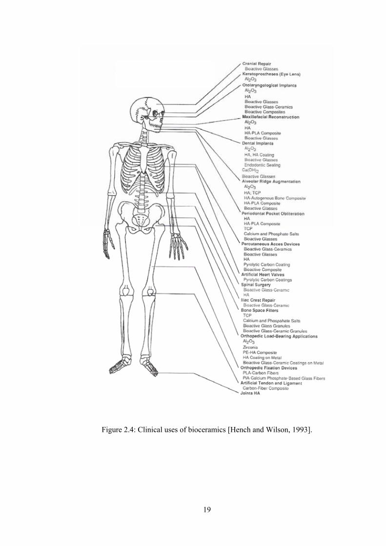
19
Figure 2.4: Clinical uses of bioceramics [Hench and Wilson, 1993].
20
2.3 Calcium Phosphate (CaP) Bioceramics
Calcium phosphate (CaP) has been synthesized and used for manufacturing
various forms of implants, as well as for solid coatings on other implants. There are
several forms of calcium phosphate and they typically form a wide group of
compounds called apatite. It is said that an Australian mineral researcher Werner
named a mineral as apatite in 1786 based on the Greek word “apataw”, which means
puzzled, since it was confused with several other similar looking minerals.
Calcium phosphate bioceramics include hydroxyapatite from synthetic,
natural (from coral) and biological (from bovine bone) origin, calcium-deficient
apatite (CDA), tricalcium phosphate (β-TCP), biphasic calcium phosphate (BCP) and
calcium phosphate cements consisting of mixtures of different CaP phases, e.g., β-
TCP, tetracalcium phosphate (TTCP), monocalcium phosphate monohydrate
(MCPM), dicalcium phosphate dehydrate (DCPD or brushite), dicalcium phosphate
anhydrous (DCPA or monetite) and octacalcium phosphate (OCP) [LeGeros and
LeGeros, 1996].
The types of calcium phosphate phase formed depend on the Ca/P ratio,
presence of water, impurities and temperature [Hench, 1998]. For example,
hydroxyapatite is more likely to form in a wet environment and at lower temperature
(<900oC), while in a dry atmosphere and at higher temperature, β-TCP will be
formed [Park and Lakes, 1992]. Table 2.4 gives the summary of various calcium
phosphate ceramics and their Ca/P ratio.
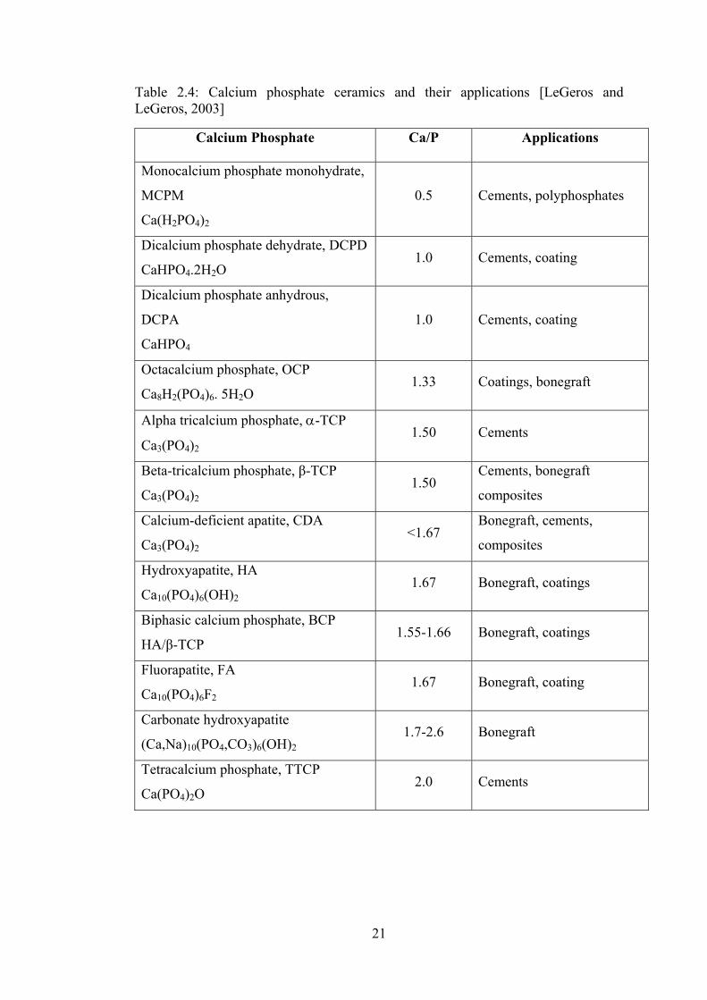
21
Table 2.4: Calcium phosphate ceramics and their applications [LeGeros and LeGeros, 2003]
Calcium Phosphate Ca/P Applications
Monocalcium phosphate monohydrate,
MCPM
Ca(H2PO4)2
0.5 Cements, polyphosphates
Dicalcium phosphate dehydrate, DCPD
CaHPO4.2H2O 1.0 Cements, coating
Dicalcium phosphate anhydrous,
DCPA
CaHPO4
1.0 Cements, coating
Octacalcium phosphate, OCP
Ca8H2(PO4)6. 5H2O 1.33 Coatings, bonegraft
Alpha tricalcium phosphate, α-TCP
Ca3(PO4)2 1.50 Cements
Beta-tricalcium phosphate, β-TCP
Ca3(PO4)2 1.50
Cements, bonegraft
composites
Calcium-deficient apatite, CDA
Ca3(PO4)2 <1.67
Bonegraft, cements,
composites
Hydroxyapatite, HA
Ca10(PO4)6(OH)2 1.67 Bonegraft, coatings
Biphasic calcium phosphate, BCP
HA/β-TCP 1.55-1.66 Bonegraft, coatings
Fluorapatite, FA
Ca10(PO4)6F2 1.67 Bonegraft, coating
Carbonate hydroxyapatite
(Ca,Na)10(PO4,CO3)6(OH)2 1.7-2.6 Bonegraft
Tetracalcium phosphate, TTCP
Ca(PO4)2O 2.0 Cements
22
2.3.1 Hydroxyapatite
Hydroxyapatite (HAp) is the dominant inorganic component if the hard tissue
of the human body, such as dental and bone, and it represent the bioactive ceramics.
The term hydroxyapatite is used generally to represent Ca10(PO4)6(OH)2 in the field
of biomaterials, although the term apatite originally indicates a much wider
composition [Shackelford, 1999].
Although the term HAp with the stoichiometric chemical formula,
Ca10(PO4)6(OH)2, are used generally for simplicity, apatite found in enamel, dentin,
and bone show more complex composition [Ishikawa et. al., 2003]. In spite of a large
variety of compositions, calcium hydroxyapatite belongs to the hexagonal system,
with a P63/m space group. The unit cell contains a complete representation of the
apatite crystal, consisting of Ca2+, PO43-, and OH- groups closely packed together in
arrangement shown in Figure 2.5. The substitute ion of OH- with fluoride gives HAp
greater chemical stability due to the closer coordination of fluoride (symmetric
shape) as compared to hydroxyl (asymmetric) by the nearest calcium. This is why
fluoridation of drinking water helps in resisting caries of the teeth [Park and Lakes,
1992].
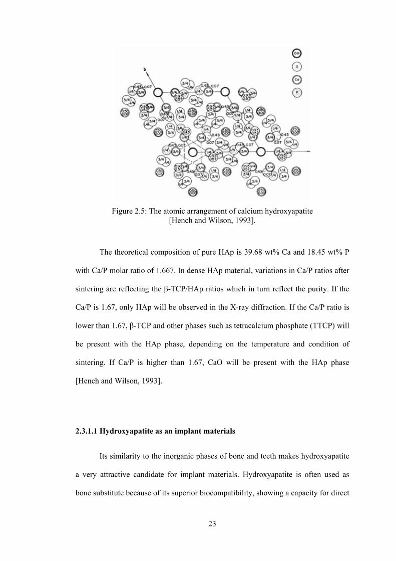
23
Figure 2.5: The atomic arrangement of calcium hydroxyapatite [Hench and Wilson, 1993].
The theoretical composition of pure HAp is 39.68 wt% Ca and 18.45 wt% P
with Ca/P molar ratio of 1.667. In dense HAp material, variations in Ca/P ratios after
sintering are reflecting the β-TCP/HAp ratios which in turn reflect the purity. If the
Ca/P is 1.67, only HAp will be observed in the X-ray diffraction. If the Ca/P ratio is
lower than 1.67, β-TCP and other phases such as tetracalcium phosphate (TTCP) will
be present with the HAp phase, depending on the temperature and condition of
sintering. If Ca/P is higher than 1.67, CaO will be present with the HAp phase
[Hench and Wilson, 1993].
2.3.1.1 Hydroxyapatite as an implant materials
Its similarity to the inorganic phases of bone and teeth makes hydroxyapatite
a very attractive candidate for implant materials. Hydroxyapatite is often used as
bone substitute because of its superior biocompatibility, showing a capacity for direct
24
chemical bonding with bone [Muster, 1992]. Hydroxyapatite is not resorbed in the
human body upon implantation and will stay in the human body while positively
influencing bone formation and thus speeding up recovery [Ishikawa et. al., 2003].
Hydroxyapatite is often used as coatings on metallic implants, usually
titanium, titanium alloys, and stainless steels, due to its surface reactivity. The body
responds to metallic implants by surrounding it with fibrous tissues in order to isolate
it. The coating of HAp provide a bioactive surface which allows the implant to bond
with the bone tissues while retaining the mechanical properties of the metal within
[Oonishi, 1991, Lim et. al., 1999]. Hydroxyapatite is also commonly applied as filler
materials and scaffolds for bone reconstruction. The need for these fillers occur when
there is a bone loss due to diseases or accident or when bone augmentations or
replacement of fragments on non-loaded bones are required, such as ridge
augmentations, ear implants and repair of periodontal defects [Muster, 1992].
Hydroxyapatite acts as a frame or scaffold that facilitates the growth of tissues across
the void. It is readily incorporated into the bone structure even as it encourages bone
growth and speeds up the healing process [Akao et. al., 1981].
2.3.1.2 Mechanical properties and manufacturing techniques of hydroxyapatite
In order to fabricate HAp bioceramic implants with desired shapes and
properties, researchers investigates the use of conventional and advanced ceramic
manufacturing techniques. The techniques used to fabricate HAp will depend on the
application of the implants and the desired properties. For applications as hard tissue
replacement, the most important and immediate property is the strength since the