panduan baca foto torax
DESCRIPTION
panduan baca foto toraxTRANSCRIPT
BASICS OF CHEST X-RAY INTERPRETATION:
AN INTRODUCTION TO THE PRINCIPLES OF CHEST X-RAY
INTERPRETATION
By Barbara Ritter, EdD, FNP, CNS with assistance from Leslie Muma, RN, MSN, NP
SECTION 1: VIEWS OF THE CHEST
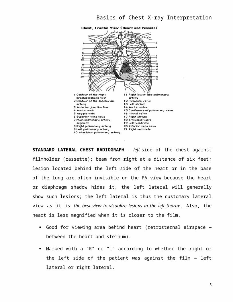
Standard Frontal Chest Radiograph
Standard Lateral Chest Radiograph
Portable Chest X-Ray
Other Views
PA oblique views
decubitus views
cross-table lateral
lordotic views
expiratory views
Bucky films
tomography
Densities
SECTION 2A: INTERPRETATION
Skeletal Structures
Soft Tissues
mediastinum
Diaphragm
Heart and Great Vessels
Lungs
SECTION 2B: ASSESSMENT
I: Skeletal Structures
II: Soft Tissues
Basics of Chest X-ray InterpretationIII: Diaphragm
IV: Heart and Great Vessels
SECTION 3: RADIOLOGIC SIGNS OF CARDIAC DISEASE
Projections
Technical Factors
Extracardiac Structures
CHF
Chamber Enlargement
Enlargement of LA
Enlargement of LV
Enlargement of Right Side
Myocardial Dysfunction
Myocardial Ischemia
Valvular Dysfunction
Poor Exercise Capacity
Arrhythmias
SECTION 4: RADIOLOGIC SIGNS OF PULMONARY DISEASE
Structures
Pulmonary Nodules
Alveolar Lung Disease
Interstitial Lung Disease
Kerley’s Lines
viral pneumonia
drug-induced pneumonia
pulmonary edema
DDX of Interstitial Lung Disease
GLOSSARY
CHEST RADIOLOGY ARTICLES
2
Basics of Chest X-ray InterpretationSection One
VIEWS OF THE CHEST:
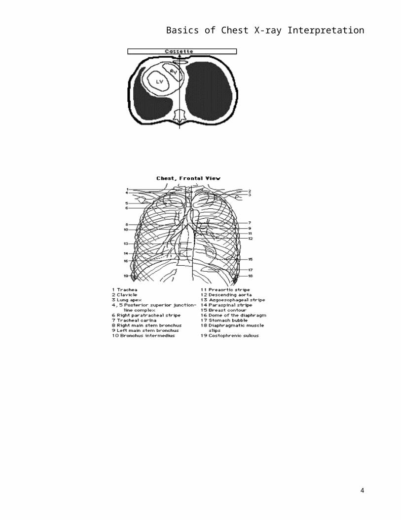
STANDARD FRONTAL CHEST RADIOGRAPH (Roentgenogram) — upright; PA or
posterior -anterior (film in front of patient, beam behind at a distance of six feet; patient usually
upright; distance of beam determines magnification and clarity or sharpness
Place the films on the view box as though you were facing the patient with his left on
your right side.
An AP film, taken from the same distance (6') enlarges the shadow of the heart which is
far anterior in the chest and makes the posterior ribs appear more horizontal.
In a supine film, the diaphragm will be higher and the lung volumes less than in a
standing patient.
3
Basics of Chest X-ray Interpretation
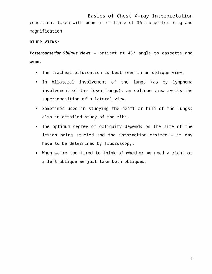
STANDARD LATERAL CHEST RADIOGRAPH — left side of the chest against filmholder
(cassette); beam from right at a distance of six feet; lesion located behind the left side of the heart
4
Basics of Chest X-ray Interpretationor in the base of the lung are often invisible on the PA view because the heart or diaphragm
shadow hides it; the left lateral will generally show such lesions; the left lateral is thus the
customary lateral view as it is the best view to visualize lesions in the left thorax. Also, the heart
is less magnified when it is closer to the film.
Good for viewing area behind heart (retrosternal airspace — between the heart and
sternum).
Marked with a "R" or "L" according to whether the right or the left side of the patient was
against the film — left lateral or right lateral.
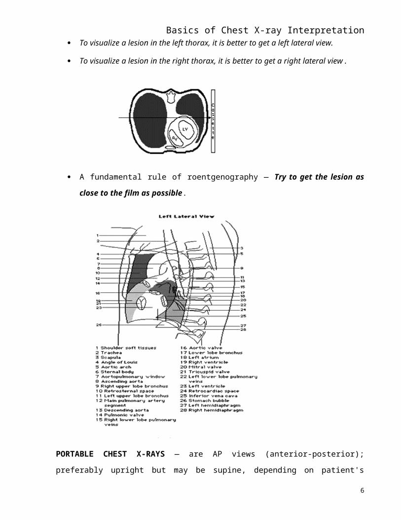
To visualize a lesion in the left thorax, it is better to get a left lateral view.
To visualize a lesion in the right thorax, it is better to get a right lateral view.
A fundamental rule of roentgenography — Try to get the lesion as close to the film as
possible.
5
Basics of Chest X-ray Interpretation
PORTABLE CHEST X-RAYS — are AP views (anterior-posterior); preferably upright but
may be supine, depending on patient's condition; taken with beam at distance of 36 inches-
blurring and magnification
OTHER VIEWS:
Posteroanterior Oblique Views — patient at 45° angle to cassette and beam.
The tracheal bifurcation is best seen in an oblique view.
In bilateral involvement of the lungs (as by lymphoma involvement of the lower lungs),
an oblique view avoids the superimposition of a lateral view.
Sometimes used in studying the heart or hila of the lungs; also in detailed study of the
ribs.
The optimum degree of obliquity depends on the site of the lesion being studied and the
information desired — it may have to be determined by fluoroscopy.
6
Basics of Chest X-ray Interpretation When we're too tired to think of whether we need a right or a left oblique we just take
both obliques.
7
Basics of Chest X-ray Interpretation
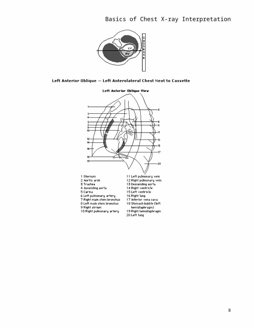
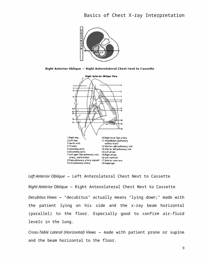
Left Anterior Oblique — Left Anterolateral Chest Next to Cassette
Right Anterior Oblique — Right Anterolateral Chest Next to Cassette
Decubitus Views — "decubitus" actually means "lying down;" made with the patient lying on his
side and the x-ray beam horizontal (parallel) to the floor. Especially good to confirm air-fluid
levels in the lung.
Cross-Table Lateral (Horizontal) Views — made with patient prone or supine and the beam
horizontal to the floor.
8
Basics of Chest X-ray InterpretationLordotic Views — formerly made in the upright AP position with the patient leaning backward at
an angle of ~ 30° from the vertical which was very awkward; now made with the patient facing
the film as for an upright PA view but the tube is elevated and angled downward 45°.
Projects the lung apices of the lungs below the clavicles and causes the ribs to project
more horizontally.
Especially good for viewing the apices of the lungs, lesions that are partially obscured by
ribs, or the right middle lobe or lingula of the left lung.
Expiratory Views — on expiration the lungs "cloud up" and the heart appears larger.
If the air on one side cannot be readily expelled, the lung on the obstructed side remains
expanded and radiolucent on expiration.
Useful in detecting unilateral obstructive emphysema (as from a unilateral obstruction of
a bronchus).
A pneumothorax always appears larger on expiration than on inspiration.
Since the thorax is smaller on expiration, the unchanged volume of pleural air spreads out
in the smaller thoracic space.
Occasionally a small pneumothorax is only visible on expiration.
Bucky Films — made with a moving grid between the patient and the film which absorbs excess,
scattered radiation.
Scattered radiation produces a hazy, unsharp image, or fog, and detracts from film clarity.
Used to delineate a thick pulmonary or pleural lesion, bony structures, or to more clearly
see structures in an obese patient.
Bucky technique also used whenever the abdomen, spine, mediastinum, pelvis, or heavy
long bones are studied.
Tomography (Laminagraphy)
An apparatus moves the tube and film synchronously in opposite directions; the
adjustable fulcrum is set to the plane of the lesion to be studied; blurs structures in the
planes above and below the level being studied.
Especially helpful in evaluating pulmonary nodules, demonstrating cavities, and depicting
bronchial obstruction.
9
Basics of Chest X-ray Interpretation If you can't think of the exact name for a view, be descriptive or draw a picture (i.e., "Get
me a cross-table view with the patient lying on his right side facing the tube.") or consult
with the radiologist.
There are all sorts of ingenious projections and fascinating special procedures in the
armamentarium of the radiologist.
DENSITIES Air < fat < liver < blood < muscle < bone < barium < lead.
Air — least dense; most transparent or radiolucent; unobstructed beam or air-filled
densities appear black
Lungs, gastric bubble, trachea, ? bifurcation of bronchi
Fat — breasts
Fluid — most of what you see; vessels, heart, diaphragm, soft tissues, mediastinal
structures
Mineral — most dense (or radiopaque) of body structures; mostly Ca++; bones (marrow
is aerated), aortic calcifications such as the aortic knob, ? calcification of the coronary
arteries, old granulomas; bullets, safety pins, etc.
Structures which are perpendicular to the plane of the film appear as they were much
more dense as the shadows represent the sum of the densities interposed between the
beam source and the film. Learn to think in terms of those parts that are relatively parallel
to the film and those that are roughly perpendicular to it. Think about it three-
dimensionally.
Thickness as well as composition determine radiodensity. The shadow cast by a thick
mass of soft tissues will approach that of bone.
Section Two
A. PROCEDURE FOR INTERPRETATION OF CHEST FILMS
Develop a systematic approach and use it consistently.
(Usually external-internal.)
I. LABEL — Read the label on every film to verify the patient's name, age, and sex.
10
Basics of Chest X-ray InterpretationII. ORIENTATION — Identify the patient's right side, his position, and determine if he is
rotated.
Symmetrical spacing of the clavicles and other structures on either side of the sternum;
clavicles esp. will show whether or not patient is straight or rotated. Symmetry of the clavicles
and ribs gives you assurance that no rotation is present. Even slight rotation is undesirable in a
chest film as the heart and mediastinum are then radiography obliquely and their shadows appear
enlarged and distorted.
III. QUALITY — In a film of good technical quality in a patient without gross cardiomegaly,
you should be able to see the outlines of the vertebral bodies within the heart shadow; notice
linearity of spine — is it straight?
IV. INTERPRETATION: the following should be identified:
A. SKELETAL STRUCTURES — what you see of the bones is incidental as the technique
used for chest films has been designed for study of the lungs. Always compare for symmetry.
1. Scapulae — PA and lateral; are there two of each?
With hands on hips, palms out, and elbows forward the scapulae are rotated to the sides to
prevent their superimposition upon the upper lung fields. Therefore only their medial margins are
seen.
2. Humeri and Shoulder Joints — PA and lateral.
Little of the shoulder girdle and humerus will be seen in films of broad-chested
individuals.
Coracoid is seen through the spine of the scapula because they superimpose.
Head of humerus and the acromium are also seen additively.
Are fractures or abnormal calcifications (dense white shadows) seen?
3. Clavicles — PA; symmetrical spacing on either side of sternum only if there is no rotation of
the chest. Turned even a few degrees, the clavicles will exhibit a remarkable degree of
asymmetry.
4. Ribs — count on every film to level of diaphragm.
Identify the first rib carefully by finding its anterior junction with the manubrium and
following this rib backward to the spine. Then count down the posterior ribs.
11
Basics of Chest X-ray Interpretation Begin at the origin of the first rib at its junction with the first thoracic vertebra and trace
each rib as far anteriorly as you can to the beginning of the radiolucent (and hence
invisible) costal cartilage.
Interspaces are useful in identifying the location of a precise shadow and are named for
the posterior rib above the interspace unless the anterior rib is specified as the marker.
Number of ribs helps you determine how much lungs are inflated → 9 or more ribs =
good inflation.
Transverse cardiac shadow smallest — used for measurement.
Lungs better filled with air; therefore relatively minor disease is seen better.
10 or more ribs = ? hyperinflated
Expiratory film — see < 9 ribs.
Diaphragm higher; lung bases less well seen; transverse diameter of heart is larger.
Minimal pneumothorax can be seen better. Also, obstructive emphysema.
Compare both sides for symmetry,
Note width of the intercostal spaces. Are they equal?
Are they continuous or is there a fracture?
Beam only "sees" what is parallel to it; anterior ribs are more perpendicular and thus not
seen very well.
5. Spine — notice linearity — is it straight?
Spine and sternum are superimposed upon each other and upon the dense shadows of the
mediastinal structures in the PA view.
Scoliosis may mask margin of RA; don't mistake for RA with mediastinal shift.
B. SOFT TISSUES — Symmetry of Density.
1. Chest wall (outside of lung fields).
2. Neck.
3. Mediastinum.
MEDIASTINAL STRUCTURES
12
Basics of Chest X-ray Interpretation Identify trachea — is it midline, not shifted?
Identify bifurcation and position.
Should not be able to follow airways any further out as they are very thin walled; if visible
(air bronchogram sign) - ?? pulmonary edema.
4. Breasts — symmetrical in size, shape, position; nipples may possibly be visible.
Be sure to check whether there are two breasts.
The lung field under a missing breast will appear a little darker than the other lung field.
C. DIAPHRAGM
1. Difference in the Level of the Hemidiaphragms
Right hemidiaphragm is normally a bit higher.
Impaired mobility of diaphragm — may be from paralysis of either phrenic nerve,
disease in abdomen such as a subdiaphragmatic abscess, pleurisy, pulmonary
infarction, etc.
2. Normal Position
Distance from gastric bubble (if it is visible) to diaphragm should be very small.
3. Shape of the Diaphragm.
4. Identification of Left and Right Diaphragms — lateral film.
5. Costophrenic Angles
Should be sharp and clear.
No fluid density should be visible.
6. Cardiophrenic angle should be fairly clear.
7. Inferior vena cava adds its own little shadow.
D. Heart and Great Vessels
13
Basics of Chest X-ray Interpretation
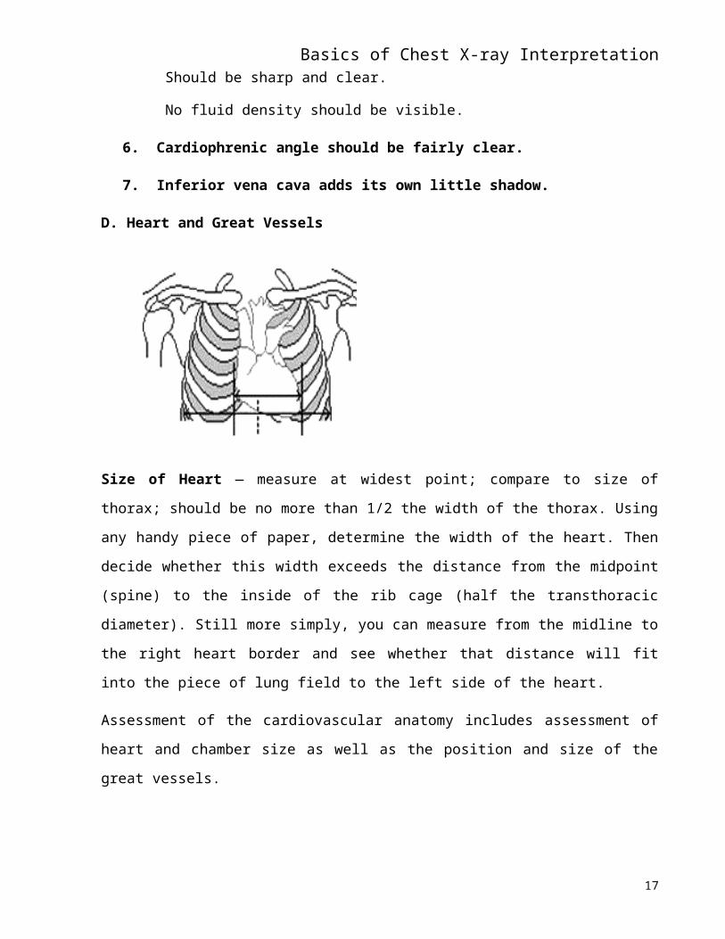
Size of Heart — measure at widest point; compare to size of thorax; should be no more than 1/2
the width of the thorax. Using any handy piece of paper, determine the width of the heart. Then
decide whether this width exceeds the distance from the midpoint (spine) to the inside of the rib
cage (half the transthoracic diameter). Still more simply, you can measure from the midline to the
right heart border and see whether that distance will fit into the piece of lung field to the left side
of the heart.
Assessment of the cardiovascular anatomy includes assessment of heart and chamber size as well
as the position and size of the great vessels.
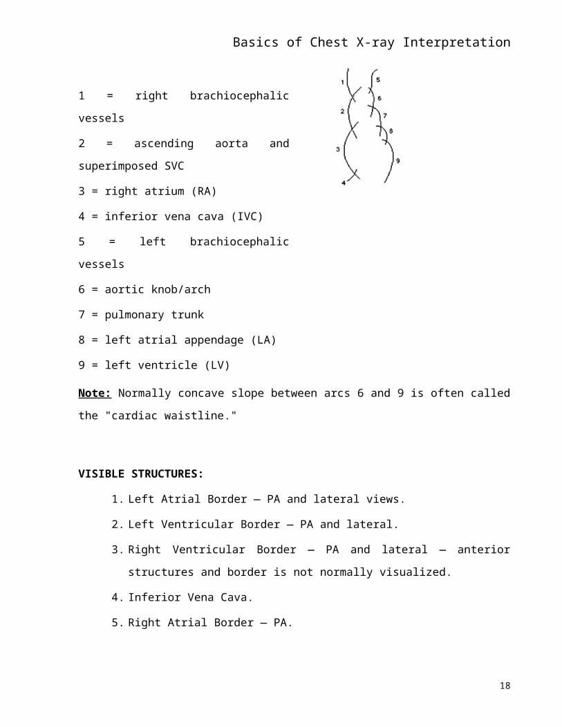
1 = right brachiocephalic vessels
2 = ascending aorta and superimposed SVC
3 = right atrium (RA)
4 = inferior vena cava (IVC)
5 = left brachiocephalic vessels
6 = aortic knob/arch
7 = pulmonary trunk
8 = left atrial appendage (LA)
9 = left ventricle (LV)
Note: Normally concave slope between arcs 6 and 9 is often called the "cardiac waistline."
VISIBLE STRUCTURES:14
Basics of Chest X-ray Interpretation1. Left Atrial Border — PA and lateral views.
2. Left Ventricular Border — PA and lateral.
3. Right Ventricular Border — PA and lateral — anterior structures and border is not
normally visualized.
4. Inferior Vena Cava.
5. Right Atrial Border — PA.
1. Scoliosis, if present, may mask border of the right atrium.
6. Superior Vena Cava — PA.
7. Ascending Aorta — PA and lateral.
8. Aortic Knob — position, calcification.
9. Main Pulmonary Artery — lateral.
10. Relative position of left and right main branches of pulmonary arteries — in relation
to left and right main bronchi.
11. Esophagus — PA and lateral.
12. Note cardiac size — normal is 1/2 or less of the thoracic width on a PA film.
E. Lungs
VISIBLE STRUCTURES:
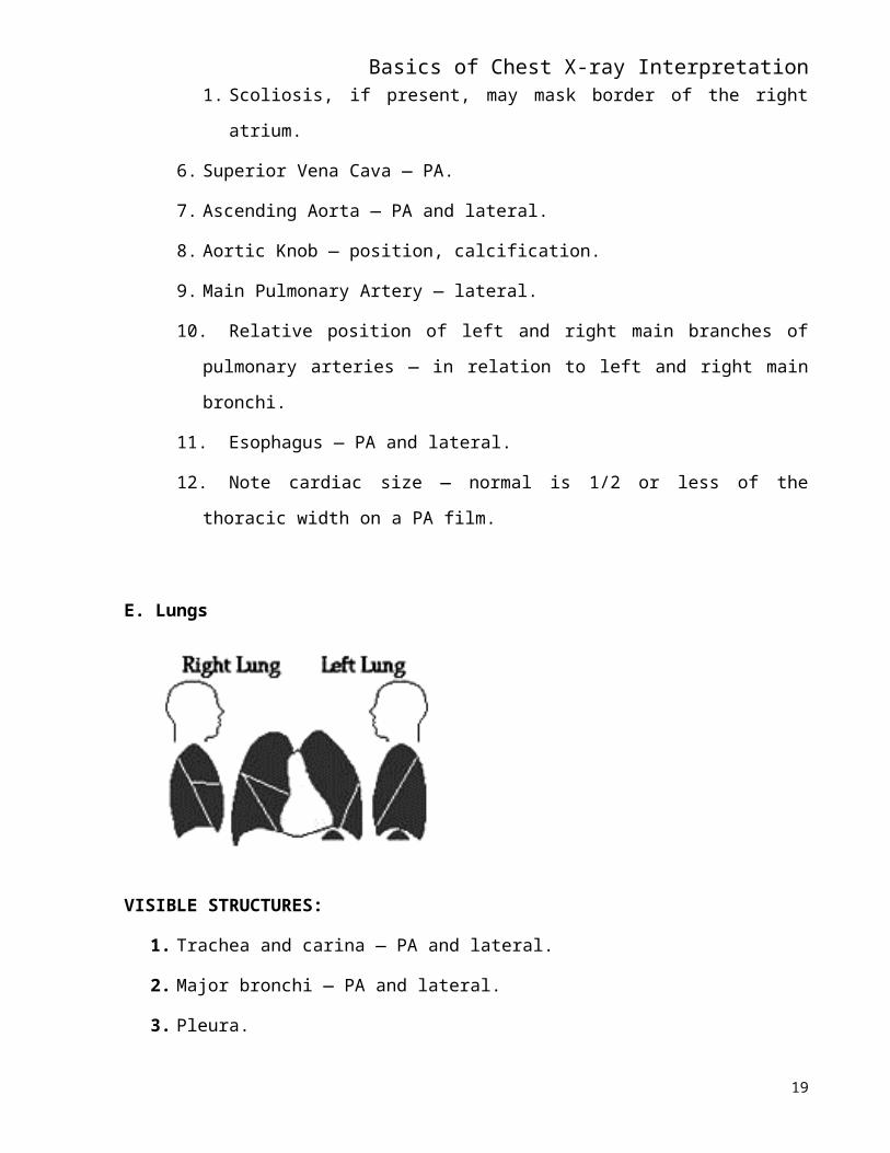
1. Trachea and carina — PA and lateral.
2. Major bronchi — PA and lateral.
3. Pleura.
15
Basics of Chest X-ray Interpretation4. Left — upper and lower lobe representation — PA and lateral.
Minor fissure on left — between ribs 6 and 8. Only one fissure.
Lingula (tongue-shaped) — area adjacent to LV; not a separate lobe.
5. Right — upper, middle, and lower lobe representation — PA and lateral.
Oblique or major fissure — T3 - T10.
6. Differences in density, upper and lower lung fields.
In a PA film the peripheral vasculature is normally seen out to the lateral one inch of
the films and is more clearly delineated in the lower lobes than the apices.
Upright — most of perfusion goes to lower lungs so you should see it all the way out.
_ PAP-reversal of blood flow with enhancement of apical vascularity.
Older smoker and vasculature not visible all the way out = ? emphysema.
Younger person and not visible all the way out = ? pneumothorax.
Pneumothorax = about the only thing that can be diagnosed with absolute
certainty with CXR.
7. Peripheral vasculature — follow it out as far as you can see it.
Hilum (pl. = hila).
Position — higher or lower.
Symmetry
o Lung fields — symmetry re: amount of density.
8. Silhouette Sign
Two densities that are alike with margins adjacent to each other — borders will be
masked.
If margin is obliterated, whatever is masked and it has to be in the same plane.
Masking of RA — would be from R middle lobe.
Masking of posterior diaphragm — would be from R lower lobe.
Masking of LV — would be from L upper lobe (anterior).
Masking of descending aorta — would be from L lower lobe.
Masking of IVC and SVC — would be from R lower and middle lobes.
If you can see heart — comes from posterior.
16
Basics of Chest X-ray Interpretation9. Air Bronchogram Sign — "butterfly" distribution of the abnormal densities or an
anatomic distribution of abnormal densities restricted to lobar or sublobar portions of the
lung.
Temporally rapid (reckoned in days) changes in the appearance of the lung infiltrate.
Indicative of alveolar disease.
See airways out past bifurcation.
Air-filled airway superimposed on air-filled densities.
Demonstration of the air-filled bronchus as a radiolucent "tube" is dependent on its
close association with alveoli that are fluid-filled rather than air-filled.
Two contrasting densities make it visible.
Airways OK, surround tissues not OK.
10. Kerley's Lines
Kerley's B Lines — short, thin horizontal lines at the periphery of the lung near the
costophrenic angles; formed by thickening of the interlobular septa 2° to fibrosis (e.g.,
pneumoconiosis), fluid accumulation, or distended lymphatics-venules Kerley's A Lines
— long, linear densities, more centrally located in the upper portions of the lungs near the
hila; may be seen in interstitial lung disease and CHF; represent swollen lymphatic
channels.
F. Iatrogenics
1. ECG leads
2. Endotracheal tube — positioning
3. CVP and PA lines
B: ASSESSMENT OF CHEST FILMS
I. Skeletal Structures
A. Scapulae
B. Humeri
C. Clavicles — symmetrical spacing on either side of sternum
D. Ribs
17
Basics of Chest X-ray InterpretationII. Soft Tissues — symmetry of density.
A. Chest wall
B. Neck
C. Mediastinum
Trachea — is it midline, not shifted.
Identify bifurcation and position.
Should not be able to follow airways any further out as they are very thin walled; if
visible (air bronchogram sign) - ?? pulmonary edema.
D. Breasts — symmetrical in size, shape, position; nipples may ? be visible.
III. Diaphragm
A. Difference in the level of the hemidiaphragms.
B. Normal position.
C. Shape of the diaphragm.
D. Identification of left and right diaphragms — lateral.
E. Costophrenic angles.
IV. Heart and Great Vessels — Assessment of the cardiovascular anatomy includes assessment
of heart and chamber size as well as the position and size of the great vessels.
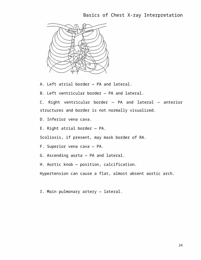
A. Left atrial border — PA and lateral.
B. Left ventricular border — PA and lateral.
18
Basics of Chest X-ray InterpretationC. Right ventricular border — PA and lateral — anterior structures and border is not
normally visualized.
D. Inferior vena cava.
E. Right atrial border — PA.
Scoliosis, if present, may mask border of RA.
F. Superior vena cava — PA.
G. Ascending aorta — PA and lateral.
H. Aortic knob — position, calcification.
Hypertension can cause a flat, almost absent aortic arch.
I. Main pulmonary artery — lateral.
J. Relative position of L and R main branches of pulmonary arteries — in relation to L &
R main bronchi
K. Esophagus — PA and lateral
19
Basics of Chest X-ray Interpretation
L. Cardiac Size — normal is 1/2 or less of the thoracic width on a PA film.
Simulation of cardiac enlargement — PA films made in expiration (high
diaphragm-heart tilted upward bringing apex closer to the lateral chest wall + less
flare of ribs which alters the apparent cardiothoracic ratio); also any abdominal
distention (late pregnancy, ascites, intestinal obstruction) produces similar results;
diaphragm also likely to be higher in supine views; portable chest films and other
AP views place heart farther away from the film.
Rotation of the patient produces appearance of widening of the heart and
mediastinal shadows.
20
Basics of Chest X-ray Interpretation Deformity of the thoracic cage — severe scoliosis; depressed sternum (pectus
excavatum) usually displaces heart to the left + right heart border not visible.
Difference between heart volumes in systole and diastole usually not enough to
affect rough estimate of the cardiothoracic ratio in adults.
Simulation of deceptively small heart — overdistention of the lungs for any reason
(dyspneic patient with low diaphragm or emphysematous patient) compresses the
heart and mediastinal structures from both sides and narrows their PA shadow.
Mediastinal disease, pulmonary disease, or any density (consolidation, effusions,
true mediastinal shift) may render the dimensions of the heart unobtainable.
Section 3
RADIOLOGIC SIGNS OF CARDIAC DISEASE:
PROJECTIONS
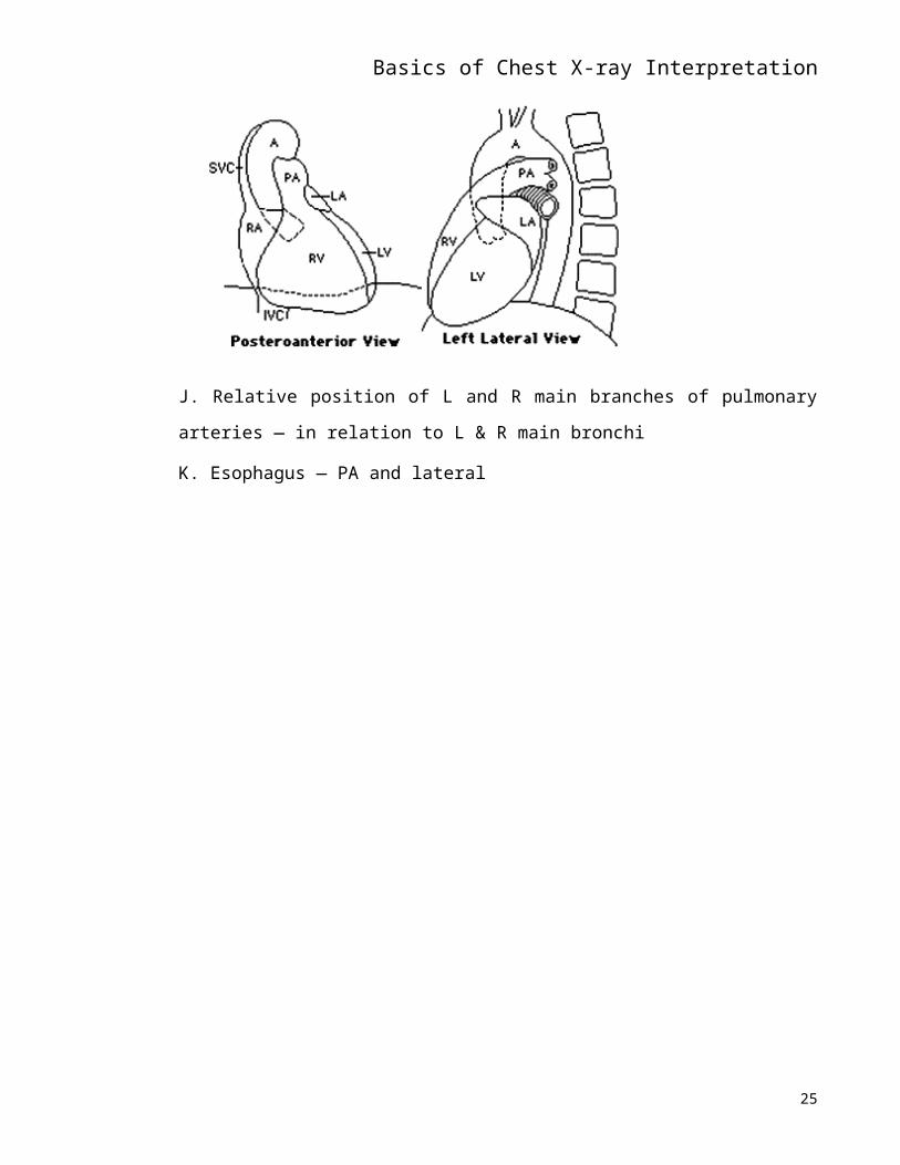
Posteroanterior Projection
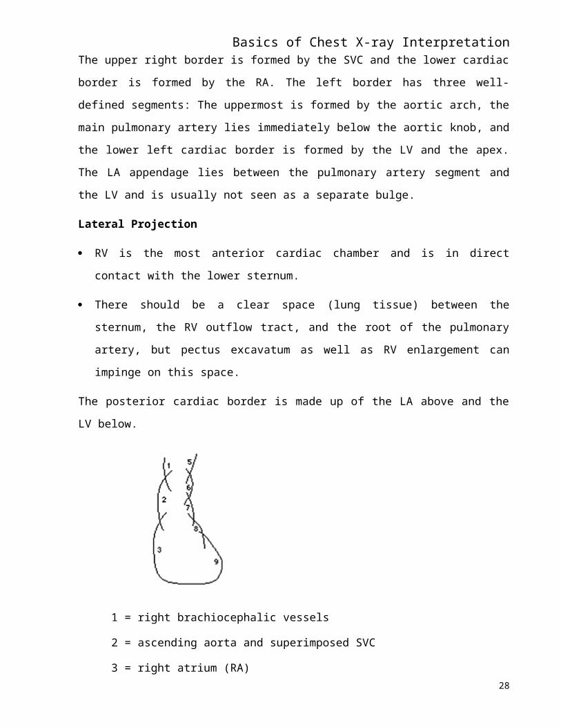
The upper right border is formed by the SVC and the lower cardiac border is formed by the RA.
The left border has three well-defined segments: The uppermost is formed by the aortic arch, the
main pulmonary artery lies immediately below the aortic knob, and the lower left cardiac border
is formed by the LV and the apex. The LA appendage lies between the pulmonary artery segment
and the LV and is usually not seen as a separate bulge.
Lateral Projection
RV is the most anterior cardiac chamber and is in direct contact with the lower sternum.
There should be a clear space (lung tissue) between the sternum, the RV outflow tract, and
the root of the pulmonary artery, but pectus excavatum as well as RV enlargement can
impinge on this space.
The posterior cardiac border is made up of the LA above and the LV below.
21
Basics of Chest X-ray Interpretation
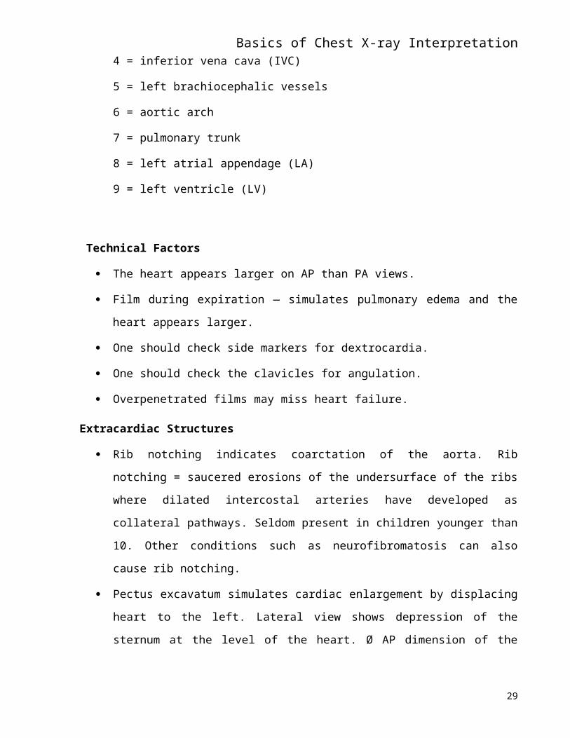
1 = right brachiocephalic vessels
2 = ascending aorta and superimposed SVC
3 = right atrium (RA)
4 = inferior vena cava (IVC)
5 = left brachiocephalic vessels
6 = aortic arch
7 = pulmonary trunk
8 = left atrial appendage (LA)
9 = left ventricle (LV)
Technical Factors
The heart appears larger on AP than PA views.
Film during expiration — simulates pulmonary edema and the heart appears larger.
One should check side markers for dextrocardia.
One should check the clavicles for angulation.
Overpenetrated films may miss heart failure.
Extracardiac Structures
Rib notching indicates coarctation of the aorta. Rib notching = saucered erosions of the
undersurface of the ribs where dilated intercostal arteries have developed as collateral
pathways. Seldom present in children younger than 10. Other conditions such as
neurofibromatosis can also cause rib notching.
22
Basics of Chest X-ray Interpretation Pectus excavatum simulates cardiac enlargement by displacing heart to the left. Lateral
view shows depression of the sternum at the level of the heart. Ø AP dimension of the
chest at heart level and the heart is displaced posteriorly (posterior margin behind the
inferior vena cava).
Straight back is a/w mitral valve prolapse and aortic insufficiency.
Right-sided pleural effusion occurs with CHF.
Physiologic Analysis of the Pulmonary Vasculature — appearance of the hilar and pulmonary
vessels is an excellent indicator of the physiologic state of the heart.
Congestive Heart Failure
_ size, shapelessness of heart, + evidence of pulmonary venous engorgement — the
vessels are seen to extend farther than normal into the lung field.
Bronchi become "framed" in the interstitial fluid accumulating around them and, when
seen end-on, appear as white rings. This is often called "peribronchial cuffing" and can be
observed to decrease as the patient improves.
Pleural effusion in cardiac failure may be bilateral or unilateral and is more frequent on
the right.
Lungs appear hazy and less radiolucent than normal because of retained water; lattice
pattern.
Kerley's B lines appear — short, horizontal white linear densities very close to the
peripheral margin of the lung; have been proven to represent the thickened, edematous
interlobular septa; also seen in lymphangitic spread of malignancies within the lung
parenchyma and interstitial pulmonary disease.
Rapid accumulation of fluid spills over into the alveoli and causes the development of
alveolar (air-space) pulmonary edema.
Pulmonary edema -- the so-called "bat-wing" appearance about both hila; superimposed
shadows of innumerable fluid-filled alveoli may cause disappearance of the vessels of the hilum;
interstitial pulmonary edema-blurring of pulmonary vasculature; perihilar haze; may appear
rapidly after sudden LV failure or it may be superimposed on the more gradual CXR findings of
CHF.
Pulmonary edema can also occur in noncardiac conditions such as fluid overload, renal failure,
heroin overdose, and inhalation injury or burns.
23
Basics of Chest X-ray InterpretationCXR findings can lag behind hemodynamic Ds but the following patterns can predict pulmonary
artery wedge pressure:
Grade 0: normal — PAWP < 12 mm Hg.
Grade 1: pulmonary venous HTN, pulmonary vascular redistribution to the apices (venous
markings Æinto the upper lobes), and loss of the right hilar angle — PAWP 12-19 mm
Hg.
Grade 2: interstitial edema (Kerley's B lines), hilar haze or blurriness, peribronchial
vascular thickening — PAWP 20-25 mm Hg.
Grade 3: generalized or perihilar alveolar edema — PAWP > 25 mm Hg.
Distinguishing Between Cardiac Hypertrophy, Dilatation, and Pericardial
Effusion:
Plain films may show ventricular enlargement but do not differentiate between
hypertrophy and dilatation.
If heart is decompensating, it will tend to shapelessness and extend to both the R and L in
the PAview, suggesting either failure or pericardial effusion. A review of the patient's old
films is probably the best way to assess development of cardiac enlargement, in and out of
failure.
Sudden shapeless _in size should suggest pericardial effusion.
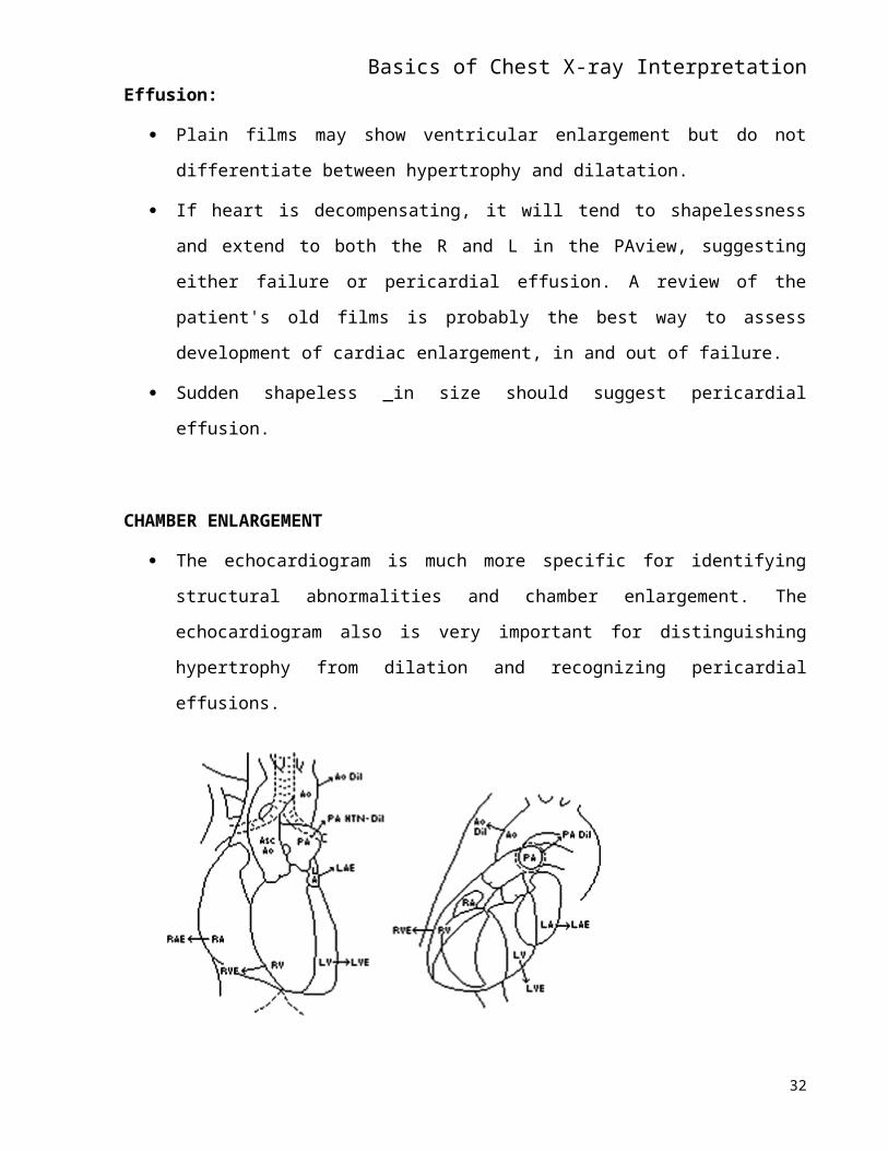
CHAMBER ENLARGEMENT
The echocardiogram is much more specific for identifying structural abnormalities and
chamber enlargement. The echocardiogram also is very important for distinguishing
hypertrophy from dilation and recognizing pericardial effusions.
24
Basics of Chest X-ray Interpretation
AP View Lateral View
Ao Dil = Aortic Dilatation
Asc Ao = Ascending Aorta
LAE = Left Atrial Enlargement
LVE = Left Ventricular Enlargement
PA Dil = Pulmonary Artery Dilatation
PA HTN-Dil = Pulmonary Artery Bulging due to
Pulmonary Hypertension
RVE = Right Ventricular Enlargement
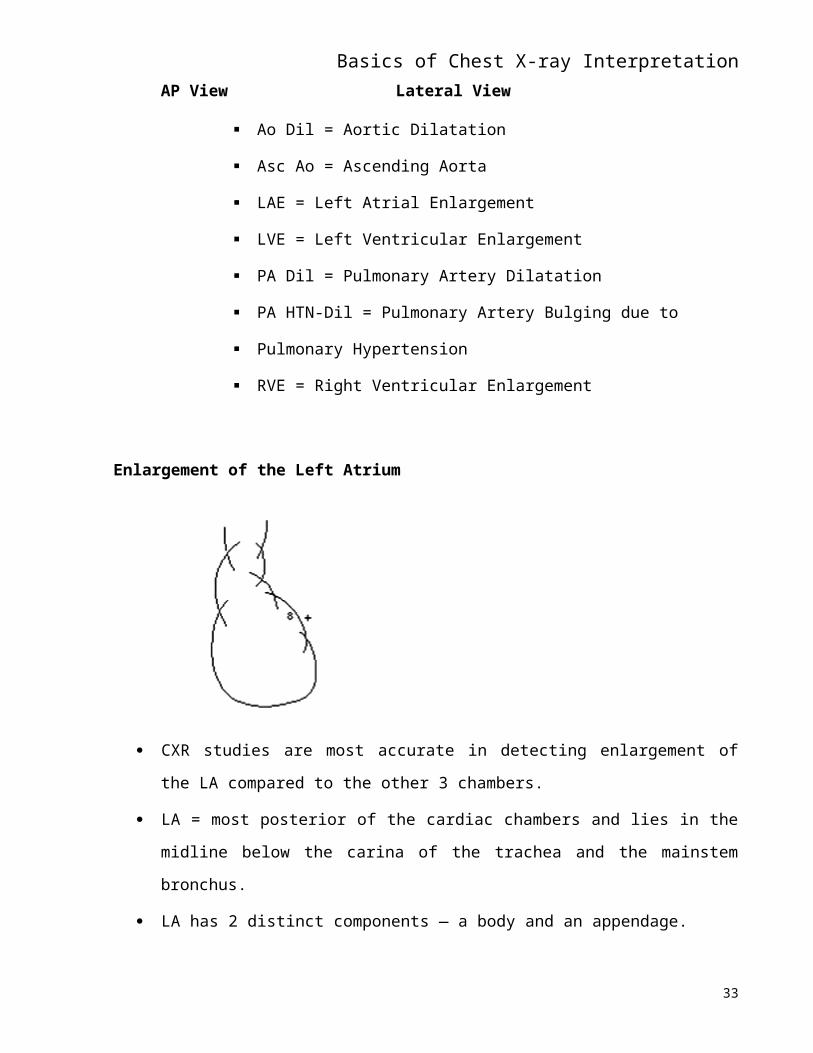
Enlargement of the Left Atrium
25

Basics of Chest X-ray Interpretation
CXR studies are most accurate in detecting enlargement of the LA compared to the other
3 chambers.
LA = most posterior of the cardiac chambers and lies in the midline below the carina of
the trachea and the mainstem bronchus.
LA has 2 distinct components — a body and an appendage.
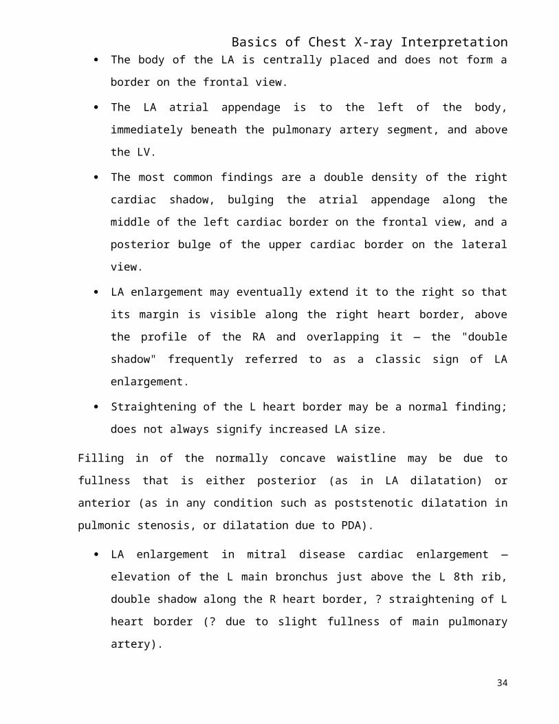
The body of the LA is centrally placed and does not form a border on the frontal view.
The LA atrial appendage is to the left of the body, immediately beneath the pulmonary
artery segment, and above the LV.
The most common findings are a double density of the right cardiac shadow, bulging the
atrial appendage along the middle of the left cardiac border on the frontal view, and a
posterior bulge of the upper cardiac border on the lateral view.
LA enlargement may eventually extend it to the right so that its margin is visible along
the right heart border, above the profile of the RA and overlapping it — the "double
shadow" frequently referred to as a classic sign of LA enlargement.
Straightening of the L heart border may be a normal finding; does not always signify
increased LA size.
Filling in of the normally concave waistline may be due to fullness that is either posterior (as in
LA dilatation) or anterior (as in any condition such as poststenotic dilatation in pulmonic
stenosis, or dilatation due to PDA).
LA enlargement in mitral disease cardiac enlargement — elevation of the L main
bronchus just above the L 8th rib, double shadow along the R heart border, ? straightening
of L heart border (? due to slight fullness of main pulmonary artery).
26
Basics of Chest X-ray Interpretation
Enlargement of the Left Ventricle
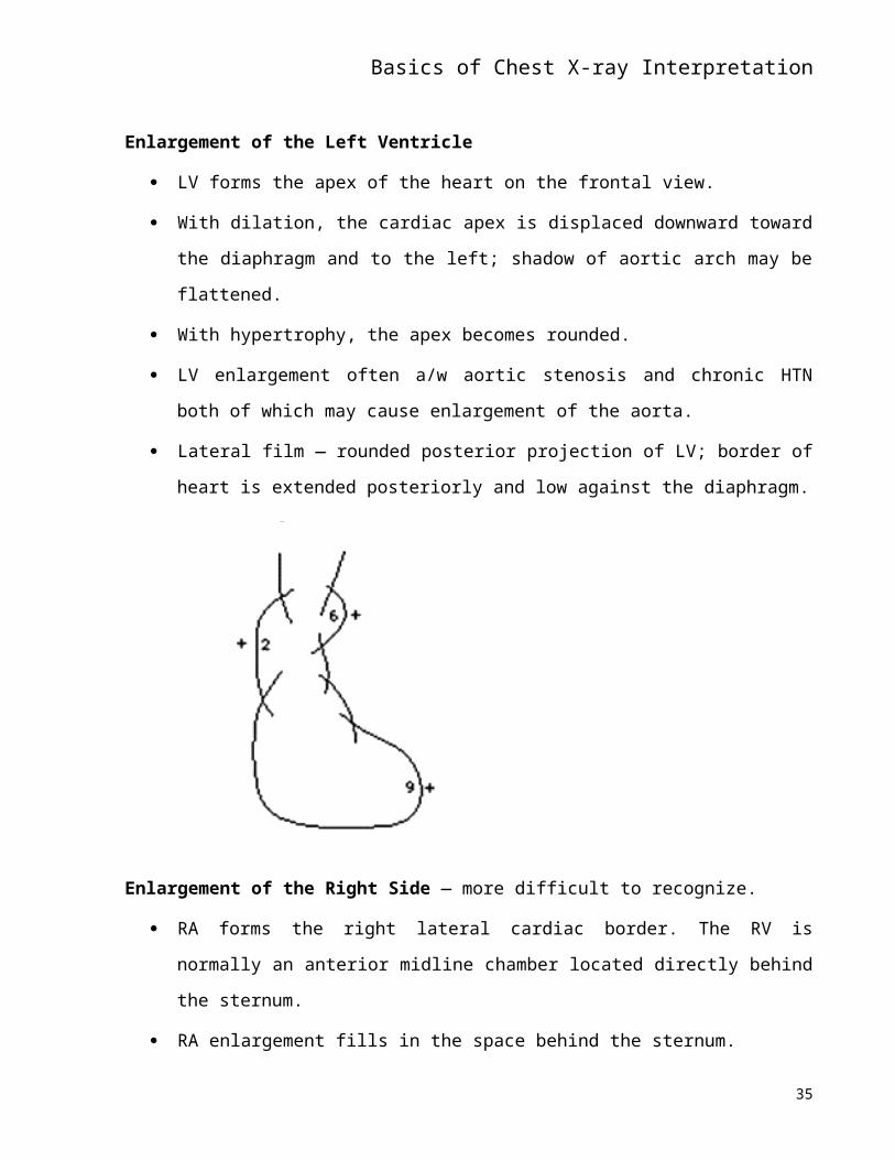
LV forms the apex of the heart on the frontal view.
With dilation, the cardiac apex is displaced downward toward the diaphragm and to the
left; shadow of aortic arch may be flattened.
With hypertrophy, the apex becomes rounded.
LV enlargement often a/w aortic stenosis and chronic HTN both of which may cause
enlargement of the aorta.
Lateral film — rounded posterior projection of LV; border of heart is extended posteriorly
and low against the diaphragm.
Enlargement of the Right Side — more difficult to recognize.
RA forms the right lateral cardiac border. The RV is normally an anterior midline
chamber located directly behind the sternum.
RA enlargement fills in the space behind the sternum.
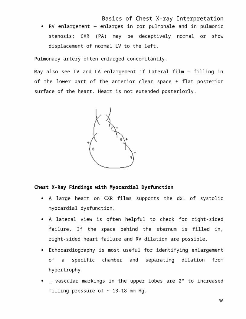
RV enlargement — enlarges in cor pulmonale and in pulmonic stenosis; CXR (PA) may
be deceptively normal or show displacement of normal LV to the left.
Pulmonary artery often enlarged concomitantly.
27
Basics of Chest X-ray InterpretationMay also see LV and LA enlargement if Lateral film — filling in of the lower part of the anterior
clear space + flat posterior surface of the heart. Heart is not extended posteriorly.
Chest X-Ray Findings with Myocardial Dysfunction
A large heart on CXR films supports the dx. of systolic myocardial dysfunction.
A lateral view is often helpful to check for right-sided failure. If the space behind the
sternum is filled in, right-sided heart failure and RV dilation are possible.
Echocardiography is most useful for identifying enlargement of a specific chamber and
separating dilation from hypertrophy.
_ vascular markings in the upper lobes are 2° to increased filling pressure of ~ 13-18 mm
Hg.
Interstitial edema (Kerley's B lines) suggests a LV end-diastolic pressure of 19-25 mm
Hg.
Alveolar infiltrates (pulmonary edema) are consistent with a LVEDP > 25 mm Hg.
Blunting of the margins is due to effusion.
CXR can help rule in or out other causes of dyspnea such as pulmonary fibrosis or COPD.
Chest X-Ray Findings with Myocardial Ischemia
Special x-ray imaging (fluoroscopy or CT) can demonstrate coronary artery calcification,
but this is an uncertain marker. It has not had the test characteristics that were originally
28
Basics of Chest X-ray Interpretationanticipated because calcification of the arterial walls is not necessarily a/w luminal
occlusion, particularly in older individuals.
Chest X-Ray Findings with Valvular Dysfunction
Signs of CHF and chamber enlargement can be detected using chest x-ray studies.
Valvular calcification can sometimes be seen.
Chest X-Ray Findings with Poor Exercise Capacity
Signs of pulmonary disease can suggest a noncardiac limitation to exercise and a large
heart could suggest cardiac disease.
Signs of CHF can offer the possibility of a cardiac cause for a change in exercise capacity.
Chest X-Ray Findings with Arrhythmias
Films are of little use in the diagnosis of arrhythmias. However, finding problems that are
often a/w arrhythmias, such as cardiac enlargement and lung disease, should alter one to
the possibility of arrhythmias.
The straight back syndrome or pectus excavatum was thought to be a/w with mitral valve
prolapse and arrhythmias.
29
Basics of Chest X-ray InterpretationSection 4
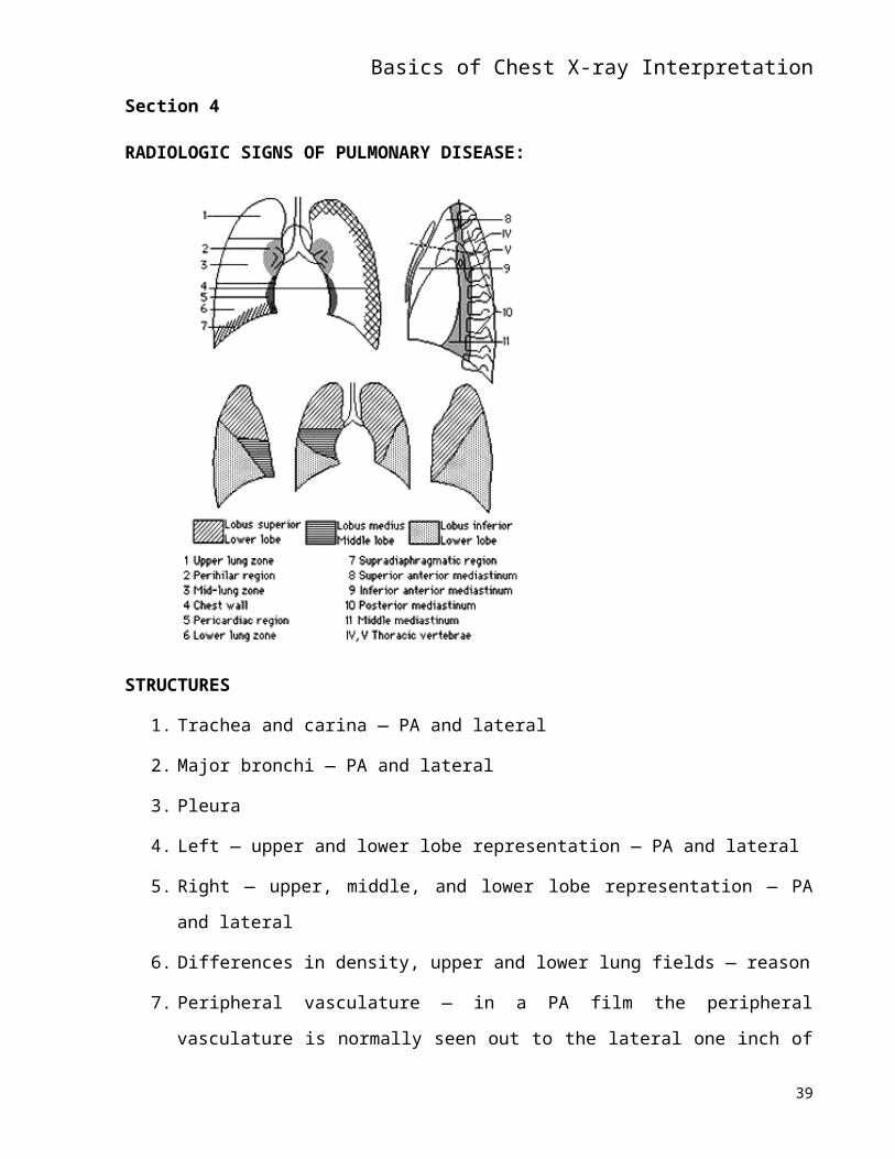
RADIOLOGIC SIGNS OF PULMONARY DISEASE:
STRUCTURES
1. Trachea and carina — PA and lateral
2. Major bronchi — PA and lateral
3. Pleura
4. Left — upper and lower lobe representation — PA and lateral
5. Right — upper, middle, and lower lobe representation — PA and lateral
6. Differences in density, upper and lower lung fields — reason
7. Peripheral vasculature — in a PA film the peripheral vasculature is normally seen out to
the lateral one inch of the films and is more clearly delineated in the lower lobes than the
apices
8. Silhouette sign
9. Air bronchogram sign
30
Basics of Chest X-ray Interpretation10. Solitary Pulmonary Nodules
Well-circumscribed, approximately round lesion that is < 4-6 cm. in diameter on
CXR.
By definition, it is completely surrounded by aerated lung.
AKA a "coin lesion."
Pulmonary masses are > 4-6 cm. in diameter.
Calcification of the lesion, absence of a history of tobacco use, and age < 35 years are
important factors that strongly correlate with benign nodules.
Noncalcified lesions can be benign or malignant.
Even benign calcification does not exclude the presence of coincidental malignancy in
adjacent tissue or the subsequent degeneration of a previously benign process into a
malignant lesion.
Close observation with serial CXRs every 6 mo. for at least 2 years is prudent.
Cavitating lesions, lesions with multilobulated or spiculated contours, and lesions with
shaggy or extremely irregular borders tend to be malignant.
Benign nodules tend to grow at either very slow or very rapid rates.
In contrast, malignant processes grow at steady, predictable, exponential rates.
The growth of a nodule is conventionally defined as the doubling time (time required
for its volume to double) and corresponds to an increase in diameter by a factor of
1.26.
In general, doubling times > 16 months or < 1 month are associated with benign
processes.
If a nodule has not increased in size over a 2-year period, the probability that it is
benign is > 99%.
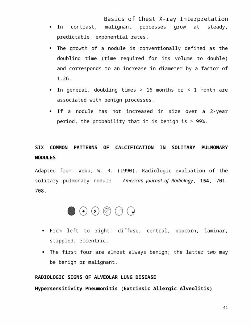
SIX COMMON PATTERNS OF CALCIFICATION IN SOLITARY PULMONARY
NODULES
Adapted from: Webb, W. R. (1990). Radiologic evaluation of the solitary pulmonary nodule.
American Journal of Radiology, 154, 701-708.
31
Basics of Chest X-ray Interpretation
From left to right: diffuse, central, popcorn, laminar, stippled, eccentric.
The first four are almost always benign; the latter two may be benign or malignant.
RADIOLOGIC SIGNS OF ALVEOLAR LUNG DISEASE
Hypersensitivity Pneumonitis (Extrinsic Allergic Alveolitis)
Perihilar haziness and peripheral alveolar infiltrates.
Chronic disease — abnormalities indistinguishable from fibrosing alveolitis are
commonly found — reticulonodular parenchymal infiltrates, dense fibrotic areas, and
decreased lung volumes.
Hilar adenopathy is not found.
Signs/Symptoms of Acute Exposure — fever, chills, anorexia, shortness of breath, dry cough;
tachypnea, pyrexia, tachycardia, dry basilar inspiratory rales without rhonchi; occasionally,
cyanosis or restlessness indicating hypoxemia.
Signs/Symptoms of Chronic Exposure — shortness of breath, mild fever, weight loss, fatigue,
malaise, dry cough, dyspnea on exertion, tachypnea; above signs + ? cor pulmonale (neck vein
distention, hepatojugular reflex, hepatomegaly, ankle edema, ascites, loud P2, increased RV
activity with a parasternal lift and parasternal S4 gallop).
RADIOLOGIC SIGNS OF INTERSTITIAL LUNG DISEASE
Reticular — lung parenchyma replaced by many thin-walled cysts (lesions less than 10 mm
in diameter), hence the term "honeycomb" lung, these microcysts may be barely perceptible,
round or oval, giving the lung the radiologic appearance of a fine network.
Seen in disseminated interstitial diseases such as eosinophilic granuloma of the lung,
scleroderma, pneumoconiosis (diseases caused by inhalation of organic or inorganic matter),
idiopathic pulmonary fibrosis, sarcoidosis, and other, less common disorders.
Miliary, Nodular — numerous discrete, tiny (< 5 mm), uniform densities; evenly distributed
throughout the lungs; quite uniform in size.
Seen in miliary tuberculosis, other fungal diseases (histoplasmosis), pneumoconiosis,
histiocytosis X (early stage), pulmonary hemosiderosis (late stage) and primary amyloidosis.
32
Basics of Chest X-ray Interpretation Reticulonodular — mixture of the two previously described patterns.
May predominate in one or another portion of the lung in the diseases described.
Kerley's Lines — most commonly encountered in CHF and interstitial pulmonary edema;
may be quite transient in these conditions.
May represent a constant, irreversible finding in other interstitial disease, esp. pneumoconiosis,
lymphatic spread of neoplasm, lymphatic mitral valve disease, and COPD.
Kerley B Lines — usually < 2 cm in length and about 1 mm in thickness.
Not confined to the margins of the lung.
Attributed to increased tissue and/or fluid accumulation in interlobular septa; also referred to
as septal lines.
Kerley A Lines — usually ~ 4 cm in length, relatively straight, linear densities.
Tend to be oriented perpendicular to the nearest pleural surface.
Attributed to increased tissue and/or fluid accumulation in communicating lymphatics
between veins and bronchi.
Exaggerated Bronchovascular Markings — ill-reputed sign; lacks specificity in terms of
pathologic correlation.
May refer to a lack of crispness of the margins of structures initially giving rise to the linear
densities within aerated lung. Caused by excessive tissue or fluid displacing air-filled lung from
the interstitial structures.
o Inability to detect radiologic signs of alveolar consolidation on abnormal CXRs such
as the air bronchogram sign.
o Majority of interstitial diseases are chronic. Principal exceptions are viral pneumonia,
drug-induced pneumonia, and pulmonary edema.
33
Basics of Chest X-ray Interpretation
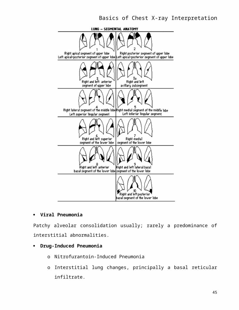
Viral Pneumonia
Patchy alveolar consolidation usually; rarely a predominance of interstitial abnormalities.
Drug-Induced Pneumonia
o Nitrofurantoin-Induced Pneumonia
o Interstitial lung changes, principally a basal reticular infiltrate.
o Presents acutely with chills, fever, quite severe dyspnea, and nonproductive cough
within hours or days of the initiation of nitrofurantoin therapy.
o Likely to have moderate peripheral eosinophilia.
34
Basics of Chest X-ray InterpretationPulmonary Edema
o Mixed alveolar and interstitial edema.
o Apical redistribution of blood flow — results in increased size of upper lung
vasculature and background veiling of the pulmonary parenchyma initially.
o Subpleural edema, peribronchial cuffing, bronchiolar cuffing, hilar haziness, haziness
of vessel detail, reticular pattern, and basilar septal lines.
o Kerley B lines are present at the periphery of the lung bases and may be quite
prominent — represent thickened interlobular septa.
o Usually, enlargement of the heart (if cardiogenic in origin) and redistribution of the
pulmonary vasculature (appears esp. engorged in the upper lung zones).
Idiopathic Pulmonary Fibrosis (Hamman-Rich disease)Reticular pattern (honeycombing).
Most common "etiology" of disseminated pulmonary fibrosis.
Differential Diagnosis of Interstitial Lung Disease
Pneumoconiosis Primary Lung Diseases
Silicosis
Sarcoidosis
Asbestosis
Histiocytosis X
Coal Worker's pneumoconiosis
Lymphangiomyomatosis
Berylliosis
Lymphangitic carcinomatosis
Organic dusts (pigeons, turkey, duck, chicken, humidifier) Lipoidosis
Emphysema
Drugs
Cystic fibrosis
Chemotherapeutic agents (busulfan, bleomycin, methotrexate)
Antibiotics (nitrofurantoin, sulfonamides, INH)
35
Basics of Chest X-ray InterpretationAlveolar Filling Disease
Diffuse alveolar bleeding (Goodpasture's Amiodarone syndrome, lupus, mitral stenosis,
Penicillamine idiopathic pulmonary hemosiderosis)
Lupus-like reactions (hydralazine, procainamide)
Alveolar proteinosis
Radiation
Alveolar cell carcinoma
Eosinophilic pneumonia
Connective Tissue Disease
Lipid pneumonia
Systemic lupus erythematosus
Rheumatoid arthritis
Infectious Diseases
Scleroderma
Miliary tuberculosis
Polymyositis
Some fungal and viral infections
Other Cardiovascular Diseases
Idiopathic pulmonary fibrosis
Interstitial pulmonary edema
Bronchiolitis obliterans organizing
Pulmonary hemosiderosis 2° to mitral pneumonia stenosis
Lymphocytic interstitial pneumonia
Amyloidosis
Pulmonary Sarcoidosis — lymphadenopathy always precedes or presents concurrently with
pulmonary changes of the disease.
± Hilar and paratracheal adenopathy.
Intrathoracic lymphadenopathy (75%)
Diffuse parenchymal disease (50%)
36
Basics of Chest X-ray InterpretationExclusively hilar lymphadenopathy initially (33%)
Pulmonary disease without hilar lymph node enlargement (25%)
Lung involvement varies from a miliary nodular pattern, to a reticulonodular pattern, to a purely
reticular pattern (honeycombing).
Occasionally patients exhibit mx. large granulomas simulating metastatic neoplasm.
Likely that a miliary nodular form precedes the reticular pattern.
Progression to marked pulmonary fibrosis of bullous emphysema with disabling functional
impairment, development of cor pulmonale, and death occurs in a small % of cases.
Majority of patients remain relatively asymptomatic.
Scleroderma
Great majority of patients with abnormal pulmonary function studies do not exhibit radiologically
discernible pulmonary changes.
?? Relatively fine network of reticular infiltrates (honeycombing); generally restricted to the
lower lung zones.
Radiologic demonstration of abnormalities of esophagus, duodenum, small bowel, or terminal
phalanges more likely to be seen.
Recurrent or chronic aspiration of ingested material may be underlying cause of pulmonary
fibrosis.
Histiocytosis X — includes Letterer-Siwe disease, Hand-Schüller-Christian disease, and
eosinophilic granuloma. Only eosinophilic granuloma occurs in adults.
Coarse, reticular interstitial pattern.
Individual cysts comprising the coarse reticular or honeycomb pattern are generally less than 5
mm in greatest dimension, although large cysts of up to 6 cm in diameter have been reported.
Pneumothorax = relatively frequent complication.
2/3 deny dyspnea; 2/3 have dry cough; systemic symptoms = lassitude, weight loss, and less
commonly, fever may predominate in 1/3.
Diabetes insipidus may be and associated disorder.
Systemic form — ? involvement of bone, liver, CNS, kidneys, and alimentary tract.
Pneumonia
37
Basics of Chest X-ray InterpretationIATROGENIC RADIOLOGIC SIGNS
A. ECG leads
B. Endotracheal tube — positioning
C. CVP and PA lines
38
Basics of Chest X-ray InterpretationGLOSSARY
Air bronchogram — Surrounding consolidation will sometimes allow more peripheral bronchi
to be seen as tubular or branching lucencies. Normally only the trachea, mainstem bronchi, and
occasionally the origins of the lobar bronchi, are visible on CXRs as air-filled tubular structures.
Visualization of the more peripheral bronchi with air in them is usually not possible.
Alveolar (consolidative) densities — An abnormal density caused by the collapse or, more
often, the filling of air spaces with abnormal material (blood, pus, water, protein, or cells).
Alveolar densities characteristically have irregular, hazy margins except where they are bounded
by a pleural surface. (Also referred to as "acinar pattern".) Segmental distribution and air
bronchograms are also characteristic of this pattern.
Atelectasis — Collapse and volume loss are synonymous terms. Very small areas of atelectasis
often produce a linear shadow, which is often, but not always, horizontal. This is referred to as
"plate-like", "linear", or "subsegmental" atelectasis. Lobar and total lung atelectasis also occur.
These larger varieties of atelectasis are usually associated with increased density in the involved
portion of lung so that there is, in fact, consolidation present as well. To diagnose atelectasis,
there must be a specific evidence of volume loss such as displacement of a fissure, the
mediastinum, or a hilum. Elevation of the hemidiaphragm and decreased space between ribs can
also be signs of atelectasis.
Bleb — A small, thin-walled, air-containing structure. This term is frequently reserved for such
small areas which are frequently intrapleural. This term may be used synonymously with "bulla"
but often is reserved for smaller air spaces.
Bronchiectasis — Dilatation of a bronchus or bronchi, usually secreting large amounts of
offensive pus. Dilatation may be in an isolated segment or spread throughout the bronchi.
Bulla — See "bleb" or "cavity." These abnormal air spaces may or may not be associated with
diffuse pulmonary emphysema.
Caseous — cheese-like.
Cavity — Another form of air space in the lung. This term is usually reserved for those which are
the result of tissue necrosis, unlike bullae. Thickness and irregularity of the walls often the
distinguishing feature separating cavities from bullae or blebs.
39
Basics of Chest X-ray InterpretationConsolidation — Filling of pulmonary air space with some abnormal material. May also be
referred to as "alveolar disease."
Density — A nonspecific term that can be used to describe any area of whiteness on the chest
film.Normal structures such as the heart as well as abnormalities in the lungs may be called
densities. This term is often used when the nature or cause of an abnormal shadow is not known.
It is a useful term in that situation, since other terms (e.g., "mass" or "infiltrate") frequently imply
more specific entities which may or may not be present.
Extra-pleural — Anything that is outside both the parietal and the visceral pleura but that
impinges on the lungs. The heart is the most obvious example. Since normal or abnormal
structures in this location are separated by two layers of pleura from the lung, the margins of
these densities are characteristically sharp and smoothly tapering.
Hilum (pleural = hila.) — "lung root;" medusa-like tangle of arteries and veins on either side of
the heart shadow. Irregular medial shadow in each lung where the bronchi and pulmonary arteries
enter. Other structures in these areas, particularly lymph nodes, are normally so small as to be
inapparent. The normal hilar shadow is almost entirely composed of the central pulmonary
arteries. R hilar vessels seem to extend out farther than those on the L because a part of the L
hilum is obscured by the shadow of the more prominent L side of the heart. The L hilum on a
normal CXR is a little higher than the R one because of the slightly higher take-off of the L
pulmonary artery.
Interminate or mixed lung disease — This category of diffuse lung disease is frequently used
when the radiographic criteria to designate a specific pattern (consolidative, interstitial, etc.) may
not be present, or when there may be elements of several types of diffuse lung disease in the same
patient.
Infiltrate — A poorly defined abnormal pulmonary density or any such density sharply bounded
by pleura and fissures. This is a confusing term, since it may be used to indicate any abnormal
lung density or, by others, as a synonym for consolidation. Synonymous with "fluid density."
Interstitial — The portion of the pulmonary parenchyma that consists of the actual lung tissue as
opposed to the air spaces. Includes alveolar walls, septa, bronchovascular structures, and pleura.
Involvement of this tissue is a frequent form of diffuse lung disease.
Kerley's lines — most commonly encountered in CHF and interstitial pulmonary edema; may be
quite transient in these conditions; may represent a constant, irreversible finding in other
40
Basics of Chest X-ray Interpretationinterstitial disease, esp. pneumoconiosis, lymphatic spread of neoplasm, lymphatic mitral valve
disease, and COPD.
Kerley B (septal) lines — usually < 2 cm in length and about 1 mm in thickness; not confined to
the margins of the lung; attributed to _tissue and/or fluid accumulation in interlobular septa;
thickening of interlobular septa for any reason may allow them to be seen as narrow, straight
shadows, especially at the periphery of the bases; another form of interstitial abnormality.
Kerley A lines — Usually ~ 4 cm in length, relatively straight, linear densities; tend to be
oriented perpendicular to the nearest pleural surface; attributed to _tissue and/or fluid
accumulation in communicating lymphatics between veins and bronchi.
kVp — Peak kilovoltage; the peak voltage across the radiographic tube. An increase in this factor
allows increased tissue penetration by higher energy roentgens.
Lingula — (tongue-shaped) area of left lung adjacent to the left ventricle not a separate lobe.
Lucency — An increase in blackness of an area on the radiograph. In the lung, it may imply that
air is being trapped, that lung tissue has been destroyed, or that there is decreased blood supply.
Artifacts, changes in position, and soft tissue abnormalities can also cause areas of lucency.
milliampere/seconds (mAs) — This is the amount of current through the radiographic tube. The
amount of current and the length of time during which the current flows control the quantity of x-
rays generated. Increasing the mA causes an increase in patient exposure to ionizing radiation and
produces more x-rays to create an image on the film.
Mass — A solid-appearing, reasonably well-defined soft tissue density usually larger than 3 or 4
cm in diameter.
Mediastinal — Referring to the structures or a lesion between the lungs. Unless the lungs are
actually invaded by a mediastinal lesion, the lesion's x-ray shadow will be extra-pleural and,
therefore, usually will have sharp demarcation from the lung.
Miliary — A form of diffuse lung disease consisting of countless very tiny nodular densities.
Nodule — A well-defined, more or less round density in the lung; smaller than a mass. No rigid
size distinction between a "mass" and "nodule" is possible.
Opacity — Synonym for "density."
41
Basics of Chest X-ray InterpretationPleural — Refers to an abnormality arising in the pleura or pleural space. Most commonly this is
free of loculated fluid.
Pneumothorax — Free air in the pleural space; may be modified by the following descriptive
terms" hydro-, pyo-, hemo-, chylo-, tension.
Pulmonary edema — defined radiographically as diffuse, bilateral consolidation by fluid' other
materials can fill air spaces bilaterally and give the same radiographic pattern.
Reticular — A fine branching pattern with lines radiating in all directions; one of the signs of the
interstitial pattern.
Segmental — Limited to specific bronchopulmonary segments or lobes. Segmental distribution
of disease usually indicated bronchial or vascular involvement and is most common in
consolidation.
Septal lines — see Kerley B lines.
Silhouette sign — Normally an interface is seen between areas of different density as between
shadows of the heart and lung. Loss of air on the pulmonary side, usually because of
consolidation, may cause obliteration or "silhouetting" of this normal interface. This sign is
useful in localizing an abnormality or confirming the presence of abnormality. Occasionally the
silhouette sign will be the only definite indication of consolidation next to the heart or diaphragm.
42
Basics of Chest X-ray InterpretationCHEST RADIOLOGY ARTICLES
Baumstark, A., Swensson, R. G., Hessel, S. J., et al. (1984). Evaluating the radiographic
assessment of pulmonary venous hypertension in chronic heart disease. American Journal of
Radiology, 142, 877.
Chakko, S., Woska, D., Martinez, H., et al. (1991). Clinical, radiographic, hemodynamic
correlations in chronic congestive heart failure: Conflicting results may lead to inappropriate
care. American Journal of Medicine, 90, 353.
Chen, J. T. T., Beliar, V. S., Morris, J. J., et al. (1968). Correlation of roentgen findings with
hemodynamic data in pure mitral stenosis. American Journal of Roentgenology, 102, 280.
Cigarroa, J. E., Isselbacher, E. M., DeSanctis, R. W., & Eagle, K. A. (1993). Diagnostic imaging
in the evaluation of suspected aortic dissection: Old standard and new directions. New England
Journal of Medicine, 328, 35-43.
Crystal, R. G., Bitterman, P. B., Rennard, S. I., et al. (1984). Interstitial lung disease of unknown
cause. Disorders characterized by chronic inflammation of the lower respiratory tract: Parts 1 and
2. New England Journal of Medicine, 310, 154.
Friedman, B. J., et al. (1985). Comparison of magnetic resonance imaging and echocardiography
in determination of cardiac dimensions in normal subjects. Journal of the American College of
Cardiology, 5, 1369.
Goodman, L. R. Radiology of asbestos disease. (1983). Journal of the American Medical
Association, 269, 465.
Gyssenhoven, E. J., et al. (1986). Transesophageal two-dimensional echocardiography: Its role in
solving clinical problems. Journal of the American College of Cardiology, 8, 975.
Haupt, M., Moore, G. W., & Hutchins, G. M. The lung in systemic lupus erythematosus. (1981).
American Journal of Medicine, 71, 791.
Hunninghake, G. W., & Fauci, A. S. (1979). Pulmonary involvement in the collagen vascular
diseases. American Review of Respiratory Disease, 119, 471.
Jay, S. J., Johannson, W. G., & Pierce, A. K. (1975). The radiographic resolution of
Streptococcus pneumoniae pneumonia. New England Medicine 293, 798.
Khouri, N. F., Meziane, M. A., Zerhouni, E. A., et al. (1987). The solitary pulmonary nodule:
Assessment, diagnosis, and management. Chest, 91, 128-133.
43
Basics of Chest X-ray InterpretationLillington, G. A. (May 15, 1993). Management of the solitary pulmonary nodule. Hospital
Practice, 41-48.
Meaney, J. F. M., Weg, J. G., Chenevert, T. L., et al. (1997). Diagnosis of pulmonary embolus
with magnetic resonance angiography. New England Journal of Medicine, 336, (20), 1422-1427
Mehlman, D. J., & Resnekov, L. (1978). A guide to the radiographic identification of prosthetic
heart valves. Circulation, 57, 613.
Nienaber, C. A., von Kodolistch, Y., Nicolas, V., et al. (1993). The diagnosis of thoracic aortic
dissection by noninvasive imaging procedures. New England Journal of Medicine, 328, 1-9.
O'Keefe, M. E., Good, C. A., & McDonald, J. R. Calcification in solitary nodules of the lung.
American Journal of Radiology, 77, 1023-1033.
Pratt, P. C. (1987). Role of conventional chest radiography in diagnosis and exclusion of
emphysema. American Journal of Medicine, 82, 998.
Ritchie, J. I., et al. (1995). Guidelines for clinical use of cardiac radionuclide imaging. A report of
the American Heart Association/American College of Cardiology Task Force. Circulation, 91 (4),
1278-1303.
Rosenow III, E. C. (1998). Interpreting chest films: Tricks of the trade. Consultant, 38 (3), 553-
567.
Schapiro, R. L., & Musallam, J. J. (1977). A radiologic approach to disorders involving the
interstitium of the lung. Heart & Lung, 6 (4), 635-643.
Seward, J. B. (1992). Transesophageal echocardiography: ACC Position Statement. Journal of
the American College of Cardiology, 20, 506.
Shuford, W. H. (1992). Detection of cardiac chamber enlargement with the chest roentgenogram.
Heart Disease and Stroke, 2, 341-347.
Stein, P. D., et al. (1992). Relation of plain chest radiographic findings to pulmonary arterial
pressure and arterial blood oxygen levels in patients with acute pulmonary embolism. American
Journal of Cardiology, 69, 394.
Tajik, A. J., et al. (1978). Two-dimensional real-time ultrasonic imaging of the heart and great
vessels. Mayo Clinic Proceedings, 53, 271.
Tapson, V. R. (1997). Pulmonary embolus - New diagnostic approaches.New England Journal of
Medicine, 336 (20), 1449-1451.
44
Basics of Chest X-ray Interpretation» Theodore, J., & Robin, E. D. (1975). Pathogenesis of neurogenic pulmonary edema. Lancet, 2,
749.
» Webb, W. R. (1990). Radiologic evaluation of the solitary pulmonary nodule. American
Journal of Radiology, 154, 701-708.
Winterhauer, R. H., Belic, N., & Moores, K. D. (1973). Clinical interpretation of bilateral hilar
adenopathy. Annals of Internal Medicine, 78, 65.
Woodring, J. H. (1990). Lung cancer. Radiology Clinics of North America, 28, 489.
Zelefsky, M. N. (1977). A simplified approach to reading x-rays of the heart. Modern Medicine,
October 30, 33-36.
45