microfungal community in sandy beaches located in kedah ...corresponding author: nur ain izzati m....
TRANSCRIPT
Submitted 12 June 2018, Accepted 28 September 2018, Published 9 October 2018
Corresponding Author: Nur Ain Izzati M. Z. – e-mail – [email protected] 321
Microfungal Community in Sandy Beaches Located in Kedah, Pahang
and Sabah, Malaysia
Salleh SL, Raup R, Azman N and Mohd Zainudin NAI Department of Biology, Faculty of Science, Universiti Putra Malaysia, 43400 Serdang, Selangor, Malaysia
Salleh SL, Raup R, Azman N, Mohd Zainudin NAI 2018 – Microfungal Community in Sandy
Beaches Located in Kedah, Pahang and Sabah, Malaysia. Studies in Fungi 3(1), 321–332, Doi
10.5943/sif/3/1/32
Abstract
Microfungi are fundamental organisms help in balancing the ecosystem. The aims of this
study are to identify microfungi isolated from sandy beaches based on internal transcribed spacers
(ITS) sequence analysis and to determine the phylogenetic relationship among the locations of
sampling areas. Fifty-seven isolates of microfungi were obtained from six beaches located in Kedah
(Chenang Beach), Pahang (Balok Beach) and Sabah (Mamutik Island, Manukan Island, Sapi Island
and Tanjung Aru Beach) to be used in this study. From the finding, the highest identified species
were Aspergillus species (27 isolates) followed by Trichoderma species (12 isolates), Penicillium
species (10 isolates), Fusarium species (6 isolates), Bipolaris species (1 isolate) and Chaetomium
species (1 isolate). ITS fragment of all isolates was amplified in within the size range of 533–658
bp. A phylogenetic tree was constructed using maximum likelihood (ML) method; the isolates were
grouped into three clades according to their classes. In conclusion, diverse microfungi isolated from
sandy beach soil depicting 19 species were discovered. Future studies are highly recommended to
focus on intra- and interspecies diversity based on microsatellite markers analysis, consequently the
originality and distribution of the fungal isolates. The outcome of this study includes a baseline data
on the occurrence and species-diversity as well as important information on the status of
microfungi in Malaysia beaches located in Kedah, Melaka and Sabah. It can be used as a checklist
for future studies related to fungi distribution in the tropical beaches.
Key words – Ecology – environmental mycology – fungi – identification – microbial
phylogenetics
Introduction
Beach is a reservoir for microfungi including Ascomycetes that act as decomposers that retain
sand ecosystem. However, concern had arose when a number of beachgoers were seen increasing.
The predicted possibilities of decreased or increased microfungi diversity in the sandy beach soil
could indicate some current situations and threat of health to the beachgoers by the pathogenic
microfungi. This study will provide an information or inventory on the ecology and distribution of
microfungi as well as early prediction on the conditions of particular beaches, which can improve
the efficiency of beach management. Sabino et al. (2014) collected growing evidence that microbes
including fungi could be harmful to humans in various beach sands worldwide and often in
concentrations greater than the beach water. Even though there were few cases reported regarding
this pathogenic microbe on public health, consideration should be made on people often exposed to
Studies in Fungi 3(1): 321–332 (2018) www.studiesinfungi.org ISSN 2465-4973
Article
Doi 10.5943/sif/3/1/32
322
the beach through professional activities since there is a high risk for them to face health problems
from the pathogenic microbes. Other than pathogenic effect, some microfungi in sandy beach soil
can also be considered as a source of industrial products with the ability to balance the ecosystem
biotic elements. Exploring the species diversity of microfungi in sandy beach soil, there is a
possibility to harvest microfungi of interest from sandy beach soil to be used as a source for
industrial production or with commercial potential.
Discovering the diversity of microfungi in sandy beach soil is important to find a source of
microfungi-based industrial products, for us to appreciate their existence in maintaining ecosystem
stability, introduce new biological indicator from microfungi and most importantly to make aware
on the existence of potentially pathogenic fungi. In the meantime, some researchers suggested that
the potential public health issues concerning both sand and water quality of beaches should be
considered in future policy decisions (Sabino et al. 2014).
To date, there have been no comprehensive reports on microfungi isolated from sandy beach
soil in Malaysia except in Penang Island by Zakaria et al. (2011), Teh & Latiffah (2015). However,
studies on the diversity of soil microfungi in sandy beach ecosystem of Malaysia as well as other
Asian countries are still limited. Thus, renowned beaches or islands were aimed in this study since
they are often visited by beachgoers and most well-known by tourists and local people in Malaysia.
These beaches include Balok Beach in Pahang, Langkawi Island in Kedah and another four islands
in Sabah comprising Mamutik Island, Sapi Island, Manukan Island and Tanjung Aru beach.
Discovering the diversity and distribution of microfungi present in sandy beach soil in Malaysia
can give significant information on Malaysian beaches mycoflora, which is useful for an efficient
management of the beach ecosystem, raising awareness concerning public health on beach and to
be used in various fields of science.
This study was conducted with findings that can be used as references for the future studies
and to give an idea on the diversity of microfungi from the six selected areas. The objectives of this
study are to isolate and identify the microfungi isolates obtained from sandy beaches based on
Internal Transcribed Spacers (ITS) sequence analysis and to determine the phylogenetic
relationship among isolated fungi from various locations.
Materials & Methods
Sampling and fungal isolation
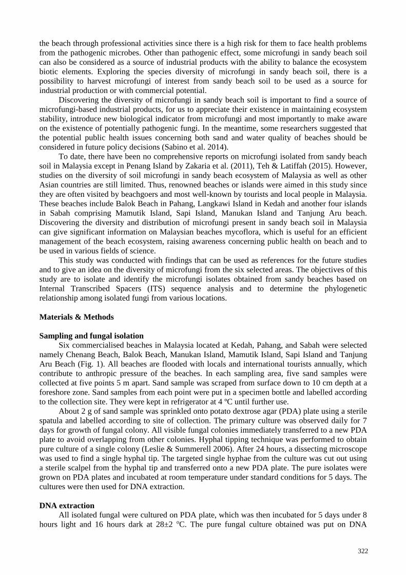
Six commercialised beaches in Malaysia located at Kedah, Pahang, and Sabah were selected
namely Chenang Beach, Balok Beach, Manukan Island, Mamutik Island, Sapi Island and Tanjung
Aru Beach (Fig. 1). All beaches are flooded with locals and international tourists annually, which
contribute to anthropic pressure of the beaches. In each sampling area, five sand samples were
collected at five points 5 m apart. Sand sample was scraped from surface down to 10 cm depth at a
foreshore zone. Sand samples from each point were put in a specimen bottle and labelled according
to the collection site. They were kept in refrigerator at 4 ºC until further use.
About 2 g of sand sample was sprinkled onto potato dextrose agar (PDA) plate using a sterile
spatula and labelled according to site of collection. The primary culture was observed daily for 7
days for growth of fungal colony. All visible fungal colonies immediately transferred to a new PDA
plate to avoid overlapping from other colonies. Hyphal tipping technique was performed to obtain
pure culture of a single colony (Leslie & Summerell 2006). After 24 hours, a dissecting microscope
was used to find a single hyphal tip. The targeted single hyphae from the culture was cut out using
a sterile scalpel from the hyphal tip and transferred onto a new PDA plate. The pure isolates were
grown on PDA plates and incubated at room temperature under standard conditions for 5 days. The
cultures were then used for DNA extraction.
DNA extraction
All isolated fungal were cultured on PDA plate, which was then incubated for 5 days under 8
hours light and 16 hours dark at 28±2 oC. The pure fungal culture obtained was put on DNA
323
extraction. Genomic DNA of pure culture was extracted using UltraClean® Microbial DNA
Isolation Kit (MO BIO, Carlsbad, CA, USA) following the manufacturer’s protocol. The 50 µl
gDNA of each fungal isolated was obtained and stored in -20 ºC freezer for further use.
Fig. 1 – The map of sampling sites with red dotted. A Malaysia map. B Langkawi Island map. C
Shore of Chenang Beach. D Shore of Balok Beach, Pahang. E Map of Sapi Island, Mamutik Island,
Manukan Island and Tanjung Aru Beach in Sabah.
Gel electrophoresis
Each PCR products was gel electrophoresed by mixing 0.3 g of agarose powder with 20 ml of
TBE buffer to make a final concentration of 1.5%. The cooled agarose gel was placed in the gel
electrophoresis box with buffer solution poured onto it. DNA was inserted into the holes and a 100
bp DNA ladder was used. The voltage was set at 90 V for 30-40 mins. The gel was visualized
under a UV trans-illuminator to view the amplicon size. The successful amplification of ITS region
was observed between 500 to 700 bp. Next, gel purification was carried out to obtain the purified
ITS regions.
Gel purification and sequencing
All PCR products were purified using QIAquick Gel Extraction Kit (QIAGEN, USA)
following the manufacturer’s instructions. The DNA fragment was cut from gel with scalpel. Then,
ratio of 3 to 1 was used to add 3 volumes of Buffer QG to one volume gel. The tube was incubated
at 50 ºC for 10 mins until the gel was dissolved. Tube was vortexed for every two to three minutes
to help dissolving the gel. Isopropanol was added to sample and mixed. Finally, the sample was
added to QIAquick spin column. Then, the purified PCR products were sent for DNA sequencing
using an Applied Biosystems 3730xl DNA Analyzer (MyTACG Bioscience Company, MY).
Phylogenetic analysis and sequence deposition
The sequences obtained were complemented and aligned. The sequencing alignments and
phylogenetic tree analysis were performed using the Molecular Evolutionary Genetics Analysis
(MEGA) 7.0 software. MEGA is one of the computer software for conducting statistical analysis on
molecular evolution and for constructing phylogenetic trees (Kumar et al. 2016). All complemented
sequences were BLAST at the GenBank database NCBI (https://blast.ncbi.nlm.nih.gov).
324
Phylogenetic tree was constructed using Maximum Likelihood (ML) with test of phylogeny of
Bootstrap method. Maximum likelihood is known to be a good statistical method for reconstructing
sequence histories and uses a more complex evolution model (Blanchet 2014). The representative
sequences of each species were obtained from GenBank and included in the tree. Saccharomyces
cerevisiae was chosen as an out-group since it was classified in different class compared to all 57
isolates. All sequences were deposited in the GenBank database ([email protected])
following the instructions.
Results
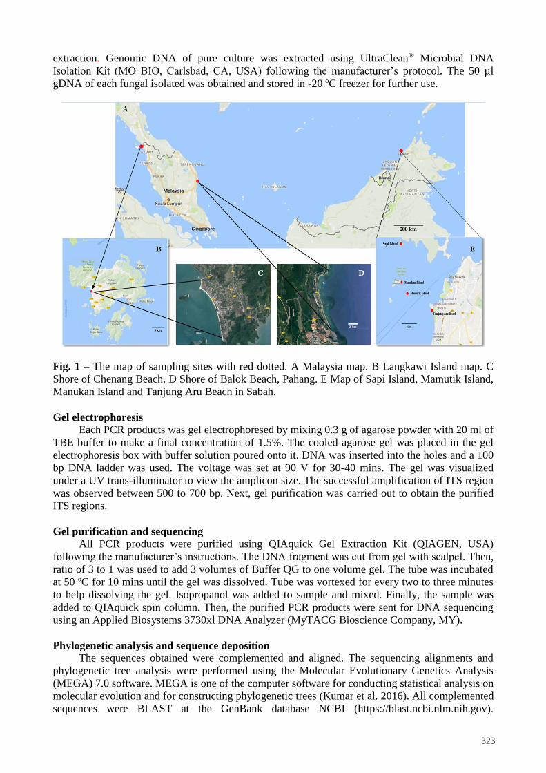
Fifty-seven isolates of Ascomycetes fungi were obtained from beach soil areas in Chenang
Beach, Mamutik Island, Manukan Island, Sapi Island, Tanjung Aru Beach, and Balok Beach from
sand beach soils samples comprising the genus Aspergillus, Bipolaris, Chaetomium, Fusarium,
Penicillium and Trichoderma species. All the fungi isolates were identified based on ITS sequence
analysis and deposited in the GenBank database (https://www.ncbi.nlm.nih.gov/genbank/) as
tabulated in Table 1. Among 57 microfungal isolates, 27 isolates were dominated by Aspergillus
species.
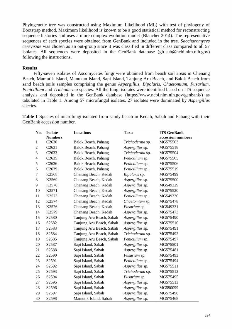
Table 1 Species of microfungi isolated from sandy beach in Kedah, Sabah and Pahang with their
GenBank accession number.
No. Isolate
Numbers
Locations Taxa ITS GenBank
accession numbers 1 C2630 Balok Beach, Pahang Trichoderma sp. MG575503
2 C2631 Balok Beach, Pahang Aspergillus sp. MG575518
3 C2633 Balok Beach, Pahang Trichoderma sp. MG575504
4 C2635 Balok Beach, Pahang Penicillium sp. MG575505
5 C2636 Balok Beach, Pahang Penicillium sp. MG575506
6 C2639 Balok Beach, Pahang Penicillium sp. MG575519
7 K2568 Chenang Beach, Kedah Bipolaris sp. MG575499
8 K2569 Chenang Beach, Kedah Aspergillus sp. MG575500
9 K2570 Chenang Beach, Kedah Aspergillus sp. MG549329
10 K2571 Chenang Beach, Kedah Aspergillus sp. MG575520
11 K2573 Chenang Beach, Kedah Penicillium sp. MG549330
12 K2574 Chenang Beach, Kedah Chaetomium sp. MG575478
13 K2576 Chenang Beach, Kedah Fusarium sp. MG549331
14 K2579 Chenang Beach, Kedah Aspergillus sp. MG575473
15 S2580 Tanjung Aru Beach, Sabah Aspergillus sp. MG575490
16 S2582 Tanjung Aru Beach, Sabah Aspergillus sp. MG575510
17 S2583 Tanjung Aru Beach, Sabah Aspergillus sp. MG575491
18 S2584 Tanjung Aru Beach, Sabah Trichoderma sp. MG575492
19 S2585 Tanjung Aru Beach, Sabah Penicillium sp. MG575497
20 S2587 Sapi Island, Sabah Aspergillus sp. MG575501
21 S2588 Sapi Island, Sabah Aspergillus sp. MG575481
22 S2590 Sapi Island, Sabah Fusarium sp. MG575493
23 S2591 Sapi Island, Sabah Penicillium sp. MG575494
24 S2592 Sapi Island, Sabah Aspergillus sp. MG575511
25 S2593 Sapi Island, Sabah Trichoderma sp. MG575512
26 S2594 Sapi Island, Sabah Fusarium sp. MG575495
27 S2595 Sapi Island, Sabah Aspergillus sp. MG575513
28 S2596 Sapi Island, Sabah Aspergillus sp. MG590099
29 S2597 Sapi Island, Sabah Aspergillus sp. MG575496
30 S2598 Mamutik Island, Sabah Aspergillus sp. MG575468
325
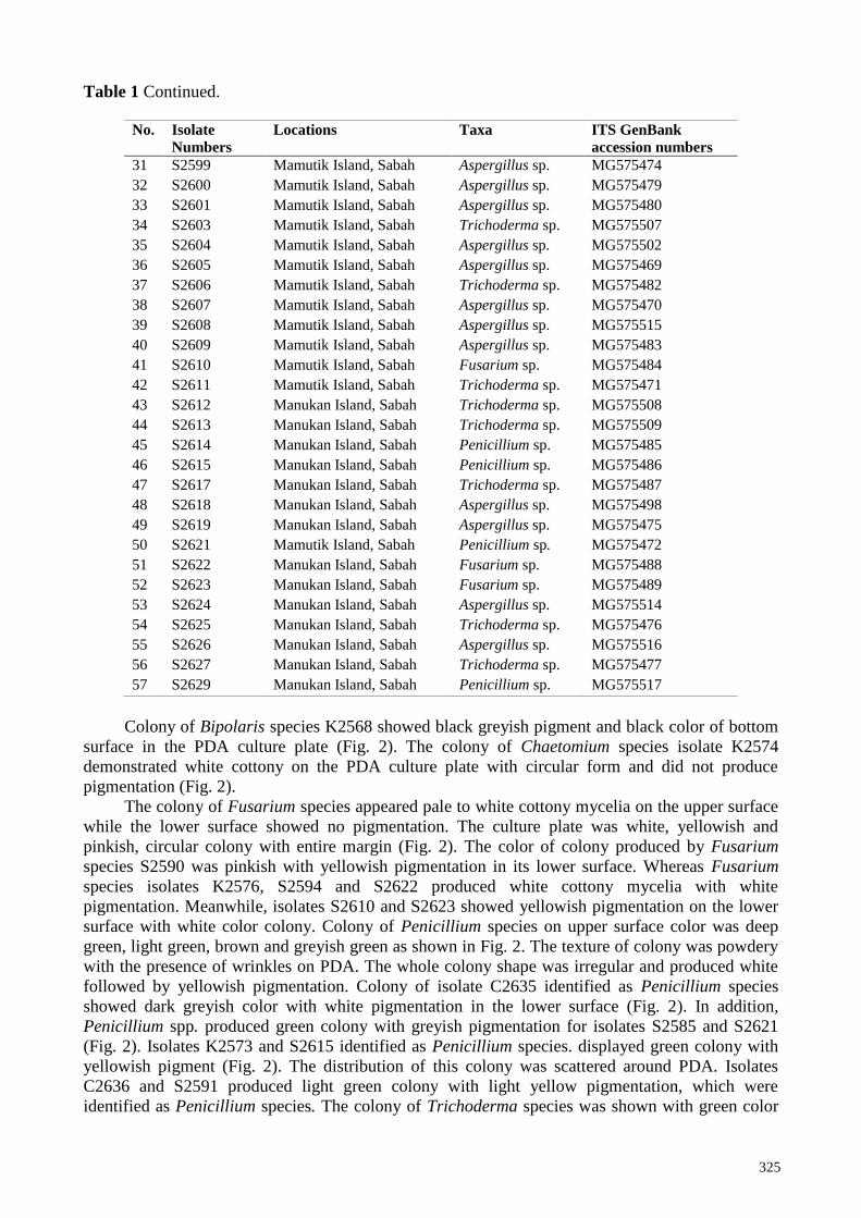
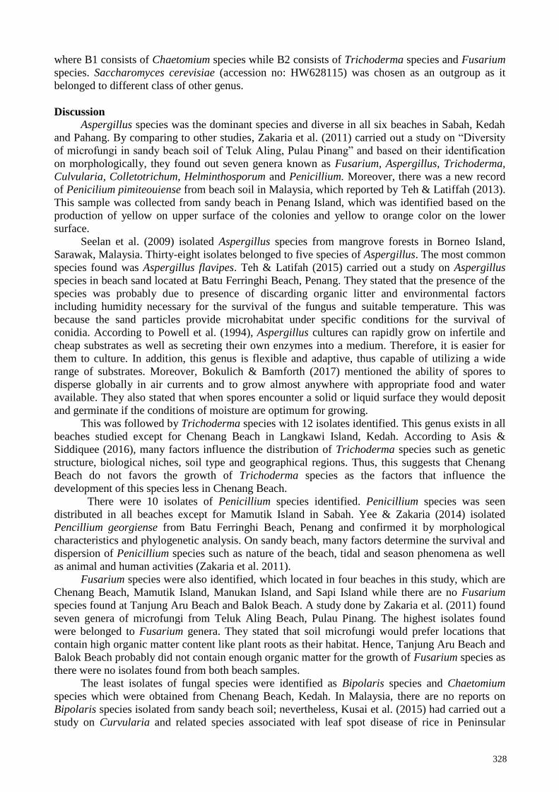
Table 1 Continued.
No. Isolate
Numbers
Locations Taxa ITS GenBank
accession numbers
31 S2599 Mamutik Island, Sabah Aspergillus sp. MG575474
32 S2600 Mamutik Island, Sabah Aspergillus sp. MG575479
33 S2601 Mamutik Island, Sabah Aspergillus sp. MG575480
34 S2603 Mamutik Island, Sabah Trichoderma sp. MG575507
35 S2604 Mamutik Island, Sabah Aspergillus sp. MG575502
36 S2605 Mamutik Island, Sabah Aspergillus sp. MG575469
37 S2606 Mamutik Island, Sabah Trichoderma sp. MG575482
38 S2607 Mamutik Island, Sabah Aspergillus sp. MG575470
39 S2608 Mamutik Island, Sabah Aspergillus sp. MG575515
40 S2609 Mamutik Island, Sabah Aspergillus sp. MG575483
41 S2610 Mamutik Island, Sabah Fusarium sp. MG575484
42 S2611 Mamutik Island, Sabah Trichoderma sp. MG575471
43 S2612 Manukan Island, Sabah Trichoderma sp. MG575508
44 S2613 Manukan Island, Sabah Trichoderma sp. MG575509
45 S2614 Manukan Island, Sabah Penicillium sp. MG575485
46 S2615 Manukan Island, Sabah Penicillium sp. MG575486
47 S2617 Manukan Island, Sabah Trichoderma sp. MG575487
48 S2618 Manukan Island, Sabah Aspergillus sp. MG575498
49 S2619 Manukan Island, Sabah Aspergillus sp. MG575475
50 S2621 Mamutik Island, Sabah Penicillium sp. MG575472
51 S2622 Manukan Island, Sabah Fusarium sp. MG575488
52 S2623 Manukan Island, Sabah Fusarium sp. MG575489
53 S2624 Manukan Island, Sabah Aspergillus sp. MG575514
54 S2625 Manukan Island, Sabah Trichoderma sp. MG575476
55 S2626 Manukan Island, Sabah Aspergillus sp. MG575516
56 S2627 Manukan Island, Sabah Trichoderma sp. MG575477
57 S2629 Manukan Island, Sabah Penicillium sp. MG575517
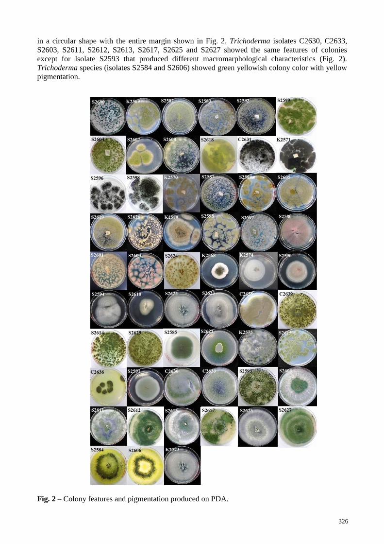
Colony of Bipolaris species K2568 showed black greyish pigment and black color of bottom
surface in the PDA culture plate (Fig. 2). The colony of Chaetomium species isolate K2574
demonstrated white cottony on the PDA culture plate with circular form and did not produce
pigmentation (Fig. 2).
The colony of Fusarium species appeared pale to white cottony mycelia on the upper surface
while the lower surface showed no pigmentation. The culture plate was white, yellowish and
pinkish, circular colony with entire margin (Fig. 2). The color of colony produced by Fusarium
species S2590 was pinkish with yellowish pigmentation in its lower surface. Whereas Fusarium
species isolates K2576, S2594 and S2622 produced white cottony mycelia with white
pigmentation. Meanwhile, isolates S2610 and S2623 showed yellowish pigmentation on the lower
surface with white color colony. Colony of Penicillium species on upper surface color was deep
green, light green, brown and greyish green as shown in Fig. 2. The texture of colony was powdery
with the presence of wrinkles on PDA. The whole colony shape was irregular and produced white
followed by yellowish pigmentation. Colony of isolate C2635 identified as Penicillium species
showed dark greyish color with white pigmentation in the lower surface (Fig. 2). In addition,
Penicillium spp. produced green colony with greyish pigmentation for isolates S2585 and S2621
(Fig. 2). Isolates K2573 and S2615 identified as Penicillium species. displayed green colony with
yellowish pigment (Fig. 2). The distribution of this colony was scattered around PDA. Isolates
C2636 and S2591 produced light green colony with light yellow pigmentation, which were
identified as Penicillium species. The colony of Trichoderma species was shown with green color
326
in a circular shape with the entire margin shown in Fig. 2. Trichoderma isolates C2630, C2633,
S2603, S2611, S2612, S2613, S2617, S2625 and S2627 showed the same features of colonies
except for Isolate S2593 that produced different macromarphological characteristics (Fig. 2).
Trichoderma species (isolates S2584 and S2606) showed green yellowish colony color with yellow
pigmentation.
Fig. 2 – Colony features and pigmentation produced on PDA.
327
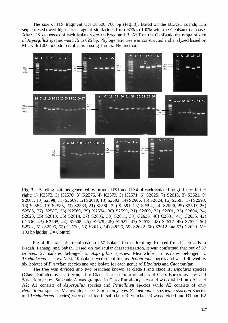
The size of ITS fragment was at 500–700 bp (Fig. 3). Based on the BLAST search, ITS
sequences showed high percentage of similarities from 97% to 100% with the GenBank database.
After ITS sequences of each isolate were analyzed and BLAST on the GenBank, the range of size
of Aspergillus species was 575 to 625 bp. Phylogenetic tree was constructed and analyzed based on
ML with 1000 bootstrap replication using Tamura-Nei method.
Fig. 3 – Banding patterns generated by primer ITS1 and ITS4 of each isolated fungi. Lanes left to
right: 1) K2573, 2) K2570, 3) K2576, 4) K2579, 5) K2571, 6) S2625, 7) S2615, 8) S2621, 9)
S2607, 10) S2598, 11) S2609, 12) S2610, 13) S2603, 14) S2606, 15) S2624, 16) S2595, 17) S2593.
18) S2584, 19) S2585, 20) S2583, 21) S2580, 22) S2591, 23) S2594, 24) S2590, 25) S2597, 26)
S2588, 27) S2587, 28) K2569, 29) K2574, 30) S2599, 31) S2600, 32) S2601, 33) S2604, 34)
S2623, 35) S2619, 36) S2614, 37) S2605, 38) S2611, 39) C2633, 40) C2631, 41) C2635, 42)
C2636, 43) K2568, 44) S2608, 45) S2629, 46) S2627, 47) S2613, 48) S2617, 49) S2592, 50)
S2582, 51) S2596, 52) C2630, 53) S2618, 54) S2626, 55) S2622, 56) S2612 and 57) C2629. M=
100 bp ladder. C= Control.
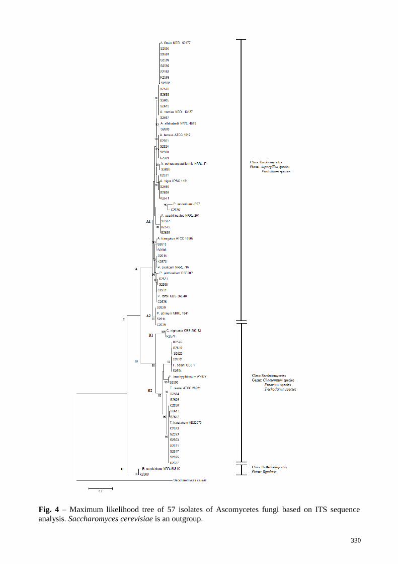
Fig. 4 illustrates the relationship of 57 isolates from microfungi isolated from beach soils in
Kedah, Pahang, and Sabah. Based on molecular characterization, it was confirmed that out of 57
isolates, 27 isolates belonged to Aspergillus species. Meanwhile, 12 isolates belonged to
Trichoderma species. Next, 10 isolates were identified as Penicillium species and was followed by
six isolates of Fusarium species and one isolate for each genus of Bipolaris and Chaetomium.
The tree was divided into two branches known as clade I and clade II; Bipolaris species
(Class Dothideomycetes) grouped in Clade II, apart from members of Class Eurotiomycetes and
Sardariomycetes. Subclade A was grouped in Class Eurotiomycetes and was divided into A1 and
A2; A1 consists of Aspergillus species and Penicillium species while A2 consists of only
Penicillium species. Meanwhile, Class Sardariomycetes (Chaetomium species, Fusarium species
and Trichoderma species) were classified in sub-clade B. Subclade B was divided into B1 and B2
328
where B1 consists of Chaetomium species while B2 consists of Trichoderma species and Fusarium
species. Saccharomyces cerevisiae (accession no: HW628115) was chosen as an outgroup as it
belonged to different class of other genus.
Discussion
Aspergillus species was the dominant species and diverse in all six beaches in Sabah, Kedah
and Pahang. By comparing to other studies, Zakaria et al. (2011) carried out a study on “Diversity
of microfungi in sandy beach soil of Teluk Aling, Pulau Pinang” and based on their identification
on morphologically, they found out seven genera known as Fusarium, Aspergillus, Trichoderma,
Culvularia, Colletotrichum, Helminthosporum and Penicillium. Moreover, there was a new record
of Penicilium pimiteouiense from beach soil in Malaysia, which reported by Teh & Latiffah (2013).
This sample was collected from sandy beach in Penang Island, which was identified based on the
production of yellow on upper surface of the colonies and yellow to orange color on the lower
surface.
Seelan et al. (2009) isolated Aspergillus species from mangrove forests in Borneo Island,
Sarawak, Malaysia. Thirty-eight isolates belonged to five species of Aspergillus. The most common
species found was Aspergillus flavipes. Teh & Latifah (2015) carried out a study on Aspergillus
species in beach sand located at Batu Ferringhi Beach, Penang. They stated that the presence of the
species was probably due to presence of discarding organic litter and environmental factors
including humidity necessary for the survival of the fungus and suitable temperature. This was
because the sand particles provide microhabitat under specific conditions for the survival of
conidia. According to Powell et al. (1994), Aspergillus cultures can rapidly grow on infertile and
cheap substrates as well as secreting their own enzymes into a medium. Therefore, it is easier for
them to culture. In addition, this genus is flexible and adaptive, thus capable of utilizing a wide
range of substrates. Moreover, Bokulich & Bamforth (2017) mentioned the ability of spores to
disperse globally in air currents and to grow almost anywhere with appropriate food and water
available. They also stated that when spores encounter a solid or liquid surface they would deposit
and germinate if the conditions of moisture are optimum for growing.
This was followed by Trichoderma species with 12 isolates identified. This genus exists in all
beaches studied except for Chenang Beach in Langkawi Island, Kedah. According to Asis &
Siddiquee (2016), many factors influence the distribution of Trichoderma species such as genetic
structure, biological niches, soil type and geographical regions. Thus, this suggests that Chenang
Beach do not favors the growth of Trichoderma species as the factors that influence the
development of this species less in Chenang Beach.
There were 10 isolates of Penicillium species identified. Penicillium species was seen
distributed in all beaches except for Mamutik Island in Sabah. Yee & Zakaria (2014) isolated
Pencillium georgiense from Batu Ferringhi Beach, Penang and confirmed it by morphological
characteristics and phylogenetic analysis. On sandy beach, many factors determine the survival and
dispersion of Penicillium species such as nature of the beach, tidal and season phenomena as well
as animal and human activities (Zakaria et al. 2011).
Fusarium species were also identified, which located in four beaches in this study, which are
Chenang Beach, Mamutik Island, Manukan Island, and Sapi Island while there are no Fusarium
species found at Tanjung Aru Beach and Balok Beach. A study done by Zakaria et al. (2011) found
seven genera of microfungi from Teluk Aling Beach, Pulau Pinang. The highest isolates found
were belonged to Fusarium genera. They stated that soil microfungi would prefer locations that
contain high organic matter content like plant roots as their habitat. Hence, Tanjung Aru Beach and
Balok Beach probably did not contain enough organic matter for the growth of Fusarium species as
there were no isolates found from both beach samples.
The least isolates of fungal species were identified as Bipolaris species and Chaetomium
species which were obtained from Chenang Beach, Kedah. In Malaysia, there are no reports on
Bipolaris species isolated from sandy beach soil; nevertheless, Kusai et al. (2015) had carried out a
study on Curvularia and related species associated with leaf spot disease of rice in Peninsular
329
Malaysia. They found 33 isolates identified as Bipolaris sorokiniana, whereas others as Curvularia
species. Moreover, Ashok et al. (2015) discovered Bipolaris species, which was isolated from
coastal area of Tuticorin Dt., Tamil Nadu, and India. Similar to Bipolaris species, there are no
study done on Chaetomium in Malaysia. Nonetheless, Migahed (2003) done a study on
“Distribution of Fungi in the Sandy Soil of Egyptian Beaches”, which isolated fungi from nine
different localities in Egypt. Four highest genera found by the study were Chaetomium,
Penicillium, Aspergillus and Trichoderma, respectively.
The size of ITS band amplified in DNA of Bipolaris species was at 575 bp compared to the
study done by Peterson & Balbalian (2010), which was 572 bp of sequence for Bipolaris isolates
using primer ITS1 and ITS4. Based on phylogenetic tree constructed, all the genera were classified
based on their classes as each of genus in the same subclades. In subclade A on A1, all of the
Aspergillus species and Penicillium species were successfully classified on the same branch of
same species. However, there were mixed-up between the sequence of branching for Penicillium
species and Aspergillus species. This shows that these two genera have a high similarity in terms of
ITS sequence since these two genera came from the same class. In addition, these two genera were
difficult to be differentiated as both genera have quite a similarity in their morphological. Volk
(2010) mentioned that there were difficulty for distinguishing between genera Aspergillus and
Penicillium species. Therefore, differentiating both genera was done based on their shapes and
arrangements of their conidia on their conidiophores. In a study done by Castrillo et al. (2012) on
“Taxonomy and phylogenetic analysis of Aspergillus section Nigri isolated from Yerba Mate in
Misiones (Argentina)”, they had constructed phylogenetic tree of Aspergillus species with
Penicillium species included in the same subclade of Aspergillus species.
Moreover, Chaetomium species was included in subclade B on B1. The bootstrap value
showed 99% similarities between subclades B1 and B2. This shows that there were high
similarities between Chaetomium species, Fusarium species and Trichoderma species as three of
these genera were classified in the same class. Ning & Sung (2008) mentioned that
Sordariomycetes is one of the largest classes in the Ascomycota where the majority members of
this class produced perithecial ascomata. Next, Fusarium species and Trichoderma species were
classified in the same subclade, which is B2 with high bootstraps percentage (94%). Bipolaris
species was classified in clade II as it is the only one in class Dothideomycetes.
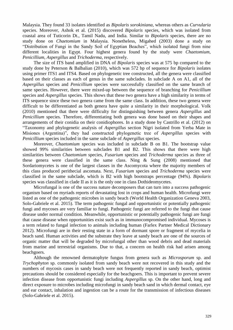
Microfungal is one of the success nature decomposers that can turn into a success pathogenic
organism based on myriads reports of devastating lost in crops and human health. Microfungi were
listed as one of the pathogenic microbes in sandy beach (World Health Organization Geneva 2003,
Solo-Gabriele et al. 2015). The term pathogenic fungal and opportunistic or potentially pathogenic
fungi and mycoses are very familiar to fungi. Pathogenic fungi are referred to the fungi that cause
disease under normal condition. Meanwhile, opportunistic or potentially pathogenic fungi are fungi
that cause disease when opportunities exist such as in immunocompromised individual. Mycoses is
a term related to fungal infection to animals including human (Farlex Partner Medical Dictionary
2012). Microfungi are in their resting state in a form of dormant spore or fragment of mycelia in
beach sand. Human activities and the substrate they leave at sandy beach are one of the sources of
organic matter that will be degraded by microfungal other than wood debris and dead materials
from marine and terrestrial organisms. Due to that, a concern on health risk had arisen among
beachgoers.
Although the renowned dermatophyte fungus from genera such as Microsporum sp. and
Trychophyton sp. commonly isolated from sandy beach were not recovered in this study and the
numbers of mycosis cases in sandy beach were not frequently reported in sandy beach, optimist
precautions should be considered especially for the beachgoers. This is important to prevent severe
infection disease from opportunistic fungi including Aspergillus sp. On the other hand, long and
direct exposure to microbes including microfungi in sandy beach sand in which dermal contact, eye
and ear contact, inhalation and ingestion can be a route for the transmission of infectious diseases
(Solo-Gabriele et al. 2015).
330
Fig. 4 – Maximum likelihood tree of 57 isolates of Ascomycetes fungi based on ITS sequence
analysis. Saccharomyces cerevisiae is an outgroup.
331
Acknowledgements
The authors would like to thank Mrs. Nor Hidayah Husain, Munirah Mohd Sattar, Sharifah
Siti Maryam Syd Abdul Rahman and Nur Baiti Abd Murad for technical assistance.
References
Ashok G, Senthilkumar G, Panneerselvam A. 2015 – Original research article diversity and
seasonal variation of soil fungi isolated from coastal area of Tuticorin Dt., Tamil Nadu, India.
International Journal Current Microbiology Application Science, 4(10), 161–178.
Asis A, Siddiquee S. 2016 – Identification of Trichoderma Species from wet paddy field soil
samples. Transactions on Science and Technology Journals, 11(1), 1–7.
Blanchet G. 2014 – Neighbor Joining or Maximum Likelihood. Retrieved from
https://www.researchgate.net/post/Neighbor_joining_or_maximum_likelihood
Bokulich NA, Bamforth CW. 2017 – Brewing Microbiology: Current Research, Omics and
Microbial Ecology. Aspergillus. Caister Academic Press Publishing. [Adobe Digital Editions
version]. doi: 10.21775/9781910190616
Castrillo ML, Fonseca MI, Bich GA, Jerke G et al. 2012 – Taxonomy and phylogenetic analysis of
Aspergillus section Nigri isolated from Yerba Mate in Misiones (Argentina). Journal of Basic
and Applied Genetics, 23(2), 19–27.
Farlex Partner Medical Dictionary. 2012 – Mycoses. Retrieved November 29, 2016, from The free
dictionary: http://medical-dictionary.thefreedictionary.com/mycoses
Kumar S, Stecher G, Tamura K. 2016 – MEGA7: Molecular evolutionary genetics analysis version
7.0 for bigger datasets. Molecular Biology Evolution, 33(7), 1870–4.
Kusai NA, Azmi MMZ, Zulkifly S, Yusof MT, Nur Ain Izzati MZ. 2015 – Morphological and
molecular characterization of Curvularia and related species associated with leaf spot disease
of rice in peninsular Malaysia. Rend. Accademia Nazionale Dei Lince, 27, 205–214.
Leslie JF, Summerell BA. 2006 – The Fusarium Laboratory Manual. Blackwell Publishing,
Hoboken, 1-2. Retrieved from https://doi.org/10.1002/9780470 278376
Migahed FF. 2003 – Distribution of Fungi in the Sandy Soil of Egyptian Beaches. Pakistan Journal
of Biological Sciences, 6(2), 860–866.
Ning Z, Sung GH. 2008 – Sordariomycetes. The Tree of Life Web Project. Retrieved from
http://tolweb.org/Sordariomycetes/29050/2008.01.14 in The Tree of Life Web Project
Peterson MT, Balbalian CJ. 2010 – First report of Bipolaris oryzae causing leaf spot of switchgrass
in Mississippi. APS Journals, 94 (5), 643–651.
Powell KA, Renwick A, Peberdy JF. 1994 – The Genus Aspergillus from Taxonomy and Genetics
to Industrial Application. Plenum Press Publishing.
Sabino R, Rodrigues R, Costa I, Carneiro C et al. 2014 – Routine screening of harmful
microorganisms in beach sands: Implications to public health. Science of the Total
Environment, 472, 1062–1069.
Seelan JSS, Ali AAKF, Muid S. 2009 – Aspergillus species isolated from mangrove forests in
Borneo Island, Sarawak, Malaysia. Journal of Threatened Taxa, 1(6), 344–346.
Solo-Gabriele HM, Harwood VJ, Kay D, Fujioka RS et al. 2015 – Beach sand and the potential for
infectious disease transmission: observations and recommendations. Journal of the Marine
Biological Association of the United Kingdom, 96(1), 1–20.
Teh LY, Latiffah Z. 2013 – A new record of Penicillium pimiteouiense from beach soil in
Malaysia. The Korean Society of Mycology, 41(4), 256–259.
Teh LY, Latiffah Z. 2015 – Occurrence and molecular characterization of Aspergillus species in
beach sand. Malaysian Applied Biology, 44(2), 119–127.
Volk T. 2010 – Aspergillus. Retrieved from Department of Biology of University of Wisconsin-La-
Crosse website: http://botit.botany.wisc.edu/toms_fungi/feb97.html
White TJ, Bruns T, Lee S, Taylor J. 1990 – Amplification and direct sequencing of fungal
ribosomal RNA genes for phylogenies. In: Innis MA, Gelfand DH, Sninsky JJ, White TJ.
332
(eds) PCR Protocols: A Guide to Methods and Applications. Academic Press, San Diego,
315–322.
World Health Organization Geneva. 2003 – Chapter 6: Microbial aspects of beach sand quality. In
Guidelines for safe recreational water environments (pp. 118–127). World Health
Organization 2003.
Yee TL, Zakaria L. 2014 – The First Report of Penicillium georgiense in Malaysia. The Korean
Society of Mycology, 42(3), 274–278.
Zakaria L, Yee TL, Zakaria M, Salleh B. 2011 – Diversity of microfungi in sandy beach soil of
Teluk Aling, Pulau Pinang. Tropical Life Sciences Research, 22(1), 71–80.