tissue culture of nicotiana plumbaginifolis ting chin yee 2007
DESCRIPTION
thesis paperTRANSCRIPT
i
PSZ 19:16 (Pind. 1/97)
UNIVERSITI TEKNOLOGI MALAYSIA
BORANG PENGESAHAN STATUS TESIS ♦ JUDUL : TISSUE CULTURE OF Nicotiana plumbaginifolis.
SESI PENGAJIAN : 2006/2007 Saya : TING CHIN YEE
(HURUF BESAR) mengaku membenarkan tesis (PSM/Sarjana/Doktor Falsafah)* ini disimpan di Perpustakaan Universiti Teknologi Malaysia dengan syarat-syarat kegunaan seperti berikut : 1. Tesis adalah hakmilik Universiti Teknologi Malaysia. 2. Perpustakaan Universiti Teknologi Malaysia dibenarkan membuat salinan untuk tujuan
pengajian sahaja. 3. Perpustakaan dibenarkan membuat salinan tesis in sebagai bahan pertukaran antara institusi
pengajian tinggi. 4. **Sila tandakan (� )
SULIT ( Mengandungi maklumat yang berdarjah keselamatan atau kepentingan Malaysia seperti yang termaktub di dalam AKTA RAHSIA RASMI 1972) TERHAD ( Mengandungi maklumat TERHAD yang telah ditentukan oleh organisasi/badan di mana penyelidikan dijalankan) TIDAK TERHAD
Disahkan oleh
________________________________ ______________________________ (TANDATANGAN PENULIS) (TANDATANGAN PENYELIA) Alamat Tetap : P.O.Box 3, Dr. Fahrul Zaman Huyop 96507, Bintangor, Sarawak. Nama Penyelia Tarikh : 7 May 2007 Tarikh : 7 May 2007
CATATAN : * Potong yang tidak berkenaan.
** Jika tesis ini SULIT atau TERHAD, sila lampirkan surat daripada pihak berkuasa/ organisasi berkenaan dengan menyatakan sekali tempoh tesis ini perlu dikelaskan sebagai SULIT atau TERHAD.
◆♦ Tesis dimaksudkan sebagai tesis bagi Ijazah Doktor Falsafah dan Sarjana secara penyelidikan, atau disertasi bagi pengajian secara kerja kursus dan penyelidikan, atau Laporan Projek Sarjana Muda (PSM).
ii
Tissue Culture of Nicotiana plumbaginifolis
This thesis is submitted in fulfillment of the requirements of
the Bachelor of Science degree in Industrial Biology, Faculty
of Science, University Technology Malaysia
By
TING CHIN YEE
2007
i
Abstract
Tissue Culture of Nicotiana plumbaginifolis
Ting Chin Yee and Fahrul Zaman Huyop
Nicotiana species is a type of plant species commonly used in plant science research,
including research of plant genetics. The main objective of this research is to grow
successfully Nicotiana plumbaginifolis through tissue culture technique. Seeds of
Nicotiana plumbaginifolis were obtained from Malaysian Palm Oil Board. Seeds
were surface sterilized using 20% hypochlorite solution. Sterilized seeds were placed
on the surface of Murashige and Skoog (MS) medium without plant growth
regulators (hormones). Young leaves started to grow from seed after two weeks.
Young leaves were taken and placed on fresh MS medium with addition of
appropriate concentration of hormone benzylaminopurine (BAP) and
naphthaleneacetic acid (NAA). Based on initial finding, 1.0 mg/L BAP for shoot
initiation and 0.1 mg/L NAA for root induction were the best concentration used to
initiate callus. When callus regenerated, callus was transferred to fresh MS medium
containing hormones (1.0 mg/L BAP and 0.1 mg/L NAA) until shoots were formed.
ii
Abstrak
Kultur Tisu Tumbuhan Nicotiana plumbaginifolis
Ting Chin Yee dan Fahrul Zaman Huyop
Spesies Nicotiana ialah salah satu spesies tumbuhan yang sering kali diggunakan
dalam kajian sains tumbuhan, termasuk kajian tentang genetik tumbuhan. Objektif
utama dalam kajian ini ialah untuk menanam Nicotiana plumbaginifolis dengan
menggunakan teknik kultur tisu. Biji benih Nicotiana plumbaginifolis diambil
daripada Malaysia Palm Oil Board (MPOB). Biji benih disterilkan dengan
menggunakan 20% larutan hypochlorite. Biji benih yang steril diletakkan atas
permukaan agar kultur tisu, iaitu Murashige dan Skoog (MS) medium tanpa
penggunaan hormon. Daun muda mula bertumbuh daripada biji benih selepas dua
minggu. Daun muda dipetik dan diletakkan dalam agar kultur tisu yang baru. Hormon
tumbuhan yang spesifik iaitu benzylaminopurine (BAP) dan naphthaleneacetic acid
(NAA) dengan kepekatan yang tertentu ditambahkan ke dalam agar. Berdasarkan
kajian yang dijalankan, didapati 1.0 mg/L BAP (pembentukkan pucuk) dan 0.1 mg/L
NAA (pembentukkan akar) adalah kepekatan yang paling sesuai untuk pembentukkan
kalus daripada explant. Selepas kalus terbentuk, kalus dipindahkan ke agar kultur tisu
yang baru yang mengandungi hormone (1.0 mg/L BAP dan 0.1 mg/L NAA) sehingga
pucuk terbentuk.
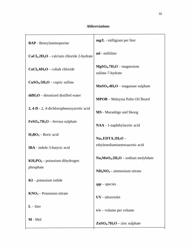
iii
BAP - Benzylaminopurine
CaCl2.2H2O – calcium chloride 2-hydrate
CoCl2.6H2O – cobalt chloride
CuSO4.5H2O – cupric sulfate
ddH2O – deionized distilled water
2, 4-D - 2, 4-dichlorophenoxyacetic acid
FeSO4.7H2O – ferrous sulphate
H3BO3 – Boric acid
IBA - indole 3-butyric acid
KH 2PO4 – potassium dihydrogen
phosphate
KI – potassium iodide
KNO 3 – Potassium nitrate
L – liter
M - Mol
Abbreviations
mg/L - milligram per liter
ml - milliliter
MgSO4.7H2O – magnesium
sulfate-7-hydrate
MnSO4.4H2O – maganase sulphate
MPOB – Malaysia Palm Oil Board
MS - Murashige and Skoog
NAA - 1-naphthylacetic acid
Na2.EDTA.2H2O –
ethylenediaminetetraacetic acid
Na2MoO4.2H2O – sodium molybdate
NH4NO3 – ammonium nitrate
spp – species
UV - ultraviolet
v/v – volume per volume
ZnSO4.7H2O – zinc sulphate

iv
“I hereby would like to state that I have read this thesis and in my opinion this thesis
fulfils the scope and quality to be awarded a bachelor degree in Industrial Science
(Industrial Biology)”.
Signature : ………………………………
Supervisor : Dr. Fahrul Zaman Huyop
Date : 7 May 2007
v
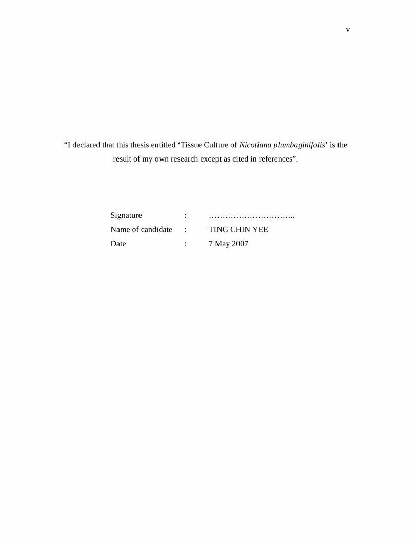
“I declared that this thesis entitled ‘Tissue Culture of Nicotiana plumbaginifolis’ is the
result of my own research except as cited in references”.
Signature : …………………………..
Name of candidate : TING CHIN YEE
Date : 7 May 2007

vi
Specially dedicated to our mighty God, my beloved family,
my loving father and mother…
Thanks for everything from you.

vii
Acknowledgements
First of all, I wish to express my special thanks and gratitude to my supervisor,
Dr. Fahrul Zaman Huyop for his invaluable supports, guidance and encouragement in
completing this project. I greatly appreciate his helpful comments, suggestions and
corrections.
Besides, I am indebted to all lecturers in biology department, all laboratory
staffs of Microbiology Laboratory, Plant Tissue Culture Laboratory and also post-
graduated students for their support and guidance.
I also wish to express my special thanks to my aunt, my friends, and all my
fellow coursemates who have directly or indirectly helped and shared information with
me in finishing my project.
Last but not least, I wish to extend my appreciation to my beloved parents, Mr.
and Mrs. Ting and family for their loving and moral support in completing my thesis.
viii
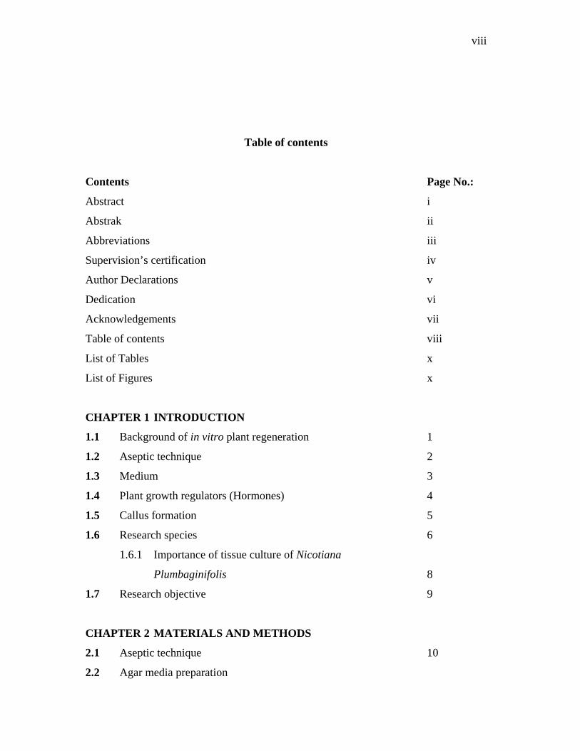
Table of contents
Contents Page No.:
Abstract i
Abstrak ii
Abbreviations iii
Supervision’s certification iv
Author Declarations v
Dedication vi
Acknowledgements vii
Table of contents viii
List of Tables x
List of Figures x
CHAPTER 1 INTRODUCTION
1.1 Background of in vitro plant regeneration 1
1.2 Aseptic technique 2
1.3 Medium 3
1.4 Plant growth regulators (Hormones) 4
1.5 Callus formation 5
1.6 Research species 6
1.6.1 Importance of tissue culture of Nicotiana
Plumbaginifolis 8
1.7 Research objective 9
CHAPTER 2 MATERIALS AND METHODS
2.1 Aseptic technique 10
2.2 Agar media preparation
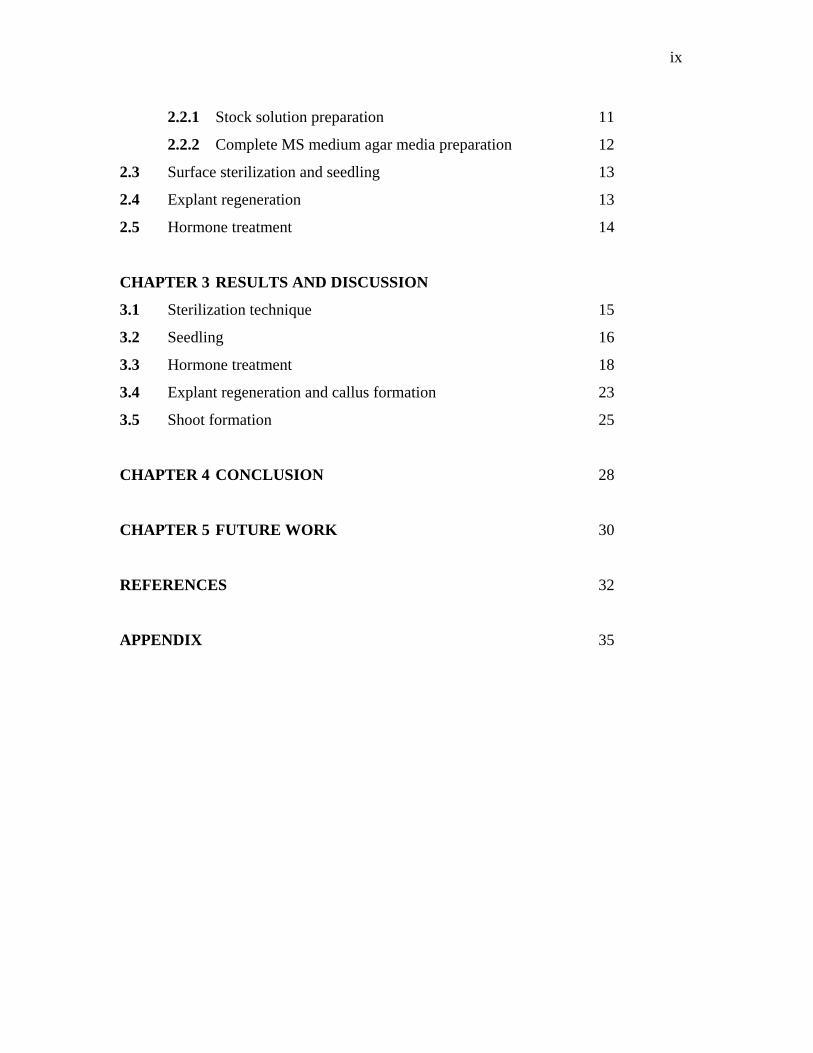
ix
2.2.1 Stock solution preparation 11
2.2.2 Complete MS medium agar media preparation 12
2.3 Surface sterilization and seedling 13
2.4 Explant regeneration 13
2.5 Hormone treatment 14
CHAPTER 3 RESULTS AND DISCUSSION
3.1 Sterilization technique 15
3.2 Seedling 16
3.3 Hormone treatment 18
3.4 Explant regeneration and callus formation 23
3.5 Shoot formation 25
CHAPTER 4 CONCLUSION 28
CHAPTER 5 FUTURE WORK 30
REFERENCES 32
APPENDIX 35
x
LIST OF TABLES Page No.:
CHAPTER 2
Table 2.1. List of components in MS medium and the concentration
in stock solution 11
Table 2.2. Different concentration of hormone treatment was designed 14
CHAPTER 3
Table 3.1. Total explants regenerated for each type of hormone
concentration 21
LIST OF FIGURES ` Page No.:
CHAPTER 1
Figure 1.1. Structural formula of some auxins and cytokinins 4
Figure 1.2. Nicotiana spp 7
CHAPTER 3
Figure 3.1. Seeds were sown on the surface of MS agar media without
addition of hormones 17
Figure 3.2. Young plants were developed from seedling 17
Figure 3.3. A graph shows the total callus formed in every types of
combination of hormone concentration 21
Figure 3.4. Different sizes of callus formed based on varies hormone
concentration after hormone treatment for three weeks 22
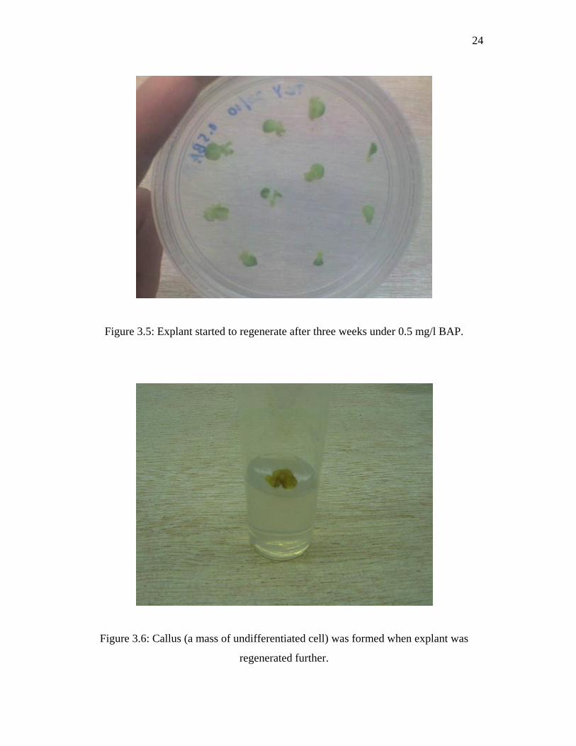
Figure 3.5. Explant started to regenerate after three weeks under
0.5 mg/L BAP 24
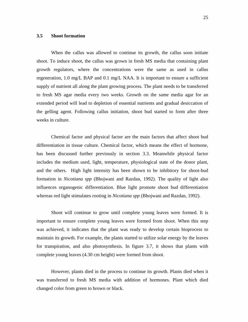
Figure 3.6. Callus was formed when explant was regenerate further 24

xi
Figure 3.7. Plants with complete young leaves (around 4.30cm) were
formed from shoot 27
Figure 3.8. Plant was died; the color has change from green to black 27
1
CHAPTER 1
Introduction
1.1 Background of in vitro plant regeneration
Nowadays, tissue culture technique (including tissue culture of cells, tissue or
organ) has become an important technique in plant research. This tissue culture
technique has been made practical and has been applied for commercial purpose.
Plant tissue culture is also called micropropagation, which was a technique
consisting of taking a piece of a plant (such as a stem tip node, meristem, embryo, or
even a seed) and placing it in a sterile nutrient medium where it multiplies. Plant tissue
can be described as plasticity and totipotency. Plasticity, allows plants to alter their
metabolism; growth and development to best suit their environment. While totipotency
can be defined as the ability of each living cell of a multicellular organism to develop
independently if provided with proper external conditions. A totipotent cell is one that
is capable of developing by regeneration into whole organisms, and this term was
probably coined by Morgan (1901).
The first experiment to culture isolated, fully differentiated plants cells in vitro
on an artificial medium was originated by Haberlandt (Dodds and Roberts, 1995). The
2
cultured cells able to survived for several months, but they were incapable of
proliferation. As a result for failure to obtain cell division, tissue culture was being
continued by others researcher, and lately being proven to be a success by White (1934).
White (1934) had successfully to culture a whole plant by using root of tomato. Further
researches had been done by a group of researchers that is Gautheret et al (1939) that
showed cells can be cultured continuously and go through differentiation step. These
finding has encouraged more research being done on plant tissue culture, from 1940 to
1960.
1.2 Aseptic technique
The importance of maintaining a sterile environment during the culture of plant
tissues was necessary. A few simple precautions to avoid contamination will save
valuable time in not repeating experiments.
The main factor for aseptic technique was selecting a right working area and
using the aseptic tools. The purpose for selecting a suitable working area was to prevent
the possible flow of unfiltered air over the disinfected working area. Most tissue culture
procedures were conducted in sterile operations, such as laminar flow cabinet. Besides
the special design of gentle flow of sterile air in cabinet, aseptic cabinet is also
equipped with germicidal lamp emitting ultraviolet (UV) light. This type of radiation is
useful in eliminating airborne contaminants and for surface disinfection.
Glassware and all the tools used for tissue culture process can also cause
contamination. It is extremely necessary to autoclave all the material before using it, so
that all the microbial contaminants are destroyed.
3
1.3 Medium
Nutritional requirements for optimal growth of a tissue in vitro may vary with
the species. Even tissues from different parts of a plant may have different requirements
for satisfactory growth (Murashige and Skoog, 1962). No single medium can be
suggested as being entirely satisfactory for all types of plant tissues and organs. When
starting with a new system, it is essential to work out a medium that would fulfill the
specific requirements of that tissue.
The components of a plant tissue culture medium included macronutrients,
micronutrients, a separate iron supplement, vitamins, a carbon source, and usually plant
growth regulators. Amino acids and various nitrogenous compounds may be present in
the vitamin mixture. Macronutrients were nitrogen, phosphorus, potassium, calcium,
magnesium, and sulfur. These macronutrients are also called inorganic chemicals, and
were essential elements required in relatively large amounts. Micronutrients, were
traces of certain elements required by all plant cells. Micronutrient elements include
iron, manganese, zinc, boron, copper, molybdenum, iodine, cobalt and chlorine.
Meanwhile, vitamins have catalytic functions in enzyme systems and were required
only in trace amounts.
The need to culture diverse tissue and organs has led to the development of
several recipes of nutrient medium. White (1943) has created a new medium that is low
in salt and free of ammonium ion. White’s medium was the earliest control medium
that consists of all needed nutrients, and applied widely for root cultivation. On the
other hand, Murashige and Skoog (1962) have developed MS medium that consist of
ammonium, nutrient and others mineral includes inorganic nutrient that needed by plant
to establish growth. Until now, the most known mediums that researchers used in plant
tissue culture were Murashige and Skoog medium (1962) that is high salt or B5
(Gamborg et al., 1968).
4
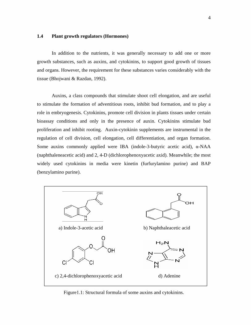
1.4 Plant growth regulators (Hormones)
In addition to the nutrients, it was generally necessary to add one or more
growth substances, such as auxins, and cytokinins, to support good growth of tissues
and organs. However, the requirement for these substances varies considerably with the
tissue (Bhojwani & Razdan, 1992).
Auxins, a class compounds that stimulate shoot cell elongation, and are useful
to stimulate the formation of adventitious roots, inhibit bud formation, and to play a
role in embryogenesis. Cytokinins, promote cell division in plants tissues under certain
bioassay conditions and only in the presence of auxin. Cytokinins stimulate bud
proliferation and inhibit rooting. Auxin-cytokinin supplements are instrumental in the
regulation of cell division, cell elongation, cell differentiation, and organ formation.
Some auxins commonly applied were IBA (indole-3-butyric acetic acid), α-NAA
(naphthaleneacetic acid) and 2, 4-D (dichlorophenoxyacetic axid). Meanwhile; the most
widely used cytokinins in media were kinetin (furfurylamino purine) and BAP
(benzylamino purine).
Figure1.1: Structural formula of some auxins and cytokinins.
a) Indole-3-acetic acid b) Naphthaleacetic acid
c) 2,4-dichlorophenoxyacetic acid d) Adenine
5
Plant growth regulators can be subdivided into natural occurring and synthetic
types. BAP, NAA, 2, 4-D were some example of synthetic growth regulators. These
synthetic growth regulators have several advances feature. They help to increase yields,
to make uniform ripening and facilitate harvesting, increased resistance against some
unfavorable factors (drought, frost), and regulated rooting, fruit setting (Sebanek, 1992).
In ornamental horticulture, they were used for regulation of plant habit, time of
flowering, and size. Another advance of using synthetic growth regulator is it would not
be degraded easily. Natural occurring hormones were rapidly degraded by light and
enzymatic oxidation (Dodds and Roberts, 1995).
1.5 Callus formation
Callus refers to a disorganized proliferated mass of actively dividing cells. A
callus consists of an amorphous mass of loosely arranged thin-walled parenchyma cells
arising from the proliferating cells of the cultured plants. The most important function
of callus was that it has the potential to develop normal roots, shoots, and embryoids
that can form plants. In addition, it can be used to initiate a suspension culture (Dodds
and Roberts, 1995). For callus formation, pieces of cotyledon, hypocotyls, stem, leaf, or
embryo were usually used. Sinnott (1960) had described some of the early observations
on wound callus formation. The stimuli in the initiation of wound callus were the
endogenous hormones auxin and cytokinin. Using tissue culture techniques, callus
formation can be induced in numerous plant tissues and organs that do not usually
develop callus in response to an injury (Street, 1969).
Establishment of a callus from an explant was divided into three developmental
stages: induction, cell division, and differentiation. Induction phase was metabolism
stimulated prior to mitotic activity. The length of induction phase depends on the
physiological status of the explant cells as well as the cultural condition (Dodds &
Roberts, 1995). Cell division phase, a phase of active cell division as the explants cells
revert to a meristematic state. The third phase involves the appearance of cellular
6
differentiation and the expression of certain metabolic pathways that lead to the
formation of secondary products. It becomes possible to subculture the callus to a fresh
medium, after the callus had been grown for a while in association with the original
tissue.
The first successful prolonged cultures of experimentally induced callus were
achieve in 1939 almost simultaneously at the research laboratory of Gautheret in Paris,
Nobecourt in Grenoble, and White in Princeton. These cultures were originally derived
from explants of cambial tissue of carrot and tobacco. Callus appears as yellowish,
white, green, or pigmented with anthocyanin.
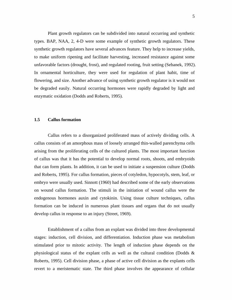
1.6 Research species
Nicotiana plumbaginifolis is related to garden vegetables, flowers, weeds and
poisonous herbs. It is also refers to a genus of broad-leafed plants of the nightshade
family. The family of plant is Solanaceae, and the genus of plant is Nicotiana. There is
about 100 species of Nicotiana. Nicotiana spp has huge green leaves and very sweet-
scented flowers with variety colors. The flowers are hermaphrodite, which has both
male and female organs. Pollination can be done through insects such as bees, moths,
and butterflies. The plant prefers sandy, loamy and clay soils and requires well-drained
soil.
7
Figure 1.2: Nicotiana spp
Nicotiana spp origin from North and South America, and it was first discovered
by South Americans. Nicotiana spp, is believed to be first used by native American by
chewing or snorting the leaves for medicinal purpose.
As it is from genus Nicotiana, this plant contains nicotine. Nicotiana tabaccum
and Nicotiana rustica having high nicotine content, and this makes them suitable as
cigarettes or cigars. Nicotiana spp has a long history in medical field. The leaves are
antispasmodic, discutient, diuretic, emetic, expectorant, irritant, narcotic, sedative and
sialagogue. The leaves were mostly used to treat rheumatic swelling, skin diseases and
scorpion stings to relieve the pain. Besides, nicotine can also be extracted and used as
an insecticide, which the leaves being take and dried. The dried leaves remain effective
for 6 months. The juice of the leaves can be rubbed on the body as an insect repellent.
The leaves have been dried and chewed as an intoxicant.
8
1.6.1 Importance of tissue culture of Nicotiana plumbaginifolis
In the research, Nicotiana spp was commonly used as the first model to study
variety aspect of plant, such as genomic transformation, metabolism, or adaptation of
plants to environmental changes. Life cycle for Nicotiana spp is about 4-5 month.
Although the life cycle is about twice as long as that of Arabidopsis thaliana (2 month)
and large growing space required, the larger size of Nicotiana reproductive structures is
an advantage for some expression and manipulation studies (McDaniel, 1999).
For example recombinant human tissue transglutaminase produced into tobacco
suspension cell cultures (Sorrentino, 2004). The cell was active and recognizes
autoantibodies in the serum of celiac patients. The recombinant enzyme was
successfully expressed in different plant cell compartments.
Another advance characteristic of Nicotiana species was the stable chromosome
numbers. As cell suspension cultures have been used frequently for mutant isolation
and protoplast fusion, the establishment of cell suspension cultures with stable
chromosome number is an essential factor for regeneration of stable haploid and diploid
plants (Evans and Gamborg, 1982). Leaf tissue of diploid tobacco has been shown to
have only diploid cells and this tissue is preferable for the initiation of cell cultures.
In previous research, several species of Nicotiana have been combined with
cultivated tobacco using sexual hybridization to introduce useful agricultural traits into
the tobacco. This had resulted in the development of several cultivated tobacco varieties
that contain genes derived from the wild species. As routine techniques were available
for plant regeneration from isolated leaf mesophyll protoplasts of numerous Nicotiana
species, more effort has been directed toward production of interspecific Nicotiana
somatic hybrids (Evans et al, 1982). New variety of plants with new characteristic had
been discovered. For example transgenic plant, developing resistant plant, plant with
high commercial value and others.
9
An example for transgenic plant using tobacco was a chimeric green fluorescent
protein gene as an embryogenic marker in transgenic cell culture of Nicotiana
plumbaginifolia (Chesnokov, 2001). In the tissue culture process, only a limited
number of cells from the in vitro culture actually undergo the transition of somatic into
embryogenic cells. The problem was that no clear evidence about the morphogenetic
changes can be observed. Therefore, specific markers with the ability to distinguish
precisely between embryogenic and non-embryogenic cells were required in order
separate out the cells. Insertion of the marker gene into transgenic Nicotiana has been
proven successful.
Nicotiana spp has also been used in the study of several enzyme activities.
Investigation on Nicotiana plumbaginifolis has been done through RNA hybridization
to distribute the differences level of enzyme production according different part of the
plant.
1.7 Research objectives
The main objective of this research was:
1. To establish plant tissue culture of Nicotiana plumbaginifolis.
10
CHAPTER 2
Materials and methods
2.1 Aseptic technique
Before starting the experiment, it is necessary to ensure all the material and
tools used are sterile. Three most important aseptic techniques that need to be done
were autoclaving, filter sterilization, and surface sterilization.
All glassware, tools and chemical materials that heat resistant were autoclaved
at temperature 121°C for 20 minutes. Microfiltration was used for media components
that were not heat resistant, such as plants growth regulators and vitamins. The working
area was generally disinfected with either ethanol or isopropanol (70% (v/v)), and also
UV light.
Surface sterilization was done before seedling. The seeds were first immersed in
20% (v/v) hypochlorite for 10 to 15 minutes. Then the seeds were rinsed in four
changes of sterile distilled water. After rinsing, the seeds were poured on a sterile filter
paper to dry off the unwanted water.
11
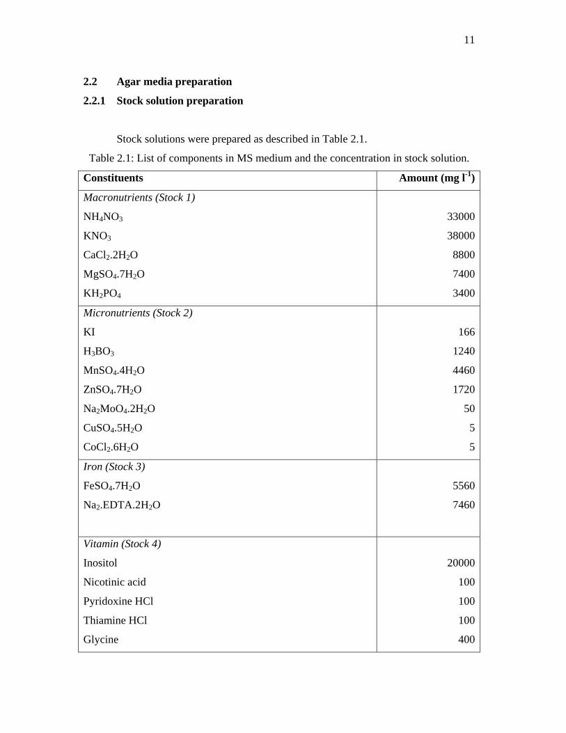
2.2 Agar media preparation
2.2.1 Stock solution preparation
Stock solutions were prepared as described in Table 2.1.
Table 2.1: List of components in MS medium and the concentration in stock solution.
Constituents Amount (mg l-1)
Macronutrients (Stock 1)
NH4NO3
KNO3
CaCl2.2H2O
MgSO4.7H2O
KH2PO4
33000
38000
8800
7400
3400
Micronutrients (Stock 2)
KI
H3BO3
MnSO4.4H2O
ZnSO4.7H2O
Na2MoO4.2H2O
CuSO4.5H2O
CoCl2.6H2O
166
1240
4460
1720
50
5
5
Iron (Stock 3)
FeSO4.7H2O
Na2.EDTA.2H2O
5560
7460
Vitamin (Stock 4)
Inositol
Nicotinic acid
Pyridoxine HCl
Thiamine HCl
Glycine
20000
100
100
100
400
12
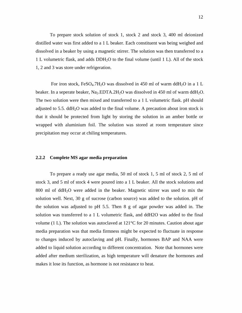
To prepare stock solution of stock 1, stock 2 and stock 3, 400 ml deionized
distilled water was first added to a 1 L beaker. Each constituent was being weighed and
dissolved in a beaker by using a magnetic stirrer. The solution was then transferred to a
1 L volumetric flask, and adds DDH2O to the final volume (until 1 L). All of the stock
1, 2 and 3 was store under refrigeration.
For iron stock, FeSO4.7H2O was dissolved in 450 ml of warm ddH2O in a 1 L
beaker. In a seperate beaker, Na2.EDTA.2H2O was dissolved in 450 ml of warm ddH2O.
The two solution were then mixed and transferred to a 1 L volumetric flask. pH should
adjusted to 5.5. ddH2O was added to the final volume. A precaution about iron stock is
that it should be protected from light by storing the solution in an amber bottle or
wrapped with aluminium foil. The solution was stored at room temperature since
precipitation may occur at chiling temperatures.
2.2.2 Complete MS agar media preparation
To prepare a ready use agar media, 50 ml of stock 1, 5 ml of stock 2, 5 ml of
stock 3, and 5 ml of stock 4 were poured into a 1 L beaker. All the stock solutions and
800 ml of ddH2O were added in the beaker. Magnetic stirrer was used to mix the
solution well. Next, 30 g of sucrose (carbon source) was added to the solution. pH of
the solution was adjusted to pH 5.5. Then 8 g of agar powder was added in. The
solution was transferred to a 1 L volumetric flask, and ddH2O was added to the final
volume (1 L). The solution was autoclaved at 121°C for 20 minutes. Caution about agar
media preparation was that media firmness might be expected to fluctuate in response
to changes induced by autoclaving and pH. Finally, hormones BAP and NAA were
added to liquid solution according to different concentration. Note that hormones were
added after medium sterilization, as high temperature will denature the hormones and
makes it lose its function, as hormone is not resistance to heat.
13
Then, liquid solution was poured to a suitable container, such as petri dishes,
culture tube, marjenta jar, or wide mouth conical flask. These tools were being
sterilized before use. After pouring, the containers were left in a sterile laminar flow
cabinet, to let the agar media to solidify. Next, the container closed tightly and wrapped
using parafilm. The solidified agar was then placed on a suitable place for storage.
2.3 Seedling
The original source of plant was the seeds of Nicotiana plumbaginifolis. The
seeds were obtained from MPOB. The sizes of seeds were very fine. Seedling, is a
process to sown the seeds and grow the desired plant. Sterile seeds were used to grow
young plants. By using a forceps, the seeds were sown carefully on the surface of MS
agar media that does not containing any plant growth regulators. Each Petri dish can be
sown with 30 seeds. The Petri dishes were closed tightly using parafilm to prevent any
cross contamination.
The Petri dishes were then placed on tray and let it grow under fluorescence
light until young plants and leaves developed. After young plants with young leaves
were developed, the leaves were taken as explant for regeneration.
2.4 Explant regeneration
Explant, is an excised fragment of plant tissue or organ used to start a tissue
culture, and it can also be called as primary explant. During this research, leaves were
used as explant. The leaves were cut from young plants. As the leaves were cut from
the plant free from contaminant, the leaves do not require surface disinfection. The
leaves were transferred to new MS agar media supplemented with plant growth
regulators. The petri dishes were closed tightly using parafilm and left on tray. The
explant were left to regenerate until callus is formed from explant.
14
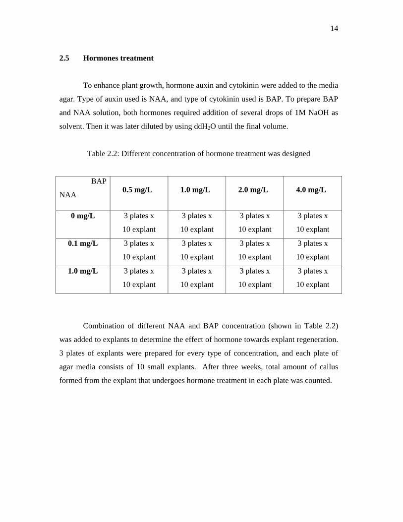
2.5 Hormones treatment
To enhance plant growth, hormone auxin and cytokinin were added to the media
agar. Type of auxin used is NAA, and type of cytokinin used is BAP. To prepare BAP
and NAA solution, both hormones required addition of several drops of 1M NaOH as
solvent. Then it was later diluted by using ddH2O until the final volume.
Table 2.2: Different concentration of hormone treatment was designed
BAP
NAA
0 mg/L 3 plates x
10 explant
3 plates x
10 explant
3 plates x
10 explant
3 plates x
10 explant
0.1 mg/L 3 plates x
10 explant
3 plates x
10 explant
3 plates x
10 explant
3 plates x
10 explant
1.0 mg/L 3 plates x
10 explant
3 plates x
10 explant
3 plates x
10 explant
3 plates x
10 explant
Combination of different NAA and BAP concentration (shown in Table 2.2)
was added to explants to determine the effect of hormone towards explant regeneration.
3 plates of explants were prepared for every type of concentration, and each plate of
agar media consists of 10 small explants. After three weeks, total amount of callus
formed from the explant that undergoes hormone treatment in each plate was counted.
0.5 mg/L 1.0 mg/L 2.0 mg/L 4.0 mg/L
15
CHAPTER 3
Results and discussion
3.1 Sterilization technique
Sterilization is an important element to ensure every thing was sterile when
doing the research of tissue culture.
In the experiment, hypochlorite was used to sterilize the seed’s surface. Seeds
were dipped in the hypochlorite solution for 10 to 15 minutes, as this is the suitable
time range. Dipping the seeds in hypochlorite solution for a long time may spoil the
seed surface, and kill the cell inside the seeds. As a result, long exposure time of seeds
with hypochlorite caused less seeds generation. Meanwhile short sterilization time was
not enough for a total elimination of contamination or microorganism. After dipping the
seeds in hypochlorite, seeds were washed with sterilized distilled water repeatedly to
wash off hypochlorite that was stuck on seed’s surface. Excessive amount of
hypochlorite within the seed’s surface give more harm than good, as it will
continuously break and kill the cells of seed.
Plant tissue culture media, which contain a high concentration of sucrose,
support the growth of many micro-organisms, such as bacteria and fungi. These
16
microbes generally grow faster than the cultured tissue and finally kill it. Apart from
that, the contaminants may also give out metabolic waste which is toxic to plant tissues.
To minimize the possibility of contamination, several steps should be done with caution,
such as hands, wrists, and forearms should be cleaned, and should not pass the hand or
arm directly over a sterile exposed surface, such as open agar plate. All sterilize open
surfaces should be placed as far back in the hood as conveniently possible. When
pouring sterile liquids, the bottle should be grasping at the base, and hands should be
kept as far as possible from the open tube or petri dishes receiving the liquids.
3.2 Seedling
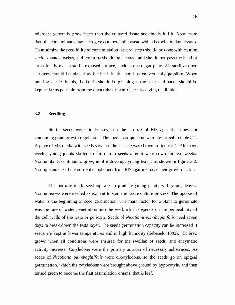
Sterile seeds were firstly sown on the surface of MS agar that does not
containing plant growth regulators. The media components were described in table 2.1.
A plate of MS media with seeds sown on the surface was shown in figure 3.1. After two
weeks, young plants started to form from seeds after it were sown for two weeks.
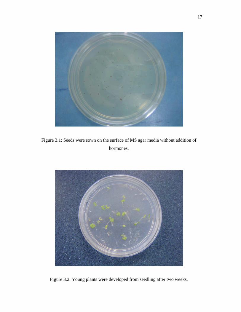
Young plants continue to grow, until it develops young leaves as shown in figure 3.2.
Young plants used the nutrient supplement from MS agar media as their growth factor.
The purpose to do seedling was to produce young plants with young leaves.
Young leaves were needed as explant to start the tissue culture process. The uptake of
water is the beginning of seed germination. The main factor for a plant to germinate
was the rate of water penetration into the seed, which depends on the permeability of
the cell walls of the testa or pericarp. Seeds of Nicotiana plumbaginifolis need seven
days to break down the testa layer. The seeds germination capacity can be increased if
seeds are kept at lower temperatures and in high humidity (Sebanek, 1992). Embryo
grows when all conditions were ensured for the swollen of seeds, and enzymatic
activity increase. Cotyledons were the primary sources of necessary substances. As
seeds of Nicotiana plumbaginifolis were dicotyledons, so the seeds go on epigeal
germination, which the cotyledons were brought above ground by hypocotyls, and then
turned green to become the first assimilation organs, that is leaf.
17
Figure 3.1: Seeds were sown on the surface of MS agar media without addition of
hormones.
Figure 3.2: Young plants were developed from seedling after two weeks.
18
3.3 Hormone treatment
This experiment was carried out to determine the suitable concentration
of hormone (shown in table 2.2) to induce callus and shoot-bud formation. In the
previous research done by other researchers; different hormone and different
concentration were used. The use of hormone for a plant species may vary under
influence of different environment, such as the weather. So, this is the purpose for us to
redo the hormone test, as plant might need different hormone concentration that varies
from what the research done before. In a paper written by Sun and Kang (2003), 1.2
mg/L BAP was used. The amount of NAA used by researchers was in the range
between 0.2 to 1 mg/L. By referring to these previous researches, a new hormone
treatment was designed. The concentration of BAP tested was 0.5 mg/l, 1 mg/l, 2 mg/l,
and 4 mg/l, while the concentration of NAA used was 0 mg/l, 0.1 mg/l, and 1.0 mg/l.
Explant (young leaves) was taken from young plant and grown on fresh MS
media containing hormones. After the explants were grown in different concentration
of hormones for four weeks, the observation was done. The number of callus formed
from explant was counted. Callus was the intermediate structure before shoot initiation.
Hormone capable of inducing shoot formation, also induce the callus formation. These
means any hormone that induces or increase the time to form callus, can also give the
same effect for shoot initiation (Sebanek, 1992). The result was shown in table 3.1.
Based on the result in table 3.1, the result was converted into figure 3.3, which is a
graph that gives a better view and analysis of the result.
The parameters used to determine the result were the amount of callus
regenerated for different set of combined BAP and NAA, and the time range needed for
explant to regenerate to form callus and the size of callus formed. Based on the
hormone treatment result, it shows that explants regenerated more when concentration
of BAP was 1.0 mg/L, and NAA was 0.1 mg/L, whereby all explants were regenerated.
The shortest time to form callus from explant was three weeks. The explant was first
regenerated at the cutting site, whereby small clumps were formed. The clumps were
19
regenerate continuously, until a big size clump in green color was formed. In this stage,
the explant loses its origin structure.
In the research, the NAA concentration used was not more that 1 mg/L as high
concentration of auxin can suppress morphogenesis. Auxin is always added in a small
quantity to affect both cell division and cellular expansion. According to the “acid
growth theory”, auxins may directly stimulate the early phase of cell elongation by
causing responsive cells to actively transport hydrogen ions out of the cell, thus
lowering the pH around the cells. This acidification of the cell wall region activates
enzymes known as expansins, which break bonds in the cell wall structure, making the
cell wall less rigid. When the cell wall is partially degraded by the action of auxins, this
now-less-rigid wall is expanded by the pressure coming from within the cell, especially
by growing vacuoles. From figure 3.3, we know that 0.1 mg/L of NAA (auxin) always
shows the best result when compared to other NAA concentrations used based on the
sizes of callus formed and time range needed for callus formation. Besides, explant
shows 100% growth when induce with 0.1 mg/L NAA. From here, it is clearly know
that 0.1 mg/L NAA was suitable for induce Nicotiana plumbaginifolis growth.
Explant was also regenerated using other hormone concentrations, but was less
efficient. High BAP concentration, such as 4.0 mg/L was unsuitable as plant growth
regulator because it suppresses shoot initiation. Another reason to explain the
unsuitable concentration of hormone that reduced the explant growth was the hormone
may have a specific activity, including binding to a receptor, which will inhibit enzyme
activity in explant.
During the four weeks observation, it can be concluded that the speed of growth
or shoot initiation was different according to different hormones concentration. The
fastest callus initiation was when the concentration of BAP was 1.0 mg/L, while the
concentration of NAA was 0.1 mg/L, as 100% explant regenerated. Figure 3.4 shows
the sizes of callus formed based on different hormone concentration. The observation
was done after treating the explant with hormone for three weeks. It was clear that the
20
callus treated with 1.0 mg/L BAP and 0.1 mg/L NAA had the biggest callus compared
to others. This means the explants generated faster under this specific hormone
concentration. On the other hand, 4.0 mg/L BAP had shown a clearly different result. It
can be seen that those explant treated with 4.0 mg/L BAP have small size callus, as it
just start to regenerate. This means longer time was needed for callus to regenerate
when unsuitable hormone concentration was used.
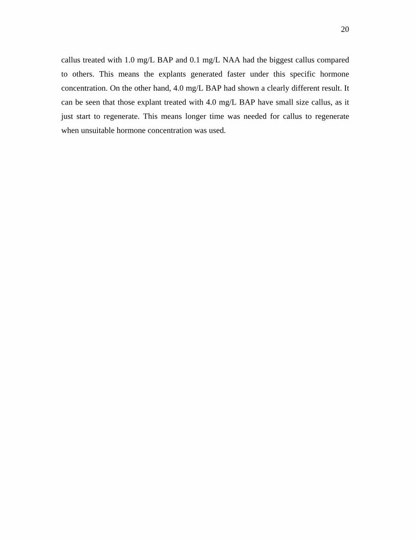
21
BAP
NAA
0 mg/L 93% explant
regenerated
90% explant
regenerated
83% explant
regenerated
33% explant
regenerated
0.1 mg/L 97% explant
regenerated
100% explant
regenerated
87% explant
regenerated
63% explant
regenerated
1.0 mg/L 87% explant
regenerated
93% explant
regenerated
87% explant
regenerated
47% explant
regenerated
Table 3.1: Total percentage of explants regenerated for each type of hormone
concentration.
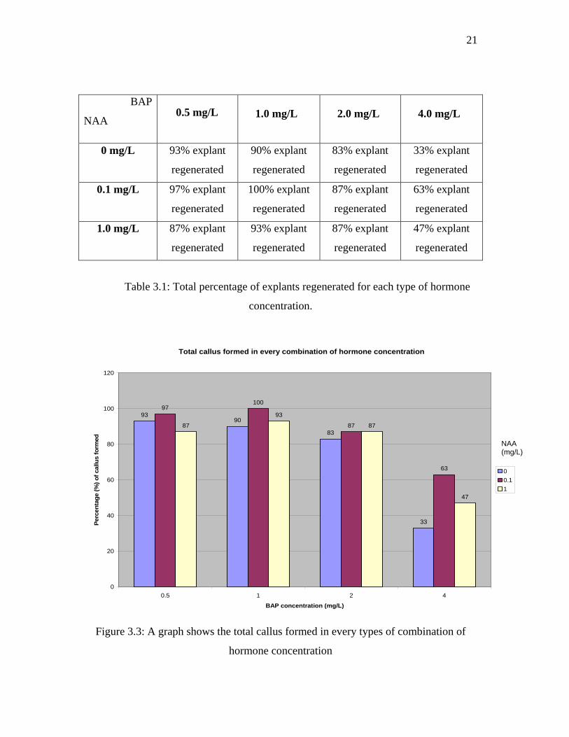
Total callus formed in every combination of hormone concentration
9390
83
33
97100
87
63
87
93
87
47
0
20
40
60
80
100
120
0.5 1 2 4
BAP concentration (mg/L)
Per
cen
tag
e (%
) o
f ca
llus
form
ed
0
0.1
1
Figure 3.3: A graph shows the total callus formed in every types of combination of
hormone concentration
1.0 mg/L 4.0 mg/L 0.5 mg/L 2.0 mg/L
NAA (mg/L)
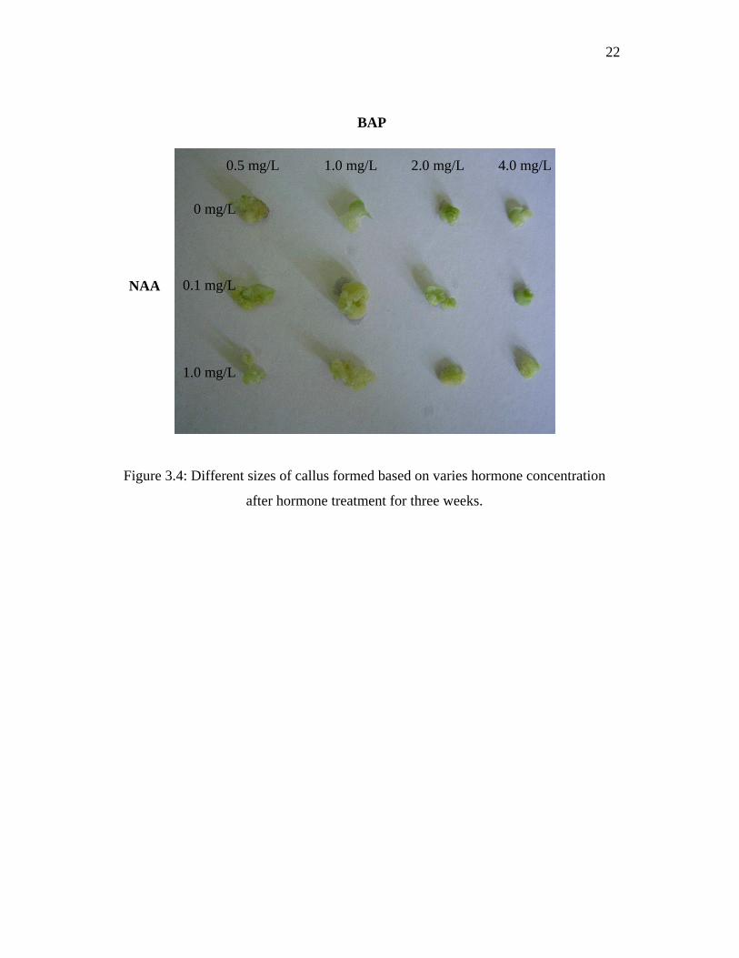
22
Figure 3.4: Different sizes of callus formed based on varies hormone concentration
after hormone treatment for three weeks.
0.5 mg/L 1.0 mg/L 2.0 mg/L 4.0 mg/L
0 mg/L
0.1 mg/L
1.0 mg/L
BAP
NAA
23
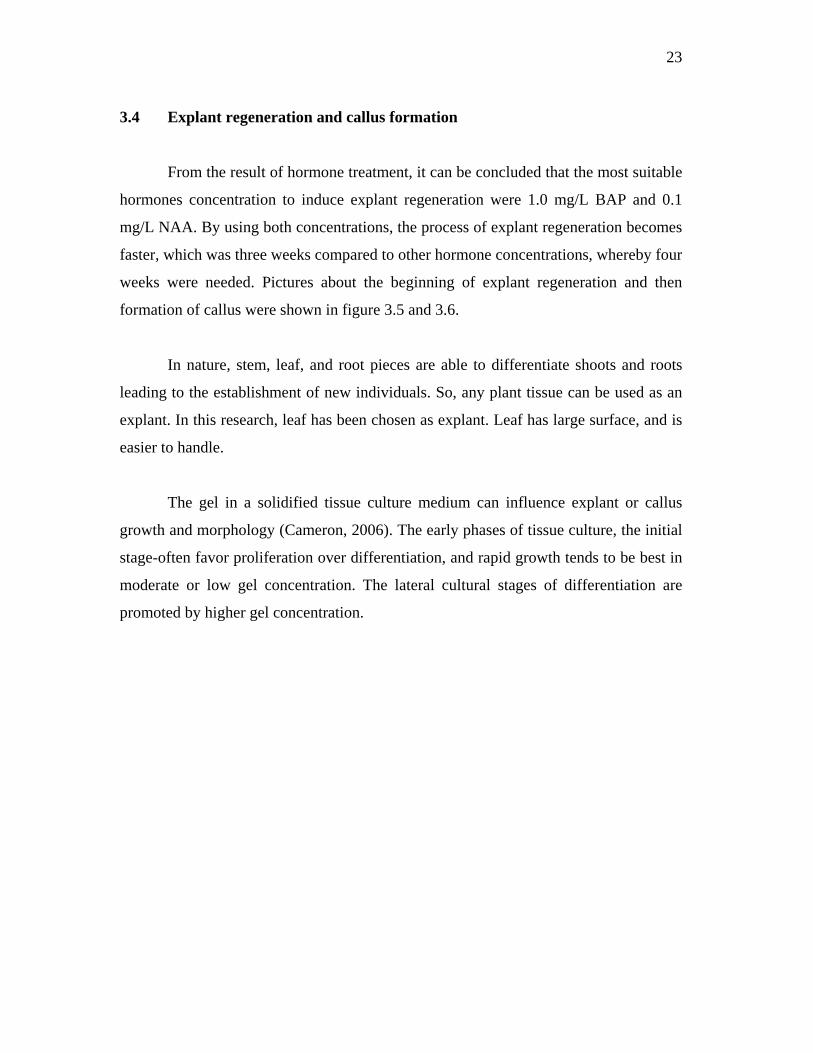
3.4 Explant regeneration and callus formation
From the result of hormone treatment, it can be concluded that the most suitable
hormones concentration to induce explant regeneration were 1.0 mg/L BAP and 0.1
mg/L NAA. By using both concentrations, the process of explant regeneration becomes
faster, which was three weeks compared to other hormone concentrations, whereby four
weeks were needed. Pictures about the beginning of explant regeneration and then
formation of callus were shown in figure 3.5 and 3.6.
In nature, stem, leaf, and root pieces are able to differentiate shoots and roots
leading to the establishment of new individuals. So, any plant tissue can be used as an
explant. In this research, leaf has been chosen as explant. Leaf has large surface, and is
easier to handle.
The gel in a solidified tissue culture medium can influence explant or callus
growth and morphology (Cameron, 2006). The early phases of tissue culture, the initial
stage-often favor proliferation over differentiation, and rapid growth tends to be best in
moderate or low gel concentration. The lateral cultural stages of differentiation are
promoted by higher gel concentration.
24
Figure 3.5: Explant started to regenerate after three weeks under 0.5 mg/l BAP.
Figure 3.6: Callus (a mass of undifferentiated cell) was formed when explant was
regenerated further.
25
3.5 Shoot formation
When the callus was allowed to continue its growth, the callus soon initiate
shoot. To induce shoot, the callus was grown in fresh MS media that containing plant
growth regulators, where the concentrations were the same as used in callus
regeneration, 1.0 mg/L BAP and 0.1 mg/L NAA. It is important to ensure a sufficient
supply of nutrient all along the plant growing process. The plant needs to be transferred
to fresh MS agar media every two weeks. Growth on the same media agar for an
extended period will lead to depletion of essential nutrients and gradual desiccation of
the gelling agent. Following callus initiation, shoot bud started to form after three
weeks in culture.
Chemical factor and physical factor are the main factors that affect shoot bud
differentiation in tissue culture. Chemical factor, which means the effect of hormone,
has been discussed further previously in section 3.3. Meanwhile physical factor
includes the medium used, light, temperature, physiological state of the donor plant,
and the others. High light intensity has been shown to be inhibitory for shoot-bud
formation in Nicotiana spp (Bhojwani and Razdan, 1992). The quality of light also
influences organogenic differentiation. Blue light promote shoot bud differentiation
whereas red light stimulates rooting in Nicotiana spp (Bhojwani and Razdan, 1992).
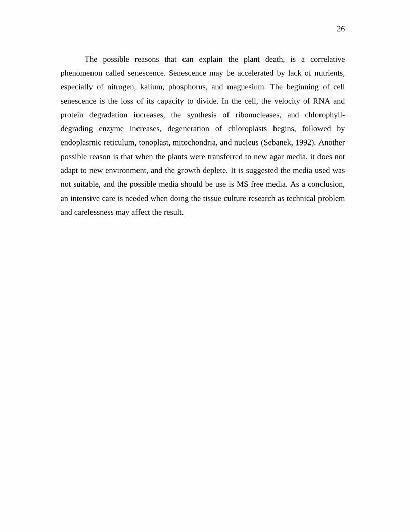
Shoot will continue to grow until complete young leaves were formed. It is
important to ensure complete young leaves were formed from shoot. When this step
was achieved, it indicates that the plant was ready to develop certain bioprocess to
maintain its growth. For example, the plants started to utilize solar energy by the leaves
for transpiration, and also photosynthesis. In figure 3.7, it shows that plants with
complete young leaves (4.30 cm height) were formed from shoot.
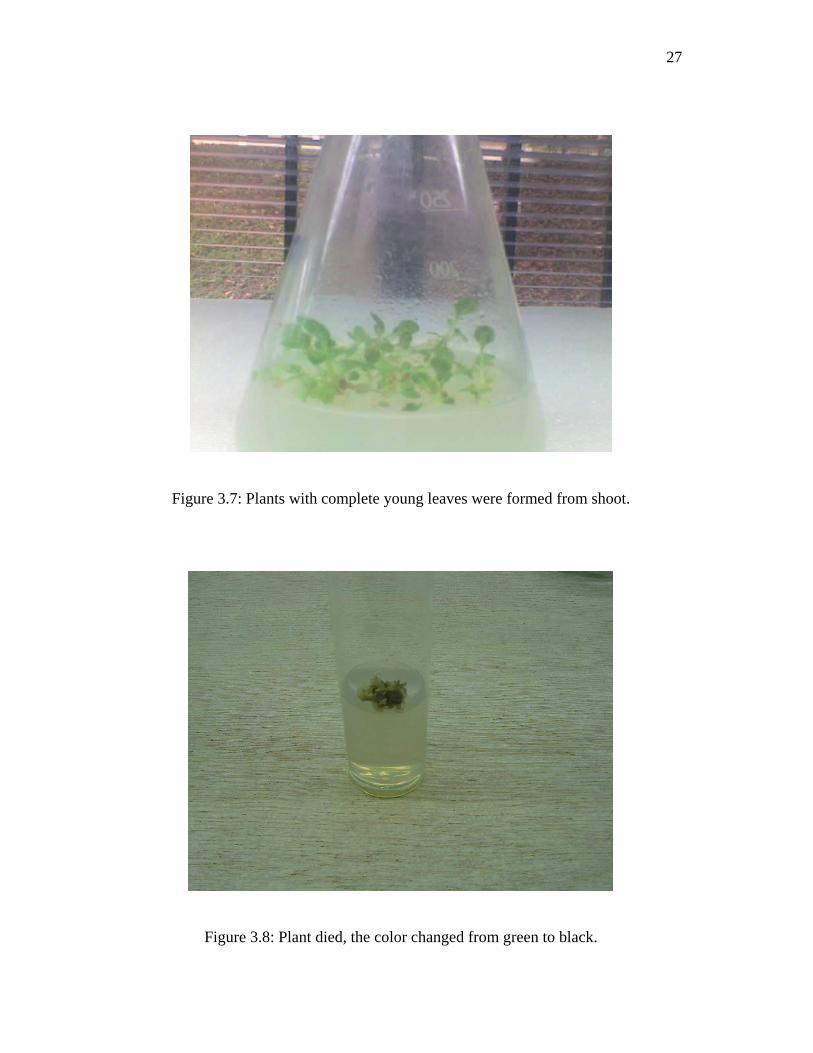
However, plants died in the process to continue its growth. Plants died when it
was transferred to fresh MS media with addition of hormones. Plant which died
changed color from green to brown or black.
26
The possible reasons that can explain the plant death, is a correlative
phenomenon called senescence. Senescence may be accelerated by lack of nutrients,
especially of nitrogen, kalium, phosphorus, and magnesium. The beginning of cell
senescence is the loss of its capacity to divide. In the cell, the velocity of RNA and
protein degradation increases, the synthesis of ribonucleases, and chlorophyll-
degrading enzyme increases, degeneration of chloroplasts begins, followed by
endoplasmic reticulum, tonoplast, mitochondria, and nucleus (Sebanek, 1992). Another
possible reason is that when the plants were transferred to new agar media, it does not
adapt to new environment, and the growth deplete. It is suggested the media used was
not suitable, and the possible media should be use is MS free media. As a conclusion,
an intensive care is needed when doing the tissue culture research as technical problem
and carelessness may affect the result.
27
Figure 3.7: Plants with complete young leaves were formed from shoot.
Figure 3.8: Plant died, the color changed from green to black.
28
CHAPTER 4
Conclusion
As with any plant transformation studies, a regeneration protocol must first be
established in order to regenerate transformed plants. Therefore, the first approach in
plant research is to carry out tissue culture experiments and figure out the most suitable
condition for plant growth.
Before starting plant tissue culture, it is evident to know well the tissue culture
process and the characteristic of the research plant. Literature reviews on tissue culture
and research on plant help gain more knowledge on the research to be done, and
prevent mistakes.
For tissue culture of Nicotiana plumbaginifolis, sterilization technique suitable
to be used was to immerse the seed in hypochlorite for 10 to 15 minutes.
The best hormone concentrations to grow the explant were at 1.0 mg/L BAP
and 0.1 mg/L NAA. Suitable hormone concentrations promote the optimum growth of
explants and also induce the callus and shoot formation. Suitable hormone
concentrations also shorten the time for plant to initiate callus and shoot.

29
Callus start to form after three weeks with the addition of plant growth
regulators. Shoot or young leaves began to form after three weeks of callus initiation.
Proper sterilization technique and experiment procedures handling can prevent the
contamination of culture.
30
CHAPTER 5
Future work
It is undeniable that tissue culture and genetic engineering goes hand in hand in
the molecular improvement of plant species. Therefore, the first step in the molecular
improvement of plant is the establishment of tissue culture technique.
Apart from leaf explant, other tissue sources can be used for tissue culture. Here,
a suitable source is through somatic organogenesis. Somatic embryos provide a ready,
long term source of tissues for transformation. It is also advantageous since somatic
embryos could arise from single cell, and this could minimize somaclonal variation.
Therefore, future studies can be focused on the establishment of somatic embryogenesis.
Manipulation of culture medium and condition must be further looked into.
Different content of culture media such as macronutrient, micronutrient, and carbon
source may give a different result of plant development. Besides, varieties of plant
growth regulator should also be test to study the effect on plant development. Study on
the possible factors and condition that can cause plant death also can be conducted.
Last but not least, with the tissue culture development for Nicotiana
plumbaginifolis, more genetic studies of this species can be conducted. As Nicotiana

31
spp allow the transformation of Agrobacterium easily, this gives the benefits to having
more studies on genetic transformation.
32
References
Bhojwani, S. S. and Razdan, M. K. (1992) Plant tissue culture: theory and practice. 6th
edition, Netherlands, Elsevier Science Publishers
Cameron, S. I. (2006) Tissue culture gel firmness: measurement and effects on growth,
In plant tissue culture engineering. Springer, 329-337
Charles-Edwards, D. A. (1986) Modeling plant growth and development. 1st, edition,
Academic Press
Chesnokov, Y. V. (2001) A chimeric green fluorescent protein gene as an embryogenic
marker in transgenic cell Culture of Nicotina Plumbaginifolis Viv. J. Plant Science
162 .59-77
Dodds, J. H. and Roberts, L. W. (1995) Experiments in plant tissue culture. 3rd.edition,
Cambridge University press, 1-65
Evans, D. A., Bravo, J. E., Kut, S. A., and Flick, C. E. (1982) Genetic behavior of
somatic hybrids in the genus Nicotiana: N. otophora + N. tabaccum and N. sylvestris +
N. tabaccum. J. Theory Application Genetics 65: 93-101
Evans, D. A. and Gamborg, O. L. (1982) Chromosome stability of cell suspension
cultures of Nicotiana spp. J. Plant Cell Reports 1: 104-107
33
Galis, I. (2004) Agrobacterium tumefacians AK-6b gene modulates phenolic
Compound Metabolism in Tobacco. J. Phytochemistry 65. 169-179
Gamborg, O. L., Miller. A., and Ojima, K. (1968) Nutrient requirements of suspension
cultures of soybean root cells. Exp. Cell Res. 50,151-8
Kung, S. D. (1989) Plant biotechnology. 1st edition, Butterworth Publishers. 374-389
Laetsch, W. M. (1967) Papers on plant growth and development. 1st edition, Little,
Brown and Company (Canada) Limited. 417-423, 215-223
McDaniel, C. N. (1999) Rapid flowering Nicotiana tabaccum L. J. Plant Reproduction
12: 123-124
Morgan, T.H. (1901) Regeneration. London, MacMillan
Murashige, T. and Skoog, F. (1962) Clonal crops through tissue culture pp392-403.
Berlin, Springer-Verlag
Murch, S. J. and Saxena, P. K. (2004) Journey of a single cell to a plant. USA, Science
Publishers
Sebanek, J. (1992) Plant physiology. Elsevier Science Publishers
Sinnott, H. E. (1960) Plant morphogenesis. New York, McGraw-Hill
Smith, R. H. (1992) Plant tissue culture techniques and experiments. Academic Press 1-
67
34
Sorrentino, A. (2004) Recombinant human tissue transglutaminase produced into
tobacco cell cultures is active and recognizes autoantibodies in the serum of celiac
patients. J. Biochemistry & Cell Biology, 845-851
Street, H. E. (1969) Plant Tissue and cell culture. H. E. Street, pp 1-10. Oxford,
Blackwell Scientific Publications
Sun, E. J. and Kang, H. W. (2003) Tobacco clones derived from tissue culture with
supersensitivity to ozone. J. Environmental Pollution 125 .111-115
White, P.R. (1934) Potentially unlimited growth of excised plant callus in an artificial
medium. J. Plant Physiol. 9, 585-600
White, P. R. (1939) Potentially unlimited growth of excised plant callus in an artificial
medium. J. Plant Physiol. 2, 231-44
White, P. R. (1943) A handbook of plant tissue culture. J. Plant Physiol. 9, 585-600