adrenal histoplasmosis in cushing's syndrome with ... with bilateral adrenocortical nodular ......
TRANSCRIPT

Adrenal histoplasmosis in Cushing's Syndrome with bilateral adrenocortical nodular hyperplasia
T.T. Tan, MRCP Lecturer
Y.W. Choy,MBBS Registrar
M.A. Norizan+, MBBS, Dip. Path. Lecturer
F. Meah *, FRACS Assoc. Pro/.
B.A.K. Khalid, FRACP, PhD
Assoc. Pro/.
Departments of Medicine, Patho!ogy+ and Surgery *, Universiti Kebangsaan Malaysia, Kuala Lumpur
Summary
Adrenal histoplasmosis in Cushing's syndrome of adrenal origin is rare. A patient with Cushing's disease with bilateral nodular hyperplasia and histoplasmosis of both the adrenal glands is described. The diagnosis of histoplasmosis was only made post operatively as the constitutional manifestations, besides being partially masked by hypercortisolism also resemble those of tuberculosis.
Key words: Adrenal histoplasmosis, adrenocortical nodular hyperplasia, Cushing's syndrome.
Introduction
In Cushing's syndrome due to adrenal hyperfunction associated with adrenal nodular hyperplasia, two distinct subtypes are recognised. 1 The commoner of the two, Le. macroscopic and nodular hyperplasia, is suggested to represent a process of transition from pituitary dependence to adrenal autonomy.2-4 Compared to classic Cushing's disease, the patients have a longer history, are more resistant to dexamethasone suppression, and have lower corticotropin levels. Pigmented multinodular adrenocortical dysplasia, the second subtype, is less common than the macroscopic form. It is often familial, occurs in children and young adults, and is non-corticotropin dependent. 5,6
Adrenal involvement is common in chronic disseminated histoplasmosis. 7 This often results in adrenal failure. Disseminated histoplasmosis occuring in immuno-defective patients due to glucocorticoid excess had also been reported. '7 , 8 The latter is often iatrogenically induced. The occurrence of histoplasmosis in both the adrenal glands that are hyperfunctioning has, to our knowledge, never been reported. In this report we describe a patient with Cushing's syndrome due to bilateral adrenocortical nodular hyperplasia with concommitant infection of the glands with histoplasmosis.
154
Case report
A 45 year old male rubber tapper was seen at the Kuala Lumpur General Hospital with a four year history of low back ache worsened by physical activity. He was found to have hypertension two years previously. For the past one year, there was increased skin pigmentation, excessive weight gain, proximal muscle weakness and deterioration of his back pain, associated with lethargy and intermittent low grade fever. He smoked ten cigarettes per day for the past 20 years. His' past history included pulmonary tuberculosis 25 years ago for which he had completed treatment.
On examination he had classical features of Cushing's syndrome with truncal obesity, facial plethora, moon facies, buffalo hump, supraclavicular fat pads, purplish abdominal striae and .generalised hyperpigmentation. The supine blood pressure was 150/105 mm Hg. His body temperature was 37.8°C. There was restriction of forward bending with tenderness over the lumbosacral spine.
Chest X-ray showed apical fibrosis consistent with old pUlmonary tuberculosis. Lumbosacral radiographs revealed generalised osteopaenia with biconcavity of vertebral bodies, compression fracture of lumbar four and five vertebral bodies, and spondylosis at lumbar five and sacral one levels. Skull radiograph showed normal pituitary fossa. Mantoux test produced a 5 mm induration. Three sputum specimens taken on three consecutive mornings for acid fast bacilli were negative on direct smear and culture.
Plasma cortisol levels at 0800 h and 2300 h were 530 nmol/l (normal range (NR) 214-497 nmol/l) and 401 nmol/l (NR 32 - 381 nmol/l) respectively. Twenty-four hour urinary free cortisol was 1711 nmol (NR 357 - 851 nmol). Both low dose dexamethasone (2 mg/day for two days) and high dose dexamethasone (8 mg/day for two days) failed to suppress plasma cortisol and 24 hour urinary free cortisol to less than 50% of basal levels (Table 1). Computerised tomographic (CT) scan of the pituitary gland was normal but a nodule measuring 14 mm by 14 mm in the left adrenal was seen. The right adrenal appeared normal.
Table 1 Low and ffigh Dose Dexamethasone Suppression Tests'
Dexamethasone dose (mg/6h x 2 days)
Basal
0.5
2.0
NR = Normal Range
0800 h Plasma cortisol (nmol/L)
(NR: 214-497)
530
467
270
Urine free cortisol (nmol/day)
(NR: 357-851)
1711
1815
917
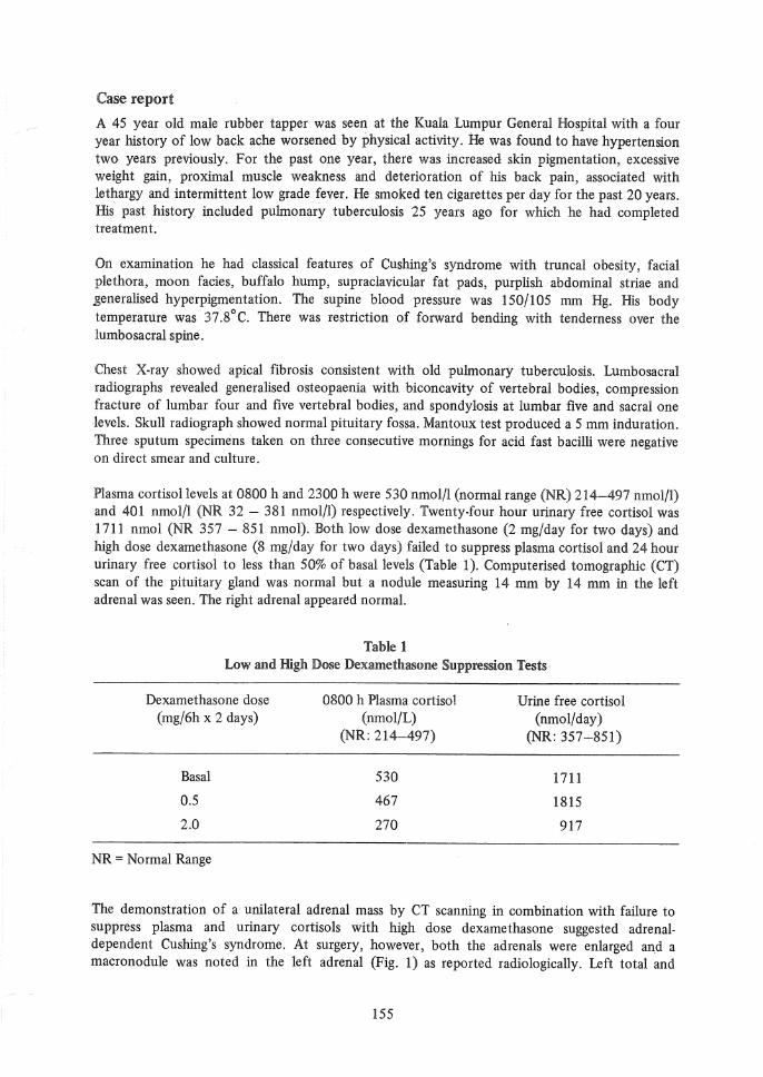
The demonstration of a unilateral adrenal mass by CT scanning in combination with failure to suppress plasma and urinary cortisols with high dose dexamethasone suggested adrenaldependent Cushing's syndrome. At surgery, however, both the adrenals were enlarged an,d a macronodule was noted in the left adrenal (Fig. 1) as reported radiologically. Left total and
155
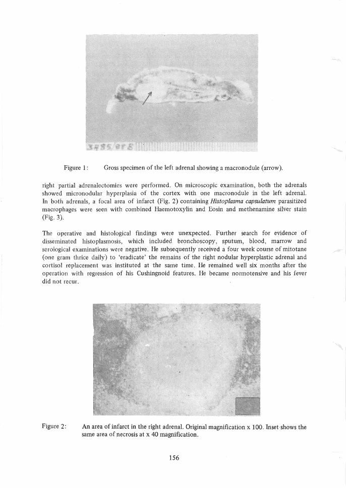
Figure 1: Gross specimen of the left adrenal showing a macronodule (arrow).
right partial adrenalectomies were performed. On microscopic examination, both the adrenals showed micronodular hyperplasia of the cortex with one macro nodule in the left adrenal. In both adrenals, a focal area of infarct (Fig. 2) containing Histoplasma capsulatum parasitized macrophages were seen with combined Haemotoxylin and Eosin and methenamine silver stain (Fig. 3).
The operative and histological findings were unexpected. Further search for evidence of disseminated histoplasmosis, which included bronchoscopy, sputum, blood, marrow and serological examinations were negative. He subsequently received a four week course of mitotane (one gram thrice daily) to 'eradicate' the remains of the right nodular hyperplastic adrenal and cortisol replacement was instituted at the same time. He remained well six months after the operation with regression of his Cushingnoid features. He became normotensive and his fever did not recur.
Figure 2: An area of infarct in the right adrenal. Original magnification x 100. Iriset shows the same area of necrosis at x 40 magnification.
156
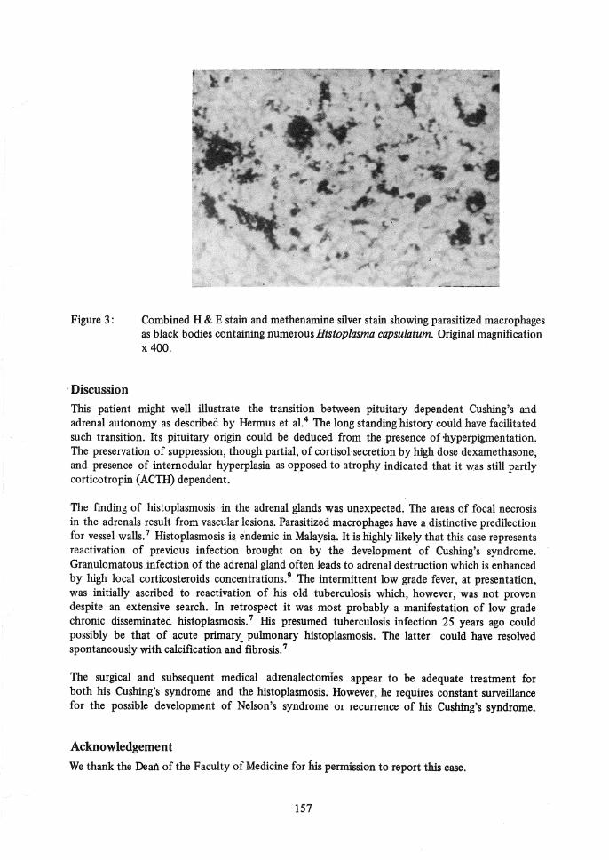
Figure 3:
Discussion
Combined H & E stain and me then amine silver stain showing parasitized macrophages as black bodies containing numerous Histoplasma capsulatum. Original magnification x 400.
This patient might well illustrate the transition between pituitary dependent Cushing's and adrenal autonomy as described by Hermus et al.4 The long standing history could have facilitated such transition. Its pituitary origin could be deduced from the presence of ·hyperpigmentation. The preservation of suppression, though partial, of cortisol secretion by high dose dexamethasone, and presence of internodular hyperplasia as opposed to atrophy indicated that it was still partly corticotropin (ACTH) dependent.
The fmding of histoplasmosis in the adrenal glands was unexpected. The areas of focal necrosis in the adrenals result from vascular lesions. Parasitized macrophages have a distinctive predilection for vessel walls. 7 Histoplasmosis is endemic in Malaysia. It is highly likely that this case represents reactivation of previous infection brought on by the development of Cushing's syndrome. Granulomatous infection of the adrenal gland often leads to adrenal destruction which is enhanced by high local corticosteroids concentrations.9 The intermittent low grade fever, at presentation, was initially ascribed to reactivation of his old tuberculosis which, however, was not proven despite an extensive search. In retrospect it was most probably a manifestation of low grade chronic disseminated histoplasmosis. 7 His presumed tuberculosis infection 25 years ago could possibly be that of acute primary pulmonary histoplasmosis. The latter could have resolved spontaneously with calcification and fibrosis. 7
The surgical and subsequent medical adrenalectomIes appear to be adequate treatment for both his Cushing's syndrome and the histoplasmosis. However, he requires constant surveillance for the possible development of Nelson's syndrome or recurrence of his Cushing's syndrome ..
Acknowledgement
We thank the Dean of the Faculty of Medicine for ius permission to report this case.
157
References
1. Nodular hyperplasia and Cushing's syndrome. Editorial. Lancet 1988; 2: 434.
2. Aron DC, Findling JW, Fitgerald PA, et at. Pituitary ACTH dependency of nodular adrenal hyperplasia in Cushing's syndrome: report of two cases and review of the literature. Am J Med 1981; 71: 302-6.
3.. Smals AGH, Pieters GFFM, Van Haelst UJG, Kloppenborg PWC. Macronodular adrenocortical hyperplasia in long-standing Cushing's disease. J CIin Endocrinol Metab 1984; 58: 25-31.
4. Hermus AR, Pieters GF, Smals AGH, et al. Transition from pituitary dependent to adrenal dependent Cushing's Syndrome. N EnglJ Med 1988; 318: 966-70.
5. Primary adrenocortical nodular dysplasia as a cause of Cushing's syndrome in infants and children. Mayo CIin Proc 1982; 57: 58-63.
158
6. Jennifer LL, Cathey WJ, William DO. Primary adrenocortical nodular dysplasia, a distinct subtype of Cushing's syndrome. Am J Med 1986: 80: 976-84.
7. Goodwin RA, Shapiro JL, Thurman GH, Thurman SS, Des Pres RM. Disseminated histoplasmosis: Clinical and Pathologic correlations. Medicine (Baltimore) 1980; 59: 1-33.
8. Davies SF, Khan M, Sarosi GA. Disseminated histoplasmosis in irnmunologically suppressed patients. Am J Med 1978; 64: 94-9.
9. Frenkel JK. Pathogenesis of infection of the adrenal gland leading to Addison's disease in man: The role of corticoids in adrenal and generalised infection. Ann N Y Acad Sci 1960; 84: 393.