1. icu protocol management cover - msic
TRANSCRIPT
Published by Malaysian Society of Intensive Care
Printed by Malaysian Society of Intensive Care (MSIC)Unit 1.6, Level 1, Enterprise 3BTechnology Park MalaysiaJalan Innovasi 1, Bukit Jalil57000 Kuala Lumpur, Wilayah PersekutuanWebsite: www.msic.org.my
In collabration with Ministry of Health Malaysia
Copyright © Malaysian Society of Intensive Care
Pusat Kebangsaan ISBN MalaysiaISBN 978-967-11415-4-0
Cover design by Nabil bin Ali
Disclaimer: The content of this book has been produced in good faith to guide medical practitioners. However practitioners are advised to keep abreast the current evidence-based practices that are constantly evolving and to take into account the local issues and limitations.
ICUManagement
Protocols

ForewordThere are many aspects in the care and management of the critically ill patient. As clinicians we need to keep abreast with the most current evidence-based practices to ensure optimal patient care and safety.
This is an update of the management protocol book written in 2012, to facilitate clinicians in the management of the critically ill. Each protocol was developed with careful consideration of current evidence as well as the practical application and cost containment within our institutions. The algorithms in the protocols are simple to use and can be easily implemented.
There are great concerns on the rise of multi-drug resistant organisms. We know that critically ill patients are at high risk of acquiring infections. To address this, a protocol on prevention and control of multi-drug organisms is included.
This protocol materialised due to the many hours of discussion and exchange of opinions. I hope the protocol will serve as a guide to ICU management that will enhance the quality of patient care.
I like to express my gratitude to the writing committee for their effort in publishing this excellent management protocol book.
Dr Melor bin Mohd MansorHead of Anaesthetic and Intensive Care ServicesMinistry of Health Malaysia
i

Writing CommitteeDr Shanti Rudra Deva(Chairperson and Editor)IntensivistDepartment of Anaesthesia and Intensive CareHospital Kuala Lumpur, Kuala Lumpur
Dr Tai Li Ling(Co-Chairperson)IntensivistDepartment of Anaesthesia and Intensive CareHospital Kuala Lumpur, Kuala Lumpur
Dr Azmin Huda Abdul RahimIntensivistDepartment of Anaesthesia and Intensive CareHospital Sultan Ismail, Johor Bahru, Johor
Dr Foong Kit WengIntensivistDepartment of Anaesthesia and Intensive CareHospital Raja Permaisuri Bainun, Ipoh, Perak
Dr Ismail Tan Mohd Ali TanIntensivistDepartment of Anaesthesia and Intensive CareHospital Kuala Lumpur, Kuala Lumpur
Dr Khoo Tien MengIntensivistDepartment of Anaesthesia and Intensive CareHospital Queen Elizabeth 1, Kota Kinabalu, Sabah
Dr Lee See PhengIntensivistDepartment of Anaesthesia and Intensive CareHospital Tengku Ampuan Rahimah, Klang, Selangor
ii

iii
Writing CommitteeDato’ Dr Lim Chew HarIntensivistDepartment of Anaesthesia and Intensive CareHospital Pulau Pinang, Pulau Pinang
Dr Mahazir bin KassimIntensivistDepartment of Anaesthesia and Intensive CareHospital Sultanah Aminah, Johor Bahru, Johor
Dr Mohd Ridhwan Mohd NoorIntensivistDepartment of Anaesthesia and Intensive CareHospital Sultanah Nur Zahirah, Kuala Terengganu, Terengganu
Dr Muhammad Zihni AbdullahIntensivistDepartment of Anaesthesia and Intensive CareHospital Tengku Ampuan Afzan, Kuantan, Pahang
Dr Nahla Irtiza IsmailIntensivistDepartment of Anaesthesia and Intensive CareHospital Melaka, Melaka
Dr Noor Airini IbrahimSenior Lecturer and IntensivistFaculty of Medicine and Health SciencesUniversiti Putra Malaysia, Serdang, Selangor
Dr Wan Daud bin Wan KadirIntensivistDepartment of Anaesthesia and Intensive CareHospital Umum Sarawak, Kuching, Sarawak

Table of ContentsContents Page
Foreword i
Writing Committee ii
Admission, Discharge and Triage 1
Vasoactive Agents in Acute Circulatory Failure 6
Severe Hypoxaemic Respiratory Failure 14
Weaning from Mechanical Ventilation 21
Pain, Sedation and Delirium 29
Nutritional Therapy 39
Early Mobilisation 47
Stress Ulcer Prophylaxis 54
Venous Thromboprophylaxis 57
Prevention and Control of Multi-Drug Resistant Organisms 62
Withholding and Withdrawing Life-Sustaining Treatment 67
Invasive Mechanical Ventilation in Non-Critical Care Areas 74
iv

1
Admission, Discharge and TriageIntroduction
Appropriate utilisation of ICU bed is essential as intensive care resources are limited and expensive. Demand for intensive care will continue to exceed supply, hence clear and rationale decision-making regarding admission and discharge is required.
Principles
1. The decision to admit a patient to ICU should be based on the concept of potentialbenefit.
2. Critically ill patients with a reversible medical condition, having a reasonable prospect of meaningful recovery should be admitted.
3. A combination of criteria should be used to determine ICU admission or discharge.
4. ICU triaging is necessary to ensure optimal and equitable use of limited intensive care resources.
Admission policy
1. It is the responsibility of the patient’s attending clinician to request for ICU admission.
2. It is the responsibility of the ICU specialist to decide on admission based on his/her clinical judgement guided by the admission criteria.
3. The decision to admit or deny admission shall not be made at the level of a medicalofficer.
4. Admission from the Emergency and Trauma Department or from another hospital shall have a primary unit.
5. Admission from other hospitals shall be discussed with the ICU specialist prior to transfer.
6. It is the responsibility of the primary team to continue resuscitation of patients while awaiting ICU admission.
2
7. Patients in ICU remain under the responsibility of the primary unit. Transfer of caretoadifferentunitshallbearrangedbytheprimaryunit.
8. Family shall be informed of patient’s admission to ICU as soon as possible, with further updates when required.
ICU admission criteria
To optimise ICU resources and improve outcomes, ICU admissions should be guided on the basis of a combination of factors: a. Prioritisation according to the patient’s severity of illness b. Specificpatientneedssuchaslife-supportivetherapies c. Diagnosis d. Prognosis e. Potentialbenefitfrominterventions f. Objective parameters at the time of referral g. Available clinical expertise h. Bed availability
ICU admission based on priority
In evaluating the appropriateness of ICU admission, the priority should be based on theneedsof thepatient and the likelihoodof benefitting fromadmission.ThisprioritisationdefinesthosewhowillbenefitmostfromICU(Priority1)tothosewhowillnotbenefitatall(Priority3).
1. Priority 1 a. Critically ill, unstable
b. Require life support for organ failure, intensive monitoring and therapies that cannot be provided elsewhere. This includes invasive ventilation, renal replacement therapy, invasive haemodynamic monitoring and other interventions
c. Do not have limitations of treatment
d.Highlikelihoodofbenefit
2. Priority 2
Priority 2A a. Acutely ill, relatively stable
b. Requires intensive monitoring and/or therapies for organ dysfunction, that can bemanaged in an intermediate care facility (high dependency unit or post anaestheticcareunit)
3
c. Admit to ICU, if early management fails to prevent deterioration or there is no intermediate care facility in the hospital
d. Examples include: i. post-operative patients who require close monitoring ii. respiratoryinsufficiencyonintermittentnon-invasiveventilation
Priority 2B a. Critically ill, unstable
b. Require life support for organ failure
c. Withsignificantlylowerprobabilityofrecoverybecauseofadvancedunderlying disease
d.Mayhavespecificlimitationsofcaree.g.nocardiopulmonaryresuscitation
e. Lowerlikelihoodofpotentialbenefit
f. Examples include: i. metastatic cancer in septic shock secondary to hospital acquired pneumonia but with some limitations of therapy e.g. no CPR ii. decompensated heart failure with deteriorating functional status and multiple hospital admissions
3. Priority 3 a. Terminally ill or moribund patients with no possibility of recovery
b. Not appropriate for ICU admission
c. Maybenefitfrompalliativecareratherthanintensivecare
d. Examples include: i. severe irreversible brain pathology impairing cognition and consciousness or in a persistent vegetative state ii. metastatic cancer unresponsive to chemotherapy and/or radiotherapy iii. end-stage cardiac, respiratory or liver disease with no options for transplant iv. severe disability with poor quality of life v. advanced disease of a progressive life-limiting condition e.g. - motor neuron disease with rapid decline in physical status, - severe Parkinson’s disease with reduced independence and needs assistance for activities of daily living vi. poor response to current treatment e.g. - bowel leak despite multiple laparotomies, - recurrent soft tissue or musculoskeletal infections despite multiple surgical intervention, - chronic medical conditions that fail to respond to treatment such as SLE or HIV
4
vii. end-stage renal disease with no option or refusal for renal replacement therapy viii. those who have explicitly stated their wish not to receive life-support therapy
Triage
Triage is the process of placing patients at their most appropriate level of care. It is often needed as the number of potential ICU patients exceeds the availability of ICU beds.Appropriatetriagingallowseffectivebedutilisationandresourcemanagement.Factors to consider when triaging include: a. Likelihoodofbenefit b. Prognosis c. Life expectancy due to disease d. Anticipated quality of life
Discharge policy
1. The decision to discharge a patient shall be made by the ICU specialist.
2. Prior to discharge: a. The primary team shall be informed of the management plan including any limitation of treatment.
b. A discharge summary shall be completed.
c. Any limitation of treatment shall be clearly documented including why and amongst whom these decisions are made.
d. Family shall be informed.
e. It is the responsibility of the primary team to receive and review patients promptly in the ward.
f. Patients who require a higher level of nursing care may benefit from admission to a step down unit, if available.
ICU discharge criteria
InordertomaximisetheefficientuseofICUresources,patientsshouldbeassessedcontinuously to identify those who may no longer need ICU care. This includes patient with: a. Stable physiological status and no longer needing ICU monitoring and treatment.
b. Stable haemodynamic parameters on low dose inotropic support.
5
c. Stable respiratory status with oxygen requirement not more than 60%.
d. Neurological stability.
e. Tracheostomy, not requiring frequent suctioning.
f. Chronic mechanical ventilation (e.g. motor neuron disease, cervical spine injury)andtheacutecriticalproblemisresolved.
g. Deteriorating or irreversible physiological status where active interventions are no longer beneficial. Withdrawal of therapy should be initiated, however, patient may be discharged to the ward if ICU bed is required.
References
1. Nates JL, et al. ICU admission, discharge, and triage guidelines: a framework to enhance clinical operations, development of institutional policies, and further research. Crit Care Med 2016;44(8):1553-1602
2. Guidelines for intensive care unit admission, discharge, and triage. Task force of the American College of Critical Care Medicine, Society of Critical Care Medicine. Crit Care Med 1999;27(3):633-638
3. White ST, et al. What every intensivist should know about intensive care unit admission criteria. Rev Bras Ter Intensiva 2017;29(4):414-417
6
Vasoactive Agents in Acute Circulatory FailureIntroduction
The aim of treatment in acute circulatory failure is to maintain adequate perfusion pressure for tissue oxygenation. Vasopressors and inotropes are cornerstones in the management of shock syndromes. It is important to identify the type of shock for the choice of vasoactive agent is determined by its mechanism of action targeting the underlying pathophysiology.
Principles
1. Initiate vasopressors promptly in severe hypotension especially in undifferentiatedshock,afterexcludingobstructiveshock.
2. It is appropriate to initiate vasoactive agents while ensuring optimal fluid resuscitation in severe hypotension.
3. Titrate the dose of vasoactive agent to achieve adequate tissue perfusion.
4. Aim for MAP 60 - 65 mmHg (or > 70 mmHg in chronic hypertension) if there is evidence of inadequate tissue perfusion.
5. The main route of administration of vasoactive drugs is via a central venous catheter,however,peripheraladministrationmaybeconsideredinlowdoses.
6. Invasive blood pressure monitoring is preferred to allow immediate recognition of change in blood pressure and precise titration of vasoactive agents.
7. Monitor regularly for potential complications e.g. arrhythmias and peripheral ischaemia.
8. Echocardiography is useful in the diagnosis and management of shock.
9. Consider advanced haemodynamic studies e.g. cardiac output monitoring in patients with refractory shock.
7
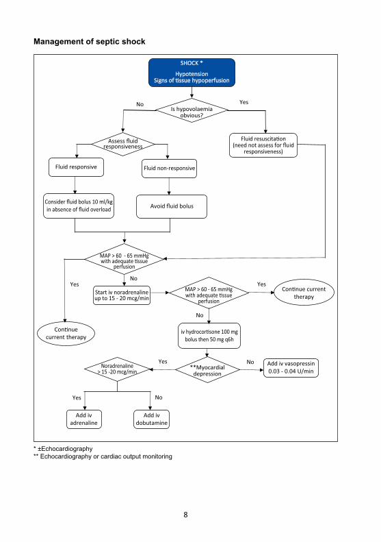
Vasoactive agents in distributive shock
Septic shock
1. Septic shock is predominantly caused by vasodilatation and relative hypovolaemia. Myocardial depression is often present although cardiac output is elevated.
2. Noradrenalineisthevasoactiveagentofchoicewithitseffectsofvenoconstriction (increasepreload),arterialvasoconstriction,positiveinotropy, improvedcardiac output and improved renal perfusion.
3. Consider adding a second vasoactive agent (e.g. vasopressin or adrenaline) when noradrenaline dose of > 15 - 20 mcg/min does not achieve targets.
4. Consider adding iv hydrocortisone 50 mg q6h following a bolus of 100 mg when noradrenaline dose reaches 15 - 20 mcg/min.
5. Consider adding iv dobutamine (2 - 5 mcg/kg/min) in presence of myocardial depression,andifnoradrenalinedoseis<15-20mcg/min.
6. Wean vasoactive agents once targeted goals are optimised. Consider weaning noradrenalinefirstbeforevasopressin.
Anaphylactic shock
1. Anaphylacticshockisanacute,multi-systemdisorder(predominantlyheart,lung andvasculature)resultingfromthesuddenreleaseofmediatorsfrommastcells, basophils and macrophages into the circulation.
2. Stopthesuspectedoffendingagent.
3. Administerivadrenalineinbolusesof0.05-0.1mg.Ifnoresponse,administer infusion adrenaline at 0.1 mcg/kg/min. Adrenaline is preferred as it reverses peripheral vasodilatation and reduces oedema.
4. Administer iv crystalloid of 20 ml/kg rapidly. Avoid colloids.
5. MayconsiderH1anti-histamine,ivchlorpheniramine10mgtocounterhistamine- mediated vasodilatation and bronchoconstriction. There is no evidence on the use of H2 anti-histamine.
6. May consider iv hydrocortisone 200 mg stat to shorten the protracted reaction.
8
Is hypovolaemiaobvious?
Fluid resuscita�on(need not assess for fluid
responsiveness)
Assess fluidresponsiveness
Fluid responsive Fluid non-responsive
Consider fluid bolus 10 ml/kgin absence of fluid overload Avoid fluid bolus
Start iv noradrenalineup to 15 - 20 mcg/min
**Myocardialdepression
Add iv vasopressin0.03 - 0.04 U/min
Noradrenaline> 15 -20 mcg/min
Add ivadrenaline
Add ivdobutamine
MAP > 60 - 65 mmHgwith adequate �ssue
perfusion
YesNo
Yes
Yes
Yes
No
No
Con�nue currenttherapy
iv hydrocor�sone 100 mgbolus then 50 mg q6h
Yes
MAP > 60 - 65 mmHgwith adequate �ssue
perfusion
Con�nuecurrent therapy
No
No
SHOCK *
HypotensionSigns of �ssue hypoperfusion
Management of septic shock
* ±Echocardiography** Echocardiography or cardiac output monitoring
9
b. Right ventricular failure c. Structural heart disease d. Arrhythmias
3. Early echocardiography is recommended for diagnostic and monitoring of therapy.
4. Early cardiology consult is recommended if there is an indication for rescue revascularisation.
5. Performtestforfluidresponsivenessandcorrecthypovolaemiainthosewhoare fluidresponsive.
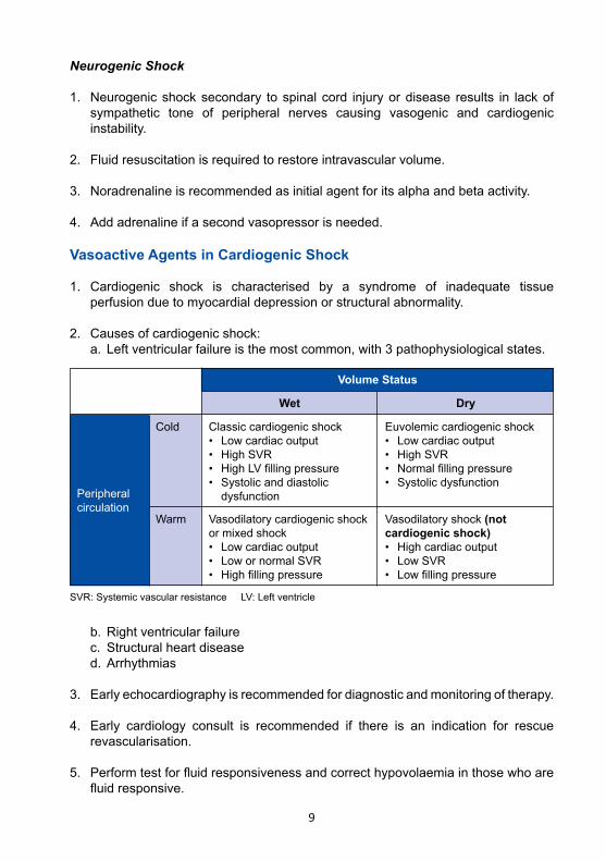
Peripheral circulation
SVR: Systemic vascular resistance LV: Left ventricle
Cold
Warm
Classic cardiogenic shock• Lowcardiacoutput• HighSVR• HighLVfillingpressure• Systolicanddiastolic dysfunction
Vasodilatory cardiogenic shock or mixed shock• Lowcardiacoutput• LowornormalSVR• Highfillingpressure
Vasodilatory shock (not cardiogenic shock)• Highcardiacoutput• LowSVR• Lowfillingpressure
Wet
Volume Status
Euvolemic cardiogenic shock• Lowcardiacoutput• HighSVR• Normalfillingpressure• Systolicdysfunction
Dry
Neurogenic Shock
1. Neurogenic shock secondary to spinal cord injury or disease results in lack of sympathetic tone of peripheral nerves causing vasogenic and cardiogenic instability.
2. Fluid resuscitation is required to restore intravascular volume.
3. Noradrenaline is recommended as initial agent for its alpha and beta activity. 4. Add adrenaline if a second vasopressor is needed.
Vasoactive Agents in Cardiogenic Shock
1. Cardiogenic shock is characterised by a syndrome of inadequate tissue perfusion due to myocardial depression or structural abnormality.
2. Causes of cardiogenic shock: a. Leftventricularfailureisthemostcommon,with3pathophysiologicalstates.
10
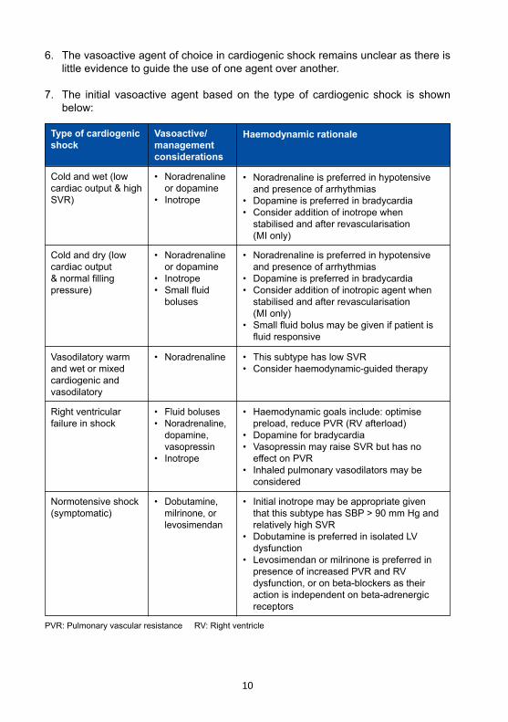
Type of cardiogenic shock
Cold and wet (low cardiac output & high SVR)
Cold and dry (low cardiac output &normalfillingpressure)
Vasodilatory warm and wet or mixed cardiogenic and vasodilatory
• Noradrenaline or dopamine• Inotrope
• Noradrenaline or dopamine• Inotrope• Smallfluid boluses
• Noradrenaline
• Noradrenalineispreferredinhypotensive and presence of arrhythmias• Dopamineispreferredinbradycardia• Consideradditionofinotropewhen stabilised and after revascularisation (MI only)
• Noradrenalineispreferredinhypotensive and presence of arrhythmias• Dopamineispreferredinbradycardia• Consideradditionofinotropicagentwhen stabilised and after revascularisation (MI only)• Smallfluidbolusmaybegivenifpatientis fluidresponsive
• ThissubtypehaslowSVR• Considerhaemodynamic-guidedtherapy
Vasoactive/management considerations
Haemodynamic rationale
Right ventricular failure in shock
Normotensive shock (symptomatic)
• Fluidboluses• Noradrenaline, dopamine, vasopressin• Inotrope
• Dobutamine, milrinone,or levosimendan
• Haemodynamicgoalsinclude:optimise preload,reducePVR(RVafterload)• Dopamineforbradycardia• VasopressinmayraiseSVRbuthasno effectonPVR• Inhaledpulmonaryvasodilatorsmaybe considered
• Initialinotropemaybeappropriategiven that this subtype has SBP > 90 mm Hg and relatively high SVR• DobutamineispreferredinisolatedLV dysfunction• Levosimendanormilrinoneispreferredin presence of increased PVR and RV dysfunction,oronbeta-blockersastheir action is independent on beta-adrenergic receptors
PVR: Pulmonary vascular resistance RV: Right ventricle
6. The vasoactive agent of choice in cardiogenic shock remains unclear as there is little evidence to guide the use of one agent over another.
7. The initial vasoactive agent based on the type of cardiogenic shock is shown below:
11
References
1. RhodesA,etal.Survivingsepsiscampaign:Internationalguidelinesformanagementofsepsis and septic shock: 2016. Crit Care Med 2017;45(3):486-552
2. SingerM,etal.Thethirdinternationalconsensusdefinitionsforsepsisandsepticshock(Sepsis-3). JAMA 2016;315(8):801-810
3. MonnetX,etal.Assessmentoffluidresponsiveness: recentadvances.Curr Opin Crit Care 2018;24(3):190-195
4. AnnaneD,etal.Aglobalperspectiveonvasoactiveagentsinshock.Intensive Care Med 2018:44(6):833-846
5. DiepenSV,etal.Contemporarymanagementofcardiogenicshock:Ascientificstatementfrom the American Heart Association. Circulation 2017;136:e232-e268
12
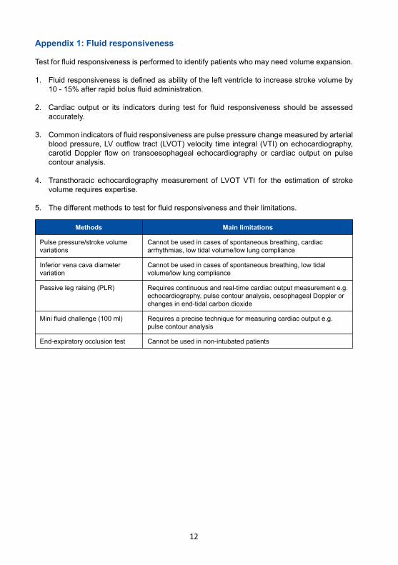
Appendix 1: Fluid responsiveness
Testforfluidresponsivenessisperformedtoidentifypatientswhomayneedvolumeexpansion.
1. Fluidresponsivenessisdefinedasabilityoftheleftventricletoincreasestrokevolumeby 10-15%afterrapidbolusfluidadministration.
2. Cardiac output or its indicators during test for fluid responsiveness should beassessed accurately.
3. Commonindicatorsoffluidresponsivenessarepulsepressurechangemeasuredbyarterial bloodpressure,LVoutflowtract(LVOT)velocitytimeintegral(VTI)onechocardiography, carotidDoppler flow on transoesophageal echocardiography or cardiac output on pulse contour analysis.
4. Transthoracic echocardiographymeasurement of LVOTVTI for the estimation of stroke volume requires expertise.
5. Thedifferentmethodstotestforfluidresponsivenessandtheirlimitations.
Methods
Pulse pressure/stroke volume variations
Inferior vena cava diameter variation
Passive leg raising (PLR)
Minifluidchallenge(100ml)
End-expiratory occlusion test
Cannotbeusedincasesofspontaneousbreathing,cardiacarrhythmias,lowtidalvolume/lowlungcompliance
Cannotbeusedincasesofspontaneousbreathing,lowtidalvolume/low lung compliance
Requires continuous and real-time cardiac output measurement e.g. echocardiography,pulsecontouranalysis,oesophagealDopplerorchanges in end-tidal carbon dioxide
Requires a precise technique for measuring cardiac output e.g. pulse contour analysis
Cannot be used in non-intubated patients
Main limitations
13
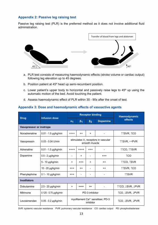
Appendix 2: Passive leg raising test
Passive leg raising test (PLR) is thepreferredmethodas itdoesnot involveadditionalfluidadministration.
a. PLRtestconsistsofmeasuringhaemodynamiceffects(strokevolumeorcardiacoutput) following leg elevation up to 45 degrees.
b. Position patient at 45º head up semi-recumbent position.
c. Lower patient’s upper body to horizontal and passively raise legs to 45º up using the automatic motion of the bed. Avoid touching the patient.
d. AssesshaemodynamiceffectofPLRwithin30-90saftertheonsetoftest.
Appendix 3: Dose and haemodynamic effects of vasoactive agents
Transfer of blood from legs and abdomen
45ᵒ 45ᵒ
Drug
Noradrenaline
Dobutamine
Adrenaline
Dopamine
Phenylephrine
Vasopressin
Levosimendan
Milrinone
Vasopressor or inotrope
Inodilators
0.01-1.5μg/kg/min
2.5-20μg/kg/min
0.01-1.5μg/kg/min
0.5-2μg/kg/min
5-10μg/kg/min
10-20μg/kg/min
0.1-10μg/kg/min
0.03 - 0.04 U/min
0.05-0.2μg/kg/min
0.125-0.75μg/kg/min
++++
+
++++
-
+
+++
+++
stimulates V1 receptors in vascular smooth muscle
myofilamentCa2+sensitiser,PD-3inhibitor
PD-3inhibitor
++
++++
++++
+
+++
++
-
+
++
+++
-
+
-
-
-
-
-
+++
++
++
-
↑↑SVR,↑CO
↑↑CO,↓SVR,↓PVR
↑↑CO,↑↑SVR
↑CO
↑↑CO,↑SVR
↑↑SVR,↑CO
↑↑SVR
↑↑SVR,↔PVR
↑CO,↓SVR,↓PVR
↑CO,↓SVR,↓PVR
Infusion doseReceptor binding
α1 β1 β2 DopamineHaemodynamic
effects
SVR:systemicvascularresistancePVR:pulmonaryvascularresistanceCO:cardiacoutputPD:phosphodiasterase
14
Severe Hypoxaemic Respiratory FailureIntroduction
PaO2/FiO2 is one of the most convenient and widely used bedside oxygenation parameter to quantify severity of hypoxaemic failure. Severe hypoxaemic respiratory failure is defined by PaO2/FiO2 < 100-150 mmHg. The primary aim of ventilatory support is to ensure adequate gas exchange while minimising the risk of ventilator-induced lung injury (VILI).
Principles
1. Currently there is no outcome advantage of using either volume or pressure controlled ventilation.
2. Protective lung ventilation (PLV) strategy should be instituted to minimise VILI. Target a tidal volume of ≤ 6 ml/kg of ideal body weight with plateau pressure (Pplat) of ≤ 30 cmH2O and accept a lower PaO2.
3. A lung recruitment manoeuvre should be reserved for patients who show ‘PEEP responsiveness’.
4. Prolonged prone positioning (> 16 hours) should be considered in early phase.
5. Muscle paralysis with neuromuscular blocking agent (NMBA) may be considered in the early phase of ventilation.
6. Extracorporeal membrane oxygenation (ECMO) may be considered as a rescue therapy. It should be based on clinician expertise on a case to case basis.
7. Non-invasive ventilation (NIV) should be avoided in patients with PaO2/FiO2 < 200 as it is associated with a high failure rate.
Ventilatory strategy and adjunctive therapies
1. Adhering to protective lung ventilation strategy2. Optimising mean alveolar pressure3. Performing lung recruitment manoeuver if applicable and selecting optimal PEEP 4. Prone positioning 5. Use neuromuscular blocking agent
15
I. Protective lung ventilation strategy
1. Calculate ideal body weight (IBW) Male = 50 + 0.91 [height (cm) - 152.4] kg Female = 45.5 + 0.91 [height (cm) - 152.4] kg
2. Mode: Pressure-controlled ventilation (PCV) or volume-controlled ventilation (VCV)
3. Target tidal volume (Vt) of 6 ml/kg IBW with Pplat ≤ 30 cmH2O.
4. Set initial PEEP level at 10 - 15 cmH2O.
5. In PCV, aim for a driving pressure of not more than 15 cmH2O.
6. In VCV a. Pplat should be measured every 4 hours if VCV is used in passively ventilated patients (no spontaneous breathing).
b. if Pplat > 30 cmH2O, decrease Vt by 1 ml/kg till Pplat target achieved or a minimum 4 ml/kg Vt is reached.
7. In patients with high respiratory drive resulting in Vt > 6 ml/kg and patient- ventilator dyssynchrony, muscle paralysis should be considered to prevent patient self-inflicted lung injury (P-SILI).
8. Use the lowest FiO2 to achieve adequate oxygenation. Allow permissive hypoxaemia, accepting PaO2 ≥ 55 mmHg or SpO2 ≥ 88%.
9. Accept permissive hypercapnia with pH > 7.2. The respiratory rate may be increased to a maximum of 35/min. Contraindications to permissive hypercapnia include intracranial hypertension, acute coronary artery disease, arrhythmias, right heart failure and worsening pulmonary hypertension.
II. Mean alveolar pressure
1. Optimise mean alveolar pressure: a. Increase inspiratory time (Ti). i. in VCV, decrease inspiratory flow rate or add inspiratory pause ii. in PCV, Ti is directly set in seconds or adjusted as I:E ratio iii. may require deep sedation as it has the potential to cause air trapping with haemodynamic consequences or barotrauma
b. Optimise PEEP and inspiratory pressure
16
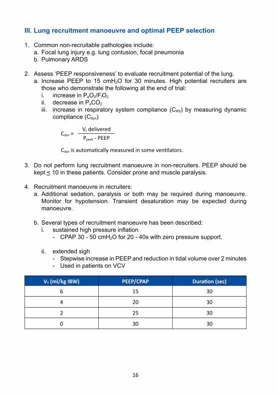
III. Lung recruitment manoeuvre and optimal PEEP selection
1. Common non-recruitable pathologies include: a. Focal lung injury e.g. lung contusion, focal pneumonia b. Pulmonary ARDS
2. Assess ‘PEEP responsiveness’ to evaluate recruitment potential of the lung. a. Increase PEEP to 15 cmH2O for 30 minutes. High potential recruiters are those who demonstrate the following at the end of trial: i. increase in PaO2/FiO2
ii. decrease in PaCO2
iii. increase in respiratory system compliance (CRS) by measuring dynamic compliance (Cdyn)
3. Do not perform lung recruitment manoeuvre in non-recruiters. PEEP should be kept < 10 in these patients. Consider prone and muscle paralysis.
4. Recruitment manoeuvre in recruiters: a. Additional sedation, paralysis or both may be required during manoeuvre. Monitor for hypotension. Transient desaturation may be expected during manoeuvre.
b. Several types of recruitment manoeuvre has been described: i. sustained high pressure inflation - CPAP 30 - 50 cmH2O for 20 - 40s with zero pressure support.
ii. extended sigh - Stepwise increase in PEEP and reduction in tidal volume over 2 minutes - Used in patients on VCV
Cdyn =
Cdyn is automatically measured in some ventilators.
Vt deliveredPpeak - PEEP
VT (ml/kg IBW) PEEP/CPAP Duration (sec)
6 15 30
4 20 30
2 25 30
0 30 30
17
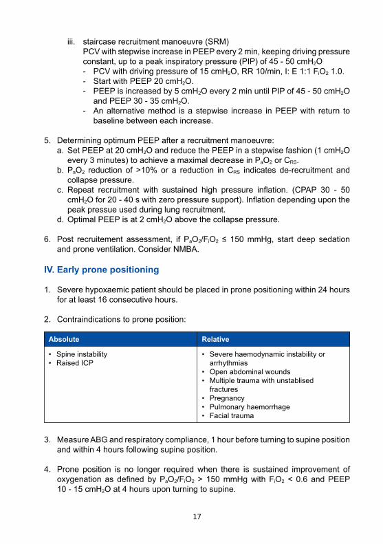
3. Measure ABG and respiratory compliance, 1 hour before turning to supine position and within 4 hours following supine position.
4. Prone position is no longer required when there is sustained improvement of oxygenation as defined by PaO2/FiO2 > 150 mmHg with FiO2 < 0.6 and PEEP 10 - 15 cmH2O at 4 hours upon turning to supine.
Absolute Relative
• Spine instability• Raised ICP
• Severe haemodynamic instability or arrhythmias• Open abdominal wounds• Multiple trauma with unstablised fractures• Pregnancy• Pulmonary haemorrhage• Facial trauma
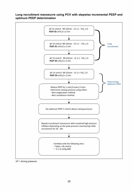
iii. staircase recruitment manoeuvre (SRM) PCV with stepwise increase in PEEP every 2 min, keeping driving pressure constant, up to a peak inspiratory pressure (PIP) of 45 - 50 cmH2O - PCV with driving pressure of 15 cmH2O, RR 10/min, I: E 1:1 FiO2 1.0. - Start with PEEP 20 cmH2O. - PEEP is increased by 5 cmH2O every 2 min until PIP of 45 - 50 cmH2O and PEEP 30 - 35 cmH2O. - An alternative method is a stepwise increase in PEEP with return to baseline between each increase.
5. Determining optimum PEEP after a recruitment manoeuvre: a. Set PEEP at 20 cmH2O and reduce the PEEP in a stepwise fashion (1 cmH2O every 3 minutes) to achieve a maximal decrease in PaO2 or CRS. b. PaO2 reduction of >10% or a reduction in CRS indicates de-recruitment and collapse pressure. c. Repeat recruitment with sustained high pressure inflation. (CPAP 30 - 50 cmH2O for 20 - 40 s with zero pressure support). Inflation depending upon the peak pressue used during lung recruitment. d. Optimal PEEP is at 2 cmH2O above the collapse pressure.
6. Post recruitement assessment, if PaO2/FiO2 ≤ 150 mmHg, start deep sedation and prone ventilation. Consider NMBA.
IV. Early prone positioning
1. Severe hypoxaemic patient should be placed in prone positioning within 24 hours for at least 16 consecutive hours.
2. Contraindications to prone position:
18
5. Patient may be placed in prone position again at anytime before the 4 hour assessment if oxygenation criteria is not met.
6. Complications of prone include desaturation, hypotension, pressure ulcers, unplanned extubation, facial and airway oedema and catheter dislodgement.
V. Neuromuscular blocking agents
1. Muscle paralysis may be considered in the early phase of severe hypoxaemia in selected patients e.g. patients with ventilatory dyssnchrony or in prone position.
2. Current evidence supports using cisatracurium besylate for not more than 48 hours.
3. There is no consensus on the depth of muscle paralysis. Titrating to achieve ‘Train of Four’ count of 2/4 with peripheral nerve stimulator may be reasonable.
4. Concerns using NMB include neuropathy and the need for deep sedation.
VI. Non-ventilatory strategy
1. Conservative fluid management that utilises fluid restriction and diuretics may improve oxygenation.
2. The use of corticosteroids cannot be recommended with the current level of evidence.
3. Consider echocardiography to assess right heart function. a. Acute cor pulmonale in severe hypoxaemic failure may require right heart inotropic support and afterload reduction.
b. RV protective ventilation strategy include: i. limit Pplat < 27 cmH2O ii. limit driving P < 15 cmH2O iii. target PaCO2 < 60 mmHg iv. decrease PEEP if RV dysfunction v. prone positioning
4. Refractory hypoxaemia may be secondary to patent or reopened foramen ovale with cardiac shunt.
19
∆P 15 cmH2O RR 10/min I:E 1:1 FiO2 1.0PEEP 25 cmH2O x 2 min
∆P 15 cmH2O RR 10/min I:E 1:1 FiO2 1.0PEEP 30 cmH2O x 2 min
∆P 15 cmH2O RR 10/min I:E 1:1 FiO2 1.0 PEEP 20 cmH2O x 2 min
Reduce PEEP by 1 cmH2O every 3 minsDetermine closing pressure using either:- Best oxygena�on method- Best compliance method
Repeat recruitment manoeuvre with sustained high pressureinfla�on depending on the peak pressure used during ini�alrecruitment for 20 - 30s
Set op�mum PEEP 2 cmH2O above closing pressure
Ven�late with the following aims:– Pplat ≤ 30 cmH2O– -Vt ≤ 6 ml/kg IBW
∆P 15 cmH2O RR 10/min I:E 1:1 FiO2 1.0, PEEP 20 cmH2O x 2 min
Lungrecruitment
Determiningop�mum PEEP
Lung recruitment manoeuvre using PCV with stepwise incremental PEEP and optimum PEEP determination
∆P = driving pressure
20
References
1. Fan E, et al. Acute respiratory distress syndrome: Advances in diagnosis and treatment. JAMA 2018;319(7):698-710
2. Chuimello D, et al. Severe hypoxaemia: which strategy to choose. Crit Care 2016;20:132
3. Writing group for the alveolar recruitment for acute respiratory distress syndrome trial (ART) investigators. Effect of lung recruitment and titrated positive end-expiratory pressure (PEEP) vs low PEEP on mortality in patients with acute respiratory distress syndrome: a randomized clinical trial. JAMA 2017;318(14):1335-1345
4. Papazian L, et al. Neuromuscular blockers in early acute respiratory distress syndrome. N Engl J Med 2010;363:1107-1116
5. Guerin C, et al. Prone positioning in severe acute respiratory distress syndrome. N Engl J Med 2013;368:2159-68
6. Vieillard-Baron A, et al. Acute cor pulmonale in ARDS. Intensive Care Med 2013;39(10):1836-1838
7. Peek GJ, et al. CESAR: conventional ventilatory support vs extracorporeal membrane oxygenation for severe adult respiratory failure. BMC Health Serv Res 2006:163
21
Weaning from Mechanical VentilationIntroduction
A weaning protocol is a systematic guide to aid the process of discontinuation of patients from mechanical ventilation. It should encompass sedation optimisation, early mobilisation, systematic assessment for readiness to wean, spontaneous breathing trials (SBT), screening for post-extubation stridor in patients at risk and use of non-invasive ventilation (NIV) in those at high risk of post-extubation failure.
Principles
1. Weaning begins as soon as the underlying cause for mechanical ventilation has sufficientlyimproved,thelevelofventilationsupportisreducedandtransitionto spontaneous breathing is initiated.
2. Benefitsofearlyweaningshouldbeweighedagainstrisksassociatedwithfailed extubation.
3. Weaning requires a sedation protocol aiming for light sedation and a rehabilitation protocol directed towards early mobilisation.
4. Assess patients’ readiness to wean on a daily basis.
5. Perform SBT in patients who pass readiness to wean and assess for extubation in those who pass the SBT.
6. Performacuffleaktestinpatientsathighriskofpost-extubationstridor.
7. Initiate NIV weaning in selected patients at high risk of weaning failure.
Assessment of readiness to wean
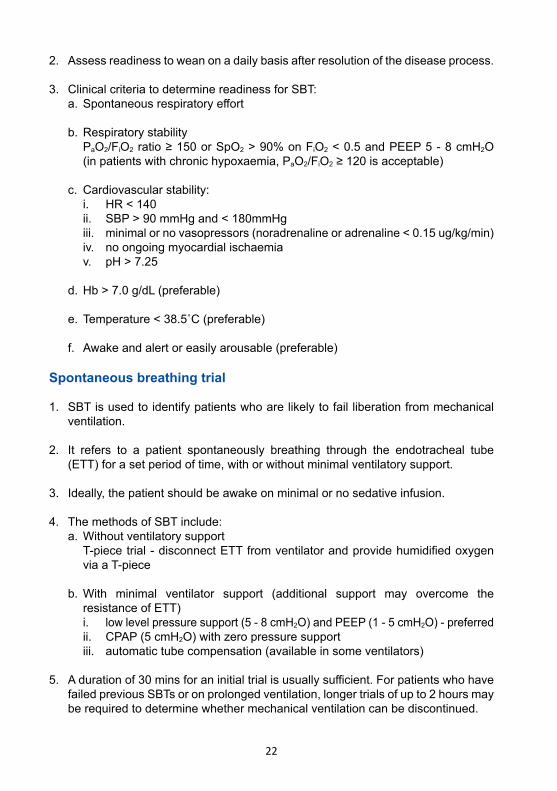
1. Weaningcanbeclassifiedassimple,difficultorprolonged.
Definition Incidence in ICU
Simple • PassfirstSBTandsuccessfullyextubated 50 - 67%Difficult • Requireupto3SBTsor
• Requireupto7daystopassaSBT26 - 39%
Prolonged • Fail>3SBTsor• Require>7daystowean
6 - 14%
22
2. Assess readiness to wean on a daily basis after resolution of the disease process.
3. Clinical criteria to determine readiness for SBT: a. Spontaneousrespiratoryeffort
b.Respiratorystability PaO2/FiO2 ratio≥150orSpO2>90%onFiO2 < 0.5 and PEEP 5 - 8 cmH2O (in patients with chronic hypoxaemia, PaO2/FiO2≥120isacceptable)
c. Cardiovascular stability: i. HR<140 ii. SBP>90mmHgand<180mmHg iii. minimal or no vasopressors (noradrenaline or adrenaline < 0.15 ug/kg/min) iv. no ongoing myocardial ischaemia v. pH>7.25
d.Hb>7.0g/dL(preferable)
e. Temperature<38.5˚C(preferable)
f. Awake and alert or easily arousable (preferable)
Spontaneous breathing trial
1. SBT is used to identify patients who are likely to fail liberation from mechanical ventilation.
2. It refers to a patient spontaneously breathing through the endotracheal tube (ETT) for a set period of time, with or without minimal ventilatory support.
3. Ideally, the patient should be awake on minimal or no sedative infusion.
4. The methods of SBT include: a. Without ventilatory support T-piecetrial-disconnectETTfromventilatorandprovidehumidifiedoxygen via a T-piece
b. With minimal ventilator support (additional support may overcome the resistance of ETT) i. low level pressure support (5 - 8 cmH2O) and PEEP (1 - 5 cmH2O) - preferred ii. CPAP (5 cmH2O) with zero pressure support iii. automatic tube compensation (available in some ventilators)
5. Adurationof30minsforaninitialtrialisusuallysufficient.Forpatientswhohave failed previous SBTs or on prolonged ventilation, longer trials of up to 2 hours may be required to determine whether mechanical ventilation can be discontinued.
23
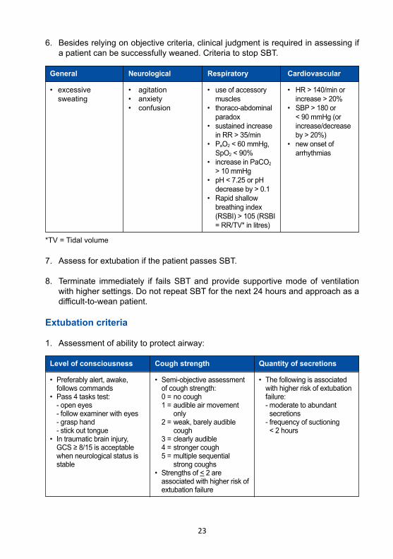
6. Besides relying on objective criteria, clinical judgment is required in assessing if a patient can be successfully weaned. Criteria to stop SBT.
General Neurological Respiratory Cardiovascular
• excessive sweating
• agitation• anxiety• confusion
• useofaccessory muscles• thoraco-abdominal paradox• sustainedincrease inRR>35/min• PaO2 < 60 mmHg, SpO2 < 90%• increaseinPaCO2 >10mmHg• pH<7.25orpH decreaseby>0.1• Rapidshallow breathing index (RSBI)>105(RSBI =RR/TV*inlitres)
• HR>140/minor increase>20%• SBP>180or < 90 mmHg (or increase/decrease by>20%)• newonsetof arrhythmias
7. Assess for extubation if the patient passes SBT.
8. Terminate immediately if fails SBT and provide supportive mode of ventilation with higher settings. Do not repeat SBT for the next 24 hours and approach as a difficult-to-weanpatient.
Extubation criteria
1. Assessment of ability to protect airway:
*TV=Tidalvolume
Level of consciousness Cough strength Quantity of secretions
• Preferablyalert,awake, follows commands• Pass4taskstest: - open eyes - follow examiner with eyes - grasp hand - stick out tongue• Intraumaticbraininjury, GCS≥8/15isacceptable when neurological status is stable
• Semi-objectiveassessment of cough strength: 0 = no cough 1 = audible air movement only 2 = weak, barely audible cough 3 = clearly audible 4 = stronger cough 5 = multiple sequential strong coughs• Strengthsof< 2 are associated with higher risk of extubation failure
• Thefollowingisassociated with higher risk of extubation failure: - moderate to abundant secretions - frequency of suctioning < 2 hours
24
2. Performacuffleaktestasasurrogatemarkeroflaryngealoedemainpatientsat high risk of post-extubation stridor. These include: a. Traumatic intubation b.Durationofintubation≥7days c. ExcessivelylargeETT(≥7.5mminwomen,≥8.5mminmen) d. Excessive ETT mobility (psychomotor agitation or ETT not anchored properly) e.Reintubationafterunplannedextubation
3. Performacuffleaktestasfollows: a. Use volume-controlled ventilation and record inspiratory tidal volume
b. Deflate cuff and average the cuff leak volume over 6 breaths
c. Calculatecuffleakvolume(CLV)i.e.thedifferencebetweentheinspiredtidal volume and average expired tidal volume. CLV < 110 ml or < 15-20% of deliveredtidalvolumeindicatesafailedcuffleaktest.
4. Inpatientswhofailacuffleaktest,theadministrationofsystemicsteroids4hours prior to extubation has been shown to reduce risks of post-extubation stridor and reintubation. a.Recommended agent is single dose iv methylprednisolone 40 mg. The alternative is iv dexamethasone 8 mg.
b. Arepeatcuffleaktestisnotrequiredafteradministrationofsystemicsteroids.
Difficult-to-wean
1. Provide ventilator management that encourages spontaneous breathing while avoiding respiratory fatigue.
2. Use pressure support (PS) as a weaning method i.e. continuous gradual reduction of PS by 2 cmH20 at a time, once or twice a day. Once PS is reduced to minimal level, repeat SBT on daily basis.
3. Consider the following strategies before resuming SBT: a. Place patient in upright position for the SBT b. Suction airway secretions c. Optimise nutrition, taking care not to over or underfeed d. Ensure adequate nocturnal rest with daytime respiratory muscle training e. Aimfornegativebalanceinpatientwithpositivecumulativefluidbalance f. Consider longer duration of SBT of up to 2 hours
4. Identify and treat causes (see appendix) of weaning failure by performing a detailed physical examination and investigations as indicated.
5. Consider tracheostomy in patients who are unable to wean within 1 - 3 weeks.
25
Non-invasive ventilation (NIV) for weaning
1. NIV for weaning can only be successful in patients with good airway protection, strong cough and manageable secretions.
2. Potential candidates for NIV weaning are those at high-risk for weaning failure: a. COPD b. Congestive heart failure c. Elderly>65years d. Hypercapnia during SBT e. Failed>1SBT
3. Start NIV immediately after extubation and maintain for at least 24 hours.
4. Do not delay reintubation in patients who fail NIV.
High-flow nasal cannula (HFNC) oxygen
1. ConsiderHFNCoxygeninpatientswithpost-extubationhypoxaemicrespiratory failure, (if available).
Prolonged mechanical ventilation (PMV)
1. Acommonconsensusdefinitionisrequirementofventilation>21daysforatleast 6 hours a day.
2. These patients will require a tracheostomy.
3. Identify factors that are potentially reversible.
4. Perform increases in duration of SBT with frequent assessment for signs of failure during SBT. A suggested approach is as follows: a. Assess for readiness to wean when PS has been gradually reduced to 10 - 12 cmH2O.
b. PerformSBTusingtrachemaskforaspecifiedduratione.g.2hours.
c. If SBT is successful, perform daily SBT of progressively longer duration e.g. 4 hours, 6 hours etc.
d. LengthentheSBTdurationifpatientfeelscomfortableandwishestocontinue at the end of the SBT.
e. Do not repeat the SBT for at least another 24 hours if SBT fails.
f. The eventual goal is to reach 24 hours and be completely liberated from the ventilator.
26
5. Patients requiring PMV should not be considered permanently ventilator- dependent until at least 3 months of weaning attempts have failed, unless the respiratory failure is due to an irreversible process.
Resolu�onof disease
Spontaneousbreathing trial
Ready to wean?
Ability to protectairway
No
Yes
Yes
Pass
No
Fail
No
High risk of post-extuba�on stridor
Invasive mechanicalven�la�on
Considertracheostomy
High risk ofextuba�on
failure
Difficult to wean(suggest PSV as
weaning method)
Yes
No
Yes Extubate to NIV(NIV weaning)
No
Extubate
Single dose systemicsteroids 4 hours
before extuba�onCuff leak test
Absent
Present
Yes
Weaning from mechanical ventilation
(modified and reproduced with permission from Professor Gavin Joynt, Chinese University of Hong Kong)
27
References
1. OuelletteDR,etal.Liberationfrommechanicalventilationincriticallyilladults:AnofficialAmerican College of Chest Physicians/American Thoracic Society clinical practice guideline. Inspiratory pressure augmentation during spontaneous breathing trials, protocols minimizing sedation, and non-invasive ventilation immediately after extubation. Chest 2017;151(1):166-180
2. Girard TD, et al. An official American Thoracic Society/American College of ChestPhysiciansclinicalpracticeguideline:Liberationfrommechanicalventilationincriticallyilladults.Rehabilitationprotocols,ventilatorliberationprotocols,andcuffleaktests.Am J Respir Crit Care Med 2017;195(1):120-133
3. BurnsKEA,etal.Useofnon-invasiveventilationtoweancriticallyilladultsoffinvasiveventilation: meta-analysis and systematic review. BMJ 2009;338:b1574
4. JaberS,etal.Effectsofsteroidsonreintubationandpost-extubationstridor inadults:meta-analysis of randomised control trials. Crit Care 2009;13(2):R49
5. HelvizY,etal.Asystematicreviewofthehigh-flownasalcannulaforadultpatients.Crit Care 2018;22:71
28
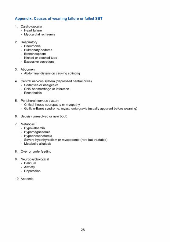
Appendix: Causes of weaning failure or failed SBT
1. Cardiovascular - Heart failure - Myocardial ischaemia
2. Respiratory - Pneumonia - Pulmonary oedema - Bronchospasm - Kinked or blocked tube - Excessive secretions
3. Abdomen - Abdominal distension causing splinting
4. Central nervous system (depressed central drive) - Sedatives or analgesics - CNS haemorrhage or infarction - Encephalitis
5. Peripheral nervous system - Critical illness neuropathy or myopathy - Guillain-Barre syndrome, myasthenia gravis (usually apparent before weaning)
6. Sepsis (unresolved or new bout)
7. Metabolic - Hypokalaemia - Hypomagnesemia - Hypophosphatemia - Severe hypothyroidism or myxoedema (rare but treatable) - Metabolic alkalosis
8. Over or underfeeding
9. Neuropsychological - Delirium - Anxiety - Depression
10. Anaemia
29
Pain, Sedation and DeliriumManagement of sedation and delirium in ICU patients has evolved, emphasizing on effective pain management and aiming for light sedation. A safe and effective strategy should be implemented to avoid complications and conflicts with other management goals e.g. weaning from mechanical ventilation and early mobilisation.
Principles
1. Focus first on analgesia, then sedation.
2. Practise analgesic-first sedation by using an analgesic (usually an opioid) prior to a sedative to reach the sedation goal.
3. Aim for light sedation unless contraindicated.
4. Titrate analgesic and sedative drugs to a defined target, using the lowest effective dose.
5. Identify risk factors and implement effective preventive measures for delirium.
6. Assess pain, sedation and delirium objectively using validated monitoring tools.
7. Employ pharmacological and non-pharmacological strategies to manage pain, agitation and delirium.
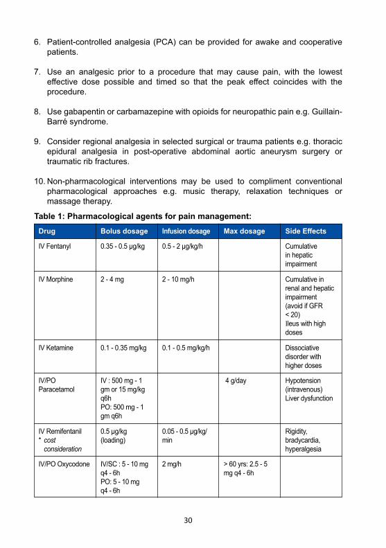
Pain
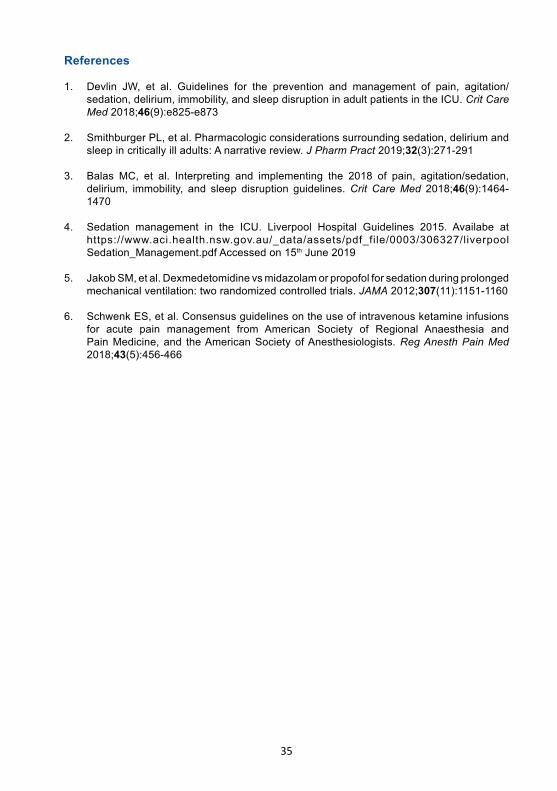
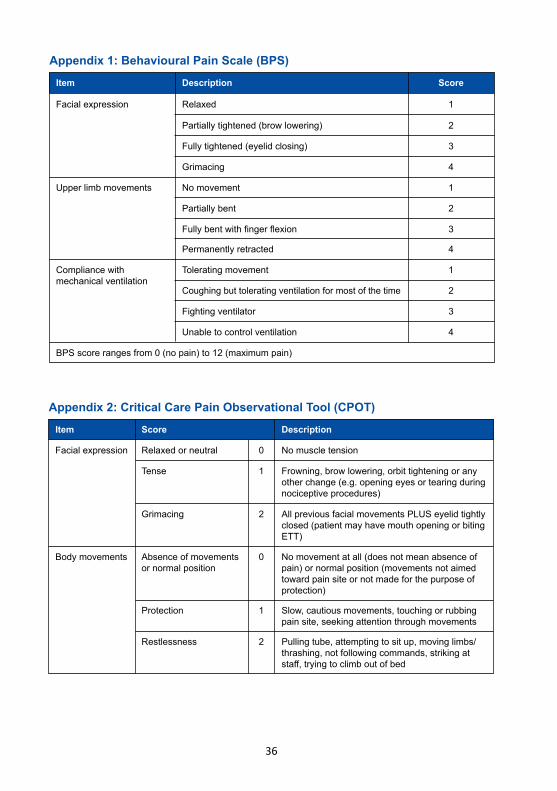
1. Use validated scales to monitor pain i.e. Behavioral Pain Score (BPS) or Critical Care Pain Observational Tool (CPOT) in the unconscious, and Visual Analogue Score (VAS) in the conscious patients.
2. Assess pain at least 4 hourly.
3. Institute pain management when pain score is: a. ≥ 5 for BPS b. ≥ 3 for CPOT c. ≥ 3 for VAS
4. Opioid based analgesia remains the mainstay of pain management.
5. Consider adjuncts to an opioid to reduce the dose of opioid and/or reduce severity of pain. a. Paracetamol either administered intravenously, orally or per rectal b. IV ketamine in post-surgical patients
30
6. Patient-controlled analgesia (PCA) can be provided for awake and cooperative patients.
7. Use an analgesic prior to a procedure that may cause pain, with the lowest effective dose possible and timed so that the peak effect coincides with the procedure.
8. Use gabapentin or carbamazepine with opioids for neuropathic pain e.g. Guillain- Barré syndrome.
9. Consider regional analgesia in selected surgical or trauma patients e.g. thoracic epidural analgesia in post-operative abdominal aortic aneurysm surgery or traumatic rib fractures.
10. Non-pharmacological interventions may be used to compliment conventional pharmacological approaches e.g. music therapy, relaxation techniques or massage therapy.
Drug Bolus dosage Infusion dosage Max dosage Side Effects
IV Fentanyl
IV Morphine
IV Ketamine
IV/PO Paracetamol
0.35 - 0.5 μg/kg
2 - 4 mg
0.1 - 0.35 mg/kg
IV : 500 mg - 1 gm or 15 mg/kg q6hPO: 500 mg - 1 gm q6h
0.5 - 2 μg/kg/h
2 - 10 mg/h
0.1 - 0.5 mg/kg/h
4 g/day
Cumulative in hepatic impairment
Cumulative in renal and hepatic impairment(avoid if GFR < 20)Ileus with high doses
Dissociative disorder with higher doses
Hypotension (intravenous)Liver dysfunction
IV Remifentanil * cost consideration
0.5 μg/kg (loading)
0.05 - 0.5 μg/kg/min
Rigidity, bradycardia, hyperalgesia
IV/PO Oxycodone IV/SC : 5 - 10 mg q4 - 6hPO: 5 - 10 mg q4 - 6h
2 mg/h > 60 yrs: 2.5 - 5 mg q4 - 6h
Table 1: Pharmacological agents for pain management:
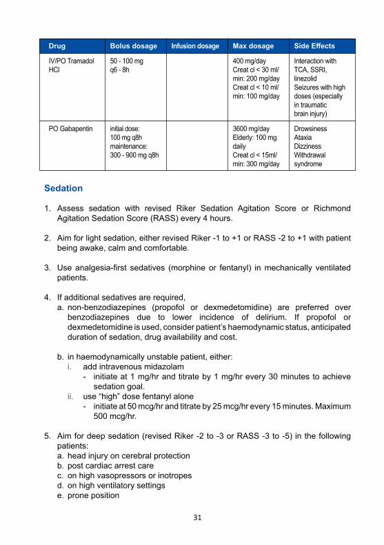
31
IV/PO Tramadol HCl
PO Gabapentin
50 - 100 mg q6 - 8h
initial dose: 100 mg q8hmaintenance: 300 - 900 mg q8h
Interaction with TCA, SSRI, linezolidSeizures with high doses (especially in traumatic brain injury)
DrowsinessAtaxiaDizzinessWithdrawal syndrome
400 mg/dayCreat cl < 30 ml/min: 200 mg/dayCreat cl < 10 ml/min: 100 mg/day
3600 mg/day Elderly: 100 mg daily Creat cl < 15ml/min: 300 mg/day
Sedation
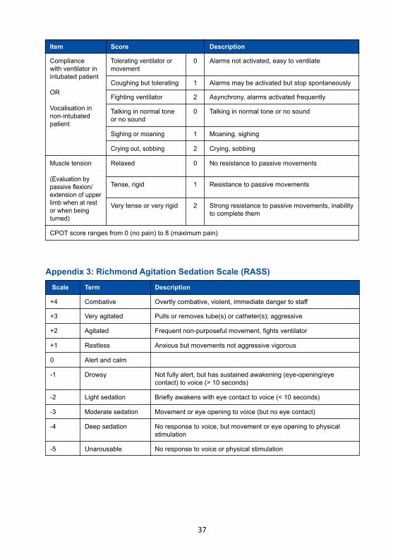
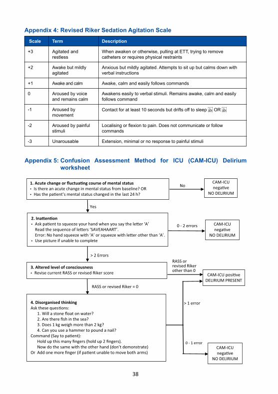
1. Assess sedation with revised Riker Sedation Agitation Score or Richmond Agitation Sedation Score (RASS) every 4 hours.
2. Aim for light sedation, either revised Riker -1 to +1 or RASS -2 to +1 with patient being awake, calm and comfortable.
3. Use analgesia-first sedatives (morphine or fentanyl) in mechanically ventilated patients.
4. If additional sedatives are required, a. non-benzodiazepines (propofol or dexmedetomidine) are preferred over benzodiazepines due to lower incidence of delirium. If propofol or dexmedetomidine is used, consider patient’s haemodynamic status, anticipated duration of sedation, drug availability and cost.
b. in haemodynamically unstable patient, either: i. add intravenous midazolam - initiate at 1 mg/hr and titrate by 1 mg/hr every 30 minutes to achieve sedation goal. ii. use “high” dose fentanyl alone - initiate at 50 mcg/hr and titrate by 25 mcg/hr every 15 minutes. Maximum 500 mcg/hr.
5. Aim for deep sedation (revised Riker -2 to -3 or RASS -3 to -5) in the following patients: a. head injury on cerebral protection b. post cardiac arrest care c. on high vasopressors or inotropes d. on high ventilatory settings e. prone position
Drug Bolus dosage Infusion dosage Max dosage Side Effects
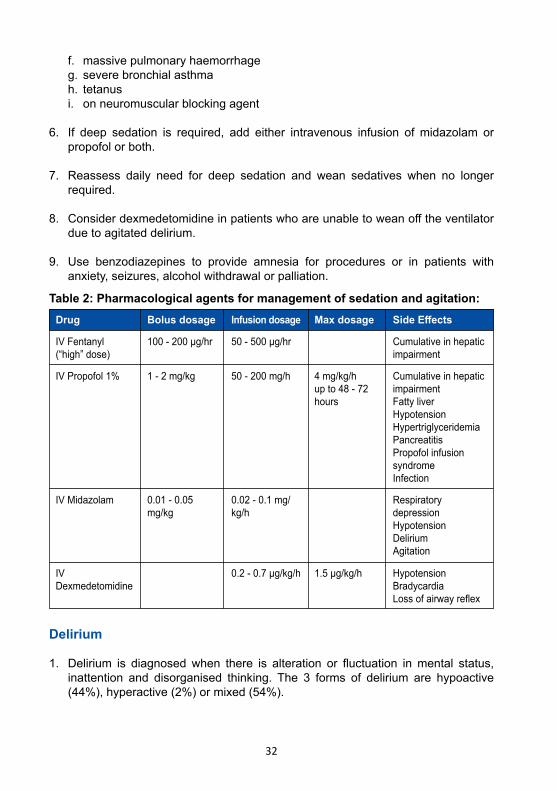
32
f. massive pulmonary haemorrhage g. severe bronchial asthma h. tetanus i. on neuromuscular blocking agent
6. If deep sedation is required, add either intravenous infusion of midazolam or propofol or both.
7. Reassess daily need for deep sedation and wean sedatives when no longer required.
8. Consider dexmedetomidine in patients who are unable to wean off the ventilator due to agitated delirium.
9. Use benzodiazepines to provide amnesia for procedures or in patients with anxiety, seizures, alcohol withdrawal or palliation.
Delirium
1. Delirium is diagnosed when there is alteration or fluctuation in mental status, inattention and disorganised thinking. The 3 forms of delirium are hypoactive (44%), hyperactive (2%) or mixed (54%).
Drug Bolus dosage Infusion dosage Max dosage Side Effects
IV Fentanyl (“high” dose)
IV Midazolam
IVDexmedetomidine
IV Propofol 1%
100 - 200 μg/hr
0.01 - 0.05 mg/kg
1.5 μg/kg/h
1 - 2 mg/kg
50 - 500 μg/hr
0.02 - 0.1 mg/kg/h
0.2 - 0.7 μg/kg/h
50 - 200 mg/h
Cumulative in hepatic impairment
Respiratory depressionHypotensionDeliriumAgitation
HypotensionBradycardiaLoss of airway reflex
Cumulative in hepatic impairmentFatty liverHypotensionHypertriglyceridemiaPancreatitisPropofol infusion syndromeInfection
4 mg/kg/hup to 48 - 72 hours
Table 2: Pharmacological agents for management of sedation and agitation:
33
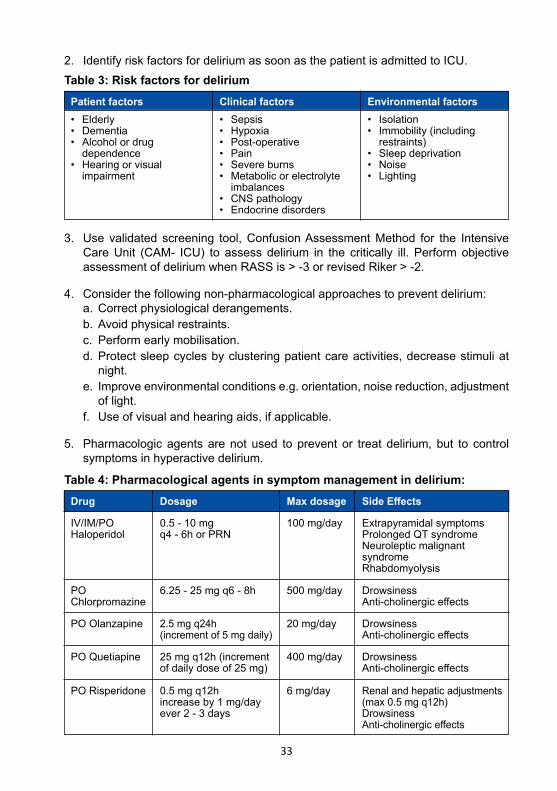
2. Identify risk factors for delirium as soon as the patient is admitted to ICU.
3. Use validated screening tool, Confusion Assessment Method for the Intensive Care Unit (CAM- ICU) to assess delirium in the critically ill. Perform objective assessment of delirium when RASS is > -3 or revised Riker > -2.
4. Consider the following non-pharmacological approaches to prevent delirium: a. Correct physiological derangements. b. Avoid physical restraints. c. Perform early mobilisation. d. Protect sleep cycles by clustering patient care activities, decrease stimuli at night. e. Improve environmental conditions e.g. orientation, noise reduction, adjustment of light. f. Use of visual and hearing aids, if applicable.
5. Pharmacologic agents are not used to prevent or treat delirium, but to control symptoms in hyperactive delirium.
Patient factors Clinical factors Environmental factors• Elderly• Dementia• Alcohol or drug dependence• Hearing or visual impairment
• Sepsis• Hypoxia• Post-operative• Pain• Severe burns• Metabolic or electrolyte imbalances• CNS pathology• Endocrine disorders
• Isolation• Immobility (including restraints)• Sleep deprivation• Noise• Lighting
Drug Dosage Max dosage Side Effects
IV/IM/PO Haloperidol
PO Chlorpromazine
PO Olanzapine
PO Quetiapine
PO Risperidone
0.5 - 10 mg q4 - 6h or PRN
6.25 - 25 mg q6 - 8h
2.5 mg q24h(increment of 5 mg daily)
25 mg q12h (increment of daily dose of 25 mg)
0.5 mg q12hincrease by 1 mg/day ever 2 - 3 days
100 mg/day
500 mg/day
20 mg/day
400 mg/day
6 mg/day
Extrapyramidal symptomsProlonged QT syndromeNeuroleptic malignant syndromeRhabdomyolysis
DrowsinessAnti-cholinergic effects
DrowsinessAnti-cholinergic effects
DrowsinessAnti-cholinergic effects
Renal and hepatic adjustments (max 0.5 mg q12h)DrowsinessAnti-cholinergic effects
Table 4: Pharmacological agents in symptom management in delirium:
Table 3: Risk factors for delirium
34
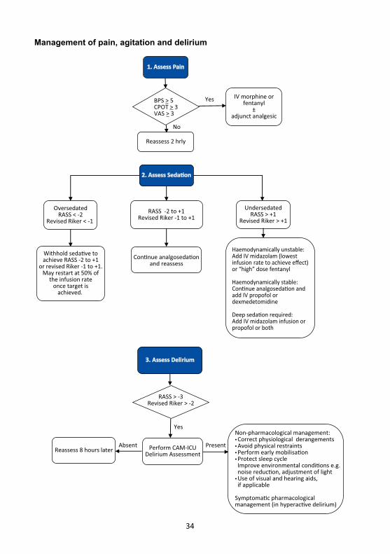
1. Assess Pain
Reassess 2 hrly
IV morphine orfentanyl
±adjunct analgesic
Yes
Yes
Absent Present
No
2. Assess Seda�on
Haemodynamically unstable:Add IV midazolam (lowestinfusion rate to achieve effect)or “high” dose fentanyl
Haemodynamically stable:Con�nue analgoseda�on andadd IV propofol ordexmedetomidine
Deep seda�on required:Add IV midazolam infusion orpropofol or both
Con�nue analgoseda�onand reassess
Withhold seda�ve toachieve RASS -2 to +1
or revised Riker -1 to +1.May restart at 50% of
the infusion rateonce target is
achieved.
OversedatedRASS < -2
Revised Riker < -1
UndersedatedRASS > +1
Revised Riker > +1
3. Assess Delirium
RASS -2 to +1Revised Riker -1 to +1
RASS > -3Revised Riker > -2
Perform CAM-ICUDelirium Assessment Reassess 8 hours later
Non-pharmacological management:• Correct physiological derangements• Avoid physical restraints• Perform early mobilisa�on• Protect sleep cycle Improve environmental condi�ons e.g. noise reduc�on, adjustment of light • Use of visual and hearing aids, if applicable
Symptoma�c pharmacologicalmanagement (in hyperac�ve delirium)
BPS > 5CPOT > 3VAS > 3
Management of pain, agitation and delirium
35
References
1. Devlin JW, et al. Guidelines for the prevention and management of pain, agitation/sedation, delirium, immobility, and sleep disruption in adult patients in the ICU. Crit Care Med 2018;46(9):e825-e873
2. Smithburger PL, et al. Pharmacologic considerations surrounding sedation, delirium and sleep in critically ill adults: A narrative review. J Pharm Pract 2019;32(3):271-291
3. Balas MC, et al. Interpreting and implementing the 2018 of pain, agitation/sedation, delirium, immobility, and sleep disruption guidelines. Crit Care Med 2018;46(9):1464-1470
4. Sedation management in the ICU. Liverpool Hospital Guidelines 2015. Availabe at https://www.aci.health.nsw.gov.au/_data/assets/pdf_file/0003/306327/liverpool Sedation_Management.pdf Accessed on 15th June 2019
5. Jakob SM, et al. Dexmedetomidine vs midazolam or propofol for sedation during prolonged mechanical ventilation: two randomized controlled trials. JAMA 2012;307(11):1151-1160
6. Schwenk ES, et al. Consensus guidelines on the use of intravenous ketamine infusions for acute pain management from American Society of Regional Anaesthesia and Pain Medicine, and the American Society of Anesthesiologists. Reg Anesth Pain Med 2018;43(5):456-466
36
Appendix 2: Critical Care Pain Observational Tool (CPOT)Item Score Description
Facial expression Relaxed or neutral
Tense
Grimacing
No muscle tension
Frowning, brow lowering, orbit tightening or any other change (e.g. opening eyes or tearing during nociceptive procedures)
All previous facial movements PLUS eyelid tightly closed (patient may have mouth opening or biting ETT)
0
1
2
Body movements Absence of movements or normal position
Protection
Restlessness
No movement at all (does not mean absence of pain) or normal position (movements not aimed toward pain site or not made for the purpose of protection)
Slow, cautious movements, touching or rubbing pain site, seeking attention through movements
Pulling tube, attempting to sit up, moving limbs/thrashing, not following commands, striking at staff, trying to climb out of bed
0
1
2
Appendix 1: Behavioural Pain Scale (BPS)Item Description Score
Facial expression
BPS score ranges from 0 (no pain) to 12 (maximum pain)
Upper limb movements
Compliance with mechanical ventilation
Relaxed
No movement
Tolerating movement
Partially tightened (brow lowering)
Partially bent
Coughing but tolerating ventilation for most of the time
Fully tightened (eyelid closing)
Fully bent with finger flexion
Fighting ventilator
Grimacing
Permanently retracted
Unable to control ventilation
1
1
1
2
2
2
3
3
3
4
4
4
37
Item Score Description
CPOT score ranges from 0 (no pain) to 8 (maximum pain)
Compliance with ventilator in intubated patient
OR
Vocalisation in non-intubated patient
Muscle tension
(Evaluation by passive flexion/extension of upper limb when at rest or when being turned)
Tolerating ventilator or movement
Relaxed
Tense, rigid
Very tense or very rigid
Talking in normal tone or no sound
Sighing or moaning
Crying out, sobbing
Coughing but tolerating
Fighting ventilator
Alarms not activated, easy to ventilate
No resistance to passive movements
Resistance to passive movements
Strong resistance to passive movements, inability to complete them
Talking in normal tone or no sound
Moaning, sighing
Crying, sobbing
Alarms may be activated but stop spontaneously
Asynchrony, alarms activated frequently
0
0
1
2
0
1
2
1
2
Appendix 3: Richmond Agitation Sedation Scale (RASS) Scale Term Description
+4
+3
+2
+1
-1
-2
-3
-4
-5
0
Combative
Very agitated
Agitated
Restless
Drowsy
Light sedation
Moderate sedation
Deep sedation
Unarousable
Alert and calm
Overtly combative, violent, immediate danger to staff
Pulls or removes tube(s) or catheter(s); aggressive
Frequent non-purposeful movement, fights ventilator
Anxious but movements not aggressive vigorous
Not fully alert, but has sustained awakening (eye-opening/eye contact) to voice (> 10 seconds)
Briefly awakens with eye contact to voice (< 10 seconds)
Movement or eye opening to voice (but no eye contact)
No response to voice, but movement or eye opening to physical stimulation
No response to voice or physical stimulation
38
Appendix 4: Revised Riker Sedation Agitation Scale Scale Term Description
+3
+2
0
-1
-2
+1
-3
Agitated and restless
Awake but mildly agitated
Aroused by voice and remains calm
Aroused by movement
Aroused by painful stimuli
Awake and calm
Unarousable
When awaken or otherwise, pulling at ETT, trying to remove catheters or requires physical restraints
Anxious but mildly agitated. Attempts to sit up but calms down with verbal instructions
Awakens easily to verbal stimuli. Remains awake, calm and easily follows command
Contact for at least 10 seconds but drifts off to sleep OR
Localising or flexion to pain. Does not communicate or follow commands
Awake, calm and easily follows commands
Extension, minimal or no response to painful stimuli
CAM-ICU posi�veDELIRIUM PRESENT
1. Acute change or fluctua�ng course of mental status• Is there an acute change in mental status from baseline? OR• Has the pa�ent’s mental status changed in the last 24 h?
2. Ina�en�on• Ask pa�ent to squeeze your hand when you say the le�er ‘A’ Read the sequence of le�ers ‘SAVEAHAART’. Error: No hand squeeze with ‘A’ or squeeze with le�er other than ‘A’.• Use picture if unable to complete
3. Altered level of consciousness• Revise current RASS or revised Riker score
4. Disorganised thinkingAsk these ques�ons: 1. Will a stone float on water? 2. Are there fish in the sea? 3. Does 1 kg weigh more than 2 kg? 4. Can you use a hammer to pound a nail?Command (Say to pa�ent): Hold up this many fingers (hold up 2 fingers). Now do the same with the other hand (don’t demonstrate)Or Add one more finger (if pa�ent unable to move both arms)
CAM-ICUnega�ve
NO DELIRIUM
CAM-ICUnega�ve
NO DELIRIUM
Yes
No
> 2 Errors
0 - 2 errors
RASS or revised Riker = 0
RASS or revised Rikerother than 0
CAM-ICUnega�ve
NO DELIRIUM
> 1 error
0 - 1 error
Appendix 5: Confusion Assessment Method for ICU (CAM-ICU) Delirium worksheet
39
Nutritional TherapyIntroduction
Early enteral nutrition attenuates metabolic response to stress, prevents oxidative cellular injury and modulates immune responses. Enteral nutrition (EN) is preferred over parenteral nutrition (PN). Parenteral nutrition is an alternative when enteral route is neither sufficient nor feasible.
Principles
1. Assess nutritional risk.
2. Commence enteral nutrition within 24 to 48 hours upon ICU admission.
3. Aim only for 70% - 80% of targeted calories within 72 hours.
4. Aim to provide protein of at least 1.2 g/kg/day.
5. Prescribe parenteral nutrition after 5 - 7 days if enteral nutrition is not feasible.
6. Avoid overfeeding.
7. Identify patients at risk of refeeding syndrome.
Nutritional risk
1. Nutritional status should be assessed clinically to identify patients with malnutrition or at risk of malnutrition.
2. The following patients are to be considered at high risk for malnutrition: a. ICU stay for > 2 days and mechanically ventilated b. Underfed for > 5 days c. Pre-existing severe chronic disease
Enteral nutrition
1. Initiate EN within 24 - 48 hours of ICU admission if gastrointestinal tract is functioning and patient adequately resuscitated.
2. EN should be delayed in: a. Shock with haemodynamic instability b. Severe hypoxaemia and acidosis c. Active upper GI bleeding d. Acute bowel ischaemic e. Abdominal compartment syndrome f. Gastric residual volume > 500 mls in 6 hours
40
3. Aim to achieve only 70 - 80% of targeted calories in the first 72 hours of admission i.e. the early phase of acute illness.
4. Provide EN via nasogastric or orogastric tube size 10 to 12Fr after confirming correct placement by the following methods: a. Chest radiograph b. Aspiration of gastric contents c. Measuring pH of aspirate using pH indicator strip (if available)
5. Ensure head of bed is elevated at least 30 degrees during feeding.
6. Enteral feeding can be administered by various means: a. Continuous: administered at an hourly rate using a feeding pump for 24 hours.
b. Intermittent: administered using a feeding pump over a few hours with rest period in between e.g. feeding over 4 hours with 2 hours rest.
c. Bolus: administered by gravity over 15 min every 3 - 4 hours.
7. Enteral formulation a. Use standard polymeric isocaloric or near isocaloric of 1 - 1.5 kcal/ml. b. Consider diabetic specific formula with low glycaemic index in diabetics. c. Consider a caloric dense formula in patients with fluid restriction.
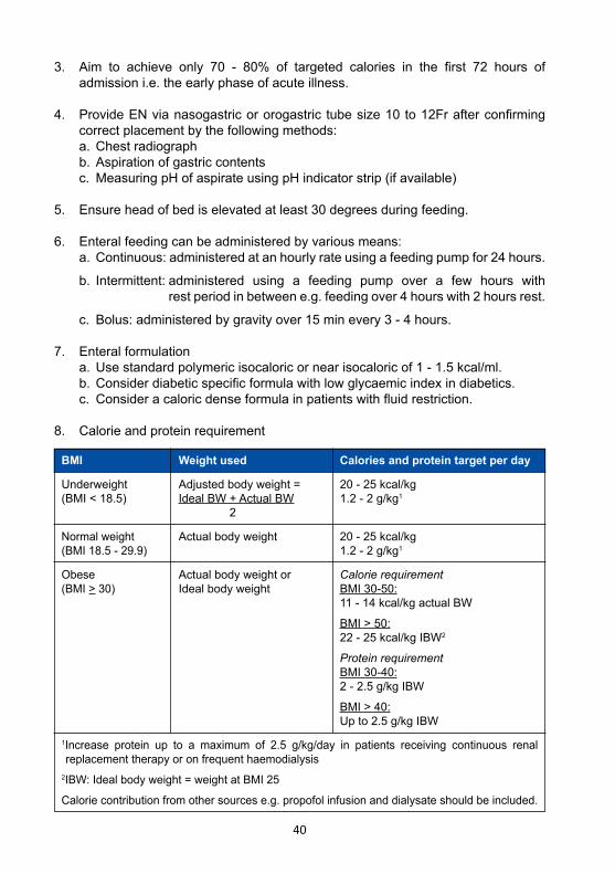
8. Calorie and protein requirement
BMI Weight used Calories and protein target per day
Underweight(BMI < 18.5)
Normal weight(BMI 18.5 - 29.9)
Obese(BMI ˃ 30)
1Increase protein up to a maximum of 2.5 g/kg/day in patients receiving continuous renal replacement therapy or on frequent haemodialysis2IBW: Ideal body weight = weight at BMI 25
Calorie contribution from other sources e.g. propofol infusion and dialysate should be included.
Adjusted body weight =Ideal BW + Actual BW 2
Actual body weight
Actual body weight or Ideal body weight
20 - 25 kcal/kg1.2 - 2 g/kg1
20 - 25 kcal/kg1.2 - 2 g/kg1
Calorie requirementBMI 30-50:11 - 14 kcal/kg actual BW
BMI > 50:22 - 25 kcal/kg IBW2
Protein requirementBMI 30-40:2 - 2.5 g/kg IBW
BMI > 40:Up to 2.5 g/kg IBW
41
9. Monitor for feeding intolerance. Feeding intolerance is defined as high gastric residual volume (GRV), abdominal distension, vomiting, diarrhoea or reduced passage of stools.
A. GRV > 300 ml a. Use a prokinetic agent: i. iv erythromycin 125 mg q6h or 250 mg q12h or ii. iv metoclopramide 10 mg q8h
b. Combination of both agents has shown to improve gastric tolerance and may be considered. Both agents are associated with QT prolongation.
c. Review the need of prokinetics after 48 hours.
B. Diarrhoea a. Defined as > 2-3 liquid stools per day or > 250 g liquid stool per day.
b. Do not routinely interrupt feeding for diarrhoea.
c. Factors that may contribute to diarrhoea: i. type and amount of fibre in formula ii. osmolality of formula iii. delivery mode iv. EN contamination v. medications (antibiotics, PPI, prokinetics, laxatives) vi. Clostridium difficile infection vii. intra-abdominal collections following abdominal surgery
d. Management includes: i. review of medications and dietary formula ii. send stool for Clostridium difficile toxin assays iii. monitor serum electrolytes iv. rule out abdominal pathology
e. If diarrhoea persists consider the addition of soluble fibre supplement into standard feeds or the use of small peptide semi-elemental formula
10. Monitor blood glucose levels a. Keep blood glucose between 8 - 10 mmol/L b. Start insulin if blood glucose > 10 mmol/L
11. Trophic feeding (defined as 10 - 20 ml/h or up to 500 kcal/day) should be considered in: a. Resolving shock (minimal or decreasing vasopressors or inotropes). b. Prone position. c. Intra-abdominal hypertension without abdominal compartment syndrome.
42
12. Fasting a. In intubated patients, do not withhold feeding for procedures in ICU or operating theatre or extubation.
b. Feeds should be aspirated prior to transport, procedures or extubation.
c. For non-intubated patients, follow the fasting protocol as per general anaesthesia.
13. Safe practice for enteral nutrition a. Use sterile water for formula reconstitution and tube flushing.
b. Feeding tubes to be flushed with at least 20 - 30 ml of water at the end of feeding.
c. Flush volume should be included in daily fluid intake.
d. Hang time duration: i. closed system (ready to hang): 24 hours or per manufacturer’s recommendation ii. sterile decanted formula: 8 hours iii. powdered reconstituted formula: 4 hours
e. Change administration set every 24 hours.
f. Opened decanted formula must be refrigerated and discarded within 12 hours if not used.
Parenteral nutrition
1. Indications for PN: a. As soon as possible in patients who are severely malnourished and EN is not feasible.
b. After 5 - 7 days in patients not deemed malnourished on admission and EN is not feasible.
c. Supplemental PN to be considered if > 60% of energy and protein requirement via EN is not met after 7 - 10 days.
2. Dosing a. Start with non-protein calories of ≤ 20 kcal/kg/day with protein of ≥ 1.2 g/kg/d. Increase gradually and aim to achieve target within 5 - 7 days.
43
b. Macronutrients required per day i. carbohydrate 2 - 5 g/kg ii. lipids 0.75 - 1.5 g/kg iii. proteins 1.2 - 2.0 g/kg
c. Carbohydrate to lipid ratio should be between 60:40 or 70:30
d. Prescribe electrolytes based on daily serum levels.
e. Add trace elements and vitamins.
3. Administration a. High osmolality via a dedicated lumen of a central line. b. Low osmolality (< 850 mOsmol/L) can be administered via peripheral line. c. Change administration set every 24 hours.
4. Monitoring a. 2 hourly blood glucose. Target level of 8 - 10 mmol/L b. Daily serum potassium, phosphate, calcium and magnesium. c. Biweekly liver function test. d. Weekly serum triglycerides. Target level < 4.5 mmol/L.
5. Discontinuation Discontinue PN when patient receives > 60% of targeted calories enterally.
Refeeding syndrome
Refeeding syndrome(RFS) describes the biochemical changes, clinical manifestations and complications that occur due to severe shift of fluid and electrolytes when a malnourished patient is fed either enterally or parenterally.
1. Risk factors a. High risk: 1 or more major risk factors - BMI < 16.5 kg/m2
- Unintentional weight loss of > 15% in the previous 3 - 6 months - Little or no nutritional intake for > 10 days
b. High risk: 2 or more minor risk factors - BMI < 18.5 kg/m2
- Unintentional weight loss of > 10% in the previous 3 - 6 months - Little or nutritional intake for > 5 days - History of alcohol abuse or drugs including insulin, chemotherapy or diuretics
c. Extreme high risk: 1 of the following - BMI < 14 kg/m2
- Little or no nutritional intake for > 15 days
44
2. Prevention and management of RFS: a. Identify patients at risk.
b. Check phosphate, potassium and magnesium levels.
c. Provide immediately before and during the first 10 days of feeding: oral thiamine 200 mg q24h, vitamin B complex 1 - 2 tablets q12h and multivitamins q24h.
d. If patients are unable to tolerate orally, administer parenteral thiamine 200 to 300 mg q24h for 3 days followed by oral thiamine.
e. Start feeding at 10 kcal/kg/day. Slowly increase by 5 kcal/kg/day every 4 - 5 days. In extreme high risk, start feeding at 5 kcal/kg/day.
f. Rehydrate carefully and monitor fluid balance.
g. Correct levels of potassium, phosphate and magnesium along with feeding.
45
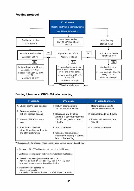
ICU admission
Intact GI tract/stable haemodynamics
Start EN within 24 - 48 h
Bolus feedingStart 50 ml/3h
Intermi�ent feedingStart 25 ml/h for 4 hours
Rest 2 h
Con�nuous feedingStart 20 ml/h
Aspirate > 300 beforenext bolus feed
Aspirate at end of 6 hrsAspirate > 300 ml
Aspirate at end of 4 hrsAspirate > 300 ml
No
Con�nue feeding at50 ml/3H
Increase feeding by 50 ml/3Hevery 12 hours
Maximum 250 ml/3h
Con�nue feeding at 24 ml/hfor 4 h. Rest 2 h then aspirate
at the end of rest periodIncrease feeding by 25 ml/h
every 12 hMaximum 100 ml/h
Con�nue feeding at 20 ml/hAspirate every 4 hrs
Increase feeding by 20 ml/hevery 12 h
Maximum: 80 ml/h
No No
**Feeding Intolerance
YesYes
Feeding protocol
** Consider post-pyloric feeding if feeding intolerance persists for more than 72 hours.
• Aim only for 70 - 80% of targeted calories in the first 72 hours.
• Continuous feeding is preferred over intermittent or bolus feeding
• Consider bolus feeding only in stable paents i.e. - non ventilated with an anticipated ICU stay of < 48 - 72 hours - previously on continuous or intermittent feeding
• Maximum volume depends on: - caloric requirements - osmolality of formula e.g. Ensure (1 kcal/ml), Nepro (2 kcal/ml)
1st episode 2nd episode 3rd episode or more
1. Check gastric tube position
2. Return aspirates up to 200 ml. Discard excess
3. Maintain EN at the same rate
4. if aspirates > 500 ml, withhold feeding for 1 cycle and start prokinetics
1. Return aspirates up to 200 ml. Discard excess.
2. Decrease rate by 20 or 25 ml/h, If patient already on 20 - 25 ml/h, reduce rate to 10 ml/h
3. Start prokinetics
4. Consider continuous or intermittent feeding if patient is on bolus feeding
1. Return aspirates up to 200 ml. Discard excess
2. Withhold feeds for 1 cycle
3. Restart at lower rate or at 10 ml/h
4. Continue prokinetics
Feeding Intolerance: GRV > 300 ml or vomiting
46
References
1. Reintam Blaser A, et al. Early enteral nutrition in critically ill patients: ESICM clinical practice guidelines. Intensive Care Med 2017;43:380-389
2. Cederholm T, et al. ESPEN guidelines on definitions and terminology of clinical nutrition. Clin Nutrition 2017;36:49-64
3. McClave SA, et al. Guidelines for the provision and assessment of nutrition support therapy in the adult critically ill patient: Society of Critical Care Medicine (SCCM) and American Society for Parenteral and Enteral Nutrition (A.S.P.E.N.). J Parenter Enteral Nutr 2016;40(2):159-211
4. Lewis K, et al. The efficacy and safety of prokinetic agents in critically ill patients receiving enteral nutrition: a systematic review and meta- analysis of randomized trials. Crit Care 2016;20(1):259
5. Singer P, et al. ESPEN guidelines on parenteral nutrition: Intensive care. Clin Nutrition 2009;28:387-400
47
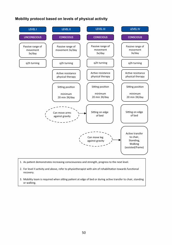
Early MobilisationIntroduction
Prolonged immobilisation of critically ill patients may lead to neuromuscular weakness, and impairment in physical and neuropsychiatric functions. Benefits of early mobilisation include improved muscle strength, physical function and quality of life. Additionally, early mobilisation may assist in weaning from mechanical ventilation, reduce the risk of deep vein thrombosis, pressure ulcer and delirium. However, there is lack of evidence benefiting neurocritical care patients. There is also inadequate evidence on the frequency and intensity of mobilisation, and devices to be used.
Principles
1. Initiate early mobility within 24 - 48 hours of admission in the absence of contraindications.
2. Assess appropriateness of early mobility by weighing risks of adverse events against benefits.
3. Step-up progression of mobilisation based on patient’s conscious level, functional capability and endurance.
4. Implement early mobilisation in combination with pain, sedation and delirium management.
General
1. Assess patients within 24 - 48 hours of ICU admission for early mobilisation.
2. Very early (less than 24 hours) and intensive out-of-bed mobilisation has been shown to be harmful in acute stroke patients.
3. Contraindications to early mobility a. Cardiovascular instability: SBP < 90 mmHg, HR > 120/min, unstable cardiac rhythm, use of 2 or more vasoactive agents
b. Neurological instability: acute traumatic brain injury, acute intracranial bleed, unstable spinal cord injury or any new neurological deterioration
c. Respiratory instability: FiO2 > 0.6, PEEP > 10 cmH2O and RR > 35/min
4. Early mobilisation activities require a multidisciplinary team comprising of clinicians, physiotherapists and nurses.
48
5. Consult the ICU specialist when there is uncertainty about safety.
6. Inform patients and families the importance of early mobility.
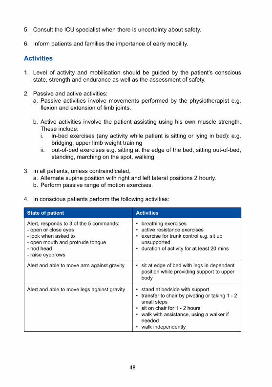
Activities
1. Level of activity and mobilisation should be guided by the patient’s conscious state, strength and endurance as well as the assessment of safety.
2. Passive and active activities: a. Passive activities involve movements performed by the physiotherapist e.g. flexion and extension of limb joints.
b. Active activities involve the patient assisting using his own muscle strength. These include: i. in-bed exercises (any activity while patient is sitting or lying in bed): e.g. bridging, upper limb weight training ii. out-of-bed exercises e.g. sitting at the edge of the bed, sitting out-of-bed, standing, marching on the spot, walking
3. In all patients, unless contraindicated, a. Alternate supine position with right and left lateral positions 2 hourly. b. Perform passive range of motion exercises.
4. In conscious patients perform the following activities:
State of patient Activities
Alert, responds to 3 of the 5 commands: - open or close eyes- look when asked to- open mouth and protrude tongue- nod head- raise eyebrows
Alert and able to move legs against gravity
Alert and able to move arm against gravity
• breathing exercises• active resistance exercises• exercise for trunk control e.g. sit up unsupported• duration of activity for at least 20 mins
• stand at bedside with support • transfer to chair by pivoting or taking 1 - 2 small steps• sit on chair for 1 - 2 hours • walk with assistance, using a walker if needed• walk independently
• sit at edge of bed with legs in dependent position while providing support to upper body
49
5. Encourage self-care activities whenever feasible.
6. Cycle ergometer exercise or neuromuscular electrical stimulation may be used as adjuncts to improve muscle strength and preserve muscle mass.
Safety
1. Terminate any physical activity if any of following signs and symptoms develop: a. Oxygen saturation < 90% b. Hypotension: SBP < 90 mmHg, associated with dizziness, and/or diaphoresis c. Hypertension: SBP > 170 mmHg d. Heart rate > 120 or presence of dysrhythmias e. Respiratory rate > 30 or change in breathing pattern with increased use of accessory muscles or nasal flaring f. Chest pain g. Patient requests to stop
2. Ensure the following safety measures for walking activities: a. Adequate staff assistance b. Tubes and catheters are secured c. Wheelchair readily available to allow resting period and safe return to bed d. Full oxygen tank
50
LEVEL I
UNCONSCIOUS
Passive range ofmovement
3x/day
q2h turning
LEVEL II
CONSCIOUS
Passive range ofmovement 3x/day
q2h turning
Ac�ve resistancephysical therapy
Si�ng posi�on
minimum20 min 3X/day
LEVEL III
CONSCIOUS
Passive range ofmovement
3x/day
q2h turning
Ac�ve resistancephysical therapy
Si�ng posi�on
minimum20 min 3X/day
Si�ng on edgeof bed
Can move armsagainst gravity
LEVEL IV
CONSCIOUS
Passive range ofmovement
3x/day
q2h turning
Ac�ve resistancephysical therapy
Si�ng posi�on
minimum20 min 3X/day
Si�ng on edgeof bed
Ac�ve transferto chair,
Standing,Walking
(assisted/frame)
Can move leg against gravity
1. As pa�ent demonstrates increasing consciousness and strength, progress to the next level.
2. For level II ac�vity and above, refer to physiotherapist with aim of rehabilita�on towards func�onal recovery.
3. Mobility team is required when si�ng pa�ent at edge of bed or during ac�ve transfer to chair, standing or walking.
Mobility protocol based on levels of physical activity
51
References
1. Hodgson CL, et al. Expert consensus and recommendations on safety criteria for active mobilisation of mechanically ventilated critically ill adults. Crit Care 2014;18:658
2. Morris PE, et al. Early intensive care unit mobility therapy in the treatment of acute respiratory failure. Crit Care Med 2008;36(8):2238-2243
3. Perme C, et al. Early mobility and walking program for patients in intensive care units: creating a standard of care. Am J Crit Care 2009;18(3):212-221
4. Parker A, et al. Early rehabilitation in the intensive care unit: preventing physical and mental health impairments. Curr Phys Med Rehabil Reports 2013;1(4):307-314
5. Maffiuletti NA, et al. Neuromuscular electrical stimulation for preventing skeletal-muscle weakness and wasting in critically ill patients: a systematic review. BMC Med 2013;11:137
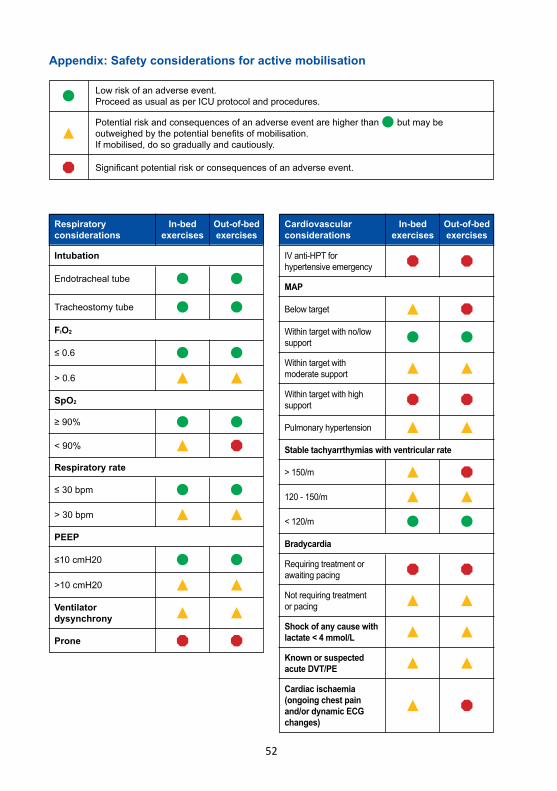
52
Appendix: Safety considerations for active mobilisation
Low risk of an adverse event.Proceed as usual as per ICU protocol and procedures.
Intubation
FiO2
SpO2
Respiratory rate
PEEP
Endotracheal tube
≤ 0.6
≥ 90%
≤ 30 bpm
≤10 cmH20
Ventilator dysynchrony
Tracheostomy tube
> 0.6
< 90%
> 30 bpm
>10 cmH20
Prone
Significant potential risk or consequences of an adverse event.
Potential risk and consequences of an adverse event are higher than but may be outweighed by the potential benefits of mobilisation.If mobilised, do so gradually and cautiously.
Respiratory considerations
Cardiovascular considerations
Out-of-bed exercises
Out-of-bed exercises
In-bedexercises
In-bedexercises
IV anti-HPT for hypertensive emergency
MAP
Stable tachyarrthymias with ventricular rate
Bradycardia
Below target
> 150/m
120 - 150/m
Not requiring treatment or pacing
Shock of any cause with lactate < 4 mmol/L
Known or suspected acute DVT/PE
Cardiac ischaemia (ongoing chest pain and/or dynamic ECG changes)
< 120/m
Within target with no/low support
Within target with moderate support
Pulmonary hypertension
Within target with high support
Requiring treatment or awaiting pacing
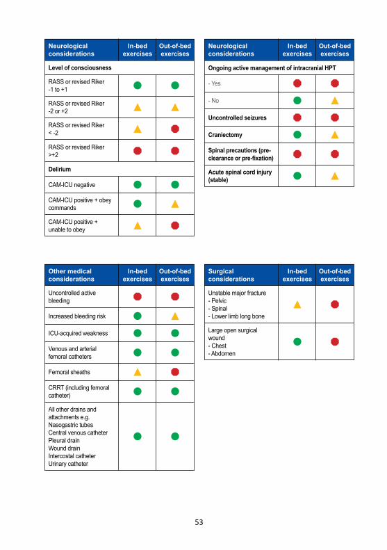
53
Level of consciousness
Delirium
RASS or revised Riker -1 to +1
CAM-ICU negative
RASS or revised Riker -2 or +2
CAM-ICU positive + obey commands
RASS or revised Riker < -2
CAM-ICU positive + unable to obey
RASS or revised Riker >+2
Ongoing active management of intracranial HPT
- Yes
Spinal precautions (pre-clearance or pre-fixation)
- No
Acute spinal cord injury (stable)
Uncontrolled seizures
Craniectomy
Neurological considerations
Other medical considerations
Neurological considerations
Surgical considerations
Out-of-bed exercises
Out-of-bed exercises
Out-of-bed exercises
Out-of-bed exercises
In-bedexercises
In-bedexercises
In-bedexercises
In-bedexercises
Uncontrolled active bleeding
Increased bleeding risk
ICU-acquired weakness
Venous and arterial femoral catheters
CRRT (including femoral catheter)
All other drains and attachments e.g.Nasogastric tubesCentral venous catheterPleural drainWound drainIntercostal catheterUrinary catheter
Femoral sheaths
Unstable major fracture- Pelvic- Spinal- Lower limb long bone
Large open surgical wound- Chest- Abdomen
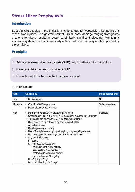
54
Stress Ulcer ProphylaxisIntroduction
Stress ulcers develop in the critically ill patients due to hypotension, ischaemic and reperfusion injuries. The gastrointestinal (GI) mucosal damage ranging from gastric erosions to ulcers results in occult to clinically significant bleeding. Maintaining adequate systemic perfusion and early enteral nutrition may play a role in preventing stress ulcers.
Principles
1. Administer stress ulcer prophylaxis (SUP) only in patients with risk factors.
2. Reassess daily the need to continue SUP.
3. Discontinue SUP when risk factors have resolved.
1. Risk factors
Risk Conditions Indication for SUP
Low
Moderate
High
• No risk factors
• Chronic NSAID/aspirin use• Peptic ulcer disease > 1 year
• Mechanical ventilation for greater than 48 hours• Coagulopathy: INR > 1.5, APTT > 2x the control, platelets < 50 000/mm3
• Traumatic brain injury with GCS < 10 or spinal cord injury• Significant burn injury (total body surface area > 30%)• Acute liver failure• Renal replacement therapy• Use of 2 antiplatelets (clopidogrel, aspirin, ticagrelor, dipyridamole)• History of upper GI bleed or gastric ulcer in the last 1 year• Any 2 of the following: i. sepsis ii. high dose corticosteroid: - hydrocortisone > 250 mg/day - prednisolone > 60 mg/day - methylprednisolone 50 mg/day - dexamethasone 10 mg/day iii. ICU stay > 7days iv. occult bleeding of > 6 days
No
To be considered
Indicated
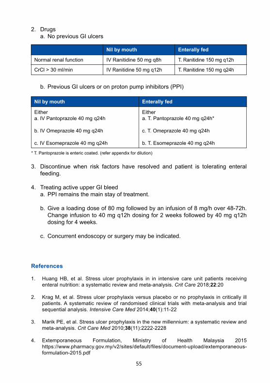
55
2. Drugs a. No previous GI ulcers
References
1. Huang HB, et al. Stress ulcer prophylaxis in in intensive care unit patients receiving enteral nutrition: a systematic review and meta-analysis. Crit Care 2018;22:20
2. Krag M, et al. Stress ulcer prophylaxis versus placebo or no prophylaxis in critically ill patients. A systematic review of randomised clinical trials with meta-analysis and trial sequential analysis. Intensive Care Med 2014;40(1):11-22
3. Marik PE, et al. Stress ulcer prophylaxis in the new millennium: a systematic review and meta-analysis. Crit Care Med 2010;38(11):2222-2228
4. Extemporaneous Formulation, Ministry of Health Malaysia 2015 https://www.pharmacy.gov.my/v2/sites/default/files/document-upload/extemporaneous-
formulation-2015.pdf
Nil by mouth Enterally fed
Normal renal function
CrCl > 30 ml/min
IV Ranitidine 50 mg q8h
IV Ranitidine 50 mg q12h
T. Ranitidine 150 mg q12h
T. Ranitidine 150 mg q24h
b. Previous GI ulcers or on proton pump inhibitors (PPI)
Nil by mouth Enterally fed
Eithera. IV Pantoprazole 40 mg q24h
b. IV Omeprazole 40 mg q24h
c. IV Esomeprazole 40 mg q24h
Eithera. T. Pantoprazole 40 mg q24h*
c. T. Omeprazole 40 mg q24h
b. T. Esomeprazole 40 mg q24h
* T. Pantoprazole is enteric coated. (refer appendix for dilution)
3. Discontinue when risk factors have resolved and patient is tolerating enteral feeding.
4. Treating active upper GI bleed a. PPI remains the main stay of treatment.
b. Give a loading dose of 80 mg followed by an infusion of 8 mg/h over 48-72h. Change infusion to 40 mg q12h dosing for 2 weeks followed by 40 mg q12h dosing for 4 weeks.
c. Concurrent endoscopy or surgery may be indicated.
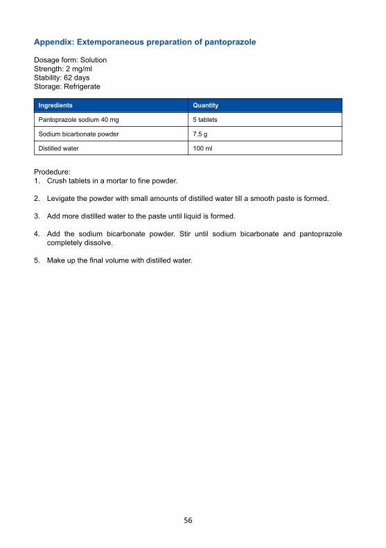
56
Appendix: Extemporaneous preparation of pantoprazole
Dosage form: SolutionStrength: 2 mg/mlStability: 62 daysStorage: Refrigerate
Prodedure:1. Crush tablets in a mortar to fine powder.
2. Levigate the powder with small amounts of distilled water till a smooth paste is formed.
3. Add more distilled water to the paste until liquid is formed.
4. Add the sodium bicarbonate powder. Stir until sodium bicarbonate and pantoprazole completely dissolve.
5. Make up the final volume with distilled water.
Ingredients Quantity
Pantoprazole sodium 40 mg
Sodium bicarbonate powder
Distilled water
5 tablets
7.5 g
100 ml
57
Venous ThromboprophylaxisIntroduction
Most patients in ICU are at moderate risk of developing venous thromboembolism (VTE), while the critically ill ones are at high risk. Pharmacological and/or mechanical thromboprophylaxis should be routinely considered in critically ill patients, unless contraindicated. However, venous thromboprophylaxis does not eliminate the risk of VTE or VTE-related deaths. Current prediction scores for VTE and models for evaluating risk of bleeding in critically ill patients require further validation. Early mobilisation is an important approach in the prevention of VTE.
Principles
1. Assess bleeding risk before initiating pharmacological prophylaxis.
2. Select a pharmocological agent based on its pharmocokinetics and the risk of developing VTE.
3. Use mechanical prophylaxis when pharmacological prophylaxis is contraindicated.
4. There is no role of routine screening for DVT.
Pharmacological Prophylaxis
1. All patients admitted to ICU should receive pharmacological prophylaxis, either unfractionated heparin (UFH) or lower molecular heparin (LMWH) within 24 hours of admission unless contraindicated.
2. Contraindications to pharmacological prophylaxis:
Absolute contraindications Relative contraindications
• activemajorbleed(atleast2unitsof blood or blood products transfused in 24 hrs)• plateletslessthan75x109/L • inheritedbleedingdisorders• activepepticulcerdisease
• neurosurgeryoreyesurgerywithinthe last 2 weeks• recentgastrointestinalorgenito-urinary bleeding• recentCNSbleed• Uncontrolledsystolichypertension (230/120mmHgorhigher)• acuteliverfailure
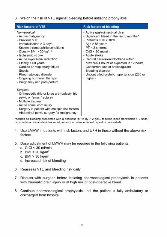
58
4. Use LMHW in patients with risk factors and UFH in those without the above risk factors.
5. DoseadjustmentofLMWHmayberequiredinthefollowingpatients: a.CrCl<30ml/min b.BMI<20kg/m2
c. BMI>30kg/m2 d. Increased risk of bleeding
6. Reassess VTE and bleeding risk daily.
7. Discuss with surgeon before initiating pharmacological prophylaxis in patients withtraumaticbraininjuryorathighriskofpost-operativebleed.
8. Continue pharmacological prophylaxis until the patient is fully ambulatory or discharged from hospital.
Risk factors of VTE Risk factors of bleedingNon-surgical:- Active malignancy- Previous VTE- Immobilisation > 3 days- Known thrombophilic conditions-ObesityBMI>30kg/m2
- Ischaemic stroke - Acute myocardial infarction-Elderly>60years- Cardiac or respiratory failure- Sepsis- Rheumatologic disorder- Ongoing hormonal therapy- Pregnancy and post-partum
Surgical:- Orthopaedic (hip or knee arthroplasty, hip, pelvic or femur fracture)- Multiple trauma-Acutespinalcordinjury- Surgery in patient with multiple risk factors- Abdominal-pelvic surgery for malignancy
*definedasbleedingassociatedwithadecrease inHbby>2g/dL, requiredblood transfusion>2units,occurred in a critical site (intracranial, intraocular, retroperitoneal, spinal or pericardial).
- Active gastrointestinal ulcer-Significantbleedinthelast3months*-Platelets<75x109/L-Age>85years- PT > 2 x normal-CrCl<30ml/min- Acute stroke- Central neuroaxial blockade within previous 4 hours or expected in 12 hours- Concurrent use of anticoagulant- Bleeding disorder-Uncontrolledsystolichypertension(230or higher)
3. Weigh the risk of VTE against bleeding before initiating prophylaxis.
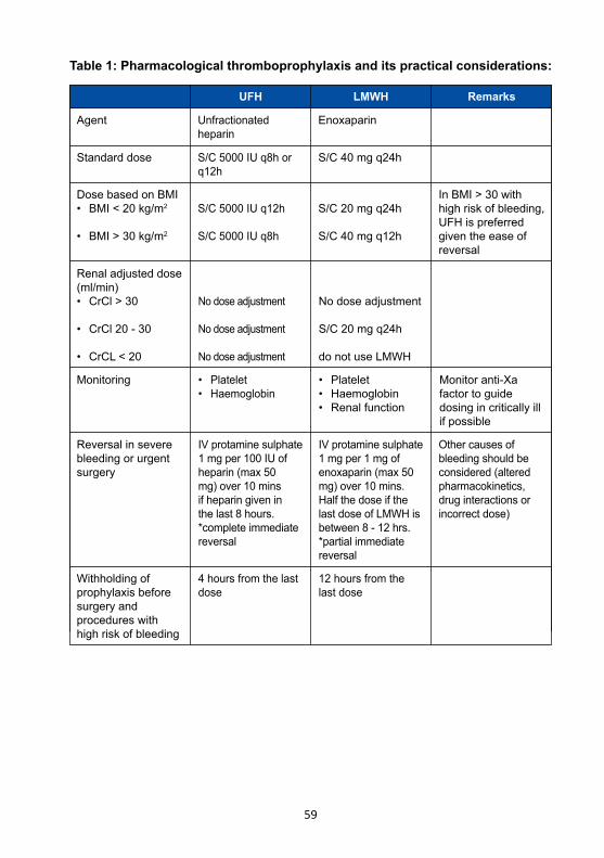
59
Table 1: Pharmacological thromboprophylaxis and its practical considerations:
UFH LMWH Remarks
Agent Unfractionated heparin
Enoxaparin
Standard dose
Dose based on BMI• BMI<20kg/m2
• BMI>30kg/m2
Renaladjusteddose(ml/min)• CrCl>30
• CrCl20-30
• CrCL<20
Reversal in severe bleeding or urgent surgery
Withholding of prophylaxis before surgery and procedures with high risk of bleeding
Monitoring
S/C5000IUq8horq12h
S/C5000IUq12h
S/C5000IUq8h
Nodoseadjustment
Nodoseadjustment
Nodoseadjustment
IV protamine sulphate 1mgper100IUofheparin(max50mg)over10minsif heparin given in the last 8 hours. *complete immediate reversal
4 hours from the last dose
• Platelet• Haemoglobin
S/C40mgq24h
S/C20mgq24h
S/C40mgq12h
Nodoseadjustment
S/C20mgq24h
do not use LMWH
IV protamine sulphate 1 mg per 1 mg of enoxaparin(max50mg)over10mins.Half the dose if the last dose of LMWH is between 8 - 12 hrs.*partial immediate reversal
12 hours from the last dose
• Platelet• Haemoglobin• Renalfunction
Monitor anti-Xa factor to guide dosing in critically ill if possible
InBMI>30withhigh risk of bleeding, UFH is preferred given the ease of reversal
Other causes of bleeding should be considered (altered pharmacokinetics, drug interactions or incorrect dose)
60
Mechanical prophylaxis
1. Provide mechanical prophylaxis with either intermittent penumatic compressor (IPC) or graduated compression stockings (GCS) when pharmacological prophylaxis is contraindicated.
2. IPC is preferred over GCS in patients at high risk of VTE.
3. Contraindications to mechanical prophylaxis: a. Severearterialinsufficiencyorperipheryarterialdisease b. Congestive heart failure c. Recent or acute DVT/PE d. Acute fractures of the lower limbs e. Loss of skin integrity f. Recent skin graft g. Post-operative venous ligation h.Morbidobesitywhichmakesitdifficulttocorrectlyfitthestockings i. Oedema or deformity of the lower limb j. Diabeticneuropathy
4. Ensureproperfitandoptimalcomplianceofmechanicaldevices.
5. Assessskinintegrityofthelowerlimbsandpressureareaseverynursingshift.
6. Initiate pharmacological prophylaxis once bleeding risk subsides.
61
References
1. Guyatt GH, et al. Executive summary: Antithrombotic therapy and prevention of thrombosis, 9th ed: American College of Chest Physicians evidence-based clinical practices. Chest2012;141(2Suppl):7S-47S
2. Duranteau J, et al. European guidelines on perioperative venous thromboembolism prophylaxis: Intensive care. Eur J Anaesthesiol2018;35:142-146
3. Afshari A, et al. European guidelines on perioperative venous thromboembolism prohylaxis: Mechanical prophylaxis. Eur J Anaesthesiol2018;35:112-115
4. SchunemannHJ,etal.AmericanSocietyofHaematology2018guidelinesformanagementof venous thromboembolism: prophylaxis for hospitalized and nonhospitalized medical patients. Blood Advances2018;2(22):3198-3225
5. Venous thrombosisprophylaxisguidelines2015;VanderbiltUniversityMedicalCenter.Available at https://www.vumc.org/trauma-and-scc/files/trauma-and-scc/public_files/Manual/VTEProphylaxisGuidelinesFinal2-10-15.Accessedon15thJune2019.
6. Guidelines for prevention of venous thromboembolism (VTE) in adult hospitalised patients,QueenslandHealth,December2018.Availableathttps://www.health.qld.gov.au/clinical-practice/guidelines-procedures/medicines/safety. Accessed on 23rdJuly2019
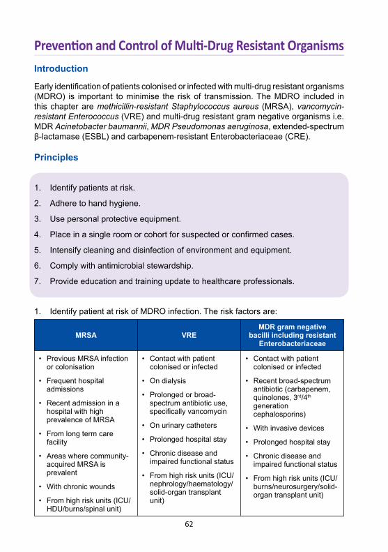
62
Prevention and Control of Multi-Drug Resistant OrganismsIntroduction
Early identification of patients colonised or infected with multi-drug resistant organisms (MDRO) is important to minimise the risk of transmission. The MDRO included in this chapter are methicillin-resistant Staphylococcus aureus (MRSA), vancomycin-resistant Enterococcus (VRE) and multi-drug resistant gram negative organisms i.e. MDR Acinetobacter baumannii, MDR Pseudomonas aeruginosa, extended-spectrum β-lactamase (ESBL) and carbapenem-resistant Enterobacteriaceae (CRE).
Principles
1. Identify patients at risk.
2. Adhere to hand hygiene.
3. Use personal protective equipment.
4. Place in a single room or cohort for suspected or confirmed cases.
5. Intensify cleaning and disinfection of environment and equipment.
6. Comply with antimicrobial stewardship.
7. Provide education and training update to healthcare professionals.
1. Identify patient at risk of MDRO infection. The risk factors are:
MRSA VREMDR gram negative
bacilli including resistant Enterobacteriaceae
• Previous MRSA infection or colonisation
• Frequent hospital admissions
• Recent admission in a hospital with high prevalence of MRSA
• From long term care facility
• Areas where community- acquired MRSA is prevalent
• With chronic wounds
• From high risk units (ICU/ HDU/burns/spinal unit)
• Contact with patient colonised or infected
• On dialysis
• Prolonged or broad- spectrum antibiotic use, specifically vancomycin
• On urinary catheters
• Prolonged hospital stay
• Chronic disease and impaired functional status
• From high risk units (ICU/ nephrology/haematology/ solid-organ transplant unit)
• Contact with patient colonised or infected
• Recent broad-spectrum antibiotic (carbapenem, quinolones, 3rd/4th generation cephalosporins)
• With invasive devices
• Prolonged hospital stay
• Chronic disease and impaired functional status
• From high risk units (ICU/ burns/neurosurgery/solid- organ transplant unit)
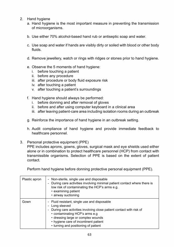
63
2. Hand hygiene a. Hand hygiene is the most important measure in preventing the transmission of microorganisms.
b. Use either 70% alcohol-based hand rub or antiseptic soap and water.
c. Use soap and water if hands are visibly dirty or soiled with blood or other body fluids.
d. Remove jewellery, watch or rings with ridges or stones prior to hand hygiene.
e. Observe the 5 moments of hand hygiene: i. before touching a patient ii. before any procedure iii. after procedure or body fluid exposure risk iv. after touching a patient v. after touching a patient’s surroundings
f. Hand hygiene should always be performed: i. before donning and after removal of gloves ii. before and after using computer keyboard in a clinical area iii. after leaving patient-care area including isolation rooms during an outbreak
g. Reinforce the importance of hand hygiene in an outbreak setting.
h. Audit compliance of hand hygiene and provide immediate feedback to healthcare personnel.
3. Personal protective equipment (PPE) PPE includes aprons, gowns, gloves, surgical mask and eye shields used either alone or in combination to protect healthcare personnel (HCP) from contact with transmissible organisms. Selection of PPE is based on the extent of patient contact.
Perform hand hygiene before donning protective personal equipment (PPE).
Plastic apron
Gown
- Non-sterile, single use and disposable- During care activities involving minimal patient contact where there is low risk of contaminating the HCP’s arms e.g. • examining patient • airway suctioning
- Fluid resistant, single use and disposable- Long sleeved- During care activities involving close patient contact with risk of • contaminating HCP’s arms e.g • dressing large or complex wounds • hygiene care of incontinent patient • turning and positioning of patient
64
4. Isolation a. Isolate patients colonised or infected with MDRO in single rooms.
b. Cohort patients if single rooms are not available. Cohorting refers to placing patients who are infected or colonised with the same organism in the same area.
c. Prioritise single room isolation to patients with: i. CRE, VRE or MRSA ii. incontinence of faeces or urine iii. open or draining wounds iv. enterostomies v. copious respiratory secretions
d. Staff nursing patients with MDRO should not have contact with other patients.
e. Appropriate signage should be placed outside the door to alert HCP of contact precautions.
5. Discontinuation of isolation precautions a. Shedding of MDRO may be intermittent and their presence may not always be detected by active surveillance culture.
b. Continue contact precautions for the duration of admission.
6. Equipment and patient care items a. Remove non-essential equipment from the room.
b. Minimise consumable items placed in the room e.g. syringes, needles and gauzes.
c. Use disposable items whenever possible.
Non-sterile gloves
Surgical mask and protective eyewear
Discard the above single used items within the room/area. Perform hand hygiene before leaving the room.
- Use clean non-sterile gloves before entering the room or cubicle- Change gloves and perform hand hygiene between different care/ treatment activities of the same patient- Attend to ‘clean’ procedures first
- When performing splash or aerosol-generating procedures e.g. • intubation • oral or airway suctioning • wound irrigation • caring for patients with open tracheostomies
65
d. Dedicate reusable equipment to a single patient e.g. thermometer, blood pressure cuff, infusion pumps, stethoscope, etc.
e. Clean and disinfect equipment that are shared between patients e.g. hoist and armchairs.
f. Use dedicated trolley for sponging and cleaning.
7. Environmental cleaning a. Increase cleaning frequency.
b. Clean and disinfect frequently touched surfaces (e.g. bedrails, drip stands, nursing tables, trolleys, doorknobs, tap handles and switches) at least once every nursing shift. This involves either: i. 2-in-1 clean: commercially prepared combination of detergent and 1000 ppm chlorine solution or impregnated wipes ii. 2-step clean: clean with detergent or detergent impregnated wipes and disinfect with 1000ppm chlorine solution or impregnated wipes
c. Perform terminal cleaning after patient discharge: i. terminal cleaning process should be monitored and supervised ii. remove curtains prior to cleaning iii. cleaning should include walls, bed, frequently touched surfaces and all equipment iv. discard unused consumables that cannot be disinfected
8. Antimicrobial stewardship Antimicrobial stewardship programme refers to coordinated interventions to ensure appropriate use of antimicrobials. When antimicrobials are prescribed ensure: a. Adequate dose and the shortest duration for efficacy. b. Use the narrowest spectrum whenever possible. c. De-escalate when cultures are available.
9. Education a. Education of all staff, including cleaning staff should be intensified. b. Reinforce standard and contact precautions.
10. Screening and surveillance a. Contact screening is done to reduce the risk of transmission of MDRO. i. screen patients who are in close contact for more than 24 hours with a CRE or VRE confirmed case. Send rectal swab
ii. screening for other MDRO depends on local hospital infection control policy
iii. isolate or cohort contacts pending screening result
66
b. Active surveillance cultures should be considered in outbreak settings. i. screen for CRE, VRE or MRSA in high risk patients admitted to the unit. Send rectal swabs for CRE or VRE and nasal swabs for MRSA
ii. screening for other MDRO depends on local hospital infection control policy
11. Other considerations a. Inform support staff (e.g. radiographer, physiotherapist, pharmacist, dialysis nurse and dietician) of patient’s status. Support staff to attend to the patient last in the unit except in emergencies.
b. Visitors i. all visitors are directed to perform hand hygiene before and after contact with patient or environment ii. visitors are not required to wear PPE unless involved in patient care
c. Patient’s movement i. patient’s movement should be kept to a minimum ii. if transportation is required such as to the radiology department, the operation theatre or another facility, the receiving unit should be informed of the patient’s status
d. Upon discharge, the receiving wards should be informed early, for isolation or cohorting purposes.
References
1. Guidelines for the prevention and control of mulit-drug resistant organisms (MDRO) excluding MRSA in the healthcare setting. Available at: http://www.hpsc.ie/a-z/microbiologyantimicrobialresistance/infectioncontrolandhai/guidelines/Guidelines; Accesses on 1st June 2019
2. A Clinical Guide for the Prevention and Control of Multidrug Resistant organisms (MDRO) including Multi-resistant Gram-negaitive Bacteria ( like ESBL) and Glycopeptide resistant enterococci (GRE/VRE). Norfolk and Norwich University Hospital 2018. Available at: http://www.nnuh.nhs.uk/publication/download/prevention-and-control-of-multidrug-resistant-organisms-ca5174-v1-1/ Accessed on 1st June 2019
3. Recommendations for the control of carbapenemase-producing Enterobacteriaceae (CPE). Australian commission on safety and quality in health care. A guide for acute care health facilities. Available at: https://www.safetyandquality.gov.au/wp-content/uploads/2017/05/Recommendations-for-the-control-of-Carbapenemase-producing-Enterobacteriaceae.pdf. Accessed on 1st June 2019
4. Management of multi-resistant organisms’ guideline. guideline. Queensland health 2017. Available at: https://www.health.qld.gov.au/_data/assets/pdf_file/0026/444626/multi-resistant-organisms.pdf. Accessed on 1st June 2019
5. Australian guideline for the prevention and control of infection in healthcare. Avavilable at: https://www.nhmrc.gov.au/about-us/publications/australian-guidelines-prevention-and
-control-infection-healthcare-2010. Accessed on 1st June 2019
67
Withholding and Withdrawing Life-Sustaining TreatmentIntroduction
The goals of intensive care are to return patients to a quality of survival that is reasonably acceptable to them and to reduce disability. If these goals are not possible, and family understands and agrees that this is not in keeping with the patient’s wishes, then compassionate care needs to be instituted to allow death with dignity.
Withholding and withdrawing life-sustaining treatment (LST) is a process where various medical interventions are either not initiated or ceased.There is no ethical or moral difference between withholding or withdrawing life-sustaining treatment.
Principles
1. Abide by the principles of medical ethics when making end-of-life decisions.
2. Assess patient’s decision-making capacity on end-of-life decisions. In patients with limited or absent capacity, families become surrogate decision-makers.
3. Respect patient’s autonomy except in cases of non-beneficial medical treatment.
4. Implement a palliative care plan once withholding or withdrawing LST is decided upon.
5. Document clearly all decisions on withholding or withdrawing LST in the clinical records.
6. Treat dying patients with respect, dignity and compassion.
Ethical principles
1. The principles that underpin end-of-life (EOL) medical decisions are: a. Respect for autonomy: acting in accordance to what the patient wants b. Beneficence: acting to benefit the patient c. Non-maleficence: causing no harm to the patient d. Distributive justice: paying attention to fairness and equity
2. These principles are either used individually or collectively to frame the discussion on EOL and one does not supersede the other.
68
3. The principles may be conflicting for example: a. between respect for autonomy and beneficence: a terminal cancer patient who insists all LST to be provided although they will not benefit him.
b. between beneficence and justice: providing invasive mechanical ventilation in a patient with decompensated heart failure who has had multiple hospital admissions in the last 6 months and is dyspnoiec at rest versus allocation of ICU bed for a polytrauma patient.
Decision-making capacity
1. Patient’s decision-making capacity should be assessed before having a discussion with him on EOL. This includes the patient’s ability to comprehend, appreciate, rationalise and express choice of treament.
2. Most ICU patients do not have decision-making capacity and hence families become the surrogate decision-maker.
3. The standards that may be used by surrogates in decision-making include: a. Substituted judgement: decision of the patient if he/she has the capacity
b. Best interest: decision based on the potential benefits vs. burdens of treatment taking into consideration patient’s values and beliefs
4. EOL decisions are shared medical decisions made by clinicians and concurred by family members.
Autonomy and obligation to treat
1. Once decision-making capacity is established, patient’s autonomy must be respected even though survival may be implicated.
2. In cases of non-beneficial medical treatment, clinicians are not obliged to initiate or continue LST.
3. The MMA Code of Ethics 2001 states: “where death is deemed to be imminent and where curative or life-prolonging treatment appears to be futile, ensure that death occurs with dignity and comfort. Such futile therapy could be withheld, withdrawn or one may allow irreversible pathology to continue without active resuscitation. One should always take into consideration any advance directives and the wishes of the family in this regard. In any circumstance, if therapy is considered to be life saving, it should never be withheld.”
69
Respect for the dying
1. All dying patients should be afforded the same standard of care as other patients.
2. They should be treated with dignity, respect and compassion.
3. Their privacy and confidentiality should be respected at all times.
Withholding and withdrawal of life-sustaining treatment
The following patients are to be considered for withholding and withdrawal of LST:
1. Imminent death This patient has a severe acute illness that is clearly not responding to therapy, and reversal or cure is unlikely despite continued optimal therapy. e.g. septic shock with multiorgan failure.
2. Terminal condition This patient has a progressive terminal disease incompatible with survival longer than 3-6 months. e.g. i. end stage respiratory disease on long term oxygen therapy with severe community acquired pneumonia ii. end stage cardiac, respiratory or liver disease with no options for transplant iii. metastatic cancer unresponsive to treatment
3. Severe and irreversible condition impairing cognition and consciousness but death may not occur in months In such cases, a patient is often planned to not receive CPR or other resuscitative measures in the event of deterioration e.g. persistent vegetative state post cardiac arrest, severe dementia or severe stroke with poor cognitive recovery.
4. Advanced age with poor functional status due to chronic organic dysfunction e.g. Multiple co-morbidities with deteriorating physical performance.
5. Severe disability with poor quality of life e.g. i. stroke with minimal conscious state or dense paralysis ii. decompensated heart failure with ongoing shortness of breath at rest despite optimal therapy
6. Advanced diseases of progressive life-limiting conditions e.g. i. motor neuron disease with rapid decline in physical status ii. severe Parkinson’s disease with reduced independence and needs assistance for activites of daily living ( ADL)
70
7. A patient who has stated his/her wish against initiation or continuation of life support therapy This will include patients who have given clear advanced care directives.
Practical issues of withholding and withdrawal of life-sustaining treatment
1. Medical team consensus The intensive care team and the primary team should agree on EOL decisions.
2. Communication with patient and relatives a. It is best that the same clinician (specialist/consultant) who is involved in the active care of the patient deals with the family. He/she should be someone who frequently communicates with the family and has established a rapport with them. A witness (nurse or doctor) should be present during these discussions.
b. Clinicians need to respect the fact that each patient and family will differ in how much input they wish to have in the decision-making process.
c. In the event of a disagreement, allow time for repeated discussions and negotiations. Failing this, consider either: i. time-limited trial which is a goal-directed trial of any intervention limited by predetermined outcomes that are evaluated at planned intervals. ii. second medical opinion from another clinician from a similar specialty iii. facilitation by a third party e.g. spiritual advisor
d. Patients and families must be given sufficient time to reach decisions on EOL.
3. Management plan for withdrawal of life-sustaining treatment A clear management plan is essential to ensure that the withdrawal process occurs smoothly. It should be conveyed to the family, with an emphasis on maintenance of comfort for the patient. The plan should include the following components: a. Maintain current support until the patient and family have had adequate time together.
b. Ensure other healthcare professionals e.g. primary team, physiotherapist, dietitian are aware of withdrawal plan. Cease all investigations e.g. blood taking and X-rays.
c. Ensure pain and other symptoms e.g. dyspnoea are well controlled. Morphine is the most commonly used opioid for analgesia and comfort. There is no maximum dose. Large doses of opiods may be required for comfort and may unintentionally hasten death. This “double effect” of opioids is acceptable.
d. Manage airway secretions by using glycopyrrolate, positioning in lateral position and frequent suctioning.
71
e. All treatment that do not contribute towards comfort should be discontinued e.g. antibiotics, blood transfusions. Feeding and intravenous fluids may be discontinued unless specifically requested by family.
f. Maintain patient’s personal hygiene and dignity at all times. e.g. diaper soiling is dealt with immediately.
g. Withdrawal of vasopressors may result in immediate death. Families should be aware and nearby.
h. Withdrawal of mechanical ventilation may be carried out either as: i. terminal weaning where ventilator settings are reduced while leaving the endotracheal tube in-situ or ii. terminal extubation
i. Disable all monitor and ventilator alarms. Demedicalise the patient and allow family members to be close by.
4. Other considerations a. Family should be given unrestricted access to the patient.
b. Non-invasive ventilation may be used as a palliative care technique to minimise dyspnoea in conscious patients.
c. Neuromuscular blockade makes assessment of comfort impossible and should not be used in intubated patients.
d. Consider terminal sedation with benzodiazepines only when high doses of opiod are inadequate for comfort.
5. Documentation Document all decisions regarding withdrawal and withholding of treatment, including the basis of the decision and amongst whom it was reached.
6. Notification of death Death should be communicated in direct language gently.
7. Bereavement Provide bereavement support to the family and healthcare providers if necessary.
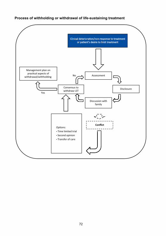
72
Clinical deteriora�on/non-response to treatmentor pa�ent’s desire to limit treatment
AssessmentNo
Yes
Discussion withfamily
Consensus towithdraw LST
Disclosure
Conflict
Op�ons:• Time limited trial• Second opinion• Transfer of care
Management plan onprac�cal aspects of
withdrawal/withholding
Process of withholding or withdrawal of life-sustaining treatment
73
References
1. Lobo SM, et al. Decision-making on withholding and withdrawing life support in the ICU: A worldwide perspective. Chest 2017;152(2):321-329
2. Connolly C, et al. End-of-life in the ICU: moving from ‘withdrawal of care’ to a palliative care, patient-centred approach. Br J Anaesth 2016;117(2)143-145
3. Mercadante S, et al. Palliative care in intensive care units: why, where, what, who, when, how. BMC Anesthesiol 2018;18:106
4. Shanawani H, et al. Meeting physicians’ responsibilities in providing EOL care. Chest 2008;133(3):776-786
5. John M Luce. End-of-life decision making in the intensive care unit. Am J Resir Crit Care Med 2010;182(1):6-11
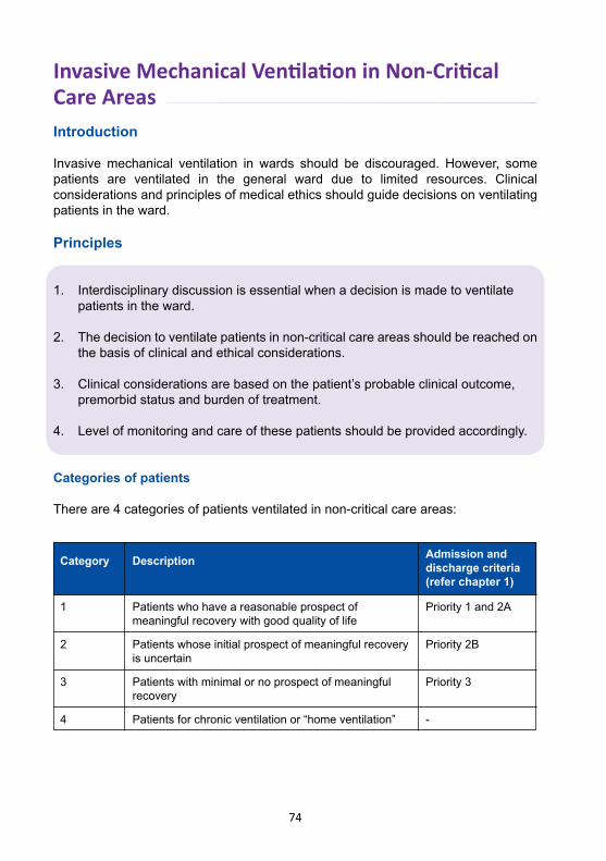
74
Invasive Mechanical Ventilation in Non-Critical Care AreasIntroduction
Invasive mechanical ventilation in wards should be discouraged. However, some patients are ventilated in the general ward due to limited resources. Clinical considerations and principles of medical ethics should guide decisions on ventilating patients in the ward.
Principles
1. Interdisciplinary discussion is essential when a decision is made to ventilate patients in the ward.
2. The decision to ventilate patients in non-critical care areas should be reached on the basis of clinical and ethical considerations.
3. Clinical considerations are based on the patient’s probable clinical outcome, premorbid status and burden of treatment.
4. Level of monitoring and care of these patients should be provided accordingly.
Categories of patients
There are 4 categories of patients ventilated in non-critical care areas:
Category Description Admission and discharge criteria (refer chapter 1)
1
2
3
4
Patients who have a reasonable prospect of meaningful recovery with good quality of life
Patients whose initial prospect of meaningful recovery is uncertain
Patients with minimal or no prospect of meaningful recovery
Patients for chronic ventilation or “home ventilation”
Priority 1 and 2A
Priority 2B
Priority 3
-
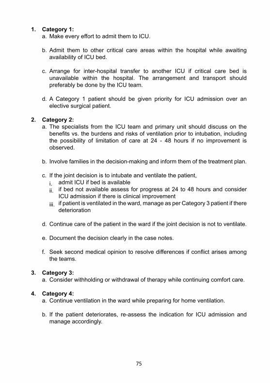
75
1. Category 1: a.MakeeveryefforttoadmitthemtoICU.
b. Admit them to other critical care areas within the hospital while awaiting availabilityofICUbed.
c. Arrange for inter-hospital transfer to another ICU if critical care bed is unavailable within the hospital. The arrangement and transport should preferablybedonebytheICUteam.
d.A Category 1 patient should be given priority for ICU admission over an elective surgical patient.
2. Category 2: a. Thespecialists from the ICU teamandprimaryunit shoulddiscusson the benefitsvs.theburdensandrisksofventilationpriortointubation,including the possibility of limitation of care at 24 - 48 hours if no improvement is observed.
b. Involve families in the decision-making and inform them of the treatment plan.
c. If the joint decision is to intubate and ventilate the patient, i. admitICUifbedisavailable ii. if bed not available assess for progress at 24 to 48 hours and consider ICUadmissionifthereisclinicalimprovement iii. if patient is ventilated in the ward, manage as per Category 3 patient if there deterioration
d. Continue care of the patient in the ward if the joint decision is not to ventilate.
e. Document the decision clearly in the case notes.
f. Seeksecondmedicalopiniontoresolvedifferencesifconflictarisesamong the teams.
3. Category 3: a. Consider withholding or withdrawal of therapy while continuing comfort care.
4. Category 4: a. Continue ventilation in the ward while preparing for home ventilation.
b. If the patient deteriorates, re-assess the indication for ICU admission and manage accordingly.
76
Responsibilities
1. The primary team is responsible for the care of the patient in the ward.
2. TheICUteamshouldreviewthepatientdaily.
Minimum requirements
1. Staffing: Nurses should be trained and privileged on basic care and safety issues in caring for patients on mechanical ventilation e.g. a. Tracheal suctioning
b. Bag-valve-mask ventilation
c. Nebulisation
d. Recognition of life threatening events e.g. desaturation, ETT blockage, disconnection or dislodgement of ETT
e. Infection control measures
2. Monitoring a. Blood pressure, pulse rate and respiratory rate hourly b. SpO2 - continuous if available c. ECG - continuous if available d. Level of consciousness e. Arterial blood gases as indicated f. Other standard nursing monitoring
3. Equipment (readily available) a. Yankauer suction b. Suction catheters c. Vacuum suction apparatus d. Laryngoscopes e. Endotracheal tubes and tapes f. Oral airways g. Bag-valve-masks h. Resuscitation trolley
Procedures of care
These should include but are not limited to the following: a. Elevate head of bed 30 degrees unless contraindicated
b. Ensureendotrachealortracheostomyandnasogastrictubesarefirmlysecured
c. Perform tracheal and oropharyngeal suctioning q4h or more often if necessary
d. Position the patient at least 4 hourly
77
e. Perform general, eye and oral hygiene regularly
f. Ensure a functioning nasogastric tube for gastric decompression and nutritional support
g. Perform chest and limb physiotherapy
INTUBATION
Referred for ven�la�on
Category4
Condi�onstable
Con�nue ven�la�on in ward ± planfor home ven�la�on
Reassess and recategorise to1, 2 or 3
Yes
No
Category3
ICU bed available
Category 1
Discussion between ICU andprimary team on intuba�on
and ven�la�on
Ini�ate discussion on withdrawal oftherapy and end-of-life care
Consensusto ven�late
Admit ICU if bed is availableor
ven�late in ward
Con�nue wardmanagement
Reassess within 24 - 48 hoursand
recategorise to category 1 or 3
Admit other cri�cal care areaswhile awai�ng ICU bed
ortransfer to another hospital
No
Yes
No
YesCategory2
No
Yes
Yes
Assess and categorise pa�ent as1, 2, 3 or 4
Admit ICU
Yes
Category 1 Category 3
No
No
Referral for ventilation
78
References
1. Zisk-Rony RY, et al. Mechanical ventilation patterns and trends over 20 years in an Israeli hospitalsystem:policyramifications.Isr J Health Policy Res 2019;8:20
2. IwashitaY,et al.Epidemiologyofmechanically ventilatedpatients treated in ICUandnon-ICUsettingsinJapan:aretrospectivedatabasestudy.Crit Care 2018;22(1):329
3. DivathiaJV,etal.Caringforthecriticallyillindevelopingcountries:AperspectivefromIndia. Rev Bras Ter Intensiva 2015;27(1):7-9
4. National operational policy of anaesthesia and intensive care services. MOH Malaysia 2013
5. The GSF Proactive Identification Guideline (PIG) version 6. The Golds StandardsFramework Centre in End of Life Care. Available at http://www.goldstandardsframework.org.uk/PIG Accessed 12thJune2019
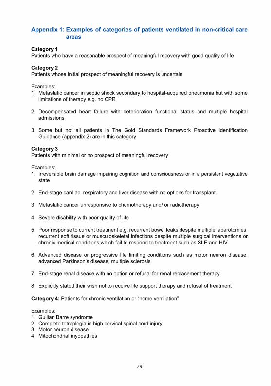
79
Appendix 1: Examples of categories of patients ventilated in non-critical care areas
Category 1Patients who have a reasonable prospect of meaningful recovery with good quality of life
Category 2Patients whose initial prospect of meaningful recovery is uncertain
Examples:1. Metastatic cancer in septic shock secondary to hospital-acquired pneumonia but with some limitations of therapy e.g. no CPR
2. Decompensated heart failure with deterioration functional status and multiple hospital admissions
3. Some but not all patients in The Gold Standards Framework Proactive Identification Guidance (appendix 2) are in this category
Category 3Patients with minimal or no prospect of meaningful recovery
Examples:1. Irreversible brain damage impairing cognition and consciousness or in a persistent vegetative state
2. End-stage cardiac, respiratory and liver disease with no options for transplant
3. Metastatic cancer unresponsive to chemotherapy and/ or radiotherapy
4. Severe disability with poor quality of life
5. Poor response to current treatment e.g. recurrent bowel leaks despite multiple laparotomies, recurrent soft tissue or musculoskeletal infections despite multiple surgical interventions or chronic medical conditions which fail to respond to treatment such as SLE and HIV
6. Advanced disease or progressive life limiting conditions such asmotor neuron disease, advanced Parkinson’s disease, multiple sclerosis
7. End-stage renal disease with no option or refusal for renal replacement therapy
8. Explicitly stated their wish not to receive life support therapy and refusal of treatment
Category 4: Patients for chronic ventilation or “home ventilation”
Examples:1. Gullian Barre syndrome2. Complete tetraplegia in high cervical spinal cord injury3. Motor neuron disease4. Mitochondrial myopathies
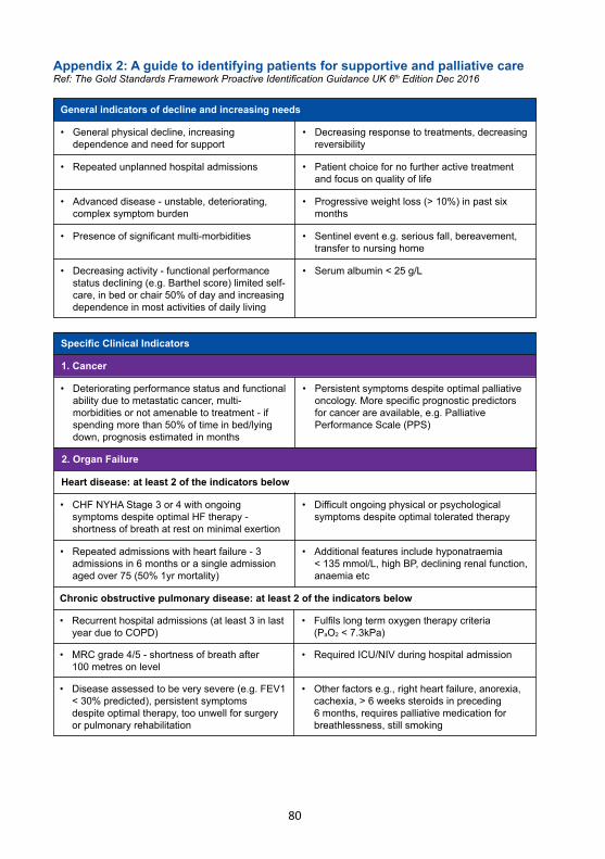
80
Appendix 2: A guide to identifying patients for supportive and palliative careRef: The Gold Standards Framework Proactive Identification Guidance UK 6th Edition Dec 2016
General indicators of decline and increasing needs
• Generalphysicaldecline,increasing dependence and need for support
• Repeatedunplannedhospitaladmissions
• Advanceddisease-unstable,deteriorating, complex symptom burden
• Presenceofsignificantmulti-morbidities
• Decreasingactivity-functionalperformance status declining (e.g. Barthel score) limited self- care, in bed or chair 50% of day and increasing dependence in most activities of daily living
• Decreasingresponsetotreatments,decreasing reversibility
• Patientchoicefornofurtheractivetreatment and focus on quality of life
• Progressiveweightloss(>10%)inpastsix months
• Sentinelevente.g.seriousfall,bereavement, transfer to nursing home
• Serumalbumin<25g/L
2. Organ Failure
• Recurrenthospitaladmissions(atleast3inlast year due to COPD)
• MRCgrade4/5-shortnessofbreathafter 100 metres on level
• Diseaseassessedtobeverysevere(e.g.FEV1 <30%predicted),persistentsymptoms despite optimal therapy, too unwell for surgery or pulmonary rehabilitation
Heart disease: at least 2 of the indicators below
Chronic obstructive pulmonary disease: at least 2 of the indicators below
• Fulfilslongtermoxygentherapycriteria (PaO2<7.3kPa)
• RequiredICU/NIVduringhospitaladmission
• Otherfactorse.g.,rightheartfailure,anorexia, cachexia,>6weekssteroidsinpreceding 6months,requirespalliativemedicationfor breathlessness, still smoking
• CHFNYHAStage3or4withongoing symptoms despite optimal HF therapy - shortness of breath at rest on minimal exertion
• Repeatedadmissionswithheartfailure-3 admissionsin6monthsorasingleadmission aged over 75 (50% 1yr mortality)
• Difficultongoingphysicalorpsychological symptoms despite optimal tolerated therapy
• Additionalfeaturesincludehyponatraemia <135mmol/L,highBP,decliningrenalfunction, anaemia etc
Specific Clinical Indicators
1. Cancer
• Deterioratingperformancestatusandfunctional ability due to metastatic cancer, multi- morbidities or not amenable to treatment - if spending more than 50% of time in bed/lying down, prognosis estimated in months
• Persistentsymptomsdespiteoptimalpalliative oncology.Morespecificprognosticpredictors for cancer are available, e.g. Palliative Performance Scale (PPS)
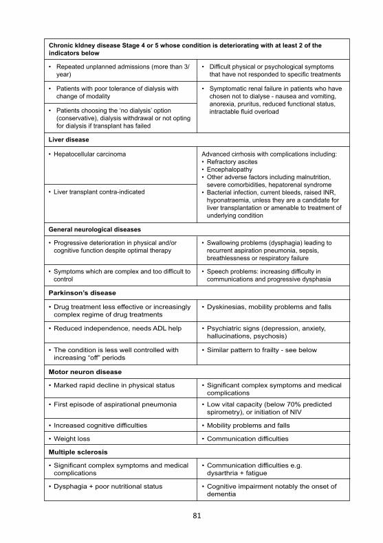
81
• Repeatedunplannedadmissions(morethan3/ year)
• Patientswithpoortoleranceofdialysiswith change of modality
• Patientschoosingthe‘nodialysis’option (conservative), dialysis withdrawal or not opting for dialysis if transplant has failed
Chronic kIdney disease Stage 4 or 5 whose condition is deteriorating with at least 2 of the indicators below
• Difficultphysicalorpsychologicalsymptoms thathavenotrespondedtospecifictreatments
• Symptomaticrenalfailureinpatientswhohave chosen not to dialyse - nausea and vomiting, anorexia, pruritus, reduced functional status, intractablefluidoverload
• Progressivedeteriorationinphysicaland/or cognitive function despite optimal therapy
• Symptomswhicharecomplexandtoodifficultto control
• Hepatocellularcarcinoma
• Livertransplantcontra-indicated
General neurological diseases
Liver disease
• Swallowingproblems(dysphagia)leadingto recurrent aspiration pneumonia, sepsis, breathlessness or respiratory failure
• Speechproblems:increasingdifficultyin communications and progressive dysphasia
Advanced cirrhosis with complications including: • Refractoryascites• Encephalopathy• Otheradversefactorsincludingmalnutrition, severe comorbidities, hepatorenal syndrome • Bacterialinfection,currentbleeds,raisedINR, hyponatraemia, unless they are a candidate for liver transplantation or amenable to treatment of underlying condition
•Drugtreatmentlesseffectiveorincreasingly complex regime of drug treatments
•Reducedindependence,needsADLhelp
•Theconditionislesswellcontrolledwith increasing“off”periods
Parkinson’s disease
•Dyskinesias,mobilityproblemsandfalls
•Psychiatricsigns(depression,anxiety, hallucinations, psychosis)
•Similarpatterntofrailty-seebelow
•Markedrapiddeclineinphysicalstatus
•Firstepisodeofaspirationalpneumonia
• Increasedcognitivedifficulties
•Weightloss
Motor neuron disease
•Significantcomplexsymptomsandmedical complications
•Lowvitalcapacity(below70%predicted spirometry), or initiation of NIV
•Mobilityproblemsandfalls
•Communicationdifficulties
•Significantcomplexsymptomsandmedical complications
•Dysphagia+poornutritionalstatus
Multiple sclerosis
•Communicationdifficultiese.g. dysarthria+fatigue
•Cognitiveimpairmentnotablytheonsetof dementia
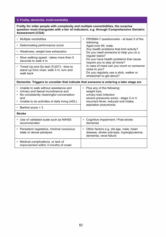
82
3. Frailty, dementia, multi-morbidity
• Multiplemorbidities
• Unabletowalkwithoutassistanceand• Urinaryandfaecalincontinenceand• Noconsistentlymeaningfulconversation and• Unabletodoactivitiesofdailyliving(ADL)
• Barthelscore>3
• Deterioratingperformancescore
• Weakness,weightlossexhaustion
• Slowwalkingspeed-takesmorethan5 seconds to walk 4 m
• TimedUpandGotest(TUGT)-timeto stand up from chair, walk 3 m, turn and walk back
Frailty for older people with complexity and multiple comorbidities, the surprise question must triangulate with a tier of indicators, e.g. through Comprehensive Geriatric Assessment (CGA)
Dementia: Triggers to consider that indicate that someone is entering a later stage are
• PRISMA-7questionnaire-atleast3ofthe following: Aged over 85, male. Any health problems that limit activity? Do you need someone to help you on a regular basis? Do you have health problems that cause require you to stay at home? In case of need can you count on someone close to you? Do you regularly use a stick, walker or wheelchair to get about?
• Plusanyofthefollowing: weight loss, urinary tract Infection severe pressures sores - stage 3 or 4 recurrent fever, reduced oral intake, aspiration pneumonia
• UseofvalidatedscalesuchasNIHSS recommended
• Persistentvegetative,minimalconscious state or dense paralysis
• Medicalcomplications,orlackof improvement within 3 months of onset
Stroke
• Cognitiveimpairment/Post-stroke dementia
• Otherfactorse.g.oldage,male,heart disease, stroke sub-type, hyperglycaemia, dementia, renal failure

ISBN 978-967-11415-4-0