phospholipid based vesicular system for …phospholipid based vesicular system for ... this drug...
TRANSCRIPT
PHOSPHOLIPID BASED VESICULAR SYSTEM FOR TRANSDERMAL AND DERMAL DELIVERY
63
PHOSPHOLIPID BASED VESICULAR SYSTEM FOR TRANSDERMAL AND DERMAL DELIVERY
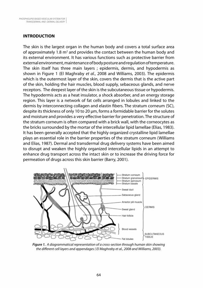
RINGKASAN: Vesikel berasaskan fosfolipid muncul sebagai sistem penghantaran dermal dan transdermal yang paling berpotensi disebabkan kelebihannya berbanding sistem-sistem penghantaran yang lain. Artikel ini mengulas mengenai jenis-jenis sistem vesikel berasaskan fosfolipid seperti liposomes, transferosomes dan ethosomes, dan mekanisma penelapan kulitnya. Ia juga menyediakan maklumat berkaitan kaedah penyediaan dan kegunaan sistem ini.
ABSTRACT: Phospholipid based vesicular appears to be most promising transdermal and dermal delivery system due to their merits over other delivery systems. This article reviews the types of phopholipid based vesicular system such as liposomes, transferosomes and ethosomes and their mechanism of skin permeation . It also provides an information on the method of preparation and application of the system.
Keywords: Phopholipid based vesicular, transdermal and dermal delivery, liposomes, transferosomes, ethosomes
Ropien JokimanMedical Technology FlagshipSIRIM Berhad1, Persiaran Dato’ Menteri,Section 2, P.O. Box 703540700 Shah Alam, SelangorMalaysia
PHOSPHOLIPID BASED VESICULAR SYSTEM FOR TRANSDERMAL AND DERMAL DELIVERY
64
INTRODUCTION
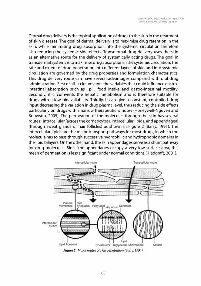
The skin is the largest organ in the human body and covers a total surface area of approximately 1.8 m2 and provides the contact between the human body and its external environment. It has various functions such as protective barrier from external environment, maintenance of body posture and regulation of temperature. The skin itself has three main layers ; epidermis, dermis, and hypodermis as shown in Figure 1 (El Maghraby et al., 2008 and Williams, 2003). The epidermis which is the outermost layer of the skin, covers the dermis that is the active part of the skin, holding the hair muscles, blood supply, sebaceous glands, and nerve receptors. The deepest layer of the skin is the subcutaneous tissue or hypodermis. The hypodermis acts as a heat insulator, a shock absorber, and an energy storage region. This layer is a network of fat cells arranged in lobules and linked to the dermis by interconnecting collagen and elastin fibers. The stratum corneum (SC), despite its thickness of only 10 to 20 µm, forms a formidable barrier for the solutes and moisture and provides a very effective barrier for penetration. The structure of the stratum corneum is often compared with a brick wall, with the corneocytes as the bricks surrounded by the mortar of the intercellular lipid lamellae (Elias, 1983). It has been generally accepted that the highly organized crystalline lipid lamellae plays an essential role in the barrier properties of the stratum corneum (Williams and Elias, 1987). Dermal and transdermal drug delivery systems have been aimed to disrupt and weaken the highly organized intercellular lipids in an attempt to enhance drug transport across the intact skin or to increase the driving force for permeation of drugs across this skin barrier (Barry, 2001).
Figure 1. A diagrammatical representation of a cross-section through human skin showing the different cell layers and appendages ( El Maghraby et al., 2008 and Williams, 2003).
PHOSPHOLIPID BASED VESICULAR SYSTEM FOR TRANSDERMAL AND DERMAL DELIVERY
65
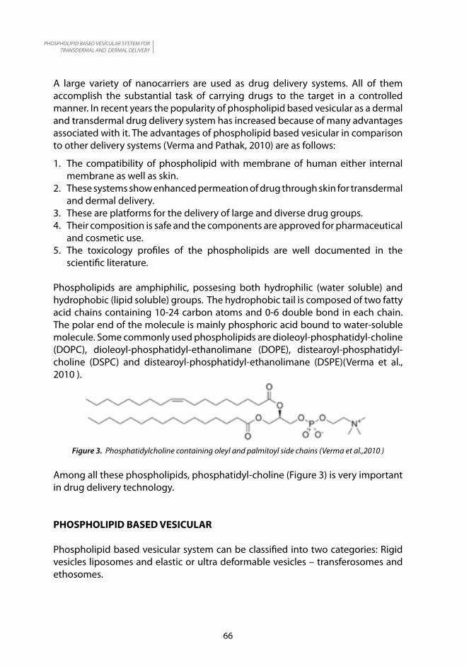
Dermal drug delivery is the topical application of drugs to the skin in the treatment of skin diseases. The goal of dermal delivery is to maximise drug retention in the skin, while minimising drug absorption into the systemic circulation therefore also reducing the systemic side effects. Transdermal drug delivery uses the skin as an alternative route for the delivery of systemically acting drugs. The goal in transdermal systems is to maximise drug absorption in the systemic circulation. The rate and extent of drug penetration into different layers of skin and into systemic circulation are governed by the drug properties and formulation characteristics. This drug delivery route can have several advantages compared with oral drug administration. First of all, it circumvents the variables that could influence gastro-intestinal absorption such as pH, food intake and gastro-intestinal motility. Secondly, it circumvents the hepatic metabolism and is therefore suitable for drugs with a low bioavailability. Thirdly, it can give a constant, controlled drug input decreasing the variation in drug plasma level, thus reducing the side effects particularly on drugs with a narrow therapeutic window (Honeywell-Nguyen and Bouwstra, 2005). The permeation of the molecules through the skin has several routes: intracellular (across the corneocytes), intercellular lipids, and appendageal (through sweat glands or hair follicles) as shown in Figure 2 (Barry, 1991). The intercellular lipids are the major transport pathways for most drugs, in which the molecule has to pass through successive hydrophilic and hydrophobic domains in the lipid bilayers. On the other hand, the skin appendages serve as a shunt pathway for drug molecules. Since the appendages occupy a very low surface area, this mean of permeation is less significant under normal conditions ( Hadgraft, 2001).
Figure 2. Major routes of skin penetration (Barry, 1991).
PHOSPHOLIPID BASED VESICULAR SYSTEM FOR TRANSDERMAL AND DERMAL DELIVERY
66
A large variety of nanocarriers are used as drug delivery systems. All of them accomplish the substantial task of carrying drugs to the target in a controlled manner. In recent years the popularity of phospholipid based vesicular as a dermal and transdermal drug delivery system has increased because of many advantages associated with it. The advantages of phospholipid based vesicular in comparison to other delivery systems (Verma and Pathak, 2010) are as follows:
1. The compatibility of phospholipid with membrane of human either internal membrane as well as skin.
2. These systems show enhanced permeation of drug through skin for transdermal and dermal delivery.
3. These are platforms for the delivery of large and diverse drug groups.4. Their composition is safe and the components are approved for pharmaceutical
and cosmetic use.5. The toxicology profiles of the phospholipids are well documented in the
scientific literature.
Phospholipids are amphiphilic, possesing both hydrophilic (water soluble) and hydrophobic (lipid soluble) groups. The hydrophobic tail is composed of two fatty acid chains containing 10-24 carbon atoms and 0-6 double bond in each chain. The polar end of the molecule is mainly phosphoric acid bound to water-soluble molecule. Some commonly used phospholipids are dioleoyl-phosphatidyl-choline (DOPC), dioleoyl-phosphatidyl-ethanolimane (DOPE), distearoyl-phosphatidyl-choline (DSPC) and distearoyl-phosphatidyl-ethanolimane (DSPE)(Verma et al., 2010 ).
Figure 3. Phosphatidylcholine containing oleyl and palmitoyl side chains (Verma et al.,2010 )
Among all these phospholipids, phosphatidyl-choline (Figure 3) is very important in drug delivery technology.
PHOSPHOLIPID BASED VESICULAR
Phospholipid based vesicular system can be classified into two categories: Rigid vesicles liposomes and elastic or ultra deformable vesicles – transferosomes and ethosomes.

PHOSPHOLIPID BASED VESICULAR SYSTEM FOR TRANSDERMAL AND DERMAL DELIVERY
67
Liposomes
Liposomes were first described by British hematologist Alec D Bangham (Bangham and Horne, 1964). Liposomes are thermodynamically stable, spontaneously formed, submicroscopic vesicular structures of amphiphilic lipids arranged in one or more concentric bilayers with an entrapped aqueous core. The lipids are natural or synthetic phosphilipids, which include phosphatidyl-choline (lecithin), phosphatidyl-ethanolimane, phosphatidyl-glycerol, phosphatidyl-serine, and phosphatidyl-ionositol (Venuganti and Perumal, 2009).
Figure 4. Liposomes contain a lipid bilayer (Lembo and Cavalli, 2010)
On mixing with an aqueous medium, the phosphate groups of the phospholipids orient themselves to the hydrophilic environment spontaneously forming unilamellar or multilamellar bilayer vesicles. Cholesterol is usually included to improve the stability of the vesicles, impart fluidity to the bilayer membrane, and prevent the leakage of vesicle contents. Hydrophilic drugs are incoporated in the aqueous core, whereas hydrophobic drugs are entrapped within the bilayer as shown in Figure 4.
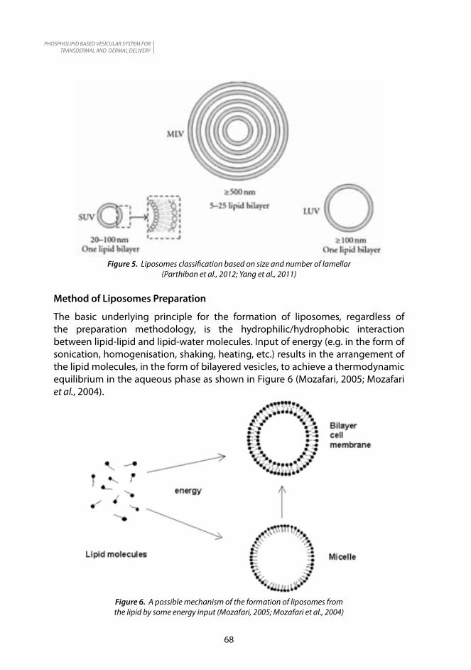
Liposomes are classified based on the size of vesicles or number of lipid bilayers. There are small unilamellar visicles (SUV), multilamellar vesicles (MLV) and large unilamellar vesicles (LUV). The diameter for SUV range from 20 nm to 100 nm, MLV is more than 500 nm, and LUV a few hundred nanometers as shown in Figure 5 (Parthiban et al., 2012; Yang et al., 2011).
PHOSPHOLIPID BASED VESICULAR SYSTEM FOR TRANSDERMAL AND DERMAL DELIVERY
68
Figure 5. Liposomes classification based on size and number of lamellar (Parthiban et al., 2012; Yang et al., 2011)
Method of Liposomes Preparation
The basic underlying principle for the formation of liposomes, regardless of the preparation methodology, is the hydrophilic/hydrophobic interaction between lipid-lipid and lipid-water molecules. Input of energy (e.g. in the form of sonication, homogenisation, shaking, heating, etc.) results in the arrangement of the lipid molecules, in the form of bilayered vesicles, to achieve a thermodynamic equilibrium in the aqueous phase as shown in Figure 6 (Mozafari, 2005; Mozafari et al., 2004).
Figure 6. A possible mechanism of the formation of liposomes from the lipid by some energy input (Mozafari, 2005; Mozafari et al., 2004)

PHOSPHOLIPID BASED VESICULAR SYSTEM FOR TRANSDERMAL AND DERMAL DELIVERY
69
The different methods used to prepare liposomes are described below:
(i) Thin-film hydration
Lipid mixture is dissolved in a suitable solvent such as methanol or chloroform. The organic solvent is removed under reduced pressure and lyophilised to remove any traces of solvent. The thin lipid film is redispersed in aqueous medium to form MLV . SUV can be prepared by sonication of MLV by using a probe or bath sonicator as shown in Figure 7 (Mozafari, 2005). Small vesicle sizes can also be achieved by passing MLV through a polycarbonate filter under high pressure (extrusion) as shown in Figure 8 (Vemuri and Rhodes, 1995).
Figure 7. Schematic representation of a probe-type sonicator (Mozafari, 2005).
PHOSPHOLIPID BASED VESICULAR SYSTEM FOR TRANSDERMAL AND DERMAL DELIVERY
70
Figure 8. Schematic representation of a small hand-held extruder (Vemuri and Rhodes, 1995).
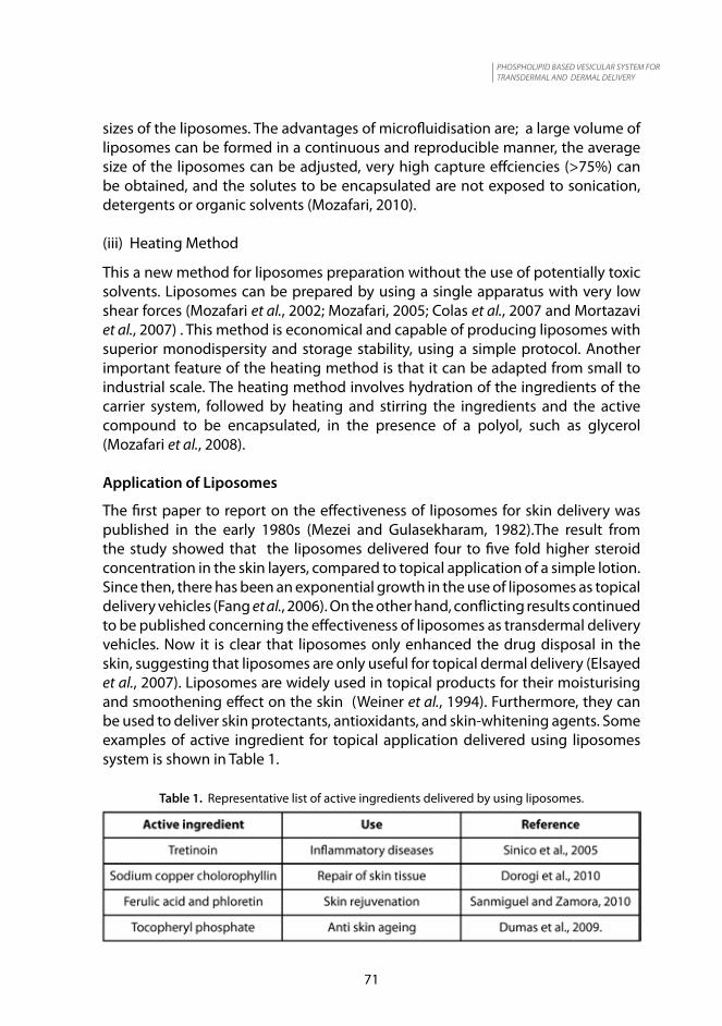
(ii) Microfluidisation
A method of liposomes preparation without using potentially toxic solvents is the microfluidisation technique using microfluidiser as shown in Figure 9 (Vemuri et al., 1990).
Figure 9. Schematic representation of a microfluidiser apparatus (Vemuri et al., 1990).
Microfluidisation is based on the principle of dividing a pressure stream into two parts, passing each part through a fine orifice, and directing the flows at each other inside the chamber of microfluidiser (Geciova et al., 2002; Jafari et al., 2006). Within the interaction chamber, cavitation, along with shear and impact, reduce particle
PHOSPHOLIPID BASED VESICULAR SYSTEM FOR TRANSDERMAL AND DERMAL DELIVERY
71
sizes of the liposomes. The advantages of microfluidisation are; a large volume of liposomes can be formed in a continuous and reproducible manner, the average size of the liposomes can be adjusted, very high capture effciencies (>75%) can be obtained, and the solutes to be encapsulated are not exposed to sonication, detergents or organic solvents (Mozafari, 2010).
(iii) Heating Method
This a new method for liposomes preparation without the use of potentially toxic solvents. Liposomes can be prepared by using a single apparatus with very low shear forces (Mozafari et al., 2002; Mozafari, 2005; Colas et al., 2007 and Mortazavi et al., 2007) . This method is economical and capable of producing liposomes with superior monodispersity and storage stability, using a simple protocol. Another important feature of the heating method is that it can be adapted from small to industrial scale. The heating method involves hydration of the ingredients of the carrier system, followed by heating and stirring the ingredients and the active compound to be encapsulated, in the presence of a polyol, such as glycerol (Mozafari et al., 2008).
Application of Liposomes
The first paper to report on the effectiveness of liposomes for skin delivery was published in the early 1980s (Mezei and Gulasekharam, 1982).The result from the study showed that the liposomes delivered four to five fold higher steroid concentration in the skin layers, compared to topical application of a simple lotion. Since then, there has been an exponential growth in the use of liposomes as topical delivery vehicles (Fang et al., 2006). On the other hand, conflicting results continued to be published concerning the effectiveness of liposomes as transdermal delivery vehicles. Now it is clear that liposomes only enhanced the drug disposal in the skin, suggesting that liposomes are only useful for topical dermal delivery (Elsayed et al., 2007). Liposomes are widely used in topical products for their moisturising and smoothening effect on the skin (Weiner et al., 1994). Furthermore, they can be used to deliver skin protectants, antioxidants, and skin-whitening agents. Some examples of active ingredient for topical application delivered using liposomes system is shown in Table 1.
Table 1. Representative list of active ingredients delivered by using liposomes.
PHOSPHOLIPID BASED VESICULAR SYSTEM FOR TRANSDERMAL AND DERMAL DELIVERY
72
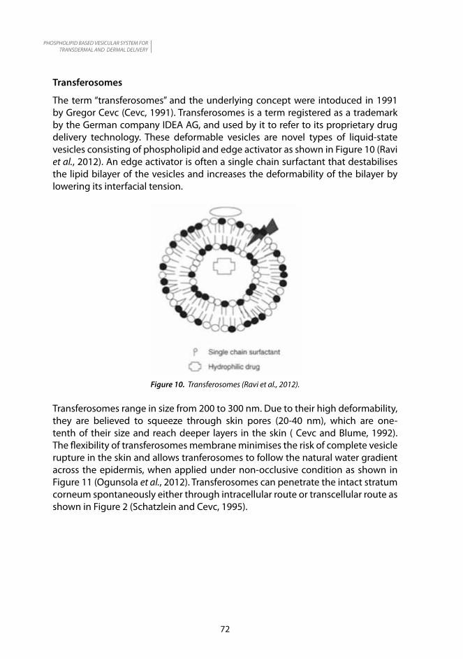
Transferosomes
The term “transferosomes” and the underlying concept were intoduced in 1991 by Gregor Cevc (Cevc, 1991). Transferosomes is a term registered as a trademark by the German company IDEA AG, and used by it to refer to its proprietary drug delivery technology. These deformable vesicles are novel types of liquid-state vesicles consisting of phospholipid and edge activator as shown in Figure 10 (Ravi et al., 2012). An edge activator is often a single chain surfactant that destabilises the lipid bilayer of the vesicles and increases the deformability of the bilayer by lowering its interfacial tension.
Figure 10. Transferosomes (Ravi et al., 2012).
Transferosomes range in size from 200 to 300 nm. Due to their high deformability, they are believed to squeeze through skin pores (20-40 nm), which are one-tenth of their size and reach deeper layers in the skin ( Cevc and Blume, 1992). The flexibility of transferosomes membrane minimises the risk of complete vesicle rupture in the skin and allows tranferosomes to follow the natural water gradient across the epidermis, when applied under non-occlusive condition as shown in Figure 11 (Ogunsola et al., 2012). Transferosomes can penetrate the intact stratum corneum spontaneously either through intracellular route or transcellular route as shown in Figure 2 (Schatzlein and Cevc, 1995).

PHOSPHOLIPID BASED VESICULAR SYSTEM FOR TRANSDERMAL AND DERMAL DELIVERY
73
Figure 11. The mechanism of skin penetration of transferosomes (Ogunsola et al., 2012).
Method of transferosomes preparation
Phospholipids, surfactant and the drug are dissolved in alcohol. The organic solvent is then removed by rotary evaporation and final traces of solvent are removed under vacuum. The deposited lipid film is hydrated with the appropriate buffer by rotation. The resulting vesicles, multilamellar lipid visicles (MLV), which are swollen at room temperature are then sonicated at room temperature. Sonication may be replaced by extrusion, low shear mixing or high shear mixing at room temperature (Jadupati et al., 2012; Elsayed et al, 2007). Application of Transferosomes
Transferosomes have been widely explored for dermal and transdermal application. Transferosomes have been widely used as carrier for the transport of proteins and peptides. Proteins and peptides are large biogenic molecules which are very difficult to transport into the body; when given orally they are completely degraded in the gastrointestinal tract. Paul et al. (1998) investigated the feasibility of non-invasive immunisation with a large membrane associated macromolecule, gap junction protein (GJP). The success of cell-mediated immune with response to the epicutaneous or subcutaneous administration of GJP in various colloidal formulation was studied. The result of the study showed that a non-invasive, transdermal immunisation with GJP-loaded transferosomes elicits 0.16 ± 0.06 and 0.37 ± 0.08 relative units of the specific IgG2a in the presence and absence of lipid A, respectively. Corresponding injections under the skin result in 0.18 ± 0.07 and 0.38 ± 0.02 relative units. It is shown that GJP is transported across the intact murine skin and processed immunologically, and the bioavaibility obtained from transferosomes is somewhat similar to that resulting from subcutaneous injection of the same protein formulation.
PHOSPHOLIPID BASED VESICULAR SYSTEM FOR TRANSDERMAL AND DERMAL DELIVERY
74
Delivery of insulin by transferosomes is the successful means of non invasive therapeutic use of such large molecular weight drugs on the skin. Cevc (Cevc, 2003) studied on vesicles containing insulin and tested them on NMRI (Nuclear Magnetic Resonance Imaging) mice and humans. Epicutaneous administration produced results similar to a subcutaneous injection in mice. Transferosome-associated insulin (Transfersulin® ) was able to reduce the blood glucose levels by 20-30 % in mice within 2-4 hours. Human data also showed very similar results but with a delay of 45-145 min from comparable subcutaneous doses. The representative list of drugs delivered using transferosomes is shown in Table 2.
Table 2. Representative list of drugs delivered by using transferosomes.
Ethosomes
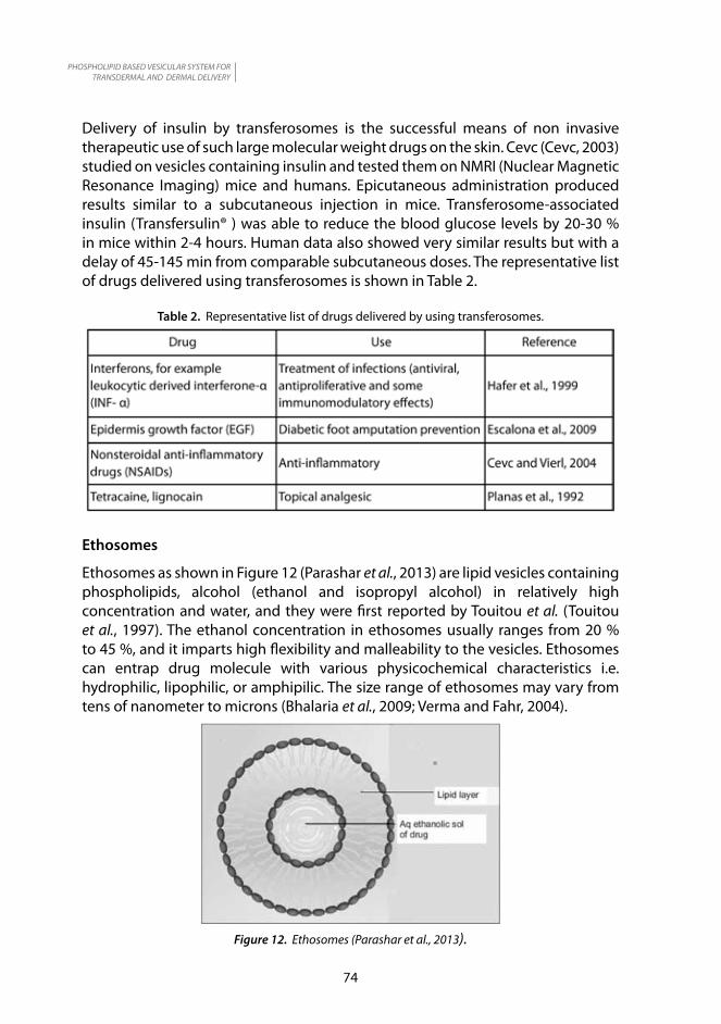
Ethosomes as shown in Figure 12 (Parashar et al., 2013) are lipid vesicles containing phospholipids, alcohol (ethanol and isopropyl alcohol) in relatively high concentration and water, and they were first reported by Touitou et al. (Touitou et al., 1997). The ethanol concentration in ethosomes usually ranges from 20 % to 45 %, and it imparts high flexibility and malleability to the vesicles. Ethosomes can entrap drug molecule with various physicochemical characteristics i.e. hydrophilic, lipophilic, or amphipilic. The size range of ethosomes may vary from tens of nanometer to microns (Bhalaria et al., 2009; Verma and Fahr, 2004).
Figure 12. Ethosomes (Parashar et al., 2013).
PHOSPHOLIPID BASED VESICULAR SYSTEM FOR TRANSDERMAL AND DERMAL DELIVERY
75
Very high encapsulation efficiencies of lipophilic drugs can be achieved due to the enhanced solubility from the presence of ethanol (Touitou et al., 2000). Unlike transferosomes, ethosomes can enhance drug delivery through skin under both non-occluding and occluding conditions (Elsayed et al., 2006).
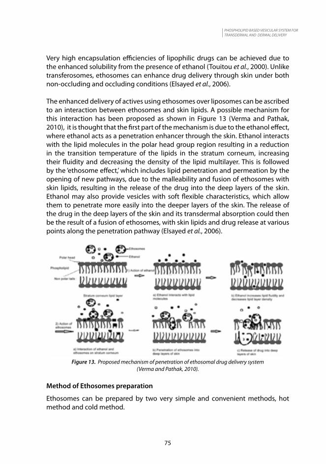
The enhanced delivery of actives using ethosomes over liposomes can be ascribed to an interaction between ethosomes and skin lipids. A possible mechanism for this interaction has been proposed as shown in Figure 13 (Verma and Pathak, 2010), it is thought that the first part of the mechanism is due to the ethanol effect, where ethanol acts as a penetration enhancer through the skin. Ethanol interacts with the lipid molecules in the polar head group region resulting in a reduction in the transition temperature of the lipids in the stratum corneum, increasing their fluidity and decreasing the density of the lipid multilayer. This is followed by the ‘ethosome effect,’ which includes lipid penetration and permeation by the opening of new pathways, due to the malleability and fusion of ethosomes with skin lipids, resulting in the release of the drug into the deep layers of the skin. Ethanol may also provide vesicles with soft flexible characteristics, which allow them to penetrate more easily into the deeper layers of the skin. The release of the drug in the deep layers of the skin and its transdermal absorption could then be the result of a fusion of ethosomes, with skin lipids and drug release at various points along the penetration pathway (Elsayed et al., 2006).
Figure 13. Proposed mechanism of penetration of ethosomal drug delivery system (Verma and Pathak, 2010).
Method of Ethosomes preparation
Ethosomes can be prepared by two very simple and convenient methods, hot method and cold method.
PHOSPHOLIPID BASED VESICULAR SYSTEM FOR TRANSDERMAL AND DERMAL DELIVERY
76
(i) Cold Method
This is the most common and widely utilised method for the preparation of ethosomes.The phospolipid, drug and other lipid materials are dissolved in ethanol in a covered vessel at room temperature with vigorous stirring. The mixture is heated up to 30 ºC in a water bath. In a separate vessel, the water is heated to 30 ºC and added to the above mixture and then stirred for five minutes in a covered vessel. The vesicle size of ethosomal formulation can be decreased using sonication or extrusion method. Finally the formulation must be properly stored under refrigeration (Nikalje and Tiwari, 2012; Dinesh et al., 2009).
(ii) Hot Method
The drug is dissolved in a mixture of ethanol and propylene glycol at 40 ºC. In a separate vessel phospholipid is dispersed in water at 40 ºC. Once both mixtures reach 40 ºC, the organic phase is added to the aqueous phase. After mixing for five minutes the preparation is sonicated at 4 ºC for three cycles of five minutes, with a rest of five minutes between each cycle, using Probe Sonicator. The formulation is then homogenised at 15,000 psi pressure, in three cycles, using high pressure homogeniser to get nano-sized ethosomes (Bhalaria et al., 2009).
Application of Ethosomes
Ethosomes are able to penetrate the deeper layers of the skin and hence appear to be vesicles of choice for transdermal drug delivery of hydrophilic and impermeable drug through skin. One of the application of ethosomes is as a vesicle for topical delivery of antibiotics. Conventional oral therapy causes several allergic reactions along with several side effects. Conventional external preparations possess low permeabilty to deep skin layers and subdermal tissue. The antibiotic (erythromycin) loaded ethosomal formulation have been prepared for dermal and intracellular delivery (Godin et al., 2005). A model for deep dermal Staphylococcus aureus infection in mice was developed. The efficiency of ethosomal erythromycin applied to the skin-infected site was compared with intraperitoneal erythromycin administration and with local application of hydroethanolic erythromycin solution. The in vivo experiments demonstrated a very efficient healing of Staphylococcus aureus-induced deep dermal infections when the mice were treated with ethosomal erythromycin. Bacterial counts and histological evaluation of the skin treated with ethosomal antibiotic revealed no bacterial growth and normal skin structure. On the contrary, no subdermal healing was observed in infected animals treated with topical hydroethanolic erythromycin solution. The results of this study showed that the ethosomal formulation of antibiotic could be highly efficient and would overcome the problems associated with conventional therapy.
Dubey, et al. (2007) prepared methotrexate (MTX) ethosomes used for the treatment of psoriasis. They also compared the extent of penetration of ethosomes and liposomes loaded with rhodamine red (RR) by CLS microscopy. They found
PHOSPHOLIPID BASED VESICULAR SYSTEM FOR TRANSDERMAL AND DERMAL DELIVERY
77
that ethosomes penetrated to a depth of 170 μm with fluorescence intensity (FI) of 160 AU, whereas liposomes penetrated only up to 80 μm with FI of 40 AU. On the other hand the flux of MTX ethosomes was found to be 57.2 ± 4.34 μg/cm2 /hour whereas the flux for hydroethanolic solution and liposomes was found to be 22.43 ± 0.24 μg/cm 2 /hour and 14.6 ± 1.65 μg/cm2 /hour, respectively. Table 3 is a short compilation of ethosomes application as a carrier for topical and transdermal delivery of a variety of drugs.
Table 3. Representative list of drugs delivered by using ethosomes
FUTURE PERSPECTIVE
Consumers nowadays are more focused on their health and appearance. As a result, there has been an increasing demand in topical anti-ageing formulation with active ingredients. Novel and innovative delivery systems are transforming the new product development in the cosmetic field because of consumer perceivable benefits and optimised sensory attributes. Liposomes are the most widely known topical delivery systems. A special characteristic of liposomes is the ability to adapt to water soluble and non-water soluble active ingredients in the liposomes membrane. Liposomes are used in a variety of skin care rejuvenation products because of their ability to encapsulate active ingredients and deliver them deep into the cells. Several excellent phytochemicals and herbal extracts have been successfully delivered via liposomes and showed some distinct advantages over conventional cosmetic products. Thus, it can be predicted that liposomes will be continously used as an efficient delivery system of active ingredients in topical products and will improve the cosmetic market even more, although a lot of research and human studies in this field is required to obtain real life data. Transdermal route is a promising alternative to drug delivery for systemic effect. Transferosomes and ethosomes have iniatiated a new area in vesicular research for transdermal drug delivery which can provide better skin permeation than liposomes. Hence, enhanced delivery of drug molecules through the skin by means of transferosomes opens new challenges and opportunities for the development of novel improved therapies. Thus, it could be concluded that the transferosomes can
PHOSPHOLIPID BASED VESICULAR SYSTEM FOR TRANSDERMAL AND DERMAL DELIVERY
78
overcome all the problems associated with transdermal delivery as transferosomes itself are specially optimised vesicles having the capability of responding to external stress by rapid and energetically inexpensive shape transformations. The high tolerability and efficiency of these vesicular systems open vast potential therapeutic uses. These nanocarriers might offer advanced local and systemic new therapies with agents that are unable to efficiently penetrate the stratum corneum via passive diffusion. IDEA AG breakthrough, proprietary Transfersome® technology enables the targeted and non-invasive delivery of drugs (including large molecules such as proteins) through the skin, with particular focus on pain relief and dermatology. The Company’s Transfersome® carriers are topically applied on the skin and can be engineered to achieve high drug concentration at or near the site of application, increasing drug potency and diminishing side effects. The non-steroidal anti-inflammatory drug (NSAID), ketoprofen, in a transferosomes formulation gained marketing approval by the Swiss regulatory agency (SwissMedic). Further therapeutic products based on the transferosome technology, according to IDEA AG, are in clinical development.
Ethosomes has shown promising result and potential for delivery of various agents more effectively. Ethosomes offers a good opportunity for non – invasive delivery of small, medium and large size drug molecules. Ethosomes can be a promising tool for dermal/transdermal delivery of various agents and an alternate formulation for problematic drugs. Further research in this area will allow better control over drug release in vivo, allowing the physician to make the therapy more effective. Therefore, it should not be long before corresponding drug formulation finds its way into clinics to be tested for widespread usage.Thus, it can be logically concluded that ethosomal formulations possess a promising future in effective dermal/transdermal delivery of bioactive agents. More formulations based on ethosomes is expected be launched in the market in the coming years, including formulations for the treatment of alopecia, deep skin infection, hormone deficiencies, inflammation and atopic dermatitis.
CONCLUSION
A number of transdermal and dermal delivery system for drugs are emerging today. The phospholipid based delivery systems are important because of intense advantages associated with them. The delivery systems described above have proved their ability and efficacy to deliver the drugs to the desired location. Liposomes are only suitable for dermal system but not suitable for transdermal delivery because they cannot reach the deeper layer of the skin as they are trapped in the superior layer of stratum corneum. Transdermal route is a promising alternative to drug delivery for systemic effect. Transferosomes and ethosomes have initiated a new area in vesicular research for transdermal drug delivey which can provide better skin permeation than liposomes.
PHOSPHOLIPID BASED VESICULAR SYSTEM FOR TRANSDERMAL AND DERMAL DELIVERY
79
REFERENCES
Bangham, A.D., Horne, R. W. (1964). Negative staining of phospholipids and their structural modification by surface-active agents as observed in the electron microscope. J. Mol.Bio. 8 : pp 660–668.
Barry, B.W. (1991). Lipid protein partitioning theory of skin penetration enhancement. J. Control. Release. 15 : pp 237–248.
Barry, B.W. (2001). Novel mechanisms and devices to enable successful transdermal drug delivery. Eur. J. Pharm. Sci. 14 : pp 101-114.
Bhalaria, M.K., Naik, S. and Misra A.N. (2009). Ethosomes: A novel delivery system for antifungal drugs in the treatment of topical fungal diseases. Indian Journal of Experimental Biology. 47 : pp 368-375.
Cevc, G. (1991). Isothermal lipid phase. Transitions chemistry and physics of lipids. 57 : pp 293-299.
Cevc, G. (2003). Transdermal drug delivery of insulin with ultradeformable carriers. Clin Pharmacokinet. 42 : pp 461-474.
Cevc, G. and Blume, G. (1992). Lipid vesicles penetrate into intact skin owing to the transdermal osmotic gradients and hydration force. Biochim Biophys Acta. 1104 : pp 226-232.
Cevc, G. and Vierl, U. (2004). Aggregate with increased deformability comprising at least three amphipats for improved transport through semi-permeable barriers and for the non-invasive drug application in vivo especially through the skin. WO Patent: 2004032900A1.
Colas, J.C., Shi, W.L., Rao, V.S.N.M., Omri, A., Mozafari, M.R. and Singh, H. (2007). Microscopical investigations of nisin-loaded nanoliposomes prepared by Mozafari method and their bacterial targeting. Micron. 38 : pp 841-847.
Dinesh, D., Amit, A.R., Maria, S., (2009). Drug vesicle based approaches of penetration enhancement. Int. J. Phar. Sci. 1(1) : pp 24-45.
Dorogi, P.L., Vasily, D.B. and McCook, J.P. (2010). Skin treatment composition containing copper pigment complex. U.S. Patent: 20100247591A1.
Dubey, V., Mishra, D., Dutta, T., Nahar, M., Saraf, D.K. and Jain, N.K. (2007). Dermal and transdermal delivery of an anti-psioriatic agent via ethanolic liposomes. J. Control Release. 123(2) : pp 148-154.
Dumas, M., Noblesse, E., Alard, V., Quiles, D. and Perrier, E. (2009). Use of tocopheryl phosphate as an agent for preventing or slowing down the appearance of the effects of skin ageing. U.S. Patent: 20090104258A1.
El Maghraby, G.M., Barry, B.W. and Williams, A.C. (2008). Liposomes and skin: From drug delivery to model membranes. Eur. J. Pharm. Sci. 34 : pp 203-222.
Elias, P.M. (1983). Epidermal lipids, barrier function, and desquamation. J. Invest. Dermatol. 80 : pp 44-49.
PHOSPHOLIPID BASED VESICULAR SYSTEM FOR TRANSDERMAL AND DERMAL DELIVERY
80
Elsayed, M.M.A., Abdallah, O.Y., Naggar, V.F., (2006). Deformable liposomes and ethosomes. Mechanism of enhanced skin delivery. Int. J. Pharm. 322 : pp 60-66.
Elsayed, M.M.A., Abdallah, O.Y., Naggar, V.F., (2006). Lipid vesicles for skin delivery of drugs. Reviewing three decades of research. Int. J. Pharm. 332 : pp 1-6.
Elsayed, M.M.A., Abdallah, O.Y., Naggar, V.F., (2007). Deformable liposomes and ethosomes as carrier for skin delivery of ketotifen. Pharmazie. 62 : pp 133-137.
Escalona, E.P., Meireles, R.P., Acosta, J.A.B. and Redriquez, B.Y.B. (2009). Use of a topical composition containing epidermal growth factor (EGF) for the diabetic foot amputation prevention. U.S. Patent: 20090074850A1.
Fang, J., Hong, C., Chiu, W. and Wang, Y. (2001). Effect of liposomes and niosomes on skin permeation of enoxacin. Int. J. Pharm. 219 : pp 61-72.
Geciova, J., Bury, D. and Jelen, P. (2002). Methods for disruption of microbial cells for potential use in the dairy industry- a review. Int. Dairy J. 12 : pp541-553.
Godin, B., Touitou, E., Rubinstein, E., Athamna, E. And Athamna, M. (2005). A new approach for treatment of deep skin infections by an ethosomal antibiotic preparation : an in vivi study. J. Antimicrob Chemother. 55: pp 989-994.
Hadgraft, J. (2001). Passive enhancement strategies in topical and transdermal drug delivery. Int. J. Pharm. 184(1) : pp 1-6.
Hafer, C., Goble, R., Deering, P., Lehmer, A. and Breut, J. (1999). Formulation of interleukin-2 and interferon-α containing ultradeformable carriers for potential transdermal application. Anticancer Res. 19(2c) : pp 1505-1512.
Honeywell-Nguyen, P.L. and Bouwstra, J.A. (2005). Vesicles as a tool for transdermal and dermal delivery. Drug Discovery Today. 2 : pp 67-74.
Jadupati, M., Amites, G and Kumar, N.A. (2012). Transferosomes: An opportunity carrier for transdermal drug delivery system. Int. Research J. Pharm. 3(3) : pp 35-38.
Jafari, S.M., He, Y.H. and Bhandari, B. (2006). Nanoemulsion production by sonication and microfluidization – a comparison. Int. J. Food Prop. 9 : pp 475-485.
Koli, J.R. and Lin, S. (2009). Development of antioxidant ethosomes for topical delivery utilizing the synergistic properties of vit A palmitate, vit E and vit C. AAPS Pharm. Sci. Tec. 11 : pp 1-8.
Lembo, D. and Cavalli, R. (2010). Nanoparticulate delivery systems for antviral drugs. Antiviral Chem. & Chemotheraphy. 21: pp 53-70.
Mezei, M. And Gulasekharam, V. (1982). Liposomes: A selective drug delivery system for the topical route of administration: Gel dosage form. J. Pharm. Pharmacol. 34 : pp 473-474.
Mortazavi, S.M., Mohammadabadi, M.R., Khosravi-Darani, K. and Mozafari, M.R. (2007). Preparation of liposomal gene theraphy vectors by a scalable method without using volatile solvents or detergents. J. Biotechnol. 129 : pp-604-613.

PHOSPHOLIPID BASED VESICULAR SYSTEM FOR TRANSDERMAL AND DERMAL DELIVERY
81
Mozafari, M.R. (2005). Liposomes: An overview manufacturing techniques . Cell Mol. Biol. Lett. 10 : pp 711 – 719.
Mozafari, M.R. (2010). Nanoliposomes: Preparation and analysis. In: Liposomes, method in molecular biology, ed. Weissig, V. Humana Press, New York pp 29-50.
Mozafari, M.R., Reed, C.J., Rostron, C., Kocum, C. and Piskin, E. (2002). Construction of stable anionic liposome-plasmid particles using the heating method: a preliminary investigation. Cell Mol. Biol. Lett. 7(3): pp 923-927.
Mozafari, M.R., Reed, C.J. and Rostron, C. (2004). Formation of the initial cell membranes under primordial earth conditions. Cell Mol. Biol. Lett. 9 (Suppl. 2): pp 97-99.
Mozafari, M.R., Johnson, C., Hatziantoniou, S. and Demetzos, C. (2008). Nanoliposomes and their applications in food nanotechnology. J. Liposome Research. 18: pp 309-327.
Nikalje, A.P. and Tiwari, S. (2012). Ethosomes: A novel tool for transdermal drug delivery. IJPRS. 2(1): pp 1-20.
Ogunsola, O.A., Kraeling, M.E., Zhong, S., Pochan, D.J., Bronaugh, R.L. and Raghavan, S.R. (2002). Structural analysis of flexible liposomes formulations: new insights into skin-penetrating ability of soft nanostructures. Soft Matter. 8: pp 10226-10232.
Parashar, T., Soniya, Sachan, R., et al (2013). Ethosomes: A recent vesicle of transdermal drug delivery system. Int. J. Res. Dev. Pharm. L. Sci. 2 (2): pp 285-292.
Parthiban C. P., Jeroen C. H. L., Pieter J. D., Marcel K. and Janine N. P. (2012). Nanomaterials for the local and targeted delivery of osteoarthritis drugs. Journal of Nanomaterials. 2012: pp 1-13.
Paul, A., Cevec, G. and Bachhavat, B.K. (1998). Transdermal immunisation with an integral membrane component, gap junction protein, by means of ultradeformable drug carriers, transferosomes. Vaccineur. 16: pp 188-195.
Planas, M.E., Gonzalez, P., Rodriguez, S., Sanchez, G. and Cevc, G. (1992). Non invasive percutaneous induction of topical analgesia by a new type of drug carrier, and prolongation of local pain insensitivity by anesthetic liposomes. Anesthia Analog: pp 615-621.
Ravi, K., Singh, M., Bala, R., Seth, N. and Rana, A.C. (2012). Transferosomes: A novel approach for transdermal drug delivery. Inter. Research J Pharm. 3(1): pp 20-24.
Sanmiguel, G.S. and Zamora, J.M. (2010). Systems and method for skin rejuvenation. Eur. Patent: 2241303A2.
Schatzlein, A. and Cevc, G. (1995). Skin penetration by phospholipids vesicles, transferosomes as visualized by means of the confocal scanning laser microscopy, in charazterization, metabolism, and novel biological application. Champaign. AOCS Press. pp 191-209.
Singh, H.P., Utreja, P., Tiwary, A.K. and Jain, S. (2008). Elastic liposomal formulations for sustained delivery of colchicine: In vitro characterization and In vivo evaluation of anti gout activity. AAPS Pharm. Sci. Tec. 11: pp 54-64.
PHOSPHOLIPID BASED VESICULAR SYSTEM FOR TRANSDERMAL AND DERMAL DELIVERY
82
Sinico, C., Manconi, M., Peppi, M., et al. (2005). Liposomes as carriers for dermal delivery of tretinon: In vitro evaluation of drug permeation and vesicle-skin intercation. J. Control. Release. 103: pp 123-136.
Tan, J., Jiang, L., Chang, T. and Zhou, Z. (2012). Bullatacin ethosome gel and preparation method thereof. CN Patent: 102552147(A).
Touitou, E. (2010). Stable compositions for nail onychomycosis treatment. WO Patent: 2010086727.
Touitou, E., Alkabes, M., and Dayan, N. (1997). Ethosomes: Novel lipid vesicular system for enhanced delivery. Pharm. Res. S14: pp 305–306.
Touitou, E., Dayan, N., Bergelson, L., et al. (2000). Ethosomes: Novel vesicular carriers: Characterization and delivery propeties. J. Control. Release. 65: pp 403-418.
Vemuri, S., Yu, C.D., Wangsatorntanakun, V. And Roodsdorp, N. (1990). Large-scale production of liposomes by a microfluidizer. Drug Dev. Ind. Pharm. 16: 2243-2256.
Vemuri, S. And Rhodes C.T. (1995). Preparation and characterisation of liposomes as therapeutic delivery systems: A review. Pharma Acta Helv. 70: pp 95-111.
Venuganti, V.V. and Perumal, O.P. (2009). Nanosystems for dermal and transdermal drug delivery. In: Drug delivery nanoparticles formulation and characterization, ed. Pathak, Y. and Thassu, D., Informa Helthcare, New York, pp 126-154.
Verma, D.D. and Fahr, A. (2004). Synergistic penetration effect of ethanol and phospholipids on the topical delivery of cyclosporin A. J. Control Release. 97: pp 55-66.
Verma, P. and Pathak, K. (2010). Therapeutic and cosmeceutical potential of ethosomes: An overview. Journal of Advanced Pharmaceutical Technology & Research. 1(3): pp 274-282.
Verma, P., Ram, A., Jha, A.K., Mishra, A. and Thakur, A. (2010). Phosphatidylcholine: A revolution in drug delivery technology. Int. J. Pharm. Sci. & Research. 1(2): pp 1-9.
Weiner, N., Lieb, L., Niemiec, S., (1994). Liposomes: A novel topical delivery system for pharmaceutical and cosmetic application. J. Drug Target. 2: pp 405-410.
Williams, A.C. (2003). Transdermal and topical drug delivery; from theory to clinical practice. Pharmaceutical Press, London.
Williams, M.L. and Elias, P.M. (1987). The extracellular matrix of stratum corneum: role of lipids in normal and pathological function. Crit. Rev. Ther. Drug Carrier Syst. 3: pp 95-122.
Yang, F., Jin, C. and Jiang, Y. (2011). Liposome based delivery systems in pancreatic cancer treatment: from bench to bedside. Cancer Treatment Reviews. 37(8): pp 633–642.