otocerebral mucormycosis a rare case in a diabetic
TRANSCRIPT

Internal Carotid Artery; GMS: Gomori Methamine Silver; ZN:Ziehl-Neelsen; AFB: Acid Fast Bacilli; KOH: Potassium Hydroxide; PAS: Periodic Acid-Schiff Staining
Pendahuluan
Mucormycosis adalah nama umum yang diberikan untuk penyakit klinis disebabkan oleh jamur dari urutan Mucoraceae dari zygomycetes kelas. Mucormycosis adalah hidup mengancam infeksi jamur yang umum mempengaruhi host immunocompromised hampir seragam dalam mengembangkan serta negara-negara industri. Mucorales didistribusikan ke enam keluarga yang semuanya dapat menyebabkan kulit dan infeksi jamur yang mendalam. Dari enam keluarga Rhizopus oryzae merupakan penyebab tersering infeksi. Diantara spesies lain yang diisolasi termasuk Rhizopus microsporus var. rhizopodiformis, Absidia corymbifera, Apophysomyces elegans, spesies Mucor, dan Rhizomucor [1]. Mucormycosis terlihat lebih umum di hidung dan sinus paranasal, sedangkan candida dan infeksi jamur aspergillus, yang agak biasa [2,3], terlihat di saluran pendengaran eksternal. Varian invasif mucormycosis umumnya terjadi pada pasien diabetes. Mucormycosis melibatkan telinga tengah adalah entitas klinis yang langka. Portal kemungkinan masuk ke telinga tengah adalah baik dari nasofaring melalui tabung Eustachio atau melalui membran timpani perforasi yang sudah ada [4]. The kelangkaan keterlibatan telinga tengah membuat sulit untuk dokter mencurigai sama dengan diagnosis utama. Klinis presentasi mungkin mirip dengan kronis Otitis Media dengan mungkinkeluhan utama nyeri telinga dominan dengan rujukan kemastoid dan leher bagian atas. Komplikasi lebih lanjut dari Mucormycosis dari keterlibatan telinga tengah dalam hal perpanjangan intrakranial dengan keterlibatan otak dan sinus vena merumitkan lanjut presentasi menambah dilema diagnostik. Awal diagnosis adalah andalan terapi sukses yang melibatkan debridement dan administrasi antijamur seperti bedah Amfoterisin B [4,5].
Laporan Kasus
Seorang pria dengan Hipertensi dan Diabetes berusia 55 tahun dibawa ke unit gawat darurat dengan riwayat hemiparesis badan sebelah kanan
Submit Manuscript | http://medcraveonline.com
Jurnal Otolaringologi-Penelitian THT
Mucormycosis Otocerebral: Kasus Langka di DiabetesPasien dengan Presentasi tidak biasa
Laporan Abstrak
Mucor adalah jamur saprofit. Meskipun umumnya menyerang hidung danlesi sinus paranasal, kulit, paru atau gastrointestinal mungkinjuga dilihat dan penyebaran hematogen ke situs lain juga bisa terjadi. itufaktor predisposisi meliputi Diabetes Mellitus dan kondisi lain yang menyebabkan negara immunocompromized. Namun, kasus yang jarang terjadi tanpa mendasari gangguan telah juga melaporkan. Kasus ini dijelaskan untuk menunjukkan jarang presentasi neurologis dari Oto-otak Mucormycosis daripada baik diakui badak-orbito-otak bentuk. Diagnosis dini dan institusipengobatan andalan untuk terapi sukses.
Kata kunci: Mucormycosis; Canal wall down mastoidectomy;
Amphotericin B; Diabetes
Volume 2 Issue 2 - 2015
I D Singh*, J R Galagali and Satish KumarDepartment of Otolaryngology Head & Neck surgery, Command Hospital (Southern Command), India
*Corresponding author: ID Singh, Department of Otolaryngology Head & Neck surgery, Command Hospital (Southern Command), Pune 411040, India, Tel: +91-7767834137; Email:
Diterima: January 29, 2015 | Dipublikasi: March 16, 2015
Singkatan CVST: Central Venous Sinus Thrombosis; ICA: dan slurring berbicara. Dia juga memiliki sejarah berbau busukmukopurulen otorrhea telinga kiri dan telinga kiri otalgia memancaruntuk hemicranium kiri dan leher bagian atas dari durasi tiga bulan.Pasien juga mengembangkan deviasi dari sudut mulut ke kananbulan sebelum rawat inap.
Thorough clinical examination revealed right hemiparesis, left Lateral rectus palsy suggestive of sixth cranial nerve palsy and deviation of angle of the mouth due to left Facial nerve Palsy (Lower Motor Neuron type) (Figure1). There was a palpable firm mass in the upper neck behind the angle of jaw on left side,post auricular lymphadenopathy and a positive Griesinger’s sign (Figure 2). Otoscopy revealed active mucopurulent discharge with granulations and necrotic tissue in external auditory canal and the middle ear (Figure 3). Magnetic resonance imaging and Contrast enhanced computed tomography temporal bones and neck revealed left coalescent mastoiditis with extension across the subperiostium with fluid collection involving the soft tissueof left postauricular region, extradural abscess from left petrous, basal meningitis and pontine infarct. Magnetic resonance venogram and angiogram revealed Central venous sinus thrombosis (CVST) of left sigmoid sinus, transverse and superior sagittal sinus, loss of luminal signal of left internal jugular veinat jugular bulb and decreased flow related to enhancementinvolving the petrous segment of the left internal carotid artery (ICA) (Figure 4).
Figure 1: Preoperative picture of the patient śhowing Cranial nerve VI and VII palsy (on the patient s left side of the face).

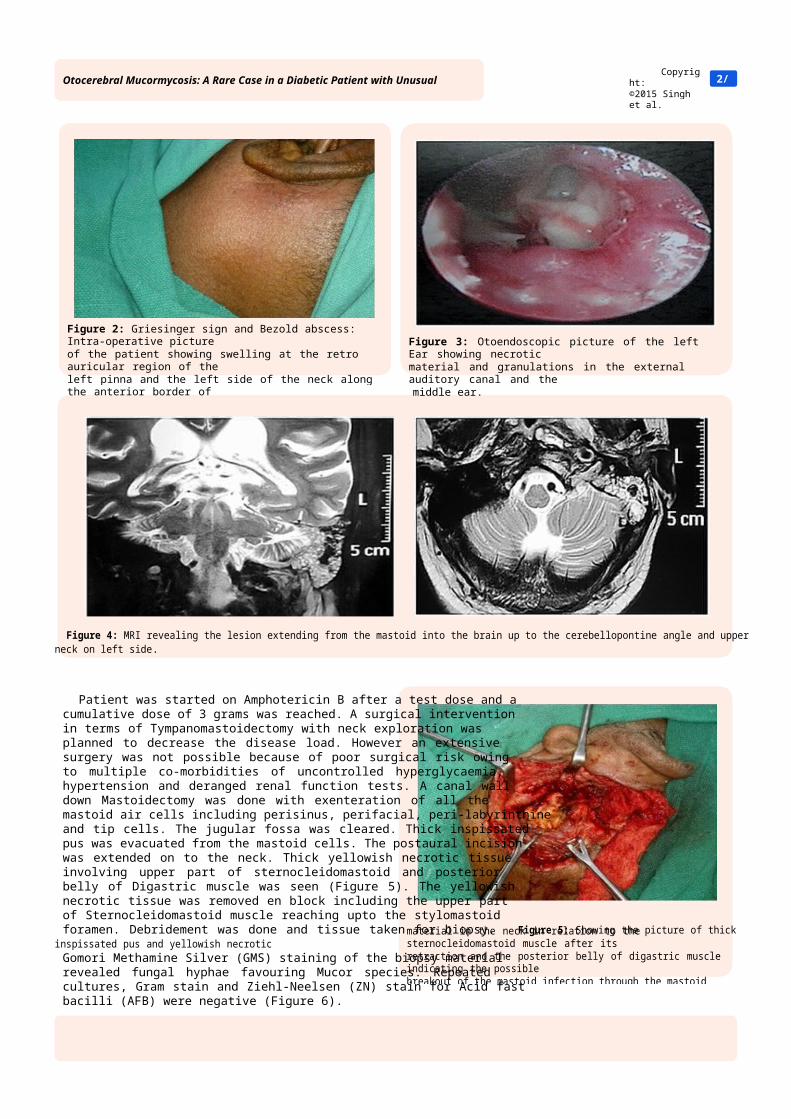
Figure 4: MRI revealing the lesion extending from the mastoid into the brain up to the cerebellopontine angle and upper neck on left side.
Patient was started on Amphotericin B after a test dose and a cumulative dose of 3 grams was reached. A surgical intervention in terms of Tympanomastoidectomy with neck exploration was planned to decrease the disease load. However an extensive surgery was not possible because of poor surgical risk owing to multiple co-morbidities of uncontrolled hyperglycaemia, hypertension and deranged renal function tests. A canal wall down Mastoidectomy was done with exenteration of all the mastoid air cells including perisinus, perifacial, peri-labyrinthine and tip cells. The jugular fossa was cleared. Thick inspissatedpus was evacuated from the mastoid cells. The postaural incision was extended on to the neck. Thick yellowish necrotic tissue involving upper part of sternocleidomastoid and posterior belly of Digastric muscle was seen (Figure 5). The yellowish necrotic tissue was removed en block including the upper part of Sternocleidomastoid muscle reaching upto the stylomastoid foramen. Debridement was done and tissue taken for biopsy. Figure 5: Showing the picture of thick inspissated pus and yellowish necrotic Gomori Methamine Silver (GMS) staining of the biopsy material revealed fungal hyphae favouring Mucor species. Repeated cultures, Gram stain and Ziehl-Neelsen (ZN) stain for Acid fast bacilli (AFB) were negative (Figure 6).
Citation: Singh ID, Galagali JR, Kumar S (2015) Otocerebral Mucormycosis: A Rare Case in a Diabetic Patient with Unusual Presentation. J Otolaryngol ENT Res 2(2): 00020. DOI: 10.15406/joentr.2015.02.00020
Otocerebral Mucormycosis: A Rare Case in a Diabetic Patient with Unusual Presentation
2/
Figure 2: Griesinger sign and Bezold abscess: Intra-operative picture of the patient showing swelling at the retro auricular region of the left pinna and the left side of the neck along the anterior border of sternocleidomastoid muscle.
Figure 3: Otoendoscopic picture of the left Ear showing necrotic material and granulations in the external auditory canal and the middle ear.
Copyright:©2015 Singh et al.
material in the neck in relation to the sternocleidomastoid muscle after its retraction and the posterior belly of digastric muscle indicating the possible breakout of the mastoid infection through the mastoid tip.
acidosis negatively impact neutrophil chemotactic activity and phagocytosis. However, neither of the two mechanisms describes preferential rhinocerebral involvement in the diabetic patient in ketoacidosis than the other forms of mucormycosis. Involvement of the blood vessels leads to infarction, hemorrhage and thrombosis. The invasive Mucormycosis is characterized by vascular invasion causing haemorrhage, thrombosis and necrosis of tissue [9]. The Temporal bone involvement in Mucormycosis is very rare and may resemble a refractory Otitis externa. McGill previously described Mucormycosis of the Temporal bone [10]. Mucormycosis of the temporal bone may involve the middle ear cavity without necrosis or it may involve even the inner ear with extensive necrosis [11]. It may involve the skull base or the intracranial compartment presenting with Cranial nerve deficitsand other neurological symptoms.
Intracranial fungal infection can be caused by the extension of a localized nasal or parana¬sal sinus infections, cavernous sinus invasion, or direct infection of the brain parenchy¬ma. Infection can invade adjacent blood vessels, including theretinal artery, oph¬thalmic artery and internal carotid artery, causing compli¬cations such as intracranial infection, systemic embolism, intracerebral hem¬orrhage, subarachnoid hemorrhage and cerebral infarction. Very rare cases have shown ICA occlusion and secondary abscess formation [12,13]. Cranial nerve involvement indicates a severe infection and a bad prognosis. Yun et al. [14] first reported a case of middle earMucormycosis with facial palsy in a diabetic patient.
Complications of relatively rare Mucormycosis in terms of intracranial involvement with cranial nerve involvement adds to the diagnostic difficulty and delay in institution of treatment.
Diagnosis of Mucormycosis necessitates a high index of suspicion, as almost half of the cases are identified in post mortemstudies [15,16]. Diagnosis can be made by imaging studies and histopathological examination of the infected material. The fungus forms fluffy white, gray, or brownish colonies onSabouraud dextrose agar within 1-7 days. Direct microscopic examination can be done with 20% potassium hydroxide (KOH), Gomori’s methenamine silver staining, hematoxilin and eosin staining, or periodic acid-Schiff staining (PAS). They are classically described as broad ribbon-like aseptate hyphae with right-angled branching. However, they are pauciseptate and the angle of branching can vary from 45° to 90°. Angioinvasion and tissue invasion are typical of this infection.
Our case was complicated by the presence of various comorbidities, and complication of the Mucormycosis of the temporal bone involving the intracranial cavity and the neck spaces. The diagnosis was clinched by the imaging studies and a positive histopathological finding.
Successful treatment of invasive mucormycosis necessitates prompt and early diagnosis, control of risk factors, surgical debridement of necrosed tissue and antifungal drugs. Amphotericin B is the antifungal of choice for the treatment of mucormycosis. Nephotoxicity, however, is a limiting adverse side effect of amphoterecin B. In our case, the patient developed deranged renal function tests during the course of treatment with Amphotericin B.
Copyright:©2015 Singh et al.
3/Otocerebral Mucormycosis: A Rare Case in a Diabetic Patient with Unusual Presentation
Figure 6: Histopathological specimen showing septate acutely branching fungal hyphae suggestive of Mucor.
Conservative measures in terms of control of blood sugar, hydration, nutrition and monitoring of renal function tests continued throughout. Post operative period was uneventful and the patient improved remarkably over a period of few days with a well healed postaural scar (Figure 7).
Figure 7: Well healed post operative retro-auricular scar.
Discussion
Mucorales are saprophytes found in the soil and decaying organic matter. Mucormycosis of the Temporal bone is rare clinical entity with only a few cases been reported [4,5]. Mucormycosis is an aggressive, opportunistic fungal infection commonly involving the rhinocerebral site causing sinonasal, orbital or deep facial infection. It is commonly seen in Immunocompromised hosts for example Diabetics in ketoacidosis typically develop rhino-orbito-cerebral disease than the less common pulmonary and disseminated form [6-8]. However, cases in patients without any comorbidity have been reported. There are six major forms of this which include rhinocerebral, pulmonary,cutaneous, disseminated, gastrointestinal and miscellaneous. The most common form of mucormycosis is rhinoorbitocerebral, followed by cutaneous and pulmonary. The spread is via the from the sequestering proteins. Also, hyperglycemia and Various studies have explained the favourable role of
Citation: Singh ID, Galagali JR, Kumar S (2015) Otocerebral Mucormycosis: A Rare Case in a Diabetic Patient with Unusual Presentation. J Otolaryngol ENT Res 2(2): 00020. DOI: 10.15406/joentr.2015.02.00020

publication and medical research. Rhinocerebral mucormycosis--case report. Neurol Med Chir (Tokyo)
References1. Ribes JA, Vanover-Sams CL, Baker DJ (2000) Zygomycetes in human tympanic mucormycosis: case report. Am J Otol 15(3): 413-414.
disease. Clin Microbiol Rev 13(2): 236-301.
2. Hotchi M, Okada M, Nasu T (1980) Present state of fungal infections Zygomycosis: two case reports and review of reported cases in the in autopsy cases in Japan. Am J Clin Pathol 74(4): 410-416. literature in Japan. Nihon Ishinkin Gakkai Zasshi 44(3): 163-179.
3. Tietz HJ, Brehmer D, Janisch W, Martin H (1998) Incidence of 16. Couch L, Theilen F, Mader JT (1988) Rhinocerebral Mucormycosis endomycoses in the autopsy material of the Berlin Charitee Hospital. with cerebral extension successfully treated with adjunctiveMycoses 41(Suppl 2): 81-85. hyperbaric oxygen therapy. Arch Otolaryngol Head Neck Surg
4. Gussen R, Canalis RF (1982) Mucormycosis of the temporal bone. Ann Otol Rhinol laryngol 91(1 Pt 1): 27-32. 17. Robb SM (1966) Reactions of fungi to exposure to 10 atmospheres
Citation: Singh ID, Galagali JR, Kumar S (2015) Otocerebral Mucormycosis: A Rare Case in a Diabetic Patient with Unusual Presentation. J Otolaryngol ENT Res 2(2): 00020. DOI: 10.15406/joentr.2015.02.00020
Copyright:©2015 Singh et al.
4/Otocerebral Mucormycosis: A Rare Case in a Diabetic Patient with Unusual Presentation
hyperbaric oxygen therapy on presumption that increased the oxygenation of the affected tissues improves the ability of the neutrophils to kill the organisms [17]. Also, high oxygen inhibits the germination of fungal spores and growth of mycelia in vitro.
Summary
presentation of Mucormycosis involving the Temporal bone and presenting with complex symptomatology including weakness left side of the body, slurred speech, diplopia, facial palsy, ear discharge and otalgia. Otocerebro mucor may involve cerebral venous sinuses and internal carotid artery in its course through Laryngoscope 107(7): 855-862.cavernous sinus and can lead to intraluminal thrombosis locally and cerebral infarcts. It can also spread to involve the cranial nerves at cavernous sinus or at ear and it can trickle to involve the deep neck spaces. A prompt diagnosis, control of predisposing factors, early surgical debridement of infective tissue and administration of antifugals like Amphotericin B are keys to successful therapy.
Ethics and Permissions Ann Otol Rhinol Laryngol 91(1
Pt 1): 27-32.
A well informed consent has been obtained from the patient regarding publication of the case report including the clinical photographs. The specific consent had also taken from thepatient to show his face photograph for the purpose of paper
5. Macdonell RA, Donan GA, Kalnins RM, Richards MJ, Blandin PF (1987) Otocerebral mucormycosis-a case report. Clin Exp Neurol 23: 225-232.
6. McNulty JS (1982) Rhinocerebral mucormycosis: predisposing factors. Laryngoscope 92(10 Pt 1): 1140-1143.
7. Nithyanandam S, Jacob MS, Battu RR, Thomas RK, Correa MA, et
analysis of clinical features and treatment outcomes. Indian J Ophthalmol 51(3): 231-236.
8. Peterson KL, Wang M, Canalis RF, Abemayor E (1997) Rhinocerebral
mucormycosis: evolution of the disease and treatment options.
9. Horger M, Hebart H, Schimmel H, Vogel M, Brodoefel H, et al. (2006) Disseminated mucormycosis in haematological patients: CT and MRI findings with pathological correlation. Br J Radiol 79(945):e88-e95.
10. McGill TJ (1978) Mycotic infection of the temporal bone. Arch Otolaryngol 104(3): 140-144.
11. Gussen R, Canalis RF (1982) Mucormycosis of the temporal bone.
12. Kikuchi K, Watanabe K, Sugawara A, Kowada M (1985) Multiple fungal aneurysms: report of a rare case implicating steroid as predisposing factor. Surg Neurol 24(3): 253-259.
13. Ochiai H, Iseda T, Miyahara S, Goya T, Wakisaka S (1993)
33(6): 373-376.
14. Yun MW, Lui CC, Chen WJ (1994) Facial paralysis secondary to
15. Mori T, Egashira M, Kawamata N, Oshimi K, Nakamura K, et al. (2003)
114(7): 791-794.
pressure of oxygen. J Gen Microbiol 45: 17-29.
This case has been described to highlight the relatively rare al. (2003) Rhino-orbito-cerebral mucormycosis. A retrospective