kat skin grp 8
TRANSCRIPT
8/4/2019 Kat Skin Grp 8
http://slidepdf.com/reader/full/kat-skin-grp-8 1/30
The skin is the largest human organ. It covers between 1.5 and 2 m2 , comprising about one sixth of total body
weight.
Function of Skin
The skin performs a complex role in human physiology: serves as a barrier to the environment, and some glands (sebaceous) may have weak anti-infective properties.
acts as a channel for communication to the outside world.
protects us from water loss, friction wounds, and impact wounds.
uses specialized pigment cells to protect us from ultraviolet rays of the sun.
produces vitamin D in the epidermal layer, when it is exposed to the sun's rays.
helps regulate body temperature through sweat glands.
helps regulate metabolism.
has esthetic and beauty qualities.
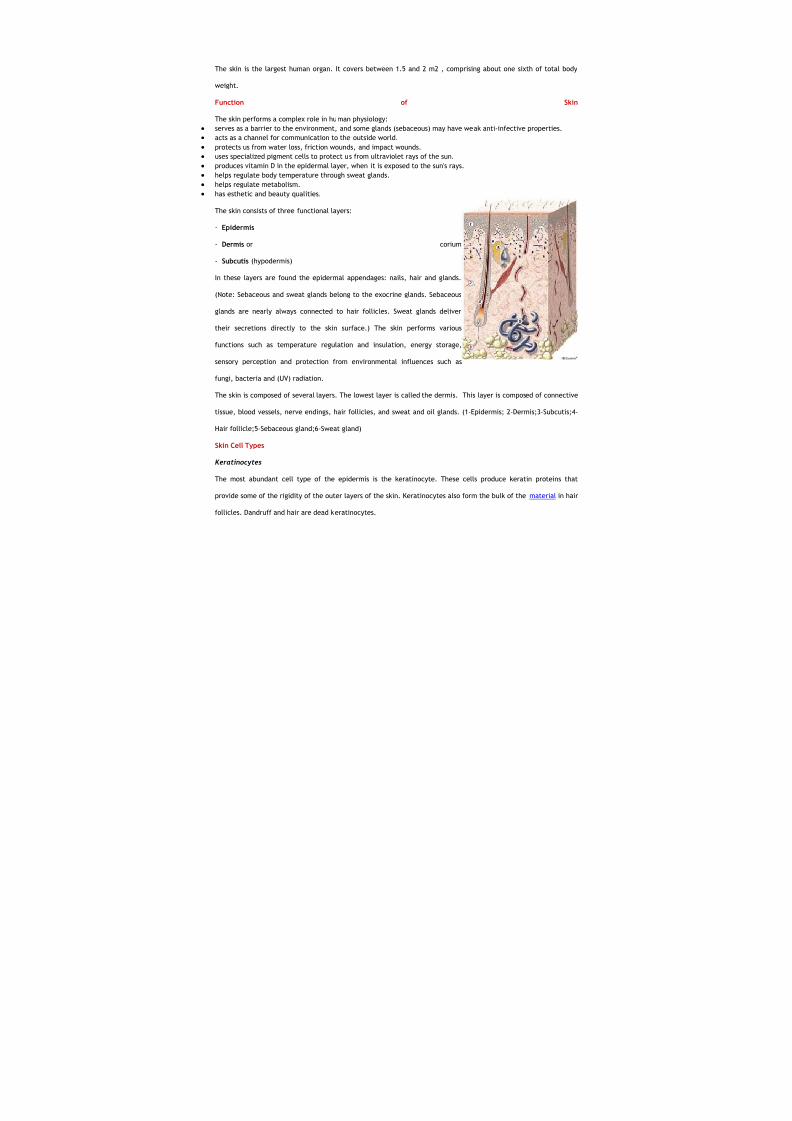
The skin consists of three functional layers:
- Epidermis
- Dermis or corium
- Subcutis (hypodermis)
In these layers are found the epidermal appendages: nails, hair and glands.
(Note: Sebaceous and sweat glands belong to the exocrine glands. Sebaceous
glands are nearly always connected to hair follicles. Sweat glands deliver
their secretions directly to the skin surface.) The skin performs various
functions such as temperature regulation and insulation, energy storage,
sensory perception and protection from environmental influences such as
fungi, bacteria and (UV) radiation.
The skin is composed of several layers. The lowest layer is called the dermis. This layer is composed of connective
tissue, blood vessels, nerve endings, hair follicles, and sweat and oil glands. (1-Epidermis; 2-Dermis;3-Subcutis;4-
Hair follicle;5-Sebaceous gland;6-Sweat gland)
Skin Cell Types
Keratinocytes
The most abundant cell type of the epidermis is the keratinocyte. These cells produce keratin proteins that
provide some of the rigidity of the outer layers of the skin. Keratinocytes also form the bulk of the material in hair
follicles. Dandruff and hair are dead keratinocytes.
8/4/2019 Kat Skin Grp 8
http://slidepdf.com/reader/full/kat-skin-grp-8 2/30
Fibroblasts
The dermis is produced largely by fibroblasts, which during embryonic development are part of the mesenchyme.
The fibroblasts produce the collagens and elastins that make skin very durable, from within.
Melanocytes
Melanocytes are cells in low abundance in the epidermis that produce the pigment melanin. The pigment made in
melanocytes is transferred to the cells of the hair or epidermis. The melanin granules are injected into (or
ingested by) the keratinocyte cells. There, the melanin granules accumulate around the nucleus of each
keratinocyte.
Melanin absorbs harmful ultraviolet (UV) light before the UV radiation can reach the nucleus. Melanin protects the
DNA in the nucleus from UV radiation damage. When melanin is produced and distributed properly in the skin,
dividing cells are protected from mutations that might otherwise be caused by harmful UV light.
Differences in skin color are due mostly to differences in the types and amount of pigment in our
keratinocytes. Skin darkening (tanning) from sun exposure is caused by the movement of existing melanin into
keratinocytes, and by increased production of melanin by the melanocyte.
During embryonic development these cells migrate from the neural crest into the skin.
Langerhans cells
These are star-shaped resident immune cells, macrophages. A macrophage is a cell that protects your body from
injury or illness. Macrophages break up or destroy (phagocytise) the invading organisms. These macrophages
process the invading organisms and present antigens to the T-lymphocytes. The T-lymphocytes are immune-system
cells which ultimately identify a substance as foreign or dangerous to the body.
Merkel's Cells
Only a few of these cells are present in skin; they are more numerous in the palms and soles (feet). These cells are
probably sensory mechanical receptors that respond to stimulus, such as pressure or touch.
Schematic Drawing of Human Skin
8/4/2019 Kat Skin Grp 8
http://slidepdf.com/reader/full/kat-skin-grp-8 3/30
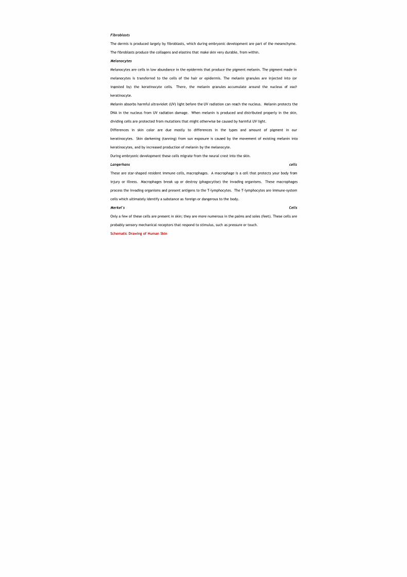
Drawing (transverse section) of human skin
illustrates the epidermis, basement membrane,
dermis, capillaries and major cellular components.
A: Epidermis || B: Dermis || C: Cornified layer of
keratinocytes (stratum corneum) || D: Suprabasal
keratinocytes || E: Basal layer of keratinocytes
(stratum basale) || F: Basement membrane G:
Collagen fibers in dermis || H: Capillary (enclosed
by a single microvascular endothelial cell) || I:
Melanocyte || J: Dermal Fibroblast
The great majority of cells in the epidermis are keratinocytes, which are arranged in stratified layers. At the
dermal-epidermal junction is a single layer of keratinocytes with a small number of interspersed melanocytes
(approximately 1/30) called the stratum basale. This basal layer of keratinocytes is also called the stratum
germinativum, because it is where new keratinocytes are generated by cell proliferation. Three types of
keratinocytes in the stratum basale have been defined by kinetic analysis: stem cells, transient-amplifying cells
and committed cells. Stem cells, which represent ~ 10% of the basal cell population, generate daughter cells from
mitosis that are either stem cells themselves or transient-amplifying cells. Transient-amplifying cells, which
represent ~40% of the basal cell population, replicate with much higher frequency than stem cells, but are capable
of only a few population doublings. Transient-amplifying cells produce daughter cells that are committed to
terminally differentiate. These committed cells detach from the basementmembrane, differentiate, and
ultimately cease to proliferate as they migrate toward the skin surface, where they are sloughed off as dead,
cornified cells called squames.
Keratinocyte stem cells (like stem cells from other tissues) are relatively undifferentiated, both biochemically and
histologically. Although keratinocyte stem cells have a high capacity for cell division, they divide with much lower
frequency than transient-amplifying cells. Thus, when labeled with 3H-thymidine, stem cells retain nuclear label
for long periods of time compared to transient-amplifying cells. Therefore, stem cells have been described as
"label-retaining" cells. Because stem cells are undifferentiated, biochemical markers of stem cells are difficult to
8/4/2019 Kat Skin Grp 8
http://slidepdf.com/reader/full/kat-skin-grp-8 4/30
identify. However, keratin 19 expression has been suggested as a marker of keratinocyte stem cells, based on
localization of keratin 19 expression to 3H-thymidine label-retaining cells. Keratinocyte stem cells may also
express higher amounts of the a2 and a3 integrins, because an approximate 1.5-fold increase in the expression of
these integrins has been observed in keratin 19-expressing cells relative to other epidermal basal cells. The
retention and expansion of keratinocyte stem cells in culture is thought to be essential for using keratinocytes in
ex vivo gene therapy.
The Epidermis
As the outermost skin layer, the epidermis forms the actual protective covering against environmental influences.
Its thickness averages 0.1 mm. On the face it is only 0.02 mm, while on the soles of the feet between 1 and 5 mm.
Though paper thin, the epidermis is composed of many layers of cells. In the basal layer (the living epidermis),
new cells are constantly being reproduced, pushing older cells to the surface. As skin cells move farther away from
their source of nourishment, they flatten and shrink. They lose their nuclei, move out of the basal layer to the
horny layer (the dead epidermis), and turn into a lifeless protein called keratin. After serving a brief protective
function, the keratinocytes are imperceptibly sloughed off. This process of a living cell's evolution, called
keratinization, takes about 4 weeks.
The epidermis consists of up to 90 percent keratinocytes, the actual epidermal cells or dead skin cells, that are
held together by what are called desmosomes. Keratinocytes function as a barrier, keeping harmful substances out
and preventing water and other essential substances from escaping the body. The other 10 percent of epidermal
cells are melanocytes, which manufacture and distribute melanin, the protein that adds pigment to skin and
protects the body from ultraviolet rays. Skin color is determined by the amount of protein produced by these cells,
not by the number of melanocytes, which is fairly constant in all races.
Hair and nails are specialized keratin structures and are considered part of the epidermis. While animals use fur
and claws for protection and defense, these corresponding structures are largely cosmetic in humans. The skin,
however, is uniquely human, since it can betray emotion by blushing (embarrassment), turning red (anger),
blanching (fear), sweating (tension), and forming goosebumps (terror).
On the skin surface are the sweat gland pores (100-200/cm2) and the openings of the sebaceous glands (50-
100/cm2). Their secretions ensure skin moisture and oiliness, and thus maintain the hydrolipid film. The epidermis
itself has no blood vessels, so the nutrients are supplied through the fine blood vessels in the dermal papillae.
The epidermis is differentiated into five layers:
8/4/2019 Kat Skin Grp 8
http://slidepdf.com/reader/full/kat-skin-grp-8 5/30
- Horny layer (stratum corneum)
- Clear layer (stratum lucidum)
- Granular layer (stratum granulosum)
- Prickle-cell layer (stratum spinosum)
- Basal layer (stratum basale)
Differentiation and skin regeneration
Through differentiation, the living, cylindrical
basal cells lose their nuclei and become
flattened cornified cells, changing their shape
and composition in the process. The cells pass
through the barrier zone, the border zone
between the living epidermal layers and the
horny layer, where the epidermal lipids are
released.
Did you know that 90% of household dust is dead skin cells? Keratinocytes contain structural protein (keratin) and
become progressively flattened as they advance upward from the basal layer to the corneal layer. The epidermis
renews itself every 28 days through continual reproduction, differentiation / cornification and desquamation
(mechanical sloughing-off of the uppermost horny cell layer).
The epidermis is a stratified squamous epithelial tissue. This means that it has several layers of epithelial cells and
that its outermost layer is made up of squamous (flat) epithelial cells.
Mitotic Activity: The layer adjacent to the dermis is known as the basal layer. The basal layer is made up of
columnar epithelial cells. Since all of the mitotic (cell-multiplying) activity of the epidermis occurs in the basal
layer, the basal layer is often called the germinative layer. This mitotic activity involves about 4 percent of the
cells in the basal layer at any given time. It occurs primarily between midnight and 0400 hours.
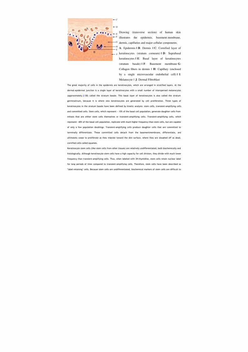
Migration of Cells to the Surface:
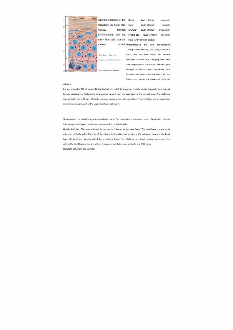
Schematic diagram of the
epidermis: the basal cells
change, through
differentiation, into flat
horny skin cells that are
without nuclei.
1 Horny layer / 2 Clear layer
3 Granular layer /4 Prickle-cell layer
5 Basal layer / 6 Basal membrane
8/4/2019 Kat Skin Grp 8
http://slidepdf.com/reader/full/kat-skin-grp-8 6/30
Over a period of weeks, new cells gradually migrate from the
basal layer to the surface. During this migration to the surface,
the cells change in shape from the original columnar to
cuboidal and then finally to squamous. As the cells
become squamous in form, they also become hardened, or
cornified, through the development of a special type of
protein. As they approach the surface, they die. Thus, the
outermost layers of the epidermis are dead, horny scales.
Keratinocytes
Keratinocytes are stratified, squamous, epithelial cells which comprise skin and mucosa, including oral,
esophageal, corneal, conjunctival, and genital epithelia. Keratinocytes provide a barrier between the host and the
environment. They prevent the entry of toxic substances from the environment and the loss of important
constituents from the host. Keratinocytes differentiate as they progress from the basal layer to the skin surface.
The normal turnover time for keratinocytes is around 30 days but epidermal turnover may be accelerated in some
skin diseases such as psoriasis.
Keratinocyte stem cells reside in the basal layer. These cells have a low rate of mitosis and give rise to a
population of transient amplifying cells. (Figure 1) Transient amplifying cells go through a limited number of
divisions, differentiate, and move up in the epidermis. The cells above the basal layer are known as the spinous
layer. Under routine microscopy small bridges, resembling spines, can be seen between the keratinocytes which
represent intercellular adhesion complexes known as desmosomes. As the cells further differentiate, they
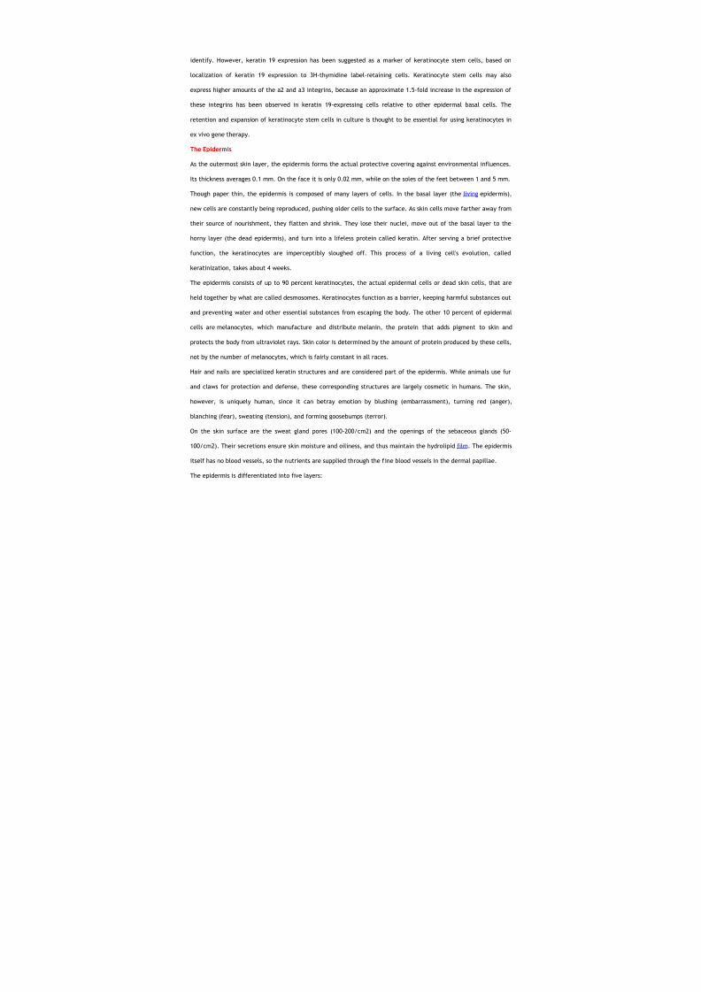
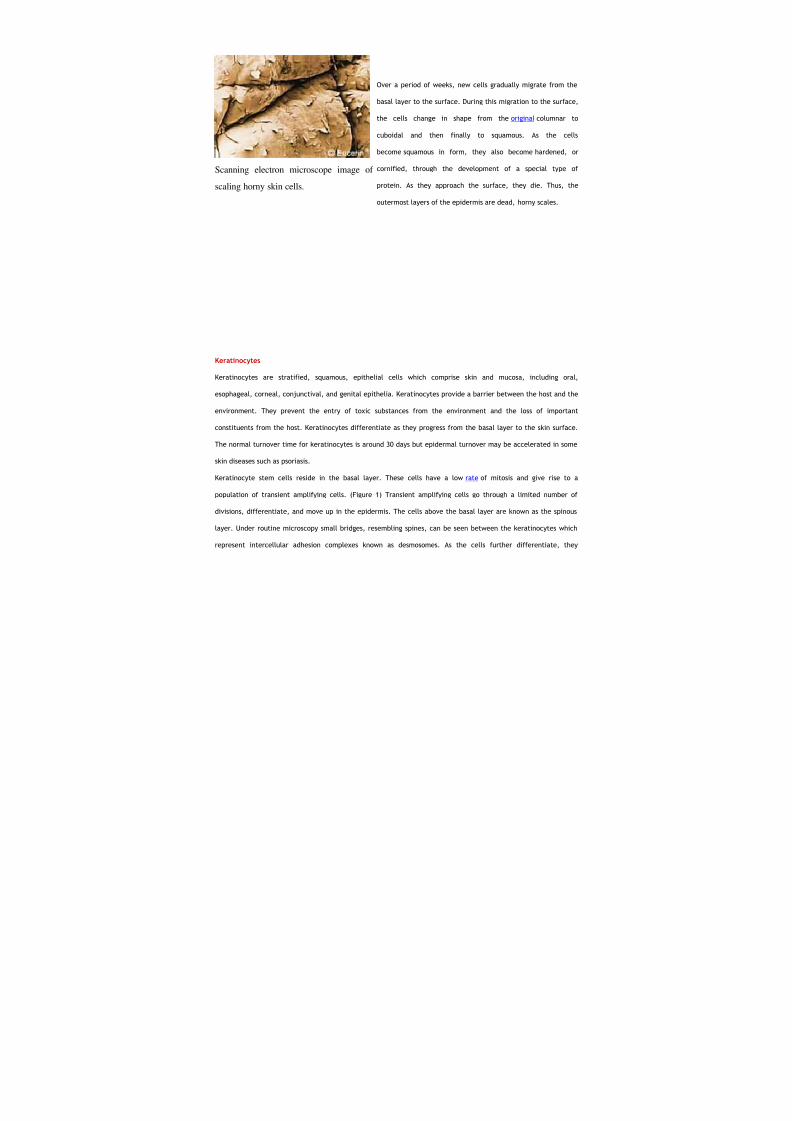
Scanning electron microscope image of
scaling horny skin cells.
8/4/2019 Kat Skin Grp 8
http://slidepdf.com/reader/full/kat-skin-grp-8 7/30
synthesize keratohyaline granules, a prominent feature of cells in the granular layer. Proteins synthesized in the
granular layer are important in the final stages of epidermal differentiation and include profilagrin, loricrin,
involucrin, and cornifin. These molecules are important in the formation of the stratum corneum, the outer most
layer of the epidermis.
Keratin
Electron microscopical examination of cells from all tissues reveals that they contain a complex, heterogenous,
intracytoplasmic system of filaments. The components of this system include actin, myosin, and tubulin, whose
diameters average approximately 60A°, 150A°, and 250A°, respectively. In addition, other intracytoplasmic
filaments were noted, and since the diameter of these latter structures was found to be between 70 and 100A°,
they were called intermediate filaments.
Intermediate filaments form a major part of the cytoskeleton of most cells and fulfill a variety of roles related to
cell shape, spatial organization, and perhaps informational transfer. The nucleus contains structures related to
these intermediate filaments and many intracellular components including polyribosomes, mitochondria, nucleic
acids, enzymes, and cyclic nucleotides are attached to the cytoskeleton.
Based on their biochemical, biophysical, and antigenic properties, a number of classes of intermediate filaments
can be recognized in different cell types: desmin (skeletin) in muscle cells, glial fibrillary acidic filaments in glial
cells, neurofilaments in neurons, vimentin in mesenchymal cells, and keratin in epithelial cells. In cultured
epidermal cells, keratins account for up to 30% of the cellular protein, while in stratum corneum, keratin accounts
for up to 85% of the cellular protein.
At least 19 keratin proteins can be identified ranging in molecular weight from approximately 40,000 to 68,000
micrograms. Moll and his coworkers published their human keratin catalogue in 1982. According to this catalogue,
there are two keratin subfamilies. The molecular weight of the members of one (the basic subfamily) is relatively
larger than that of the members of the other (the acidic subfamily). Each of the keratins is the product of a unique
gene and, in essentially all situations, the keratins are expressed as pairs containing one member of each
subfamily. The two members of each pair are in the same size rank order within their respective family, e.g., the
largest acidic keratin is expressed with the largest basic.
The type of keratin differs in different tissues, i.e, there are different types of keratin for keratinized epidermis,
hyperproliferative epidermis of palms and soles, corneal epithelium, stratified epithelium of the esophagus and
8/4/2019 Kat Skin Grp 8
http://slidepdf.com/reader/full/kat-skin-grp-8 8/30
cervix, and simple epithelium of the epidermal glands. As mentioned before, keratin is the main structural protein
of the epidermis.
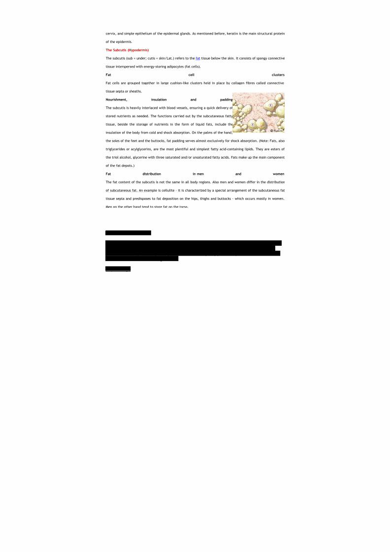
The Subcutis (Hypodermis)
The subcutis (sub = under; cutis = skin/Lat.) refers to the fat tissue below the skin. It consists of spongy connective
tissue interspersed with energy-storing adipocytes (fat cells).
Fat cell clusters
Fat cells are grouped together in large cushion-like clusters held in place by collagen fibres called connective
tissue septa or sheaths.
Nourishment, insulation and padding
The subcutis is heavily interlaced with blood vessels, ensuring a quick delivery of
stored nutrients as needed. The functions carried out by the subcutaneous fatty
tissue, beside the storage of nutrients in the form of liquid fats, include the
insulation of the body from cold and shock absorption. On the palms of the hand,
the soles of the feet and the buttocks, fat padding serves almost exclusively for shock absorption. (Note: Fats, also
triglycerides or acylglycerins, are the most plentiful and simplest fatty acid-containing lipids. They are esters of
the triol alcohol, glycerine with three saturated and/or unsaturated fatty acids. Fats make up the main component
of the fat depots.)
Fat distribution in men and women
The fat content of the subcutis is not the same in all body regions. Also men and women differ in the distribution
of subcutaneous fat. An example is cellulite - it is characterized by a special arrangement of the subcutaneous fat
tissue septa and predisposes to fat deposition on the hips, thighs and buttocks - which occurs mostly in women.
Men on the other hand tend to store fat on the torso.
Functions of the Skin
The skin protects the body. The skin protects the body from water loss. The skin isinvolved in the production of vitamin D from precursors with the aid of sunlight.There are many sensory receptors in the skin: pain, pressure, fine touch. The skinis also involved in heat regulation.
Top of Page

8/4/2019 Kat Skin Grp 8
http://slidepdf.com/reader/full/kat-skin-grp-8 9/30
Types of Skin
Thick skin is found on the palms of the hand and the sole of the feet. Thin skin isfound everywhere else.
Top of Page
Layers of the Skin
The skin is composed of two layers: the epidermis and the dermis. Underneaththese layers lies the hypodermis (subcutaneous tissue). The hypodermis is a layerof loose connective tissue.
Top of Page
Histology of the Epidermis
The epidermis is formed by stratified squamous epithelium. Keratinization is seen inthe epidermis. Keratinocytes, melanocytes Merkel cells and Langerhans cells are allfound in the epidermis. The keratinocyte is the most abundant cell in the epidermis.The melanocyte produces melanin, which is responsible for skin pigmentation. TheMerkel cell is a mechanoreceptor. The Langerhans cell is a phagocyte. Langerhanscells are macrophages seen in the skin.
The epidermis is divided into five layers: stratum basale, stratum spinosum,stratum granulosum, stratum lucidum, and stratum corneum.
Stratum Basale
The stratum basale contains the dividing cells. This layer is also called the stratumgerminativum.
Stratum Spinosum
The stratum spinosum consists of a layer several cells deep. The cells have pointyor spiny processes on them.
Stratum Granulosum
The cells in the stratum granulosum contain keratohyaline granules.
Stratum Lucidum
The stratum lucidum is present only in thick skin.
Stratum Corneum
8/4/2019 Kat Skin Grp 8
http://slidepdf.com/reader/full/kat-skin-grp-8 10/30
The stratum corneum is the outermost layer. It is also called the horny layer. Thecells in this layer are essentially bags of keratin. They contain no nuclei ororganelles.
Histology hint from Sarah Bellham: The epithelium is classified as "squamous"based on the cells of the surface layer.
Top of Page
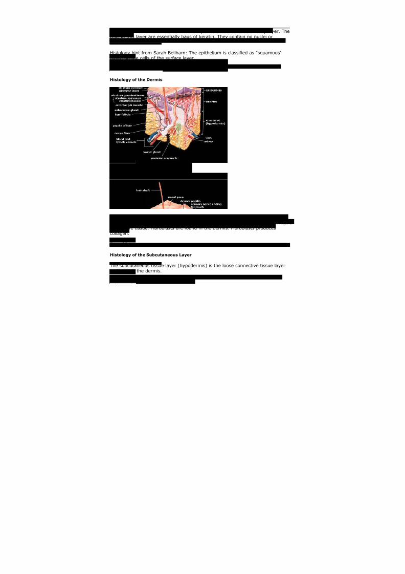
Histology of the Dermis
Beneath the epidermis is the dermis. The dermis is composed of a papillary layerand a reticular layer. The reticular layer of the dermis is made up of dense irregularconnective tissue. Fibroblasts are found in the dermis. Fibroblasts producescollagen.
Top of Page
Histology of the Subcutaneous Layer
The subcutaneous tissue layer (hypodermis) is the loose connective tissue layerunderneath the dermis.
Top of Page

8/4/2019 Kat Skin Grp 8
http://slidepdf.com/reader/full/kat-skin-grp-8 11/30
Receptors in the Skin
There are several different sensory receptors in the skin.
Ruffini endings, pacinian corpuscles, meissner's corpuscles, and merkel cells are allencapsulated sensory receptors. Free nerve endings are not encapsulated.
The most abundant sensory receptor are the free nerve endings. Free nerveendings respond to pain and temperature. Ruffini's corpuscles respond tocontinuous pressure. Pacinian corpuscles respond to vibration and rapidly changingpressure. Krause's end bulbs are a receptor for fine touch which are located inmucous membranes and the tongue. Meissner's corpuscles are also a receptor forfine touch but they are located in the dermis. Pacinian corpuscles are pressurereceptors in the skin.
Top of Page
Skin Appendages
Sweat glands, hair, nails and sebaceous glands are all considered epidermalappendages.
Nails
The lunula is the half moon shaped white area on a nail. The anatomical term forthe cuticle is the eponychium. The matrix is the region of the nails where there aredividing cells and nail growth. The nail plate rests on the nail bed. The nail root isthe proximal portion of the nail that is underneath skin.
Ceruminous Glands
The ceruminous glands of the ear are apocrine sweat glands.
Glands of Moll
The glands of Moll in the eyelid are apocrine sweat glands.
Histology of Sweat Glands
Sweat gland are also called sudoriferous glands.
8/4/2019 Kat Skin Grp 8
http://slidepdf.com/reader/full/kat-skin-grp-8 12/30
Classification of Sweat Glands
Sweat glands are divided into apocrine and eccrine. Apocrine sweat glands arefound on the areola, external genitalia, axilla, and curcumanal region. Eccrine sweatglands are distributed over most of the body.
Innervation of Sweat Glands
Eccrine sweat glands are innervated by the sympathetic nervous system. Theneurotransmitter for the eccrine sweat glands is acetylcholine. Thus, it ischolinergic.
Histology hint from Sarah Bellham: For most postganglionic sympathetic neurons,
the neurotransmitter isnorepinephrine. Eccrine sweat glands are an exception to this generalization, as theinnervation for eccrine sweat glands is cholinergic sympathetic.
Apocrine sweat glands are innervated by the sympathetic nervous system. Theneurotransmitter for theapocrine sweat glands is norepinephrine. Thus, it is adrenergic.
Hair
Hair is present over most of the body. It is not found on the palms of the hand,soles of the feet, urogenital openings, and lips. Huxley's layer is a layer in the hairfollicle. Henle's layer is a layer in the hair follicle.
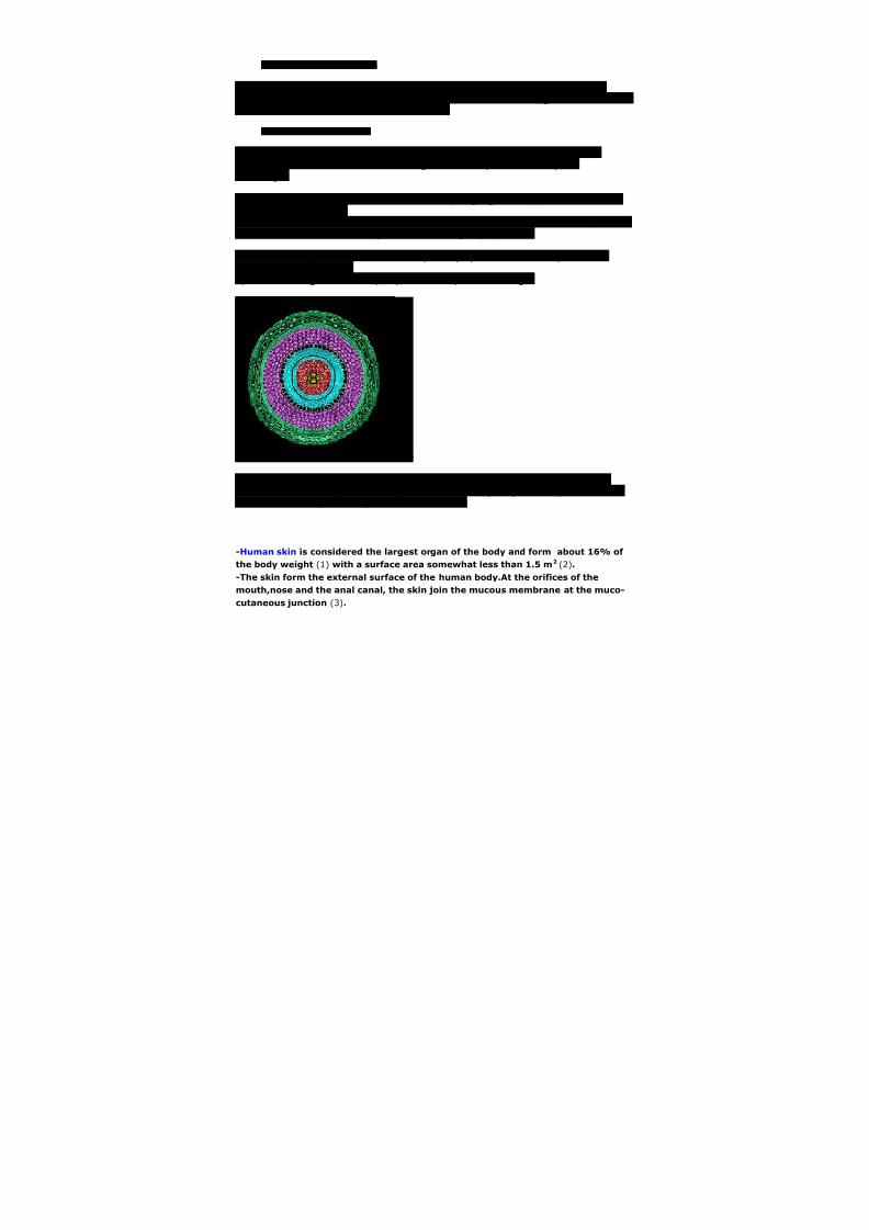
-Human skin is considered the largest organ of the body and form about 16% of
the body weight (1) with a surface area somewhat less than 1.5 m2 (2).
-The skin form the external surface of the human body.At the orifices of the
mouth,nose and the anal canal, the skin join the mucous membrane at the muco-
cutaneous junction (3).
8/4/2019 Kat Skin Grp 8
http://slidepdf.com/reader/full/kat-skin-grp-8 13/30
-Types of human skin (1):-
1. Thick skin (Non-Hairy) which has thick epidermis and found only in the
palms and soles as they are the most sites subjected to abrasions and
trauma and thick skin shows characteristic parallel ridges and grooves
which are called "Finger prints".
2. Thin skin (Hairy) which has thin epidermis and covers the rest of the body.
-Histological structure of the skin :-
skin is composed of 3 layers
1. Epidermis
2. Dermis
3. Hypodermis (Subcutaneous fatty layer)
1- Epidermis:-
It is the outer superficial epithelial layer of skin.
It is composed of Stratified squamous keratinized epithelium.
The thickness of the epidermis varies in different types of skin. It is the
thinnest on the eyelids and the thickest on the palms(0.8mm) and
soles(1.4mm).
The epidermis is ectodermal in origin.
The epidermis is devoid of blood vessels and gets its nutrition through
diffusion.
-It consists of :-
Keratinocytes (85% of cells) which are responsible for formation of keratin,
Dendertic cells which are (melanocyte and the Langerhans cell)
Merkel cells which until recently, were not consistently identified as the
third dendritic cell in human epidermis, because light microscopy and low
power electron microscopy did not readily allow the differentiation of
merkel cells from the other two dendritic cells of the epidermis.
-Keratinocytes are arranged in 5 layers from above downward:-
Stratum basale (basal cell layer)(The stratum germinatum):- which is single
layer of columnar cells with basal oval nuclei,cells of this layer are resting
on basement membrane and attached to each other and with the overlying

8/4/2019 Kat Skin Grp 8
http://slidepdf.com/reader/full/kat-skin-grp-8 14/30
cells by inter-cellular bridges called "desmosomes" these basal cells
continue to divide throughout the life so they are called mother cells of
epidermis.
Stratum spinosum (prickle cell layer):- formed of 5-7 layers of nucleated
polygonal cells attached to each other by desmosomes which are like spines
hence its name.
Stratum granulosum (Granular cell layer):- formed of 2-3 layers of
nucleated spindle shaped cells which accumlate dense basophilic
keratohyalin granules,these granules contain lipids, which along with the
desmosomal connections, help to form a waterproof barrier that functions
to prevent fluid loss from the body.
Stratum licidum (Clear layer):- only well seen in thick epidermis and
represents a transition from the stratum granulosum to the stratum
corneum and formed of dead clear non nucleated containing eleidin
granules.
Stratum corneum (horny layer):- formed of flattened non nucleated cellscalled "squames" condensed in linear manner and containing Keratin which
derived from eleidin granules.These squames are continuously shed from
the surface and replaced from the deeper layer by new ones.
-Melanocytes:- they form melanin from tyrosine under the effect of tyrosinase
enzyme and lie just under and in between basal cells and in hair matrix.
-The Langerhans' cells:- present in Stratum spinosum,they act as macrophages.
-The Merkel's cell's:- present in Stratum basale and they act as receptors for touch
sensation.
2- Dermis:-
The dermis is typically subdivided into two zones, a papillary layer and a
reticular layer.
It is 15-40 more thicker than epidermis.
Function:- act as frame work and supports for nerves,lymphatics,hair
follicles,sweat glands,Sebaceous glands and blood vessels to supply the
avascular epidermis with nutrients.
The dermis contains mostly fibroblasts which are responsible for secreting
collagen, elastin and ground substance that give the support and elasticity
of the skin. Also present are immune cells that are involved in defense
against foreign invaders passing through the epidermis.
The dermis is mesodermal in origin.
8/4/2019 Kat Skin Grp 8
http://slidepdf.com/reader/full/kat-skin-grp-8 15/30
The papillary layer lies below and interdigitates with the epidermal rete
ridges,papillary layer is formed of loose C.T with fine collagenous fibers type
II,reticular and elastic fibers . and contains the free sensory nerve endings
and structures called Meissner's corpuscles in highly sensitive areas.
The reticular layer lies below papillary layer and is formed of dense C.T
with coarse collagenous fibers type I and some elastic fibers the fibers are
irregularly arranged,this layer contain Pacinian corpuscles.
3- Hypodermis:-
- Contains adipose tissue and this layer is rich in adipose tissue except in scrotum
and eyelids.
- Skin has 4 appendages :-
Hair. Nails.
Sweat Glands.
Sebaceous glands.
INTEGUMENTARY SYSTEM
The skin or cutis covers the entire outer surface of the body. Structurally, the skin
consists of two layers which differ in function, histological appearance and theirembryological origin. The outer layer orepidermis is formed by an epithelium and is of
ectodermal origin. The underlying thicker layer, the dermis , consists of connective tissue
and develops from the mesoderm. Beneath the two layers we find a subcutaneous layer
of loose connective tissue, the hypodermis or subcutis , which binds the skin to
underlying structures. Hair, nails and sweat and sebaceous glands are of epithelial
origin and collectively called the appendages of the skin .
The skin and its appendages together are called the integumentary system .
Suitable Slides
sections of skin - H&E, trichrome or van Gieson
8/4/2019 Kat Skin Grp 8
http://slidepdf.com/reader/full/kat-skin-grp-8 16/30
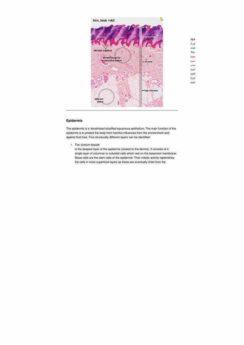
Epidermis
The epidermis is a keratinised stratified squamous epithelium . The main function of the
epidermis is to protect the body from harmful influences from the environment and
against fluid loss. Five structurally different layers can be identified:
1. The stratum basale is the deepest layer of the epidermis (closest to the dermis). It consists of a
single layer of columnar or cuboidal cells which rest on the basement membrane.
Basal cells are the stem cells of the epidermis. Their mitotic activity replenishes
the cells in more superficial layers as these are eventually shed from the
8/4/2019 Kat Skin Grp 8
http://slidepdf.com/reader/full/kat-skin-grp-8 17/30
epidermis. The renewal of the human epidermis takes about 3 to 4 weeks.
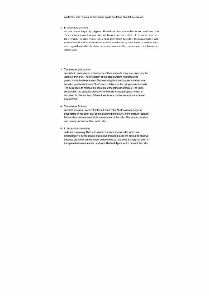
2. In the stratum spinosum,
the cells become irregularly polygonal. The cells are often separated by narrow, translucent clefts
These clefts are spanned by spine-like cytoplasmatic extensions of the cells (hence the name of
the layer and of its cells: spinous cells), which interconnect the cells of this layer. Spines of cells
meet end-to-end or side-to-side and are attached to each other by desmosomes. In addition to the
usual organelles of cells, EM shows membrane-boundlamellar granules in the cytoplasm of the
spinous cells.
3. The stratum granulosum
consists, in thick skin, of a few layers of flattened cells. Only one layer may be
visible in thin skin. The cytoplasm of the cells contains numerous fine
grains, keratohyalin granules . The keratohyalin is not located in membrane-
bound organelles but forms "free" accumulations in the cytoplasm of the cells.
The cells begin to release the contents of the lamellar granules. The lipids
contained in the granules come to fill the entire interstitial space, which is
important for the function of the epidermis as a barrier towards the external
environment.
4. The stratum lucidum
consists of several layers of flattened dead cells. Nuclei already begin to
degenerate in the outer part of the stratum granulosum. In the stratum lucidum,
faint nuclear outlines are visible in only a few of the cells. The stratum lucidum
can usually not be identified in thin skin.
5. In the stratum corneum ,
cells are completely filled with keratin filaments (horny cells) which are
embedded in a dense matrix of proteins. Individual cells are difficult to observe
because (1) nuclei can no longer be identified, (2) the cells are very flat and (3)
the space between the cells has been filled with lipids, which cement the cells

8/4/2019 Kat Skin Grp 8
http://slidepdf.com/reader/full/kat-skin-grp-8 18/30
together into a continuous membrane. In the EM, the cell membranes appear
thickened and interdigitate with those of neighbouring cells. Closest to the
surface of the epidermis, the stratum corneum has a somewhat looser
appearance. Horny cells are constantly shed from this part of the stratum
corneum.
The protection of the body by the epidermis is essentially due to the functional
features of the stratum corneum .
Variations in the thickness of the epidermis (~0.1 mm in thin skin, 1 mm or more in thick skin) are
mainly the result of variations in the thickness of the stratum corneum, although the
other layers also vary in thickness. Cells of the epidermis of the skin will at some time of
their life keratinise and are collectively also called keratinocytes .
Keratinisation should not be used as a synonym for the formation of the stratum corneum: other stratified squamous
epithelia may become keratinised but may not form a stratum corneum in which cells join to form a horny cell
membrane.
Suitable Slides
sections of skin - H&E, trichrome or van Gieson
Skin, thin - H&E and Skin, thick, trichrome
The most superficial part of the epidermis is formed by the stratum corneum. Nuclei are
not visible in this layer. Cell outlines may be visible at high magnification or, in the form
of artefacts, as cracks or clefts in the stratum corneum. The stratum granulosum is
formed by a single layer of very dark and flattened cells in thin skin. Several layers of
cells containing keratohyalin granules are visible in thick skin. Polyhedral cells with clear
outlines form the stratum spinosum. The stratum basale is formed by a single layer of
cuboidal or columnar cells and delimits the epidermis from the dermis.
At high magnification, the basal cytoplasm of the basal cells seem to interdigitate with the underlying dermis. Similar
to the dermal papilla, this irregular border at the cellular level, the dermal-epidermal junction , anchors individual basal
cells firmly to the underlying dermis.
Identify and draw the epithelium in thick and thin skin. Identify in your drawing as
many of the layers of the epidermis as possible.
8/4/2019 Kat Skin Grp 8
http://slidepdf.com/reader/full/kat-skin-grp-8 19/30
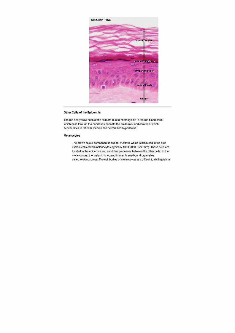
Other Cells of the Epidermis
The red and yellow hues of the skin are due to haemoglobin in the red blood cells,
which pass through the capillaries beneath the epidermis, and carotene, which
accumulates in fat cells found in the dermis and hypodermis.
Melanocytes
The brown colour component is due to melanin , which is produced in the skin
itself in cells called melanocytes (typically 1000-2000 / sqr. mm). These cells are
located in the epidermis and send fine processes between the other cells. In the
melanocytes, the melanin is located in membrane-bound organelles
called melanosomes . The cell bodies of melanocytes are difficult to distinguish in
8/4/2019 Kat Skin Grp 8
http://slidepdf.com/reader/full/kat-skin-grp-8 20/30
ordinary LM preparations, because the melanosomes are located mainly in the
processes of the cells.
Melanocytes can transfer melanin to keratinocytes - mainly to the basal cells. The fine processes o
melanocytes may invade keratinocytes and bud-off part of the melanocyte cytoplasm, including t
melanosomes, within the keratinocytes. Melanin protects the chromosomes of mitotically active b
cells against light-induced damage.
Pigmentation is not just under the control of light. Hormones produced by the pituitary and the ad
glands also affect pigmentation. Diseases of these two endocrine organs often result in changes o
pigmentation of the skin.
Although melanocytes are also ectodermal in origin, they are derived exclusively from the neural crest of the embryo, from where
migrate to all other parts of the body.
Langerhans Cells
are another cell type found within the epidermis. Morphologically they are not
unlike melanocytes, but functionally they are more closely related to
macrophages. They are important in immune reactions of the epidermis. Their
fine processes form a network between the cells of the epidermis and
phagocytose antigens which have entered the epidermis. Langerhans cells may
only be temporary residents of the skin. If they have come into contact with anantigen, they can migrate to regional lymph nodes, where they initiate an immune
response.
T-lymphocytes
are, like Langerhans cells, a group of cells functioning in the immune system. Some of them will be present
in the epidermis. Together with Langerhans cells they are sometimes referred to as SALT, i.e. skin-
associated lymphoid tissue.
Dermis
The dermis is the thick layer of connective tissue to which the epidermis is attached. Its
deepest part continues into the subcutaneous tissue without a sharply defined

8/4/2019 Kat Skin Grp 8
http://slidepdf.com/reader/full/kat-skin-grp-8 21/30
boundary. Its thickness is for this reason difficult to determine but 1-2 mm is a good
guestimate for "average" skin. The dermis may be divided into two sublayers (again without
a sharp boundary):
The papillary layer consists of loose, comparatively cell-rich connective tissue,which fills the hollows at the deep surface (dermal papillae ) of the epidermis.
Capillaries are frequent. Collagen fibres appear finer than in the reticular layer.
The reticular layer appears denser and contains fewer cells. Thick collagen
fibres (5-10 µm) often aggregate into bundles (up to 100 µm thick). The fibres form an
interlacing network, although their predominant direction is parallel to the surface
of the skin. A preferred orientation of the collagen fibres is not visible in the
sections, but the main orientation of the fibres differs in skin from different parts
of the body. Usually, their main orientation will follow the " lines of greatest
tension " in the skin (Kraissl lines). This is of some surgical importance since
incisions parallel to these lines will heal faster and with less formation of scar
tissue.
Kraissl lines have been defined in living humans. They not always coincide with the cleavage lines, which
Langer defined (Langer's cleavage lines ) about a century before Kraissl in cadavers.
Elastic fibres are found in both the papillary (fine fibres) and reticular (coarse fibres)
layers.
They can not be identified in H&E stained sections.
Suitable Slides
sections of skin - H&E, van Gieson
Van Gieson stained sections are particularly nice if the van Gieson stain has been combined with an elastin
stain.
Skin, thin - H&E and Skin, thick - van Gieson & elastin How easy it is to differentiate between the papillary and reticular layer of the dermis
depends on the preparation - you may have to look at several preparations. Immediately
beneath the epidermis you should see a layer which at low magnification appears rather
evenly stained. At high magnification the stain should resolve into a fine network of
collagen fibres, which blend with equally fine elastic fibres. Cells are more numerous in
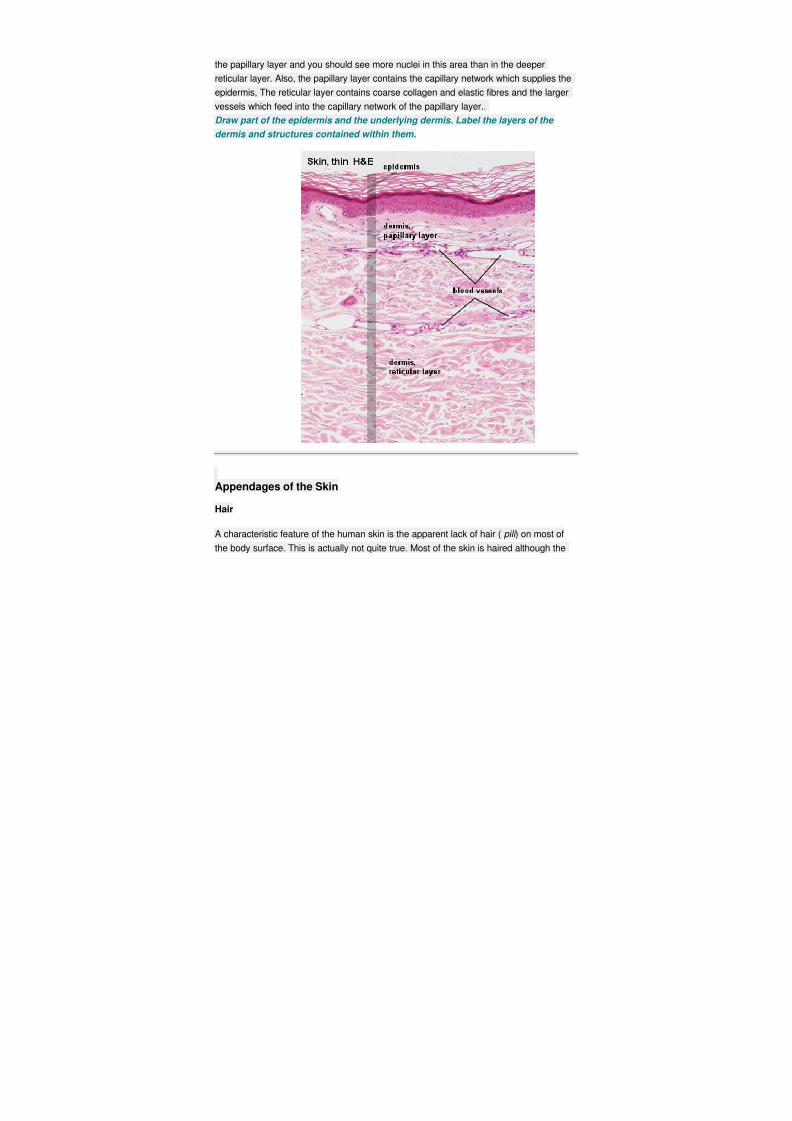
8/4/2019 Kat Skin Grp 8
http://slidepdf.com/reader/full/kat-skin-grp-8 22/30
the papillary layer and you should see more nuclei in this area than in the deeper
reticular layer. Also, the papillary layer contains the capillary network which supplies the
epidermis, The reticular layer contains coarse collagen and elastic fibres and the larger
vessels which feed into the capillary network of the papillary layer..
Draw part of the epidermis and the underlying dermis. Label the layers of the
dermis and structures contained within them.
Appendages of the Skin
Hair
A characteristic feature of the human skin is the apparent lack of hair (pili ) on most of
the body surface. This is actually not quite true. Most of the skin is haired although the
8/4/2019 Kat Skin Grp 8
http://slidepdf.com/reader/full/kat-skin-grp-8 23/30
hair in most areas is short, fine and only lightly pigmented. This type of hair is
called vellus hair .
Truly hairless are only the palms of hands and soles of feet, the distal phalanges and
sides of fingers and toes and parts of the external genitalia.
In those parts of the skin which we perceive as "hairy" we find terminal hairs . The free
part of each hair is called the shaft . The root of each hair is anchored in a tubular
invagination of the epidermis, the hair follicle , which extends down into the dermis and,
usually, a short distance into the hypodermis. The deepest end of the hair follicle forms
an enlargement, the bulb . Cells in the bulb are mitotically active. Their progeny
differentiates into the cell types which form the hair and the cells that surround its root,
the root sheath . Hair cells keratinise within the lower one-third of the hair follicle. Above
this level it is not possible to identify individual cells within the hair. Each hair follicle has
an associated bundle of smooth muscle, the arrector pili muscle . This muscle inserts
with one end to the papillary layer of the dermis and with the other end to the dermal
sheath of the hair follicle.
Hair growth is discontinuous . Hairs are lost and replaced by new ones. The hair follicle
goes through different stages that reflect the discontinuous hair growth. Anagen is the
phase of growth. The resting stage is called telogen . The length of the anagen is
variable in different regions of the body - lasting only a few months for hair of the
eyebrows and eyelashes but 2 to 5 years for hair of the scalp. Hair growth is controlled
by a number of hormonal and hereditary factors and their interactions.
Suitable Slides
Sections of hairy skin or scalp - H&E
With a few exceptions (thick skin and skin covering parts of the external genitalia), all skin sections should
contain a few hair follicles.
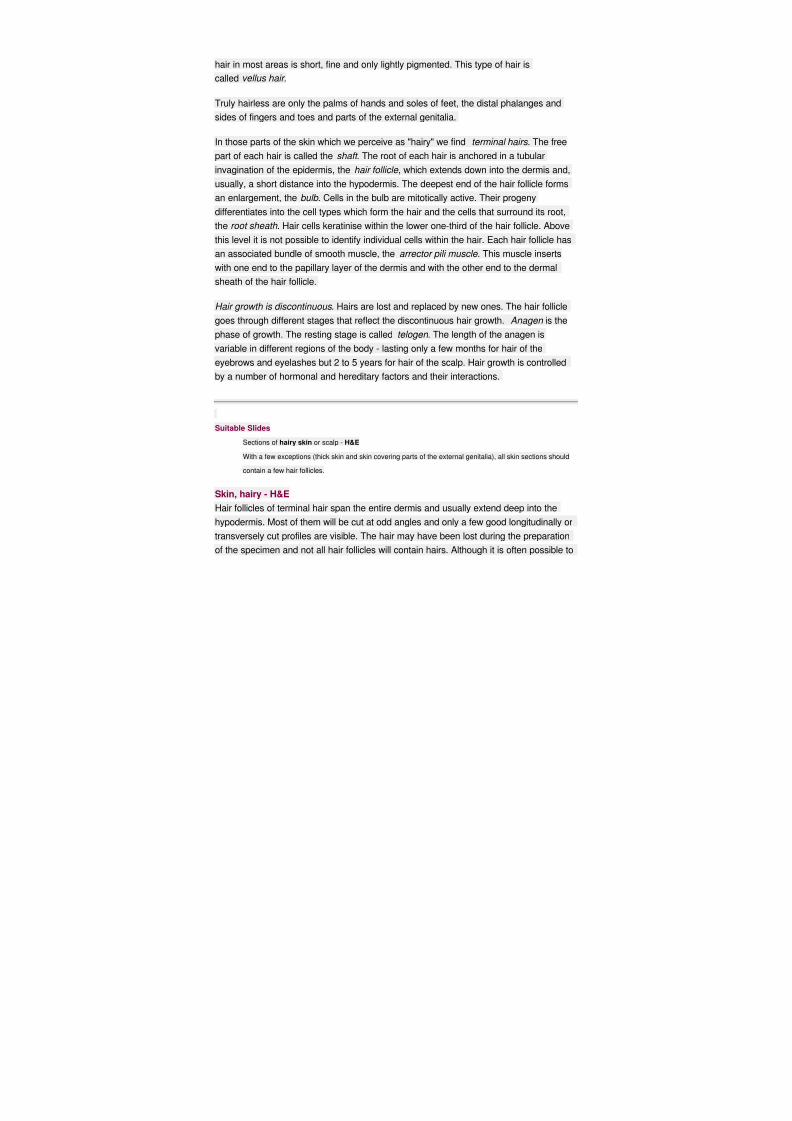
Skin, hairy - H&E
Hair follicles of terminal hair span the entire dermis and usually extend deep into the
hypodermis. Most of them will be cut at odd angles and only a few good longitudinally or
transversely cut profiles are visible. The hair may have been lost during the preparation
of the specimen and not all hair follicles will contain hairs. Although it is often possible to
8/4/2019 Kat Skin Grp 8
http://slidepdf.com/reader/full/kat-skin-grp-8 24/30
see the attachment of the arrector pili muscle into the hair follicle or the papillary layer of
the dermis, both attachments are hardly ever visible in the same section.
Draw a hair follicle at low magnification. Try to draw a composite from several
hair follicles and associated structures, which captures their appearance from the
bulb to the epidermis.
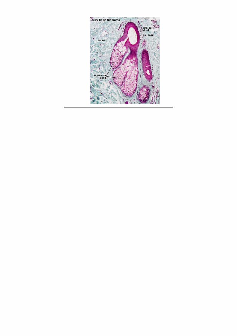
Sebaceous Glands
Sebaceous glands empty their secretory product into the upper parts of the hair follicles.
They are therefore found in parts of the skin where hair is present. The hair follicle and
its associated sebaceous gland form a pilosebaceous unit .
8/4/2019 Kat Skin Grp 8
http://slidepdf.com/reader/full/kat-skin-grp-8 25/30
Sebaceous glands are also found in some of the areas where no hair is present, for
example, lips, oral surfaces of the cheeks and external genitalia.
Sebaceous glands are as a rule simple and branched (Remember the nomenclature of
glands!). The secretory portion consists of alveoli . Basal cells in the outermost layer of thealveolus are flattened. Basal cells are mitotically active. Some of the new cells will
replenish the pool of basal cells, while the remaining cells are displaced towards the
centre of the alveolus as more cells are generated by the basal cells. The secretory
cells will gradullay accumulate lipids and grow in size. Finally their nuclei disintegrate,
and the cells rupture. The resulting secretory product of lipids and the constituents of
the disintegrating cell is aholocrine secretion .
The lipid secretion of the sebaceous glands has no softening effect on the skin, and it
has only very limited antibacterial and antifungoid activity. Its importance in humans is
unclear. Clinically the sebaceous glands are important in that they are liable to
infections (e.g. with the development of acne).
Suitable Slides
slides of hairy skin or thin skin - H&E, trichrome, van Gieson
Skin, hairy - trichrome, H&E
Sebaceous glands will be present in all types of skin other than thick skin. Their numbers should correlate with the
number of hair follicles. If your section does not contain hair follicles you are unlikely to see a good sebaceous
gland. Sebaceous glands are usually embedded in the dermis. Although they empty into
the hair canal of the hair follicle, this point will only be visible for a few of them because
of the thinness of the sections. It should however be possible to follow the fate of the
secretory cells. Deep in the sebaceous glands cells are smaller with intact nuclei. Cell
size increases with the accumulation of sebum as the cells are gradually displaced
towards the opening of the gland into the hair follicle. The nuclei condense, becomedarker and irregularly shaped.
Draw a sebaceous gland. Emphasise the appearance of the secretory cells in
different parts of the gland. If possible include part of the associated hair follicle.
8/4/2019 Kat Skin Grp 8
http://slidepdf.com/reader/full/kat-skin-grp-8 26/30
8/4/2019 Kat Skin Grp 8
http://slidepdf.com/reader/full/kat-skin-grp-8 27/30
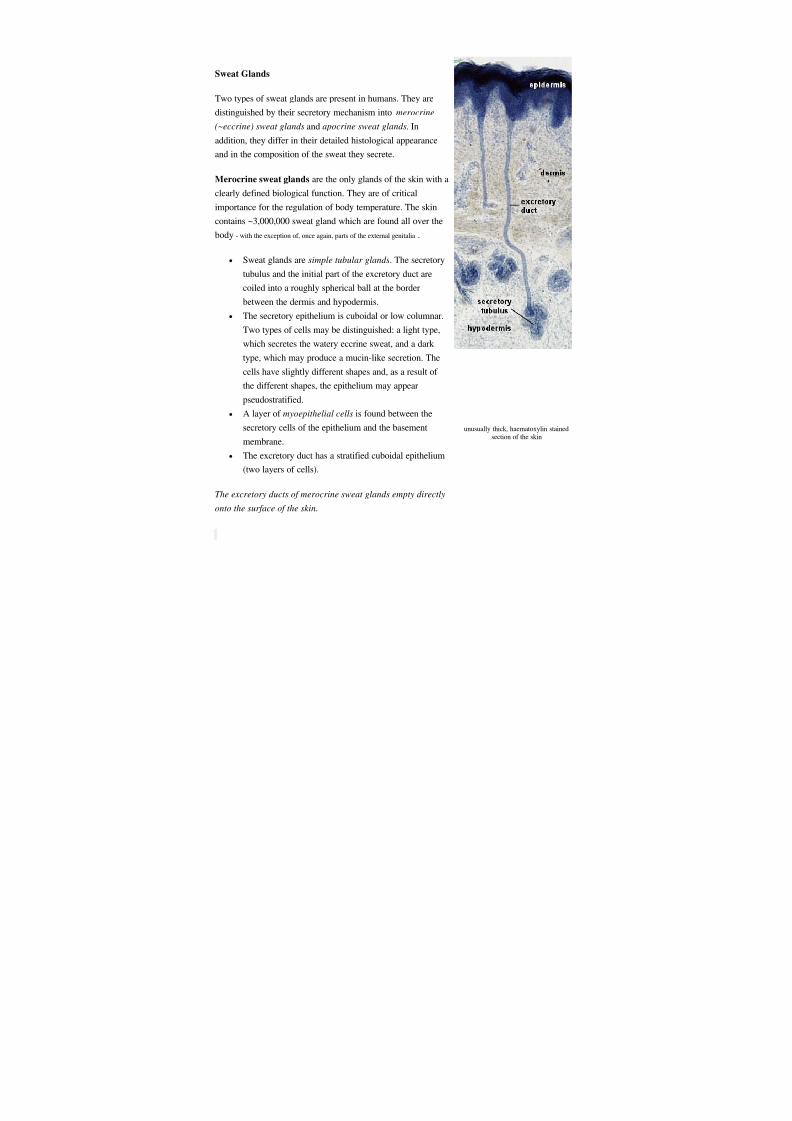
Sweat Glands
Two types of sweat glands are present in humans. They are
distinguished by their secretory mechanism into merocrine
(~eccrine) sweat glands and apocrine sweat glands. In
addition, they differ in their detailed histological appearance
and in the composition of the sweat they secrete.
Merocrine sweat glands are the only glands of the skin with a
clearly defined biological function. They are of critical
importance for the regulation of body temperature. The skin
contains ~3,000,000 sweat gland which are found all over the
body - with the exception of, once again, parts of the external genitalia .
Sweat glands are simple tubular glands. The secretory
tubulus and the initial part of the excretory duct are
coiled into a roughly spherical ball at the border
between the dermis and hypodermis.
The secretory epithelium is cuboidal or low columnar.
Two types of cells may be distinguished: a light type,
which secretes the watery eccrine sweat, and a dark
type, which may produce a mucin-like secretion. The
cells have slightly different shapes and, as a result of
the different shapes, the epithelium may appear
pseudostratified.
A layer of myoepithelial cells is found between the
secretory cells of the epithelium and the basement
membrane.
The excretory duct has a stratified cuboidal epithelium
(two layers of cells).
The excretory ducts of merocrine sweat glands empty directly
onto the surface of the skin.
unusually thick, haematoxylin stainedsection of the skin
8/4/2019 Kat Skin Grp 8
http://slidepdf.com/reader/full/kat-skin-grp-8 28/30
Apocrine sweat glands occur in, for example, the axilla. They are stimulated by sexual
hormones and are not fully developed or functional before puberty. Apocrine sweat is a
milky, proteinaceous and odourless secretion. The odour is a result of bacterial
decomposition and is, at least in mammals other than humans, of importance for sexual
attraction.
The histological structure of apocrine sweat glands is similar to that of merocrine sweat
glands, but the lumen of the secretory tubulus is much larger and the secretory
epithelium consists of only one major cell type, which looks cuboidal or low columnar.
Apocrine sweat glands as such are also much larger than merocrine sweat glands.
The excretory duct of apocrine sweat glands does not open directly onto the surface of
the skin. Instead, the excretory duct empties the sweat into the upper part of the hair
follicle . Apocrine sweat glands are therefore part of the pilosebaceous unit.
Some texts argue that the apocrine sweat glands use a merocrine or a combined merocrine / apocrine secretory
mechanism.
Suitable Slides
merocrine sweat glands: sections of thick skin or thin skin - H&E
apocrine sweat glands: sections of skin from the areolae (pigmented skin surrounding the nipples), the
axilla (arm pit) or skin covering the external genitalia - H&E
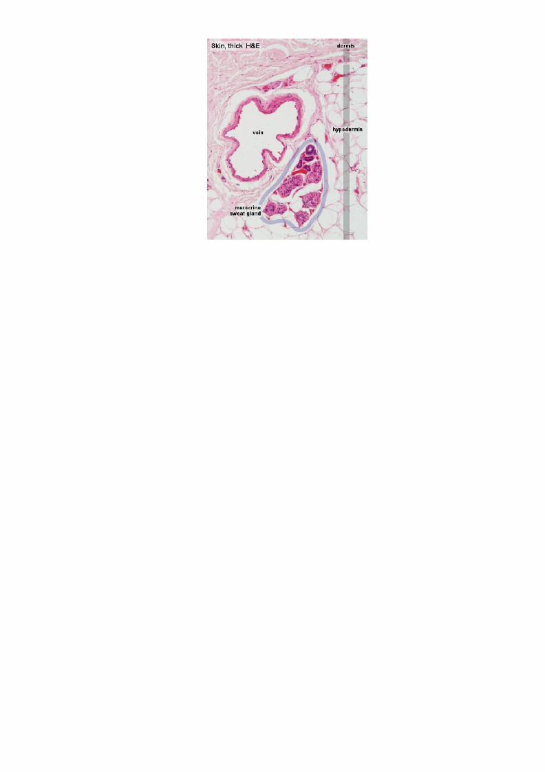
Skin, thick - H&E
Scan along the border between dermis and hypodermis and locate a sweat gland. The
secretory tubulus and the initial segment of the duct usually form a cluster of round or
irregularly shaped profiles, which stain darker than the surrounding connective tissue.
The structural preservation of the sweat glands may vary quite a bit in the different
preparations. The different cell types in the secretory epithelium of merocrine sweat
glands are only visible in well preserved glands. The red rim around the secretory
tubulus is formed by the cytoplasm of myoepithelial cells. Their small, dark nuclei may
be visible close to the periphery of the tubulus.
Draw a small schematic illustrating the relative position of the sweat gland in the
skin. Identify and draw the secretory tubulus and excretory duct. Label as many
features as can be identified.
8/4/2019 Kat Skin Grp 8
http://slidepdf.com/reader/full/kat-skin-grp-8 29/30
8/4/2019 Kat Skin Grp 8
http://slidepdf.com/reader/full/kat-skin-grp-8 30/30
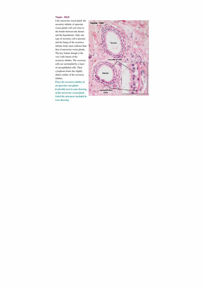
Nipple - H&E
Like merocrine sweat gland, the
secretory tubulus of apocrine
sweat glands will coil close to
the border between the dermis
and the hypodermis. Only one
type of secretory cell is present,
and the lining of the secretory
tubulus looks more uniform than
that of merocrine sweat glands.
The key feature though is the
very wide lumen of the
secretory tubulus. The secretorycells are surrounded by a layer
of myoepithelial cells. Their
cytoplasm forms the slightly
darker outline of the secretory
tubulus.
raw the secretory tubulus of
an apocrine seat gland -
referably next to your drawing
of the merocrine sweat gland.
Label the structures included in
our drawing.