iridoviridae sabah, malaysia - ijcmas.com abd razak, et al.pdf · sabah, malaysia asrazitah abd...
TRANSCRIPT

Int.J.Curr.Microbiol.App.Sci (2014) 3(3): 896-909
896
Original Research Article
First report of Megalocytivirus (Iridoviridae) in grouper culture in Sabah, Malaysia
Asrazitah Abd Razak1, Julian Ransangan1* and Ahemad Sade2
1Microbiology and Fish Disease Laboratory, Borneo Marine Research Institute, Universiti Malaysia Sabah, Jalan UMS, 88400, Kota Kinabalu, Sabah, Malaysia
2Fisheries Department Sabah, Wisma Pertanian, Jalan Tasek, 88628 Kota Kinabalu, Sabah, Malaysia
*Corresponding author
A B S T R A C T
Introduction
Groupers are popular aquaculture fish species in Sabah, Malaysia. They attain high market demand both in local and international markets and constitute the top menus for restaurants, hotels and resorts, especially during festive seasons.
However, the supply of groupers from aquaculture is often limited due to diseases, which occur throughout the production cycle (Muroga, 2001; Hyatt and Whittington, 2005; Bondad-Reantaso et al., 2005; Harikishnan et al., 2010).
ISSN: 2319-7706 Volume 3 Number 3 (2014) pp. 896-909 http://www.ijcmas.com
K e y w o r d s
Grouper; Megalo-cytivirus; ISKNV; nested-PCR; Sabah; Malaysia
Groupers are popular aquaculture species in Sabah, Malaysia. However, its aquaculture production is often limited by disease outbreaks. Although many diseases are known to affect groupers, iridovirus infection is a major concern because it causes high mortality within a short period of time. Recently, a disease resembled to iridovirus occurred and caused heavy losses to grouper aquaculture in Sabah. This has prompted us to conduct a study with the aim to determine if iridovirus present in the culture groupers. In this study, we examined 212 fish specimens, which represented all the major culture grouper species in Malaysia. The examination was carried out using single- and nested-PCR methods and followed by DNA sequencing. Two genes (major capsid protein and ATPase) were targeted for the PCR amplification and DNA sequencing. The finding showed 15.6% (33/212) of the grouper specimens were severely infected by iridovirus. Meanwhile, 17.4% of the specimens exhibited latent infection or asymptomatic carriers. Phylogenetic analysis revealed that the iridovirus in this study was clustered together with the infectious spleen and kidney necrosis virus (ISKNV) under the genus Megalocytivirus (Iridoviridae). Generally, poor biosecurity measures in the aquaculture farms seemed to be the major factor responsible for the viral occurrence. Hence, adherence to good aquaculture practices plus strict enforcement of aquaculture biosecurity policy may help control the spread of the virus in the farms.
Int.J.Curr.Microbiol.App.Sci (2014) 3(3): 896-909
897
Recently, a disease outbreak occurred in grouper aquaculture in Sabah that has resulted in heavy mortalities over a short period of time. The fish exhibited dark skin coloration and abnormal swimming behavior, suffered from skin lesion, hemorrhage and fin erosion. Assuming based on the clinical signs of affected fish; the outbreak might be due to Iridoviridae. Iridoviridae, particularly the Infectious Spleen and Kidney Necrosis Virus (ISKNV) under the genus Megalocytivirus, has been widely reported to cause high mortality amongst groupers (Chia et al., 2004; Eaton et al., 2007; Chinchar et al., 2008), and variety of other freshwater and marine fish species (Fauquet et al., 2005; Eaton et al., 2008; Murwantoko et al., 2009). However, presence of Iridoviridae in fish is difficult to determine because it can persist for very long time in host cells without manifesting any detectable effects. Under this situation, the host can become the asymptomatic carriers of the virus (Jeong et al., 2006). Hence, the ability to detect the carrier fish could help prevent future disease outbreak from occurring in the aquaculture. However, many of the existing diagnostic methods may not be able to detect when the virus exists in a minute amount. To date, the only method that has the ability to detect small amount of virus presence in tissue sample is the nested-PCR (Chao and Yang, 2002; Wang et al., 2007).
Although it has issues related to carry-over contamination, it can be avoided by strict adherence to good laboratory practices. Motivated by the occurrence of high grouper mortality with clinical signs resembled to iridovirus, this study was conducted with the aim to determine the presence of the virus in culture grouper. To the best of our knowledge, this is the
first report related to iridovirus in grouper culture in Sabah, Malaysia. Materials and Methods
Fish specimens
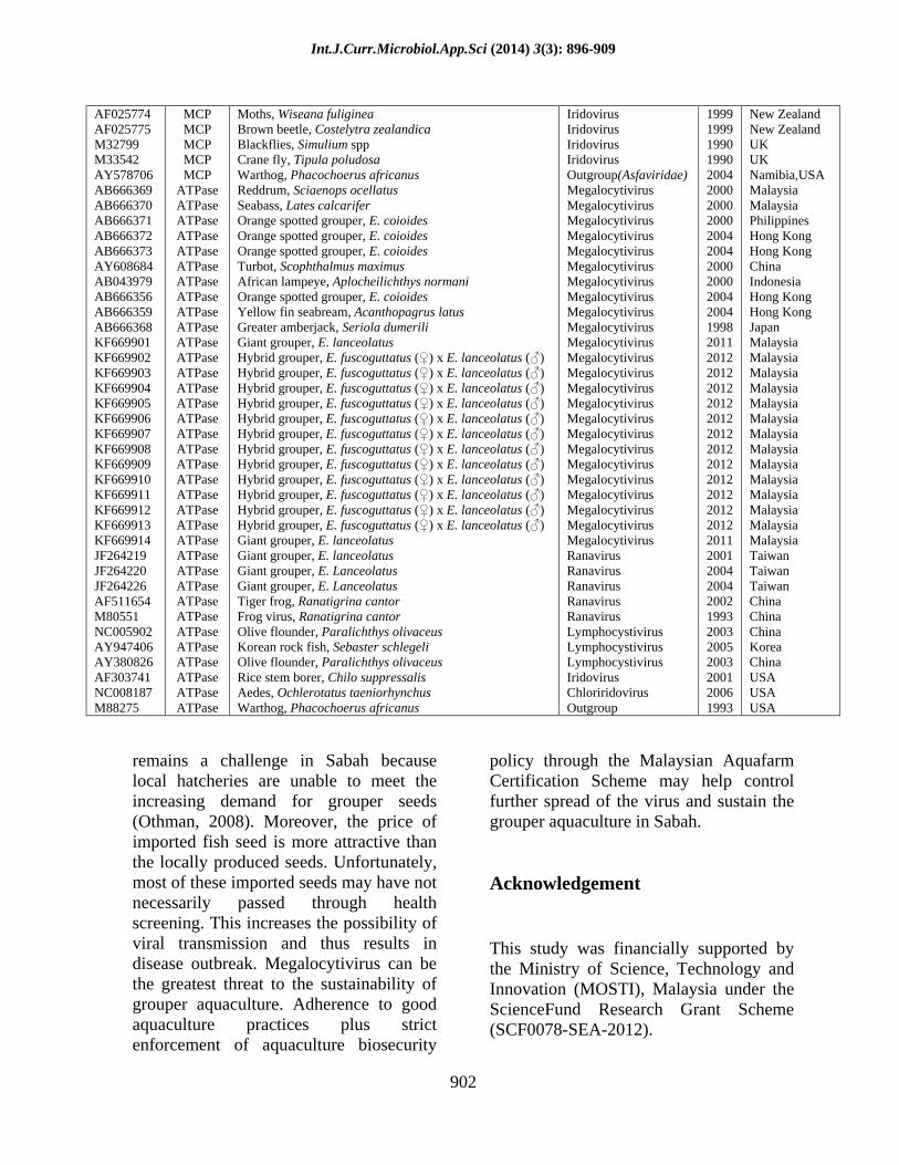
In this study, 212 specimens of groupers collected from net-cages throughout Sabah (Fig. 1) were analyzed. The specimens comprised of brown-marbled grouper (Epinephelus fuscoguttatus), humpback grouper (Cromileptes altivelis), giant grouper (E. lanceolatus), orange-spotted grouper (E. coioides) and hybrid grouper (E. fuscoguttatus ( )
x E. lanceolatus ( )). Although majority of the fish
specimens suffered from skin and gill lesions, few fish were observed to have enlarged kidney, liver and spleen.
DNA Extraction
Total genomic DNA was extracted from fish tissues (kidney, liver, spleen) using the DTAB-CTAB method described by Philips and Simons (1995). Briefly, about 30 mg of the pooled tissues were homogenized in 600µl DTAB solution followed by incubation at 75ºC for 5 minutes. Then, 700µl of chloroform solution was added into tissue homogenate before centrifuging at 12,000 rpm for 5 minutes. Subsequently, 400µl of the aqueous solution was added with 100µl CTAB and 900µl of autoclaved distilled water. The mixtures were briefly vortex and incubated at 75ºC for 5 minutes. After the incubation, the mixture was centrifuged at 12,000 rpm for 10 minutes. The supernatant was discarded while the pellet was re-suspended in 200µl of 1.2M NaCl solution, incubated at 75ºC for 5 minutes and centrifuged at 12,000 rpm for 5 minutes. Next, the clear suspension was transferred to a new tube containing 400µl
Int.J.Curr.Microbiol.App.Sci (2014) 3(3): 896-909
898
of 95% ethanol for DNA precipitation by centrifugation at 12,000 rpm for 5 minutes. Then, the DNA pellet was briefly air dried before dissolving in 30µl 1X TE buffer. Finally, the DNA solution was stored in -20ºC until use.
PCR amplification
Specific fragments of the viral major capsid protein (MCP) and ATPase genes were amplified either by single- or nested-PCR. PCR primers used in the amplification are shown in Table 1. The single-PCR amplification was carried out in 50µl reaction (5X GoTaq® Flexi PCR Buffer (Promega), 0.2mM of dNTPs (Promega), 1.7mM of MgCl2 (Promega), 10µM of each primer, 0.3 units of GoTaq®
DNA Polymerase (Promega) and ~100ng of DNA) at the following conditions: initial denaturation at 95ºC for 3 min followed by 30 cycles of denaturation at 94ºC for 1 min, annealing at 55ºC for 1 min, extension at 72ºC for 1 min and an extra extension at 72ºC was allowed for 5 min. The nested-PCR amplification was carried according to the conditions described for single-PCR except that 2µl of the PCR product generated during the single-PCR amplification was used as the DNA template. Subsequently, 5µl of the PCR products from single- and nested-PCR amplification was then separated on 1.5% of agarose gel, stained with ethidium bromide (5µg/µl) and visualized under UV light (Alpha Innotech Chemi Imager System). Based on the results of PCR amplification, the iridovirus infection was classified into two categories (severe and latent) according to the classification proposed by Jeong et al. (2006). DNA cloning and sequencing
PCR fragments of major capsid protein (MCP) and ATPase genes were first
purified using MEGAquick-spinTM PCR and Agarose Gel DNA Extraction System (iNtRON Biotechbology, Inc.) according to the manufacture s instruction before ligation into pGEM-T-Easy Vector (Promega).
Recombinant plasmids containing the gene fragments were transformed into competent E. coli strain JM109 (Promega) using the heat-shock method described by Sambrook and Russell (2001). Then, the competent cells were aseptically spread onto Luria-Bertani (LB)/ampicillin /IPTG/X-gal agar plates and incubated at 37oC overnight. After incubation, white colonies of E.coli JM109 were picked and aseptically inoculated into LB tubes containing 100µg/ml ampicillin. Then, the tubes were incubated at 37ºC with shaking at 150 rpm overnight. Subsequently, bacterial cells were harvested by centrifugation at 12,000 rpm for 5 minutes. Plasmids were extracted using alkaline lysis method (Sambrook and Russell, 2001) and purified using DNAspin Plasmid DNA Purification Kit (iNtRON Biotechnology, Inc) according to the manufacturer s procedure. DNA insert was verified by EcoR1 restriction analysis following manufacturer s instruction (New England Biolabs). Then, all plasmids containing correct DNA insert were sequenced at AIT,Singapore Pt Ltd.
Sequence alignment
Multiple alignment of DNA sequences was done against selected sequences of the genes (major capsid protein and ATPase) downloaded from the National Center for Biotechnology Information (NCBI) using the Clustal W method (Thompson, 1994). The list of sequences analyzed in the study is given in Table 2 and Table 3, respectively.
Int.J.Curr.Microbiol.App.Sci (2014) 3(3): 896-909
899
Construction of phylogenetic tree
Phylogenetic trees were constructed by the MegAlign program of the DNASTAR software package that employs the neighbor-joining algorithm of Saitou and Nei (1987). The accuracy of the phylogenetic trees was evaluated using bootsrapping analysis.
Results and Discussion
Virus detection
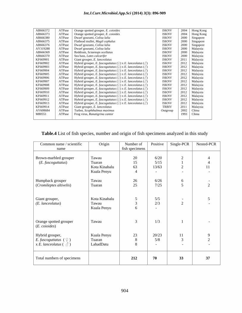
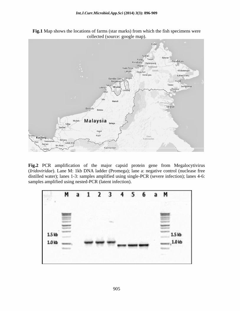
It was recorded that 33.02% (70/212) of the fish specimens were found positive for iridovirus. From the 70 infected fish specimens, 47.14% (33/70) were amplified for both genes during the single-PCR amplification and classified under severe infection. The rest of infected fish specimens (52.86%) were classified under latent infection since they were only amplified during the nested-PCR amplification (Table 4; Fig. 2). The percentage of fish specimens, according to species, which are severely and latently infected with iridovirus is given in Fig. 3.
Clinical signs of infected fish
Majority of the severely infected grouper specimens exhibited gross external clinical signs of diseases such as fin erosion, dark skin coloration, red spot in gills and skin lesion. The observation recorded were similar to the findings of Chincar et al. (2008); Murwantoko et al. (2009); Yanong and Waltzek (2010). In contrast, the specimens in the latent infection category did not exhibit apparent clinical signs of disease.
DNA Sequence analysis
DNA sequences of major capsid protein and ATPase genes examined in this study
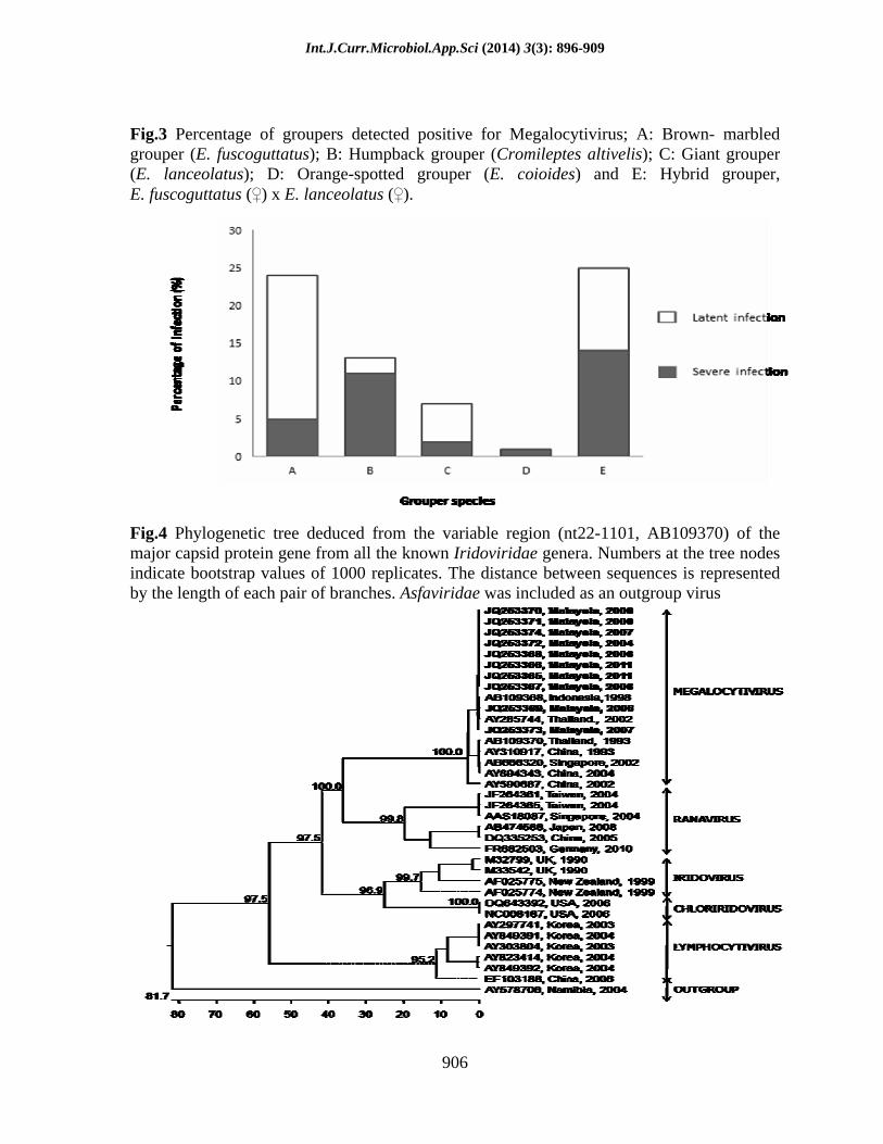
have been deposited into GenBank (http://www.ncbi.nih.gov) with the following accession numbers: MCP-JQ253365, JQ253366, JQ253367, JQ253368, JQ253369, JQ253370, JQ253371, JQ253372, JQ253373 and JQ253374; ATPase-KF669901, KF669902, KF669903, KF669904, KF669905, KF669906, KF669907, KF669908, KF669909, KF669910, KF669911, KF669911, KF669912, KF669913 and KF669914. Analysis of major capsid protein gene sequences against other genera within the family of Iridoviridae showed that the virus in this study had 96.04% nucleotide similarity to genus Megalocytivirus, 54.25% to Ranavirus, 51.21% to Chloriridovirus, 50.37% to Lymphocytivirus, 48.86% to Iridovirus and 34.4% to out-group (African Swine Fever Virus). Similar finding was also observed using ATPase gene where the DNA sequences had higher nucleotide similarity (97.73%) to Megalocytivirus compared to Ranavirus (63.41%), Chloriridovirus (52.76%), Lymphocytivirus (39.8%), Iridovirus (37.1%) and out-group (37.6%). Phylogenetic trees of the major capsid protein and ATPase genes also showed that the virus was clustered within the clade of Megalocytivirus (Fig. 4 and Fig. 5). Further phylogenetic analysis with the aim to elucidate the viral strain also revealed that the virus was clustered within the ISKNV strain (Fig. 6 and Fig. 7).
It is evident in this study that Iridoviridae, particularly the ISKNV within the genus Megalocytivirus could have responsible for the high mortality in grouper culture in Sabah. Megalocytivirus belongs to the fifth genera of Iridoviridae (Fauquet et al., 2005; Wang et al., 2007) and members of this genus are known to infect different species of freshwater and marine fishes

Int.J.Curr.Microbiol.App.Sci (2014) 3(3): 896-909
900
(Wang et al., 2007; Kurita and Nakajima, 2012). The results of this study have shown that about 33% of the grouper specimens suffered from Megalocytivirus infection. Out of these positive specimens, significant percentage (17.5%) of the fish specimens was found to be asymptomatic carriers of the virus. Furthermore, all the grouper species examined in this study have shown some degrees of susceptibility to Megalocytivirus including hybrid grouper. This is a worrying situation since these fish may potentially transmit the virus to other culture fish species or even to their counterparts in the wild.
Groupers with severe viral infection were seen lethargic, exhibited skin darkening, showed abnormal swimming behavior, increased respiration, suffered from skin hemorrhage, fin erosion, and red spots in the gills. Infected grouper also exhibited enlargement of kidney, liver and spleen. These clinical signs were similar to the one reported by Chinchar et al. (2008), Murwantoko et al. (2009) and Yanong and Waltzek (2010). Interestingly, some of the externally healthy-looking groupers were also found positive. This condition shows, under certain circumstances, groupers can become asymptomatic carriers of the virus (Choi et al., 2006; Jeong et al., 2006; Wang et al., 2007).
According to Wen et al. (2008) and Yanong and Waltzek (2010), Megalocytivirus requires high water temperature to multiply. The relatively high annual water temperature (28-320C) of the coastal waters in Sabah (Jiran and Ransangan, 2013) may provide an optimal condition for the virus to remain active all year round. Unlike temperate countries, the virus is only reported to outbreak during summer months (Joon et al., 2003).
Majority of groupers are cultured on small scale basis and are generally poorly operated. The farms generally consist of 10-40 cages per farm. Despite small in size (3m x 3m x 3m), cages are heavily stocked. From our observation, the average stocking density of grouper in Sabah was at 450 individuals (25-30cm TL) per cage. Limited space coupled with high stocking density can be stressful to groupers. Such situation can cause fish to become immunologically incompetent and eventually make them susceptible to viral infection. Poorly maintained farms also provide excellent habitats for bio-fouling organisms such as oysters and mussels. When these organisms are heavily growing on the net cage, they can easily cause skin injury to fish especially during net lifting, sorting or harvesting. The injured skin may then develop into wounds, which then provide entry points to fish pathogens, including viruses.
Viruses can also enter fish orally via the consumption of contaminated feeds such as trash fish. Trash fish have been known to carry iridovirus (Mao et al., 1999; Wang et al., 2007). Unfortunately, majority of the aquaculture farms in Sabah depends on trash fish for feeding. It was also evident to our observation that many of the aquaculture farms do not have proper storage facility for trash fish. Rotten trash fish may increase the chances of pathogens contamination. Although pellet feeds are beneficial over trash fish, availability and relatively high cost have often been the issues of their use in aquaculture.
Viruses are also reported to spread through transboundary movement of live fish (Subasinge and Bondad-Reantaso, 2008; Walker and Winton, 2010). However, this
Int.J.Curr.Microbiol.App.Sci (2014) 3(3): 896-909
901
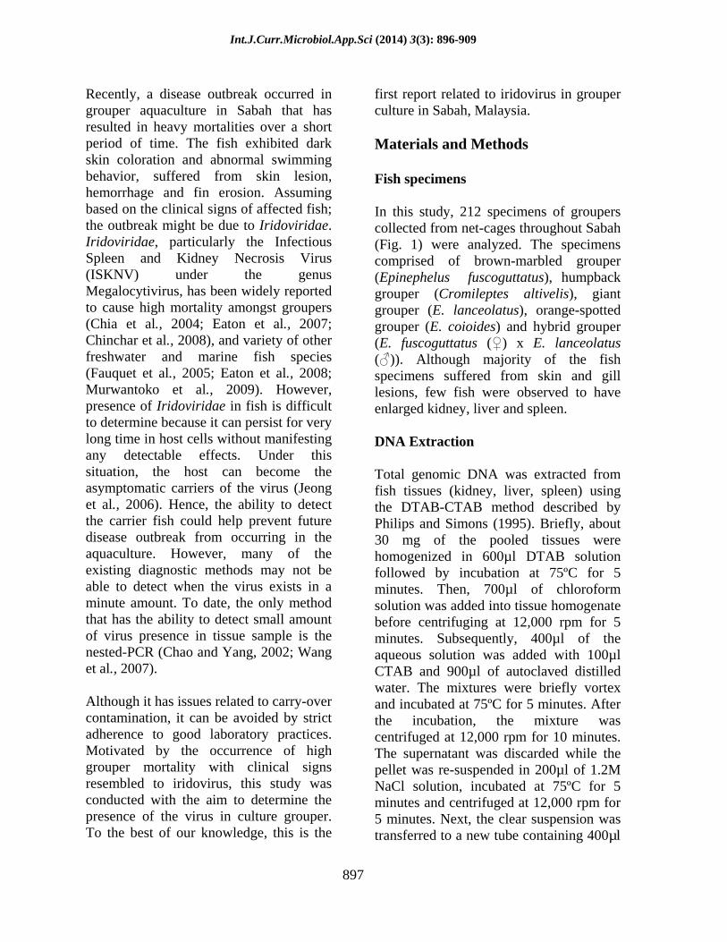
Table.1 PCR primers used for the amplification of MCP gene and ATPase genes
Degenerate bases :M (A/C); R (A/G); Y (C/T); W (A/T) *Adopted from Huang et al. (2011)
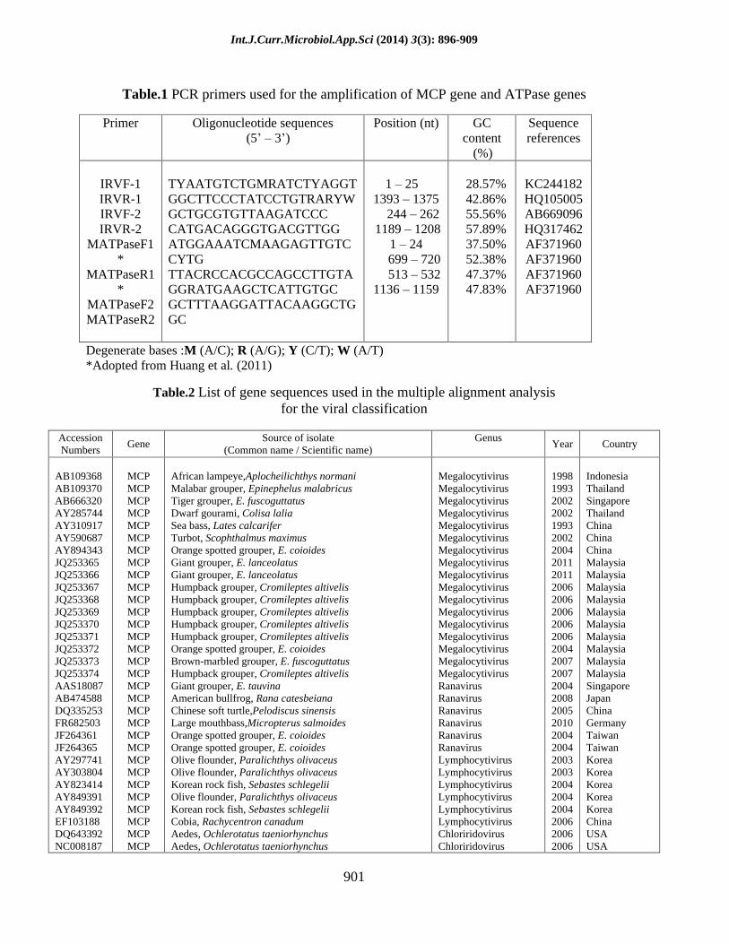
Table.2 List of gene sequences used in the multiple alignment analysis for the viral classification
Accession Numbers
Gene Source of isolate
(Common name / Scientific name) Genus
Year Country
AB109368 AB109370 AB666320 AY285744 AY310917 AY590687 AY894343 JQ253365 JQ253366 JQ253367 JQ253368 JQ253369 JQ253370 JQ253371 JQ253372 JQ253373 JQ253374 AAS18087 AB474588 DQ335253 FR682503 JF264361 JF264365 AY297741 AY303804 AY823414 AY849391 AY849392 EF103188 DQ643392 NC008187
MCP MCP MCP MCP MCP MCP MCP MCP MCP MCP MCP MCP MCP MCP MCP MCP MCP MCP MCP MCP MCP MCP MCP MCP MCP MCP MCP MCP MCP MCP MCP
African lampeye,Aplocheilichthys normani Malabar grouper, Epinephelus malabricus Tiger grouper, E. fuscoguttatus Dwarf gourami, Colisa lalia Sea bass, Lates calcarifer Turbot, Scophthalmus maximus Orange spotted grouper, E. coioides Giant grouper, E. lanceolatus Giant grouper, E. lanceolatus Humpback grouper, Cromileptes altivelis Humpback grouper, Cromileptes altivelis Humpback grouper, Cromileptes altivelis Humpback grouper, Cromileptes altivelis Humpback grouper, Cromileptes altivelis Orange spotted grouper, E. coioides Brown-marbled grouper, E. fuscoguttatus Humpback grouper, Cromileptes altivelis Giant grouper, E. tauvina American bullfrog, Rana catesbeiana Chinese soft turtle,Pelodiscus sinensis Large mouthbass,Micropterus salmoides Orange spotted grouper, E. coioides Orange spotted grouper, E. coioides Olive flounder, Paralichthys olivaceus Olive flounder, Paralichthys olivaceus Korean rock fish, Sebastes schlegelii Olive flounder, Paralichthys olivaceus Korean rock fish, Sebastes schlegelii Cobia, Rachycentron canadum Aedes, Ochlerotatus taeniorhynchus Aedes, Ochlerotatus taeniorhynchus
Megalocytivirus Megalocytivirus Megalocytivirus Megalocytivirus Megalocytivirus Megalocytivirus Megalocytivirus Megalocytivirus Megalocytivirus Megalocytivirus Megalocytivirus Megalocytivirus Megalocytivirus Megalocytivirus Megalocytivirus Megalocytivirus Megalocytivirus Ranavirus Ranavirus Ranavirus Ranavirus Ranavirus Ranavirus Lymphocytivirus Lymphocytivirus Lymphocytivirus Lymphocytivirus Lymphocytivirus Lymphocytivirus Chloriridovirus Chloriridovirus
1998 1993 2002 2002 1993 2002 2004 2011 2011 2006 2006 2006 2006 2006 2004 2007 2007 2004 2008 2005 2010 2004 2004 2003 2003 2004 2004 2004 2006 2006 2006
Indonesia Thailand Singapore Thailand China China China Malaysia Malaysia Malaysia Malaysia Malaysia Malaysia Malaysia Malaysia Malaysia Malaysia Singapore Japan China Germany Taiwan Taiwan Korea Korea Korea Korea Korea China USA USA
Primer Oligonucleotide sequences (5
3 )
Position (nt) GC content
(%)
Sequence references
IRVF-1 IRVR-1 IRVF-2 IRVR-2
MATPaseF1*
MATPaseR1*
MATPaseF2 MATPaseR2
TYAATGTCTGMRATCTYAGGT GGCTTCCCTATCCTGTRARYW GCTGCGTGTTAAGATCCC CATGACAGGGTGACGTTGG ATGGAAATCMAAGAGTTGTCCYTG TTACRCCACGCCAGCCTTGTA GGRATGAAGCTCATTGTGC GCTTTAAGGATTACAAGGCTGGC
1 25 1393 1375 244 262 1189 1208
1 24 699 720 513 532 1136 1159
28.57% 42.86% 55.56% 57.89% 37.50% 52.38% 47.37% 47.83%
KC244182 HQ105005 AB669096 HQ317462 AF371960 AF371960 AF371960 AF371960
Int.J.Curr.Microbiol.App.Sci (2014) 3(3): 896-909
902
AF025774 AF025775 M32799 M33542 AY578706 AB666369 AB666370 AB666371 AB666372 AB666373 AY608684 AB043979 AB666356 AB666359 AB666368 KF669901 KF669902 KF669903 KF669904 KF669905 KF669906 KF669907 KF669908 KF669909 KF669910 KF669911 KF669912 KF669913 KF669914 JF264219 JF264220 JF264226 AF511654 M80551 NC005902 AY947406 AY380826 AF303741 NC008187 M88275
MCP MCP MCP MCP MCP
ATPase ATPase ATPase ATPase ATPase ATPase ATPase ATPase ATPase ATPase ATPase ATPase ATPase ATPase ATPase ATPase ATPase ATPase ATPase ATPase ATPase ATPase ATPase ATPase ATPase ATPase ATPase ATPase ATPase ATPase ATPase ATPase ATPase ATPase ATPase
Moths, Wiseana fuliginea Brown beetle, Costelytra zealandica Blackflies, Simulium spp Crane fly, Tipula poludosa Warthog, Phacochoerus africanus Reddrum, Sciaenops ocellatus Seabass, Lates calcarifer Orange spotted grouper, E. coioides Orange spotted grouper, E. coioides Orange spotted grouper, E. coioides Turbot, Scophthalmus maximus African lampeye, Aplocheilichthys normani Orange spotted grouper, E. coioides Yellow fin seabream, Acanthopagrus latus Greater amberjack, Seriola dumerili Giant grouper, E. lanceolatus Hybrid grouper, E. fuscoguttatus ( ) x E. lanceolatus ( )
Hybrid grouper, E. fuscoguttatus ( ) x E. lanceolatus ( )
Hybrid grouper, E. fuscoguttatus ( ) x E. lanceolatus ( )
Hybrid grouper, E. fuscoguttatus ( ) x E. lanceolatus ( )
Hybrid grouper, E. fuscoguttatus ( ) x E. lanceolatus ( )
Hybrid grouper, E. fuscoguttatus ( ) x E. lanceolatus ( )
Hybrid grouper, E. fuscoguttatus ( ) x E. lanceolatus ( )
Hybrid grouper, E. fuscoguttatus ( ) x E. lanceolatus ( )
Hybrid grouper, E. fuscoguttatus ( ) x E. lanceolatus ( )
Hybrid grouper, E. fuscoguttatus ( ) x E. lanceolatus ( )
Hybrid grouper, E. fuscoguttatus ( ) x E. lanceolatus ( )
Hybrid grouper, E. fuscoguttatus ( ) x E. lanceolatus ( )
Giant grouper, E. lanceolatus Giant grouper, E. lanceolatus Giant grouper, E. Lanceolatus Giant grouper, E. Lanceolatus Tiger frog, Ranatigrina cantor Frog virus, Ranatigrina cantor Olive flounder, Paralichthys olivaceus Korean rock fish, Sebaster schlegeli Olive flounder, Paralichthys olivaceus Rice stem borer, Chilo suppressalis Aedes, Ochlerotatus taeniorhynchus Warthog, Phacochoerus africanus
Iridovirus Iridovirus Iridovirus Iridovirus Outgroup(Asfaviridae) Megalocytivirus Megalocytivirus Megalocytivirus Megalocytivirus Megalocytivirus Megalocytivirus Megalocytivirus Megalocytivirus Megalocytivirus Megalocytivirus Megalocytivirus Megalocytivirus Megalocytivirus Megalocytivirus Megalocytivirus Megalocytivirus Megalocytivirus Megalocytivirus Megalocytivirus Megalocytivirus Megalocytivirus Megalocytivirus Megalocytivirus Megalocytivirus Ranavirus Ranavirus Ranavirus Ranavirus Ranavirus Lymphocystivirus Lymphocystivirus Lymphocystivirus Iridovirus Chloriridovirus Outgroup
1999 1999 1990 1990 2004 2000 2000 2000 2004 2004 2000 2000 2004 2004 1998 2011 2012 2012 2012 2012 2012 2012 2012 2012 2012 2012 2012 2012 2011 2001 2004 2004 2002 1993 2003 2005 2003 2001 2006 1993
New Zealand New Zealand UK UK Namibia,USA Malaysia Malaysia Philippines Hong Kong Hong Kong China Indonesia Hong Kong Hong Kong Japan Malaysia Malaysia Malaysia Malaysia Malaysia Malaysia Malaysia Malaysia Malaysia Malaysia Malaysia Malaysia Malaysia Malaysia Taiwan Taiwan Taiwan China China China Korea China USA USA USA
remains a challenge in Sabah because local hatcheries are unable to meet the increasing demand for grouper seeds (Othman, 2008). Moreover, the price of imported fish seed is more attractive than the locally produced seeds. Unfortunately, most of these imported seeds may have not necessarily passed through health screening. This increases the possibility of viral transmission and thus results in disease outbreak. Megalocytivirus can be the greatest threat to the sustainability of grouper aquaculture. Adherence to good aquaculture practices plus strict enforcement of aquaculture biosecurity
policy through the Malaysian Aquafarm Certification Scheme may help control further spread of the virus and sustain the grouper aquaculture in Sabah.
Acknowledgement
This study was financially supported by the Ministry of Science, Technology and Innovation (MOSTI), Malaysia under the ScienceFund Research Grant Scheme (SCF0078-SEA-2012).

Int.J.Curr.Microbiol.App.Sci (2014) 3(3): 896-909
903
Table.3 List of gene sequences used in the multiple alignment analysis
for viral strain determination
Accession Numbers
Gene Source of isolate (Common name / Scientific name)
Strain Year Country
AB109368 AY285745 AB666342 AB666348 AB666337 AB666338 JQ253373 AB666340 AB666341 HM067835 HQ317461 HQ317462 HQ317460 JF264354 JF264345 JF264349 JQ253365 JQ253366 JQ253367 JQ253368 JQ253369 JQ253370 JQ253371 JQ253372 JQ253373 JQ253374 AB080362 AB461855 AB461856 AB666335 AB666326 AB666321 AB666322 AB666318 AB666319 AB666320 AY285746 AB109370 AY532608 AY532613 DQ198145 HQ263620 AY532611 EU276417 HM067603 GQ273492 JF264361 AB007367 AB666362 AB666368 AB666357 AB666354 AB666350 AB666352 AB666351 AF462344 AF462343 AY894343 GQ202217 AY779031 AB043979 AB666379
MCP MCP MCP MCP MCP MCP MCP MCP MCP MCP MCP MCP MCP MCP MCP MCP MCP MCP MCP MCP MCP MCP MCP MCP MCP MCP MCP MCP MCP MCP MCP MCP MCP MCP MCP MCP MCP MCP MCP MCP MCP MCP MCP MCP MCP MCP MCP
ATPase ATPase ATPase ATPase ATPase ATPase ATPase ATPase ATPase ATPase ATPase ATPase ATPase ATPase ATPase
African lampeye, Aplocheilichthys normani African lampeye, Aplocheilichthys normani Flatheat mullet, Mugil cephalus Dwarf gourami,Colisa lalia Reddrum, Sciaenops ocellatus Barramundi perch, Lates calcarifer Brown marbled grouper, E. fuscoguttattus Orange spotted grouper, E. coioides Orange spotted grouper, E. coioides Marble sleepy goby, Oxyeleotris marmorata Mandarin fish, Synchiropus splendidus Mandarin fish,Synchiropus splendidus Mandarin fish,Synchiropus splendidus Giant sea perch, Lates calcarifer Orange spotted grouper, E. coioides Orange spotted grouper, E. coioides Giant grouper, E. lanceolatus Giant grouper, E. lanceolatus Humpback grouper, Cromileptes altivelis Humpback grouper, Cromileptes altivelis Humpback grouper, Cromileptes altivelis Humpback grouper, Cromileptes altivelis Humpback grouper, Cromileptes altivelis Orange spotted grouper, E. coioides Brown-marbled grouper, E. fuscoguttatus Humpback grouper, Cromileptes altivelis Red sea bream, Pagrus major Japanese amberjack, Seriola quinqueradiata Red sea bream, Pagrus major Red sea bream, Pagrus major Orange spotted grouper, E. coioides Red sea bream, Pagrus major Orange spotted grouper, E. coioides Hybrid sea bass, Moronesaxatilis x Micropterussalmoides Brown marbled grouper, E. fuscoguttattus Brown marbled grouper, E. fuscoguttattus Malabar grouper, E. malabricus Malabar grouper, E. malabricus Barred knifejaw, Oplegnathus fasciatus Sea bass, Lateolabrax japonicus Olive flounder, Paralichthys olivaceus Stone flounder, Kareius bicoloratus Rock bream, Oplegnathus fasciatus Olive flounder, Paralichthys olivaceus Sea perch, Lateolabrax sp. Turbot, Scophthalmus maximus Orange spotted grouper, E. coioides Red sea bream, Pagrus major Pacific bluefin tuna, Thunnus orientalis Greater amberjack, Seriola dumerili Giant grouper, E. lanceolatus Orange spotted grouper, E. coioides Hybrid sea bass, Moronesaxatilis x Micropterussalmoides Brown marbled grouper, E. fuscoguttatus Brown marbled grouper, E. fuscoguttatus Giant sea perch, Lates calcarifer Grouper sp, Epinephelus spp Orange spotted grouper, E. coioides Spotted knifejaw, Oplegnathus punctatus Large yellow croacker, Pseudosciaena crocea African lampeye, Aplocheilichthys normani Dwarf gourami, Colisa lalia
ISKNV ISKNV ISKNV ISKNV ISKNV ISKNV ISKNV ISKNV ISKNV ISKNV ISKNV ISKNV ISKNV ISKNV ISKNV ISKNV ISKNV ISKNV ISKNV ISKNV ISKNV ISKNV ISKNV ISKNV ISKNV ISKNV RSIV RSIV RSIV RSIV RSIV RSIV RSIV RSIV RSIV RSIV RSIV RSIV RSIV RSIV RSIV RSIV
TRBIV TRBIV TRBIV TRBIV
Outgroup RSIV RSIV RSIV RSIV RSIV RSIV RSIV RSIV RSIV RSIV RSIV RSIV RSIV RSIV
ISKNV
1998 1998 2000 2000 2000 2000 2007 2004 2004 2009 2006 2007 2009 2005 2006 2008 2011 2011 2006 2006 2006 2006 2006 2004 2007 2007 1992 2004 2005 2001 2004 2004 2004 2004 2000 2002 1992 1993 2001 2001 2005 2010 2001 2007 2010 2006 2004 1992 1996 1998 2004 2004 2004 2002 2000 2001 2001 2004 2007 2002 1998 2001
Indonesia Indonesia Singapore Singapore Malaysia Malaysia Malaysia Hong Kong Hong Kong China China China China Taiwan Taiwan Taiwan Malaysia Malaysia Malaysia Malaysia Malaysia Malaysia Malaysia Malaysia Malaysia Malaysia Japan Japan Japan Japan Hong Kong Hong Kong Hong Kong Hong Kong Singapore Singapore Thailand Thailand Korea Korea Korea China Korea Korea Korea China Taiwan Japan Japan Japan Hong Kong Hong Kong Hong Kong Singapore Singapore Taiwan Taiwan China China China Indonesia Japan
Int.J.Curr.Microbiol.App.Sci (2014) 3(3): 896-909
904
AB666372 AB666373 AB666380 AB666375 AB666376 AY319288 AB666369 AB666370 KF669901 KF669902 KF669903 KF669904 KF669905 KF669906 KF669907 KF669908 KF669909 KF669910 KF669911 KF669912 KF669913 KF669914 AY608684 M80551
ATPase ATPase ATPase ATPase ATPase ATPase ATPase ATPase ATPase ATPase ATPase ATPase ATPase ATPase ATPase ATPase ATPase ATPase ATPase ATPase ATPase ATPase ATPase ATPase
Orange spotted grouper, E. coioides Orange spotted grouper, E. coioides Dwarf gourami, Colisa lalia Flathead mullet, Mugil cephalus Dwarf gourami, Colisa lalia Dwarf gourami, Colisa lalia Reddrum, Sciaenops ocellatus Sea bass, Lates calcarifer Giant grouper, E. lanceolatus Hybrid grouper, E. fuscoguttatus ( ) x E. lanceolatus ( )
Hybrid grouper, E. fuscoguttatus ( ) x E. lanceolatus ( )
Hybrid grouper, E. fuscoguttatus ( ) x E. lanceolatus ( )
Hybrid grouper, E. fuscoguttatus ( ) x E. lanceolatus ( )
Hybrid grouper, E. fuscoguttatus ( ) x E. lanceolatus ( )
Hybrid grouper, E. fuscoguttatus ( ) x E. lanceolatus ( )
Hybrid grouper, E. fuscoguttatus ( ) x E. lanceolatus ( )
Hybrid grouper, E. fuscoguttatus ( ) x E. lanceolatus ( )
Hybrid grouper, E. fuscoguttatus ( ) x E. lanceolatus ( )
Hybrid grouper, E. fuscoguttatus ( ) x E. lanceolatus ( )
Hybrid grouper, E. fuscoguttatus ( ) x E. lanceolatus ( )
Hybrid grouper, E. fuscoguttatus ( ) x E. lanceolatus ( )
Giant grouper, E. lanceolatus Turbot, Scophthalmus maximus Frog virus, Ranatigrina cantor
ISKNV ISKNV ISKNV ISKNV ISKNV ISKNV ISKNV ISKNV ISKNV ISKNV ISKNV ISKNV ISKNV ISKNV ISKNV ISKNV ISKNV ISKNV ISKNV ISKNV ISKNV TRBIV
Outgroup
2004 2004 2000 2000 2000 2000 2000 2000 2011 2012 2012 2012 2012 2012 2012 2012 2012 2012 2012 2012 2012 2011 2002 1993
Hong Kong Hong Kong Singapore Singapore Singapore Malaysia Malaysia Malaysia Malaysia Malaysia Malaysia Malaysia Malaysia Malaysia Malaysia Malaysia Malaysia Malaysia Malaysia Malaysia Malaysia Malaysia China China
Table.4 List of fish species, number and origin of fish specimens analyzed in this study
Common name / scientific name
Origin Number of fish specimens
Positive Single-PCR Nested-PCR
Brown-marbled grouper (E. fuscoguttatus)
Humpback grouper (Cromileptes altivelis)
Giant grouper, (E. lanceolatus)
Orange spotted grouper (E. coioides)
Hybrid grouper, E. fuscoguttatus ( )
x E. lanceolatus ( )
Tawau Tuaran Kota Kinabalu Kuala Penyu
Tawau Tuaran
Kota Kinabalu Tawau Kuala Penyu
Tawau
Kuala Penyu Tuaran LahadDatu
20 15 63
4
26 25
5 3 6
3
23 8 8
6/20 5/15
13/63 -
6/26 7/25
5/5 2/3 -
1/3
20/23 5/8 -
2 1 2 -
6 5
- 2
1
11 3 -
4 4
11 -
- 2
5 -
-
9 2 -
Total numbers of specimens 212 70 33 37
Int.J.Curr.Microbiol.App.Sci (2014) 3(3): 896-909
905
Fig.1 Map shows the locations of farms (star marks) from which the fish specimens were
collected (source: google map).
Fig.2 PCR amplification of the major capsid protein gene from Megalocytivirus (Iridoviridae). Lane M: 1kb DNA ladder (Promega); lane a: negative control (nuclease free distilled water); lanes 1-3: samples amplified using single-PCR (severe infection); lanes 4-6: samples amplified using nested-PCR (latent infection).
Int.J.Curr.Microbiol.App.Sci (2014) 3(3): 896-909
906
Fig.3 Percentage of groupers detected positive for Megalocytivirus; A: Brown- marbled grouper (E. fuscoguttatus); B: Humpback grouper (Cromileptes altivelis); C: Giant grouper (E. lanceolatus); D: Orange-spotted grouper (E. coioides) and E: Hybrid grouper, E. fuscoguttatus ( ) x E. lanceolatus ( ).
Fig.4 Phylogenetic tree deduced from the variable region (nt22-1101, AB109370) of the major capsid protein gene from all the known Iridoviridae genera. Numbers at the tree nodes indicate bootstrap values of 1000 replicates. The distance between sequences is represented by the length of each pair of branches. Asfaviridae was included as an outgroup virus

Int.J.Curr.Microbiol.App.Sci (2014) 3(3): 896-909
907
Fig.5 Phylogenetic tree deduced from variable region (nt146-231, AB666373) of ATPase gene from all the known Iridoviridae genera. Numbers at the tree nodes indicate bootstrap values of 1000 replicates. The distance between sequences is represented by the length of each pair of branches. Asfaviridae was included as an outgroup virus
Fig.6 Phylogenetic tree deduced from variable region (nt540-951, AB109370) of the major capsid protein gene from all the knwon strains of Megalocytivirus. Numbers at the tree nodes indicate bootstrap values of 1000 replicates. The distance between sequences is represented by the length of each pair of branches. Ranavirus was included as an outgroup virus.

Int.J.Curr.Microbiol.App.Sci (2014) 3(3): 896-909
908
Fig.7 Phylogenetic tree deduced from variable region (nt122-521, AB666373) of ATPase gene from all the known strains of Megalocytivirus. Numbers at the tree nodes indicate bootstrap values of 1000 replicates. The distance between sequences is represented by the length of each pair of branches. Ranavirus was included as an outgroup virus
References
Bondad-Reantaso, M. G., Subasinghe, R.P., Arthur, J.R., Ogawa, K., Chinabut, S., Adlard, R., Tan, Z., Shariff, M., 2005. Disease and health management in Asian aquaculture. Vet. Parasitol. 132, 249-272.
Chao,C.B., Yang, S.C.,Tsai, H.Y., Chen, C.Y., Lin, C.S., Huang, H.T., 2002. A nested PCR for the detection of grouper iridovirus in taiwan (TGIV) in cultured hybrid grouper, giant seaperch and largemouth bass. J. Aquat. Anim. Health. 14, 104-113.
Chia,C.B., Chun, Y.C., Yueh, Y.L., Chan, S.L., Hung, T.H., 2004. Histological, ultrastructural, and in situ hybridization study on enlarged cells in grouper Epinephelus hybrids infected by grouper iridovirus in Taiwan (TGIV). Dis. Aquat. Organ. 58, 127-142.
Chincar,V.G., Hyatt, A., Miyazaki,T., Williams, T., 2008. Family Iridoviridae: poor viral relations no longer. Microbiol. Immunol. 328, 123-170.
Choi, S.K., Kwon, S.R., Nam,Y.K., Kim,S.K., Kim,K.H., 2006. Organ distribution of red sea bream iridovirus (RSIV) DNA in asymptomatic yearling and fingerling rock bream (Oplegnathus fasciatus) and effects of water temperature on transition of RSIV into acute phase. Aquaculture. 256, 23-26.
Eaton, H.E., Metcalf, J., Penny, E., Tcherepanov, V., Upton, C., Brunetti, C.R. 2007.Comparative genomic analysis of the family Iridoviridae: re-annotating and defining the core set of iridovirus genes. Virol. J. 4, 11.
Eaton, H.E., Metcalf, J., Brunetti,C.R., 2008. Expression of frog virus 3 genes is impaired in mammalian cell lines. Virol. J. 5, 83.
Fauquet, C.M., Mayo, M.A., Maniloff, J., Desselberger, U., Ball, L.A., 2005. Virus Taxonomy. Eight Report of the International Commitee on Taxonomy of Viruses. Elsevier Academic Presss, USA, pp 145-161.
Harikrisnan, R., Chellam, B., Moon, S.H., 2010. Molecular studies, disease status

Int.J.Curr.Microbiol.App.Sci (2014) 3(3): 896-909
909
and prophylactic measures in grouper aquaculture: Economic importance, diseases and immunology. Aquaculture.10, 1-14.
Hyatt, A.D., Whittington, R.J., 2005. Ranaviruses of fish, amphibians and reptiles: diversity and the requirement for revised taxonomy. In P. Walker, R. Lester and M.G. Bondad-Reantaso (eds.). Disease in Asian Aquaculture V.pp 155- 170. Fish Health Section, Asian Fisheries Society, Manila.
Jeong, J.B., Jun, L.Y., Park, K.H., Kim, K.H., Chung, J.K., Komisar, J.L., Jeong, H.D., 2006. Asymptomatic iridovirus infection in various marine fishes detected by a 2-step PCR method. Aquaculture. 255, 30-38.
Jiran,V.A., Ransangan,J., 2013. Effect of water temperature on susceptibility of culture marine fish species to vibriosis. Int. J. Res. Pure Appl. Microbiol. 3(3), 48-52.
Joon, B.J., Lyu, J.J., Min, H.Y., Myoung, S.K., Jack, L.K., Hyun, D.J., 2003. Characterization of the DNA nucleotide sequences in the genome of red sea bream iridoviruses isolated in Korea. Aquaculture. 220, 119-133.
Kurita, J., Nakajima, K., 2012. Megalocytiviruses. Viruses. 4, 521- 538.
Mao, J., Green, D.E., Fellers, G., Chinchar., V.G., 1999. Molecular characterization of iridoviruses isolated from sympatric amphibians and fish. Virus Res. 63, 45-52.
Muroga, K., 2001. Viral and bacterial diseases of marine fish and shellfish in Japanese hatcheries. Aquaculture. 202, 23-44.
Murwantoko., Handayani, C.R., Pratiwe,R., 2009. Cloning and sequence analysis of capsid protein gene of iridovirus Indonesian isolates. Indonesian J. Biotechnol. 14, 1117-1123.
Othman, M.F., 2008. The FAO/NACA Regional Workshop on the future of mariculture: a regional approach for responsible development in the Asia-Pacific region. FAO Proceedings. No.11. pp. 207-224.
Philips, A.J., Simon, C., 1995. Simple, efficient and nondestructive DNA
extraction protocol for arthropods. Ann. Entomol. Soc. Am. 88, 281-283.
Saitou, N., Nei, M., 1987. The neighbour-joining method: A new method for reconstructing phylogenetic trees. Mol. Biol. Evol. 4, 406-425.
Sambrook, J., Russell, D.W. 2001. Molecular Cloning: A Laboratory Manual. Cold Spring Harbor Laboratory Press, New York.
Subasinghe, R.P., Bondad-Reantaso, M.G., 2008. The FAO/NACA Asia regional technical guidelines on health management for the responsible movement of live aquatic animals: lessons learned from their development and implementation. Rev. Sci. Tech. OIE. 27, 55-63.
Thompson, J.D., Higgins, D.G., Gibson, T., 1994. Clustal W: improving the sensitivity of progressive multiple sequence alignment through sequence wighting, position-specific gap penalties and weight matrix choice. Nucleic Acids Res. 22, 4673-4680.
Walker, P.J., Winton, J.R. 2010. Emerging viral diseases of fish and shrimp. Vet. Res. 41, 51-75.
Wang,Y.Q., Lu, L., Weng, S.P., Huang, J.N., Chan,S.M., He, J.G., 2007. Molecular epidemiology and phylogenetic analysis of a marine fish infectious spleen and kidney necrosis virus like (ISKNV-like) virus. Arch. Virol. 152, 763-773.
Wen, C.M., Lee, C.W., Wang, C.S., Cheng, Y.H., Huang, H.Y., 2008. Development of two cell lines from Epinephelus coioides brain tissue for characterization of betanodavirus and megalocytivirus infectivity and propagation. Aquaculture. 278, 14-21.
Yanong, R.E., Waltzek, T., 2010. Megalocytivirus infections in fish, with emphasis on ornamental species. The Institute of Food and Agricultural Sciences (IFAS). University of Florida IFAS Extension FA182. Available: http://edis.ifas.ufl.edu/fa182 (October 2013).