histological changes in the kidneys of experimental ... · in diabetic patients’ life expectancy...
TRANSCRIPT
Romanian Journal of Morphology and Embryology 2010, 51(1):91–95
OORRIIGGIINNAALL PPAAPPEERR
Histological changes in the kidneys of experimental diabetic rats fed with
Momordica charantia (bitter gourd) extract S. L. TEOH, AZIAN ABD LATIFF, S. DAS
Department of Anatomy, Universiti Kebangsaan Malaysia, Jalan Raja Muda Abdul Aziz, Kuala Lumpur, Malaysia
Abstract Momordica charantia (MC) or bitter gourd is widely known for its antidiabetic properties. The aim of the present study was to observe the protective effect of MC extract on the kidneys of streptozotocin-induced diabetic rats. Eighteen male Sprague–Dawley rats (n=18) weighing 200±50 g were taken for the study. The study comprised of three groups i.e. a non-diabetic, diabetic untreated and diabetic treated with MC extract, with each group comprising of six (n=6) rats. Diabetes was induced in the overnight fasted rats by intramuscular injection of streptozotocin (50 mg/kg body weight). The MC extract (50 mg/kg body weight) was administered via oral gavage. Both the kidneys were collected on the tenth day following treatment. Histological study using Verhoeff’s van Gieson (VvG) and Periodic Acid–Schiff (PAS) stains were performed. The kidneys of the diabetic rats showed thickening of the basement membrane of the Bowman’s capsule, edema and hypercellurarity of the proximal tubules, necrosis and hyaline deposits. These features were found to be reversed when the MC extract was administered to the experimental animals. The MC extract acted as an antioxidant thereby preventing the oxidative damage involved in the diabetic kidney. The administration of MC extract prevents oxidative damage in diabetic nephropathy. Keywords: Momordica charantia, bitter gourd, antioxidant, diabetes, kidney, microscopic changes.
Introduction
Diabetes mellitus (DM) is one of the most common endocrine disease in the world characterized by the state of hyperglycemia. It is a systemic disease caused by defect in the insulin secretion, insulin action or even both [1]. Interestingly, this common metabolic disorder has a prevalence rate varying between 1–50% [2].
Unfortunately, DM in the younger age group has been on the rise and there is an urgent need to combat this disease. DM patients are prone to some long-term complications like nephropathy, retinopathy and neuropathy [3]. These long-term complications resulted in diabetic patients’ life expectancy accounting to only two-thirds of the general population [4].
In our previous research, we had even created animal model of DM to observe the wound healing and liver degenerative changes [5, 6]. The popularity in using traditional medicine to treat DM has been on the rise, in the recent years. Many medicinal herbs through its hypoglycemic and antioxidant action may protect the organs involves in DM patients. This prompted us to experiment the herbal extract on the diabetic kidney.
MC has been commonly consumed as a vegetable and used as a medicinal herb in India, China and Africa [7]. The vegetable is also consumed in various parts of India, Pakistan and South East Asia. MC hails from the Cucurbitaceae family. Past reports depict that it is helpful in treating wound, ulcer, dysmenorrhea, eczema, gout, jaundice, kidney stone, leprosy, leucorrhea, piles, pneumonia, psoriasis, rheumatism and scabies [7]. Earlier studies performed with MC extract have demon-
strated its antidiabetic, antiviral, antitumor, antileuke-mic, antibacterial, antihelminthic, antimutagenic, anti-mycobacterial, antioxidant, antiulcer, anti-inflammato-ry, hypocholesterolemic, hypotriglyceridemic, hypoten-sive, immunostimulant and insecticidal properties [7, 8].
Keeping in view the protective effect of MC in various diseases especially DM, the present study was undertaken to observe the histological changes on both kidneys of streptozotocin-induced diabetic fed with MC extract and then compared to those which were either normal or untreated. Our study on the histology of the kidney damage could help in better understanding of the damage caused in the kidneys in DM and highlight the protective action of the MC extract.
Material and Methods
Study groups of animals A total of eighteen male Sprague–Dawley rats
weighing 200±50 g were obtained for the study. Prior ethical permission was obtained from the Animal Ethics Committee of Universiti Kebangsaan Malaysia (UKMAEC).
The animals were divided into three groups i.e. a non-diabetic control (group I), diabetic untreated (group II) and diabetic treated with MC extract orally (group III) with each group comprising six (n=6) rats. Prior to the study, the animals were acclimatized to their surroundings for five days. The rats were fed with commercial rat feed and water ad libitum and were maintained under controlled environmental conditions (12-hour light/dark cycle).
S. L. Teoh et al.
92
Preparation of MC extract
Fruits of the MC were purchased from local Chow Kit market in the city of Kuala Lumpur. The extract was prepared based on a previous protocol [5, 6]. The fruit was cut into slices, dried in a 450C oven and then grounded to powdered-form, which was kept, until use. The MC extract was prepared by mixing 1 kg of MC powder into 1 L of boiling water for an hour. The MC extract was then filtered using a coarse sieve and the residue was re-extracted twice under the same condi-tions. The filtrate was combined and spray-dried to obtain the powdered form of MC extract. The yield obtained was about 8.14% and was stored at room temperature until use.
Induction of diabetes
Earlier in our studies, we had used the method of IV administration of STZ [5, 6], but we embarked on this study with a different method i.e. by intramuscular (IM) route. DM was induced in the overnight fasted rats by IM injection of STZ (50 mg/kg body weight) as per an ear-lier protocol [9]. The fasting blood glucose was checked with a glucose reagent strip and a glucometer (Advan-tage, Germany) three days following the STZ injection. The rats were considered diabetic when their fasting blood glucose levels were greater than 8 mmol/L [9].
Treatment and sample collection
Treatment of the rats began 10 days after the induc-tion of diabetes. Each rat in the treated group received 50 mg/kg body weight of MC extract orally via oral gavage daily for 10 days. During the experimental period, the body weight and fasting blood glucose were monitored. Both the kidneys of all the rats were collected on the 10th day following treatment. The collected tissues were fixed in 10% formalin, dehydrated through graded alcohol series (50–100%), cleared in xylene and embedded in paraffin wax. Sections of 5-µm thickness were made and stained with Verhoeff’s van Gieson (VvG) and Periodic Acid–Schiff (PAS). These stains were used as per standard histological procedures [10]. Photomicrographs of the stained slides were taken using a light microscope (Leica, Germany) attached to a digital camera (Pixelink, Canada). The software programme used to take the photographs was VideoTesT–Master Morphology, version 4 (VideoTesT, Russia).
Statistical analysis
We used the analysis of variance (ANOVA) follo-wed by post-hoc Student–Newman–Keuls test to esta-blish allowable comparisons. In all cases, a p value of less than 0.05 was considered to be significant. Statisti-cal analysis was done using SPSS version 11.5 software.
Results
STZ-induced diabetes in rats
Following the IM administration of STZ, there was an increase in the fasting blood glucose level. On the 3rd day following the STZ administration, the fasting
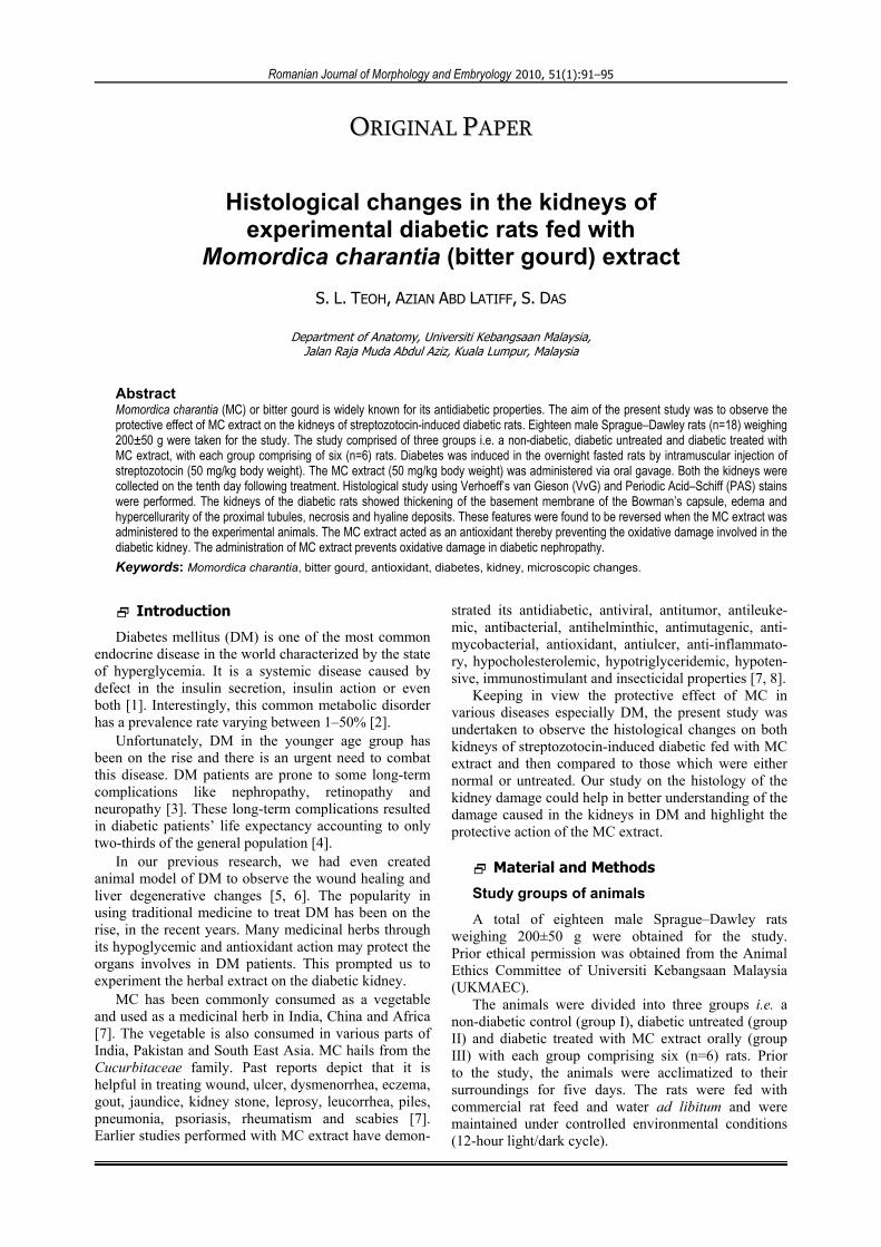
blood glucose level in the diabetic and the non-diabetic rats were found to be 13.23±0.98 mmol/L and 4.65± 0.25 mmol/L, respectively. Following 10 days of treat-ment with the MC extract, the diabetic rats showed a decrease in the fasting blood glucose level (16± 1.6 mmol/L) as compared to the untreated diabetic rats (27.3±1.49 mmol/L) (Figure 1).
Figure 1 – The effect of MC extract on fasting blood glucose level in non-diabetic rats, untreated and treated diabetic rats. The fasting blood glucose level was markedly raised in the diabetic rats as compared in the non-diabetic rats. Treatment with MC extract for 10 days significantly reduced the fasting blood glucose level. Group II and III were compared to group I. *Values were statistically significant at p<0.05.
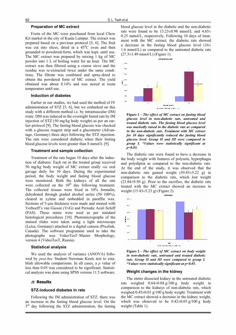
The diabetic rats were found to have a decrease in the body weight with features of polyuria, hyperphagia and polydipsia as compared to the non-diabetic rats. At the end of the study, it was observed that the non-diabetic rats gained weight (59.85±5.22 g) in comparison to the diabetic rats, which lost weight (23.84±9.50 g). Prior to the sacrifice, the diabetic rats treated with the MC extract showed an increase in weight (15.43±5.23 g) (Figure 2).
Figure 2 – The effect of MC extract on body weight in non-diabetic rats, untreated and treated diabetic rats. Group II and III were compared to group I. *Values were statistically significant at p<0.05.
Weight changes in the kidney
The entire dissected kidney in the untreated diabetic rats weighed 0.64±0.04 g/100 g body weight in comparison to the kidneys of non-diabetic rats, which weighed 0.45±0.01 g/100 g body weight. Treatment with the MC extract showed a decrease in the kidney weight, which was observed to be 0.42±0.03 g/100 g body weight (Table 1).

Histological changes in the kidneys of experimental diabetic rats fed with Momordica charantia (bitter gourd) extract
93
Table 1 – The effect of MC extract on kidney weight in non-diabetic rats, untreated and treated diabetic rats Groups Parameter
Non-diabetic Diabetic Diabetic + MC Kidney weight [g] 1.28±0.03 1.25±0.08 0.96±0.04 Kidney weight / 100 g body weight 0.45±0.01 0.64±0.04* 0.42±0.03 All data are expressed as mean±SEM. *p<0.05 vs. the non-diabetic group. Diabetic rats showed renal hypertrophy. Treatment with the MC extract prevented renal hypertrophy.
Histopathological changes
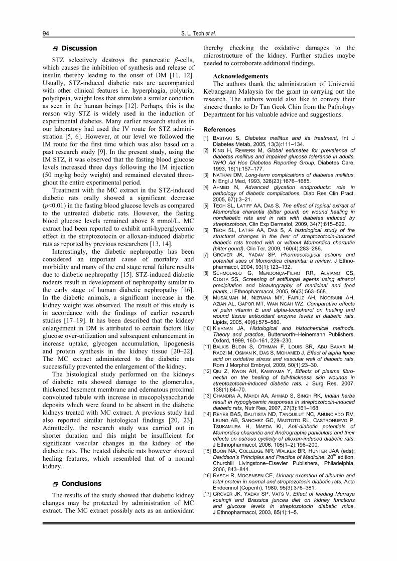
Histological study of the normal kidney of the non-diabetic rats revealed normal glomerulus surrounded by the Bowman’s capsule, proximal and distal convoluted tubules without any inflammatory changes. The kidneys of untreated diabetic rats showed degenerated glomeruli infiltrated by the inflammatory cells and thickening of the basement membrane. The proximal convoluted tubule exhibited edematous changes with deposition of mucopolysaccharide and hyaline substances. All the
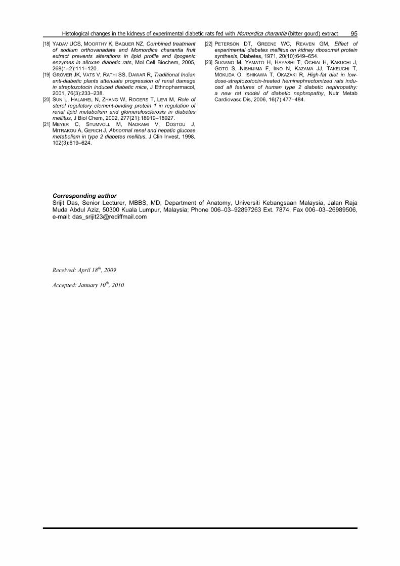
necrotic changes observed in the proximal and distal convoluted tubules along with the deposits were found to be absent in the diabetic rats treated with the MC extract. The group that was treated with MC extract showed features of healing i.e. normal glomerulus, absence of inflammatory cells, normal basement membrane and capillaries, decrease in the mucopoly-saccharide and hyaline deposit, respectively. The tissue necrosis was also observed to decrease in the group treated with MC extract (Figures 3 and 4).
Figure 3 – (a) Photograph of histological section of a normal kidney using the VvG stain (×40); (b) Photograph of histological section of the diabetic kidney (non-treated) using the VvG stain (×40). The necrotic area of the glomerulus in the diabetic kidney is shown with white arrows. The thickening in the basement membrane and the edematous proximal convoluted tubule could be well observed; (c) Photograph of histological section of the diabetic kidney (treated with MC extract) using the VvG stain (×40). Features of healing like normal basement membrane, the absence of necrotic cells in glomerulus and the absence of any edema in the proximal convoluted tubule could be easily appreciated. BC – Bowman’s capsule; G – glomerulus; P – proximal convoluted tubule.
Figure 4 – (a) Photograph of histological section of the normal kidney using the PAS stain (×40); (b) Photograph of histological section of the diabetic kidney (non-treated) using the PAS stain (×40). The proximal convoluted tubule showed hypercellularity with edema (E) and mucopolysaccharide deposits (D) were found; (c) Photograph of histological section of the diabetic kidney (treated with MC extract) using the PAS stain (×40). The edematous changes and deposition of mucopolysaccharide were not observed. G – glomerulus; P – proximal convoluted tubule; E – edematous proximal convoluted tubule; D – mucopolysaccharide deposits.
S. L. Teoh et al.
94
Discussion
STZ selectively destroys the pancreatic β-cells, which causes the inhibition of synthesis and release of insulin thereby leading to the onset of DM [11, 12]. Usually, STZ-induced diabetic rats are accompanied with other clinical features i.e. hyperphagia, polyuria, polydipsia, weight loss that stimulate a similar condition as seen in the human beings [12]. Perhaps, this is the reason why STZ is widely used in the induction of experimental diabetes. Many earlier research studies in our laboratory had used the IV route for STZ admini-stration [5, 6]. However, at our level we followed the IM route for the first time which was also based on a past research study [9]. In the present study, using the IM STZ, it was observed that the fasting blood glucose levels increased three days following the IM injection (50 mg/kg body weight) and remained elevated throu-ghout the entire experimental period.
Treatment with the MC extract in the STZ-induced diabetic rats orally showed a significant decrease (p<0.01) in the fasting blood glucose levels as compared to the untreated diabetic rats. However, the fasting blood glucose levels remained above 8 mmol/L. MC extract had been reported to exhibit anti-hyperglycemic effect in the streptozotocin or alloxan-induced diabetic rats as reported by previous researchers [13, 14].
Interestingly, the diabetic nephropathy has been considered an important cause of mortality and morbidity and many of the end stage renal failure results due to diabetic nephropathy [15]. STZ-induced diabetic rodents result in development of nephropathy similar to the early stage of human diabetic nephropathy [16]. In the diabetic animals, a significant increase in the kidney weight was observed. The result of this study is in accordance with the findings of earlier research studies [17–19]. It has been described that the kidney enlargement in DM is attributed to certain factors like glucose over-utilization and subsequent enhancement in increase uptake, glycogen accumulation, lipogenesis and protein synthesis in the kidney tissue [20–22]. The MC extract administered to the diabetic rats successfully prevented the enlargement of the kidney.
The histological study performed on the kidneys of diabetic rats showed damage to the glomerulus, thickened basement membrane and edematous proximal convoluted tubule with increase in mucopolysaccharide deposits which were found to be absent in the diabetic kidneys treated with MC extract. A previous study had also reported similar histological findings [20, 23]. Admittedly, the research study was carried out in shorter duration and this might be insufficient for significant vascular changes in the kidney of the diabetic rats. The treated diabetic rats however showed healing features, which resembled that of a normal kidney.
Conclusions
The results of the study showed that diabetic kidney changes may be protected by administration of MC extract. The MC extract possibly acts as an antioxidant
thereby checking the oxidative damages to the microstructure of the kidney. Further studies maybe needed to corroborate additional findings.
Acknowledgements The authors thank the administration of Universiti
Kebangsaan Malaysia for the grant in carrying out the research. The authors would also like to convey their sincere thanks to Dr Tan Geok Chin from the Pathology Department for his valuable advice and suggestions.
References [1] BASTAKI S, Diabetes mellitus and its treatment, Int J
Diabetes Metab, 2005, 13(3):111–134. [2] KING H, REWERS M, Global estimates for prevalence of
diabetes mellitus and impaired glucose tolerance in adults. WHO Ad Hoc Diabetes Reporting Group, Diabetes Care, 1993, 16(1):157–177.
[3] NATHAN DM, Long-term complications of diabetes mellitus, N Engl J Med, 1993, 328(23):1676–1685.
[4] AHMED N, Advanced glycation endproducts: role in pathology of diabetic complications, Diab Res Clin Pract, 2005, 67():3–21.
[5] TEOH SL, LATIFF AA, DAS S, The effect of topical extract of Momordica charantia (bitter gourd) on wound healing in nondiabetic rats and in rats with diabetes induced by streptozotocin, Clin Exp Dermatol, 2009, 34(7):815–822.
[6] TEOH SL, LATIFF AA, DAS S, A histological study of the structural changes in the liver of streptozotocin-induced diabetic rats treated with or without Momordica charantia (bitter gourd), Clin Ter, 2009, 160(4):283–286.
[7] GROVER JK, YADAV SP, Pharmacological actions and potential uses of Momordica charantia: a review, J Ethno-pharmacol, 2004, 93(1):123–132.
[8] SCHMOURLO G, MENDONÇA-FILHO RR, ALVIANO CS, COSTA SS, Screening of antifungal agents using ethanol precipitation and bioautography of medicinal and food plants, J Ethnopharmacol, 2005, 96(3):563–568.
[9] MUSALMAH M, NIZRANA MY, FAIRUZ AH, NOORAINI AH, AZIAN AL, GAPOR MT, WAN NGAH WZ, Comparative effects of palm vitamin E and alpha-tocopherol on healing and wound tissue antioxidant enzyme levels in diabetic rats, Lipids, 2005, 40(6):575–580.
[10] KIERNAN JA, Histological and histochemical methods. Theory and practice, Butterworth–Heinemann Publishers, Oxford, 1999, 160–161, 229–230.
[11] BALKIS BUDIN S, OTHMAN F, LOUIS SR, ABU BAKAR M, RADZI M, OSMAN K, DAS S, MOHAMED J, Effect of alpha lipoic acid on oxidative stress and vascular wall of diabetic rats, Rom J Morphol Embryol, 2009, 50(1):23–30.
[12] QIU Z, KWON AH, KAMIYAMA Y, Effects of plasma fibro- nectin on the healing of full-thickness skin wounds in streptozotocin-induced diabetic rats, J Surg Res, 2007, 138(1):64–70.
[13] CHANDRA A, MAHDI AA, AHMAD S, SINGH RK, Indian herbs result in hypoglycemic responses in streptozotocin-induced diabetic rats, Nutr Res, 2007, 27(3):161–168.
[14] REYES BAS, BAUTISTA ND, TANQUILUT NC, ANUNCIADO RV, LEUNG AB, SANCHEZ GC, MAGTOTO RL, CASTRONUEVO P, TSUKAMURA H, MAEDA KI, Anti-diabetic potentials of Momordica charantia and Andrographis paniculata and their effects on estrous cyclicity of alloxan-induced diabetic rats, J Ethnopharmacol, 2006, 105(1–2):196–200.
[15] BOON NA, COLLEDGE NR, WALKER BR, HUNTER JAA (eds), Davidson’s Principles and Practice of Medicine, 20th edition, Churchill Livingstone–Elsevier Publishers, Philadelphia, 2006, 843–844.
[16] RASCH R, MOGENSEN CE, Urinary excretion of albumin and total protein in normal and streptozotocin diabetic rats, Acta Endocrinol (Copenh), 1980, 95(3):376–381.
[17] GROVER JK, YADAV SP, VATS V, Effect of feeding Murraya koeingii and Brassica juncea diet on kidney functions and glucose levels in streptozotocin diabetic mice, J Ethnopharmacol, 2003, 85(1):1–5.
Histological changes in the kidneys of experimental diabetic rats fed with Momordica charantia (bitter gourd) extract
95[18] YADAV UCS, MOORTHY K, BAQUER NZ, Combined treatment
of sodium orthovanadate and Momordica charantia fruit extract prevents alterations in lipid profile and lipogenic enzymes in alloxan diabetic rats, Mol Cell Biochem, 2005, 268(1–2):111–120.
[19] GROVER JK, VATS V, RATHI SS, DAWAR R, Traditional Indian anti-diabetic plants attenuate progression of renal damage in streptozotocin induced diabetic mice, J Ethnopharmacol, 2001, 76(3):233–238.
[20] SUN L, HALAIHEL N, ZHANG W, ROGERS T, LEVI M, Role of sterol regulatory element-binding protein 1 in regulation of renal lipid metabolism and glomerulosclerosis in diabetes mellitus, J Biol Chem, 2002, 277(21):18919–18927.
[21] MEYER C, STUMVOLL M, NADKAMI V, DOSTOU J, MITRAKOU A, GERICH J, Abnormal renal and hepatic glucose metabolism in type 2 diabetes mellitus, J Clin Invest, 1998, 102(3):619–624.
[22] PETERSON DT, GREENE WC, REAVEN GM, Effect of experimental diabetes mellitus on kidney ribosomal protein synthesis, Diabetes, 1971, 20(10):649–654.
[23] SUGANO M, YAMATO H, HAYASHI T, OCHIAI H, KAKUCHI J, GOTO S, NISHIJIMA F, IINO N, KAZAMA JJ, TAKEUCHI T, MOKUDA O, ISHIKAWA T, OKAZAKI R, High-fat diet in low-dose-streptozotocin-treated heminephrectomized rats indu-ced all features of human type 2 diabetic nephropathy: a new rat model of diabetic nephropathy, Nutr Metab Cardiovasc Dis, 2006, 16(7):477–484.
Corresponding author Srijit Das, Senior Lecturer, MBBS, MD, Department of Anatomy, Universiti Kebangsaan Malaysia, Jalan Raja Muda Abdul Aziz, 50300 Kuala Lumpur, Malaysia; Phone 006–03–92897263 Ext. 7874, Fax 006–03–26989506, e-mail: [email protected] Received: April 18th, 2009
Accepted: January 10th, 2010