case series - hindawi publishing...
TRANSCRIPT

Case SeriesMultifacet of Cornea Patch Graft in Anterior Segment Diseases
Prema Chendran, Yong Meng Hsien, and Wan Haslina Wan Abdul Halim
Department of Ophthalmology, University Kebangsaan Malaysia Medical Centre (UKMMC), Jalan Yaacob Latif, 56 000 Bandar Tun Razak, Kuala Lumpur, Malaysia
Correspondence should be addressed to Wan Haslina Wan Abdul Halim; [email protected]
Received 14 June 2019; Accepted 7 September 2019; Published 11 November 2019
Academic Editor: Sandra M. Johnson
Copyright © 2019 Prema Chendran et al. �is is an open access article distributed under the Creative Commons Attribution License, which permits unrestricted use, distribution, and reproduction in any medium, provided the original work is properly cited.
Incidence of cornea melting or perforation is commonly seen in variety of cornea conditions. It can cause debilitating vision loss and impair patient’s daily activity. Several techniques have been described to surgically treat cornea perforation and melting. In this article, a series of corneal diseases treated with corneal patch gra� are highlighted to relate di�erent approach of cornea patch gra� surgery. Post-operative management and complications are discussed.
1. Introduction
Cornea patch gra� has been advocated as the surgical proce-dure to treat variety of anterior segment pathologies. It involves patching the a�ected area with full thickness or partial thick-ness corneal donor tissue. �e aim is to restore globe integrity and prevent further in�ammation that could lead to devastat-ing complications such as endophthalmitis. Cornea perfora-tion and melting can occur in infectious keratitis, autoimmune diseases such as rheumatoid arthritis, ocular trauma and ocu-lar surface disorder.
Patch gra� materials are derived from cornea, sclera, per-icardium and dura mater [1]. Scleral patch gra� is commonly used in anterior segment pathologies because it is relatively cost e�ective and easy to preserve. However due to its avascu-lar property it is o�en associated with progressive tissue necro-sis or melting [2]. Pericardial gra� is used as patch gra� in glaucoma drainage devise (GDD) exposure prevention. It is commercially available, no need to be dependent on eye bank for its availability and has high sterility but has tendency to develop gra� thinning and subsequent exposure of the GDD [3] besides being expensive.
Cornea patch gra� has been indicated in cases of corneal perforation, cornea thinning [4], scleral thinning or as proph-ylaxis to prevent the exposure of GDD [1]. �e advantages of cornea patch gra� compared to others is that: it is translucent, less chances of gra� melting and conjunctiva retraction [1]. It can provide good tectonic support for the ocular wall as the tissue has good strength and rigidity [1]. �is case series of
corneal diseases treated with corneal patch gra� in UKMMC is presented. �e author discuss about the post-operative man-agement that was tailored individually to each patient.
2. Case History
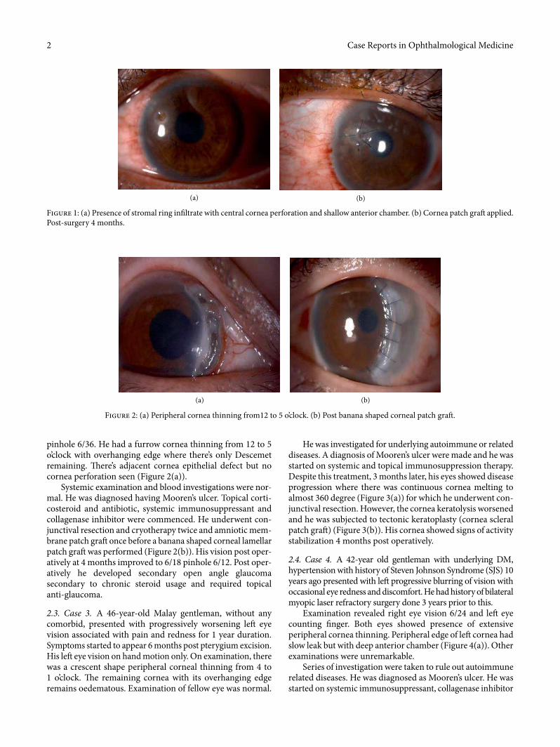
2.1. Case 1. A 32-year-old gentleman, presented to eye casualty with sudden onset le� eye redness and discomfort. He had history of foreign body entering into his le� eye while hammering 1 week prior to that. Examination revealed deeply seated rust ring on the anterior cornea stroma. Failure to remove the rust ring completely resulted in corneal ulceration 1 week later. Topical antibiotics and lubricants were initiated. However, he started to develop cornea thinning. Despite being treated with intensive antibiotics and lubricants, the cornea thinning progressed to cornea perforation with �at anterior chamber (AC) (Figure 1(a)). He underwent series of nonsurgical (cornea glue, bandage contact lens (BCL) application) and surgical (AC reformation and cornea patch gra�) intervention. He was able to regain his vision to 6/9 with presence of stable cornea patch gra� during last review (Figure 1(b)).
2.2. Case 2. A 38-year-old gentleman with underlying diabetes mellitus (DM) type I, hypertension, end stage renal failure (ESRF), right eye pseudophakia presented with right eye blurring of vision associated with photophobia and watery eye for 1 month. Examination revealed right eye vision of 6/60
HindawiCase Reports in Ophthalmological MedicineVolume 2019, Article ID 6862487, 5 pageshttps://doi.org/10.1155/2019/6862487
Case Reports in Ophthalmological Medicine2
pinhole 6/36. He had a furrow cornea thinning from 12 to 5 o’clock with overhanging edge where there’s only Descemet remaining. �ere’s adjacent cornea epithelial defect but no cornea perforation seen (Figure 2(a)).
Systemic examination and blood investigations were nor-mal. He was diagnosed having Mooren’s ulcer. Topical corti-costeroid and antibiotic, systemic immunosuppressant and collagenase inhibitor were commenced. He underwent con-junctival resection and cryotherapy twice and amniotic mem-brane patch gra� once before a banana shaped corneal lamellar patch gra� was performed (Figure 2(b)). His vision post oper-atively at 4 months improved to 6/18 pinhole 6/12. Post oper-atively he developed secondary open angle glaucoma secondary to chronic steroid usage and required topical anti-glaucoma.
2.3. Case 3. A 46-year-old Malay gentleman, without any comorbid, presented with progressively worsening le� eye vision associated with pain and redness for 1 year duration. Symptoms started to appear 6 months post pterygium excision. His le� eye vision on hand motion only. On examination, there was a crescent shape peripheral corneal thinning from 4 to 1 o’clock. �e remaining cornea with its overhanging edge remains oedematous. Examination of fellow eye was normal.
He was investigated for underlying autoimmune or related diseases. A diagnosis of Mooren’s ulcer were made and he was started on systemic and topical immunosuppression therapy. Despite this treatment, 3 months later, his eyes showed disease progression where there was continuous cornea melting to almost 360 degree (Figure 3(a)) for which he underwent con-junctival resection. However, the cornea keratolysis worsened and he was subjected to tectonic keratoplasty (cornea scleral patch gra�) (Figure 3(b)). His cornea showed signs of activity stabilization 4 months post operatively.
2.4. Case 4. A 42-year old gentleman with underlying DM, hypertension with history of Steven Johnson Syndrome (SJS) 10 years ago presented with le� progressive blurring of vision with occasional eye redness and discomfort. He had history of bilateral myopic laser refractory surgery done 3 years prior to this.
Examination revealed right eye vision 6/24 and le� eye counting nger. Both eyes showed presence of extensive peripheral cornea thinning. Peripheral edge of le� cornea had slow leak but with deep anterior chamber (Figure 4(a)). Other examinations were unremarkable.
Series of investigation were taken to rule out autoimmune related diseases. He was diagnosed as Mooren’s ulcer. He was started on systemic immunosuppressant, collagenase inhibitor
Figure 1: (a) Presence of stromal ring inltrate with central cornea perforation and shallow anterior chamber. (b) Cornea patch gra� applied. Post-surgery 4 months.
(a) (b)
Figure 2: (a) Peripheral cornea thinning from12 to 5 o’clock. (b) Post banana shaped corneal patch gra�.
(a) (b)

3Case Reports in Ophthalmological Medicine
and topical corticosteroid and antibiotic. He was scheduled for le� eye cornea banana-shaped patch gra� (Figure 4(b)). Post operatively his vision improved to 6/18 at 3 months and it remains stable without any recurrence.
2.5. Case 5. A 32-year-old Myanmar lady, complained of right eye discomfort with recurrent eye redness for the past 2 years. �e eye redness usually resolve temporarily with over the counter eye drop. She noticed the presence of bluish discoloration at the right nasal conjunctiva for the past 1 year which was increasing in size. Her vision on both eyes were 6/6. She had history of trauma to right eye during childhood.
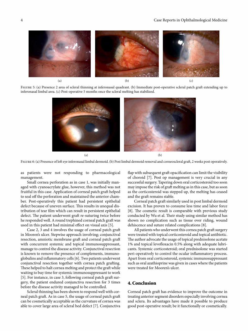
Right eye examination showed two locations of scleral thinning with visible uveal tissue at inferonasal quadrant with adjacent cornea scarring (Figure 5(a)). Other ocular exami-nation were normal. Her infectious and autoimmune disease screening were negative. She was given topical lubricants and topical antiglaucoma (prophylaxis).
She underwent cornea patch gra� for the scleral thinning (Figure 5(b)). Post-operatively she was prescribed with topical antibiotic, topical and systemic corticosteroid. Two weeks post-operatively, her gra� was melting, oral corticosteroid was stepped up, topical steroid and lubricants were continued and she was under close observation for every 2 weeks. Subsequently, no further melting was noted a�er 5 months post-operative period (Figure 5(c)).
2.6. Case 6. A 15-year-old boy with congenital right eye temporal limbal dermoid (Figure 6(a)) was seen in eye clinic. �is child has been on regular follow up with refraction. He had amblyopia on right eye. �e presence of limbal dermoid on his right eye didn’t cross his visual axis.
He underwent excision of the limbal dermoid and corneal patch gra� surgery (Figure 6(b)). Post-operatively, the gra� looked stable. However his refraction noted to have high astig-matism for which he is being followed up.
3. Discussion
Cornea melting is a sequelae of progressive keratolytic process. Untreated cases can lead to cornea perforation. Application of corneal patch gra� is another way to address this issue. Corneal patch gra� use when the cornea defect is too large until the application of cyanoacrylate tissue adhesive is not recom-mended (perforation of >1mm) [5] or when there is cyanoacr-ylate tissue adhesive application failure [5]. �is technique will allow the perseverance of globe integrity, stabilize the eye while waiting for the systemic immunosuppressant e�ect to take place, clearance of necrotic stroma, which is a source of collagenase and shields the bare stroma from the tears neutrophil [5].
In this series, the decision for patch gra� was made due to many reasons. �e author attempted corneal patch gra�
Figure 3: (a) Showing progression of cornea melt to 360 degrees. (b) Whole cornea patch gra�.
(a) (b)
Figure 4: (a) Peripheral cornea melting on le� eye. (b) Post banana shaped corneal patch gra� on le� eye.
(a) (b)
Case Reports in Ophthalmological Medicine4
�ap with subsequent gra� opacication can limit the visibility of choroid [7]. Post op management is very crucial in any successful surgery. Tapering down oral corticosteroid too soon may impose the risk of gra� melting as in this case, but as soon as the corticosteroid was stepped up, the melting has ceased and the gra� remains stable.
Corneal patch gra� similarly used in post limbal dermoid excision. It has proven to consume less time and labor force [8]. �e cosmetic result is comparable with previous study conducted by Wu et al. �eir study using similar method has shown no complication such as tissue over riding, wound dehiscence and suture related complications [8].
All patients who underwent this cornea patch gra� surgery were treated with topical corticosteroid and topical antibiotic. �e author advocate the usage of topical prednisolone acetate 1% and topical levo�oxacin 0.5% along with adequate lubri-cants. Systemic corticosteroid; oral prednisolone was started peri-operatively to control the ocular in�ammatory process. Apart from oral corticosteroid, systemic immunosuppressant such as oral azathioprine was given in cases where the patients were treated for Mooren’s ulcer.
4. Conclusion
Corneal patch gra� has evidence to improve the outcome in treating anterior segment disorders especially involving cornea and sclera. Its advantages have made it possible to produce good post-operative result; be it functionally or cosmetically.
as patients were not responding to pharmacological management.
Small cornea perforation as in case 1, was initially man-aged with cyanoacrylate glue, however, this method was not fruitful in this case. Application of corneal patch gra� helped to seal o� the perforation and maintained the anterior cham-ber. Post-operatively this patient had persistent epithelial defect because of uneven surface. �is results in unequal dis-tribution of tear lm which can result in persistent epithelial defect. �e patient underwent gra� re-suturing twice before he responded well. A round trephined corneal patch gra� was used in this patient had minimal e�ect on visual axis [5].
Case 2, 3 and 4 involves the usage of corneal patch gra� in Mooren’s ulcer. Stepwise approach involving; conjunctival resection, amniotic membrane gra� and corneal patch gra� with concurrent systemic and topical immunosuppressant, manage to control the disease activity. Conjunctival resection is known to remove the presence of complements, immuno-globulins and in�ammatory cells [6]. Two patients underwent conjunctival resection together with cornea patch gra�ing. �ese helped to halt cornea melting and protect the gra� while waiting to buy time for systemic immunosuppressant to work [5]. For instance, in case 3, following corneal patch gra� sur-gery, the patient endured conjunctiva resection for 3 times before the disease activity managed to be controlled.
Scleral thinning has been shown to respond well with cor-neal patch gra�. As in case 5, the usage of corneal patch gra� can be cosmetically acceptable as the curvature of cornea was able to cover large area of scleral bed defect [7]. Conjunctiva
Figure 5: (a) Presence 2 area of scleral thinning at inferonasal quadrant. (b) Immediate post-operative scleral patch gra� extending up to inferonasal limbal area. (c) Post-operative 5 months once the scleral melting has stabilized.
(a) (b) (c)
Figure 6: (a) Presence of le� eye inferonasal limbal dermoid. (b) Post limbal dermoid removal and corneoscleral gra�, 2 weeks post operatively.
(a) (b)

5Case Reports in Ophthalmological Medicine
Conflicts of Interest
�e authors declare that they have no conflicts of interest.
References
[1] O. Spierer, M. Waisbourd, Y. Golan, H. Newman, and R. Rachmiel, “Partial thickness corneal tissue as patch gra� material for prevention of glaucoma drainage device exposure,” BMC Opthalmology, vol. 16, no. 1, 2016.
[2] Y. Yao and V. Jhanji, “Short term observation and management of scleral patch gra� used in scleral defects,” Annals of Eye Science, vol. 2, p. 30, 2017.
[3] T. Raviv, D. S. Greenfield, J. M. Liebmann, P. A. Sidoti, H. Ishikawa, and R. Ritch, “Pericardial patch gra� in glaucoma implant surgery,” Journal of Glaucoma, vol. 7, no. 1, pp. 27–32, 1998.
[4] G. Schaab, C. Clavell, and H. Goyal, “Corneal patch gra�: a review of indication, technique and success rates. ARVO annual meeting abstract,” Investigative Ophthalmology & Visual Science, vol. 53, no. 14, p. 6044, 2012.
[5] H. K. Soong, A. A. Farjo, D. Katz, R. F. Meyer, and A. Sugar, “Lamellar corneal patch gra� in the management of cornea melting,” Cornea, vol. 19, no. 2, pp. 126–134, 2000.
[6] I. Lal, S. B. Shivanagari, M. H. Ali, and J. Vazirani, “Efficacy of conjunctival resection with cyanoacrylate glue application in preventing recurrences of Mooren’s ulcer,” British Journal of Ophthalmology, vol. 100, no. 7, pp. 971–975, 2016.
[7] C. C. de Farias, T. de Sterlenich, L. B. Souza, L. A. Vieira, and J. A. P. Gomes, “Randomized trial comparing multilayer amniotic membrane transplantation with scleral and corneal gra� for the treatment of scleral thinning a�er pterygium surgery associated with beta therapy,” Cornea, vol. 33, no. 11, pp. 1197–1204, 2014.
[8] K. I. Wu, H. S. Chu, A. S. I. Pai et al., “Surgical management of limbal dermoid using anterior corneal button from descemet stripping automated endothelial keratoplasty donor tissue as patch gra�,” Cornea, vol. 36, no. 1, pp. 64–67, 2017.

Stem Cells International
Hindawiwww.hindawi.com Volume 2018
Hindawiwww.hindawi.com Volume 2018
MEDIATORSINFLAMMATION
of
EndocrinologyInternational Journal of
Hindawiwww.hindawi.com Volume 2018
Hindawiwww.hindawi.com Volume 2018
Disease Markers
Hindawiwww.hindawi.com Volume 2018
BioMed Research International
OncologyJournal of
Hindawiwww.hindawi.com Volume 2013
Hindawiwww.hindawi.com Volume 2018
Oxidative Medicine and Cellular Longevity
Hindawiwww.hindawi.com Volume 2018
PPAR Research
Hindawi Publishing Corporation http://www.hindawi.com Volume 2013Hindawiwww.hindawi.com
The Scientific World Journal
Volume 2018
Immunology ResearchHindawiwww.hindawi.com Volume 2018
Journal of
ObesityJournal of
Hindawiwww.hindawi.com Volume 2018
Hindawiwww.hindawi.com Volume 2018
Computational and Mathematical Methods in Medicine
Hindawiwww.hindawi.com Volume 2018
Behavioural Neurology
OphthalmologyJournal of
Hindawiwww.hindawi.com Volume 2018
Diabetes ResearchJournal of
Hindawiwww.hindawi.com Volume 2018
Hindawiwww.hindawi.com Volume 2018
Research and TreatmentAIDS
Hindawiwww.hindawi.com Volume 2018
Gastroenterology Research and Practice
Hindawiwww.hindawi.com Volume 2018
Parkinson’s Disease
Evidence-Based Complementary andAlternative Medicine
Volume 2018Hindawiwww.hindawi.com
Submit your manuscripts atwww.hindawi.com