damage in the multiethnic malaysian systemic lupus ... · yaacob latiff, 56000 kuala lumpur,...
TRANSCRIPT
RESEARCHARTICLE
Damage in the Multiethnic MalaysianSystemic Lupus Erythematosus (SLE) Cohort:Comparison with Other Cohorts WorldwideSyahrul Sazliyana Shaharir1*, Heselynn Hussein1,2‡, Sakthiswary Rajalingham1‡, MohdShahrir Mohamed Said1‡, Abdul Halim Abdul Gafor1‡, Rozita Mohd1‡, Ruslinda Mustafar1‡
1 Department of InternalMedicine, Pusat Perubatan Universiti Kebangsaan Malaysia (UKMMC), JalanYaacob Latiff, 56000 Kuala Lumpur, Malaysia, 2 Department of Medicine, Putrajaya Hospital, Jalan P9,Presint 7, 62250 Putrajaya, Malaysia
‡ These authors also contributed equally to this work.* [email protected]
AbstractSystemic Lupus Erythematosus (SLE) is a chronic autoimmune disease and despite the
improvement in the survival in the past few decades, the morbidity due to disease damage
remains significant. The objectives of this study were to investigate the disease damagepat-
tern and determine the associated factors of damage in the multi-ethnicMalaysian SLE
patients. We consecutively 424SLE patients who attended a consistent follow-up at the
National University of Malaysia Medical Centre and Putrajaya Hospital were recruited.Dis-
ease damage was assessed using the SLICC/ACR (Systemic Lupus InternationalCollabo-
rating Clinics/AmericanCollege of Rheumatology)Damage Index (SDI) scores. Information
on their demographics and disease characteristicswere obtained from the clinical record.
Univariate analysis was performed and the best model of independent predictors of disease
damage was determinedby multivariate logistic regression analysis. A total of 182 patients
(42.9%) had disease damage (SDI�1). A significantly higher number of Indian patients had
disease/organ damage and they predominantly developed steroid-induced diabetesmelli-
tus (SDM). Patients with corticosteroid-induced osteoporosis (CIOP) were more likely to be
Malayswhile majority of patients who developed malignancy were Chinese (p<0.05). In theunivariate and multivariate analyses, disease damage was significantly associated with
age, Indian ethnicity, lower mean cumulative C3 level, neuropsychiatry lupus (NPSLE), and
antiphospholipid syndrome (APLS). Patients who had ever and early treatmentwith hydro-
xychloroquine(HCQ)were less likely to develop disease damage while more patients who
had received oral prednisolone�1mg/kg daily over 2 weeks had disease damage (p<0.05).In conclusion, there were inter-ethnic differences in the damage pattern and risks among
SLE patients.
PLOSONE | DOI:10.1371/journal.pone.0166270 November 15, 2016 1 / 14
a11111
OPENACCESS
Citation:Shaharir SS, Hussein H, RajalinghamS,MohamedSaid MS, Abdul Gafor AH, Mohd R, et al.(2016) Damage in the MultiethnicMalaysianSystemic Lupus Erythematosus (SLE) Cohort:Comparison with Other CohortsWorldwide. PLoSONE 11(11): e0166270. doi:10.1371/journal.pone.0166270
Editor:Masataka Kuwana, JAPAN
Received:August 16, 2016
Accepted:October 25, 2016
Published:November 15, 2016
Copyright:© 2016 Shaharir et al. This is an openaccess article distributed under the terms of theCreative Commons Attribution License, which permitsunrestricteduse, distribution, and reproduction in anymedium, provided the original author and source arecredited.
Data Availability Statement:All relevant data arewithin the paper and its Supporting Information files.
Funding: The authors received no specific fundingfor this work.
Competing Interests: The authors have declaredthat no competing interests exist.

IntroductionSystemic Lupus Erythematosus (SLE) is an autoimmune disease which is characterized bymulti-system organ inflammation. Despite a remarkable improvement in the management ofSLE and improved survival in the past few decades [1], however, the morbidity due to organdamage sequelae remains significant. In view of this, apart from disease activity and quality oflife, measurement of disease damage is very important as part of a standard assessment in themanagement of SLE patients.
The Systemic Lupus Erythematosus International CollaboratingClinics/AmericanCollegeof Rheumatology (SLICC/ACR) damage index for SLE is a well validated tool to assess accumu-lated damage index (DI) since the onset of the disease [2]. The damage includes non-reversiblechanges in organs and systems affected by the disease process itself, its therapy, or inter-currentillness. The SLICC/ACR damage index is highly reproducible and has been shown to have agood agreement with prospective and retrospectivemeasurement of DI [3]. Disease damagemeasured with SLICC/ACR damage index is well correlated with mortality [4] and quality oflife in patients with SLE [5]. In addition, a different pattern of organ damage such as renal, neu-ropsychiatry, cardiovascular and pulmonary damage also predicts poorer prognosis and mor-tality [2, 6].
Disease outcome in SLE is largely influenced by various factors which include genetic,socio-cultural, behavioural and environment [7]. Ethnicity was considered as part of a geneticmarker and it is well established that Asian and Non-Caucasian SLE patients generally exhibitmore severe lupus and poorer outcome [8–11]. Several studies have evaluated the systemicdamage among SLE patients from various ethnic backgrounds. The LUMINA (LUpus inMInorities: NAture vs nurture) cohort revealed a different disease outcome and damageamong their Caucasian, Hispanic and African-American SLE patients [8,9] while GLADELstudy group demonstrated the influence of the multi-nationality and ethnicity among SLEpatients in Latin America towards the disease outcome [12]. However, the data on morbidityand burden of SLE in Asia is still lacking and majority of the studies which addressed this issuewere mainly fromOrientals or Chinese ethnicity [13–15].
Malaysia is a multi-racial Southeast Asian country which comprises of three major ethnicgroups. The largest ethnic composition is Malay, followed by Chinese and Indian. The catch-ment area of Universiti Kebangsaan Malaysia Medical Centre (UKMMC) and Putrajaya Hospi-tal comprised of Malays (45.9%), Chinese (43.2%) and Indians (10.3%) [16].
Previous studies which were performed in a tertiary centre in Kuala Lumpur, Malaysia havedemonstrated that the prevalence of SLE was seen to be higher in Chinese population, followedby Malays. On the other hand, prevalence of SLE among Malaysian Indians are generally lowranging from 7–11.6% only [17,18]. This was in contrast to Rheumatoid Arthritis in whichIndians were predominantly affected as the reported prevalence of RA among them were54.5% [19].
Although SLE was less common among Indians in Malaysia, they have lower survival rateas compared to other ethnicities [17]. However, this study was done almost two decades agoand the trend may have changed, parallel with the progress in the overall lupus managementand care. Herein we described the differences in the disease damage pattern among our multi-ethnic SLE cohort and highlighted the factors contributing to irreversible damage among them.
MethodologyThis was a cross-sectional study involving SLE patients attending a regular follow up and mon-itoring at the Rheumatology and Nephrology/SLE Clinic in National University of MalaysiaMedical Centre (UKMMC) and Putrajaya Hospital, Malaysia. Consecutive patients with
Damage in Malaysian SLE Cohort
PLOSONE | DOI:10.1371/journal.pone.0166270 November 15, 2016 2 / 14
minimum disease duration of 6 months were enrolled from August 2013 until January 2015.All patients fulfilled at least 4 criteria from the American College of RheumatologyClassifica-tion Criteria for SLE 1997 [20]or renal biopsy consistent with lupus nephritis (LN). The localethic committee of National University Malaysia Medical Centre and Ministry of Health,Malaysia have approved the study (FF-2013-337 and NMRR-14-386-19203). Informed writtenconsent was taken from the patients and this procedure has been approved by the ethic com-mittee of National University Malaysia Medical Centre and Ministry of Health Malaysia.
Clinical and laboratoryevaluationDetailed demographic data were collected including age, gender and self-reported ethnicity.Information on the disease characteristics (onset of disease, system involvement in lupus andduration of disease) were obtained from the medical records. The presence of autoantibodiesincluding anti-DsDNA and antiphospholipid antibodies including lupus anticoagulant (LA)and anti cardiolipin (ACL) antibodies were determined from the medical records. The meancumulative of complement levels (C3 and C4) were determined by calculating the mean ofcumulative complement C3 and C4 levels which were done routinely at least 6–12 monthlyduring their clinical follow-up.
The information on the type of immunosuppressants used at any point of SLE diseasecourse or follow up such as mycophenolic acid (MMF), cyclosporineA (CyA), azathioprineand hydroxychloroquine (HCQ) were determined from the clinical records and electronicpharmacy prescriptions. The history of oral prednisolone�1mg/kg daily for over two weeksusage and early HCQ use (less than 3months after SLE diagnosis) was obtained from the medi-cal records. Cumulative exposure to glucocorticoids and cyclophosphamide (CYC) was alsocalculated. For patients with disease damage (SDI�1), the cumulative dose of corticosteroidsand CYC were calculated until the date of organ damage development was confirmed.Mean-while, for patients without disease damage, the cumulative corticosteroids and CYC were calcu-lated until the date of the last visit to the clinic.
Disease damage assessmentThe presence of disease damage was assessed by the rheumatologists and determined from themedical records. Itwas measured using the Systemic Lupus International CollaboratingClinics/American College of Rheumatology damage index (SDI) [2]. This index documents cumulativeand irreversible damage, irrespective of its cause, in 12 different organ systems. Apart fromdamage that is resulted from previous disease activity, the SLICC damage index also measuresthe irreversible damage due to treatment or medications including corticosteroid induced oste-oporosis (CIOP), diabetes mellitus and cataracts. To be scored, each manifestation must bepresent for at least 6 months. The category of damage and the onset of damage from the date ofdiagnosis were determined from the medical records. As part of a standard follow up monitor-ing protocol, a regular bone mineral density scan was performed at least every 2 years inpatients who were on long term corticosteroids.Diagnosis of osteoporotic fractures were basedclinical history of fracture in the typical sites (neck of femur, spine, wrist) with trivial traumawhich was associated with low bone mineral density (T score less than -1). Subsequently,patients were dichotomized into the presence and absence of damage based on a cutoff of theSDI score of 1.
Statistical analysisFeatures from the different parameters were compared between those with disease damage(SDI�1) and without disease damage (SDI = 0) using standard statistical tests. All normally
Damage in Malaysian SLE Cohort
PLOSONE | DOI:10.1371/journal.pone.0166270 November 15, 2016 3 / 14
distributed numerical data will be expressed as mean ±SD and continuous variables were ana-lyzed with students T test. Non-normally distributed data will be expressed as median ± Inter-quartile range (IQR) and continuous variables were analyzed using Mann-Whitney U test.Chi-square test was used for categorical variables. Significancewas taken as p<0.05. Multivari-able logistic regression analysis was performed to determine the odds ratio of the each indepen-dent predictors of disease damage in LN. All factors which were found to be significantlyassociated with disease damage (p< 0.05) during the univariate analysis were included in theregression. All statistical analyses were performed using the SPSS program version 22.0.
ResultsA total of 424patients were included into the study. The cohort’s mean age was 38.9 ± 13.5years with mean disease duration of 9.2 ± 6.8 years. Their mean age at SLEdiagnosiswas29.7 ± 13.2 years and majority of them were women (n = 390, 92%). Our cohort consistedof a multi-ethnic populations with predominant Malays (n = 265, 62.5%), followed by Chinese(n = 137, 32.3%), Indians (n = 18, 4.2%), and others (n = 4, 0.9%).
Majority of them had lupus nephritis (n = 272, 64.5%), followed by haematological(n = 264, 62.4%), musculoskeletal (n = 250, 59.4%), mucocutaneous (n = 187, 44.5%), neuro-psychiatric lupus (NPSLE) (n = 57 (13.5%) and serositis (n = 45, (10.7%). A total of36 (8.5%)patients had antiphospholipid syndrome (APLS).
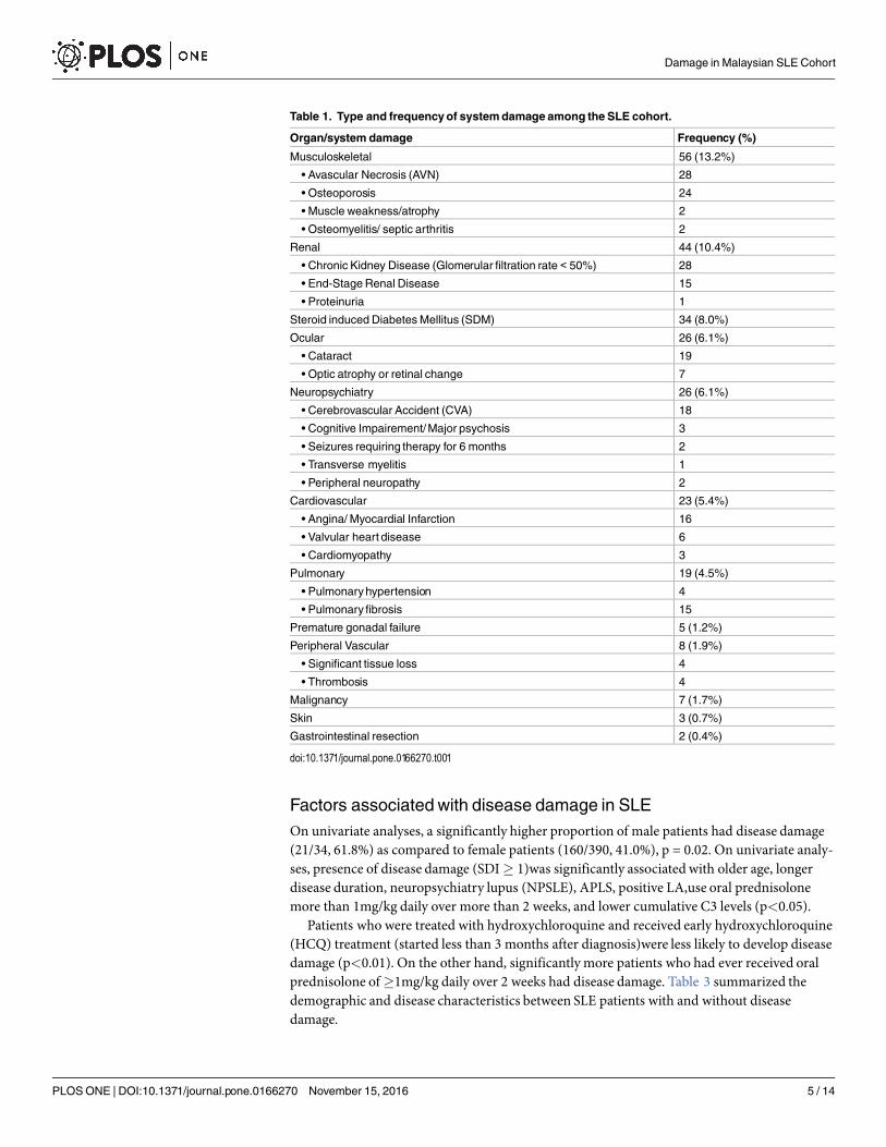
Disease Damage among SLE patientsA total of 182 patients (42.9%) had disease damage (SDI�1) with a mean of onset of damagewas 5.8 ± 4.8 years from the diagnosis of SLE. The median SLICC/ACR damage index (SDI)was 1 (IQR 1).Table 1 illustrates the system domain of disease damage in the lupus cohort.
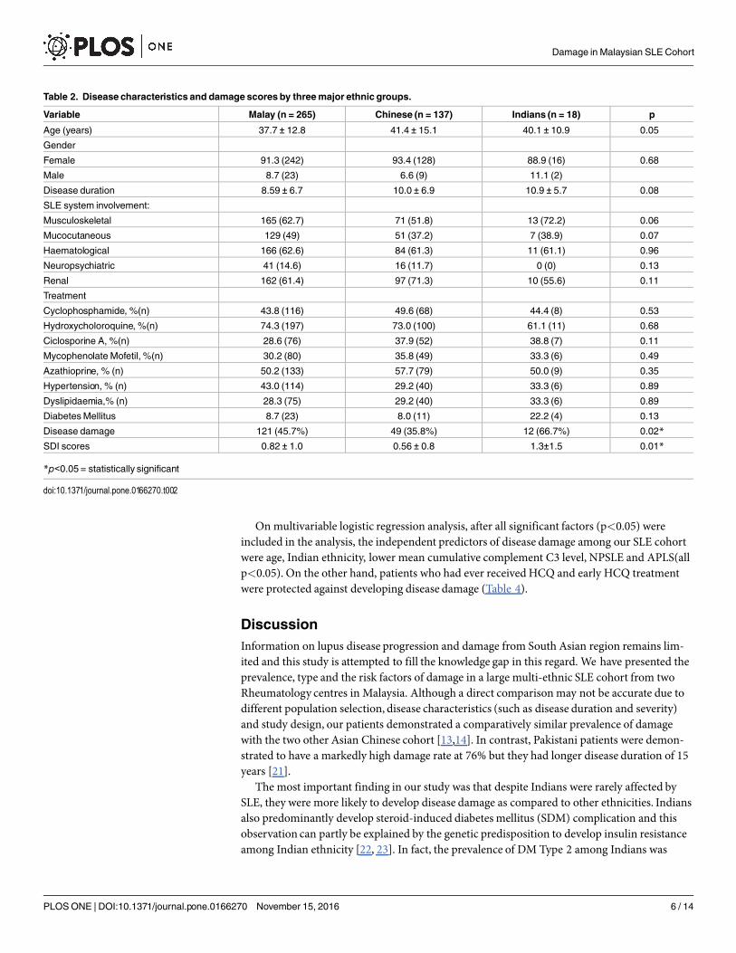
Although only 4.2% (n = 18) in our cohort was Indians, however, approximately two-third(n = 12/18, 66.7%) of them had disease damage. This proportion was significantly higher ascompared to other ethnicities(42.1%), p = 0.04. Indian patients also have a higher mean SLICCscore as compared to other ethnicities. Table 2 illustrates the comparisons of the demographics,disease characteristics, treatment, mean SDI scores and disease damage prevalence between the3 major ethnics in our cohort.
Indian patients predominantly developed steroid-inducedDiabetesMellitus (SDM) as33.3% of them developed this complication, as compared to only 6.9% from other ethnicities(p = 0.002). Malay ethnicity was associated with corticosteroid induced osteoporosis (CIOP) as79.2% (n = 19/24) of patient with CIOP were Malays as compared to only 20.8% (n = 5/24)from other ethnicities (p = 0.02). Majority of the osteoporotic fractures occurred at the spinalregion (n = 22/24 cases) while two of them had neck of femur fractures. 85.7% (n = 6) ofpatients who developedmalignancy were Chinese while one of them was Malay (14.3%),p = 0.01.
Since our cohort consisted of a minority of Indian ethnicity, comparisons was also madebetween the two major ethnic groups, ie Malays and Chinese, while excluding the Indianpatients. Disease damage prevalence was higher among Malays (n = 121, 45.7%) compared tothe Chinese patients (n = 49, 35.8%), p = 0.07. More Malay patients developedCIOP(n = 19,7.2%) compared to the Chinese (n = 4, 2.9%), p = 0.06. Apart from that, pulmonarydamage was more also more prevalent among the Malays (n = 16, 6%) as compared to the Chi-nese patients (n = 3, 2.2%), p = 0.07. Otherwise, there was no significant difference in the prev-alence of other disease damage domains betweenMalays and Chinese patients.
Damage in Malaysian SLE Cohort
PLOSONE | DOI:10.1371/journal.pone.0166270 November 15, 2016 4 / 14
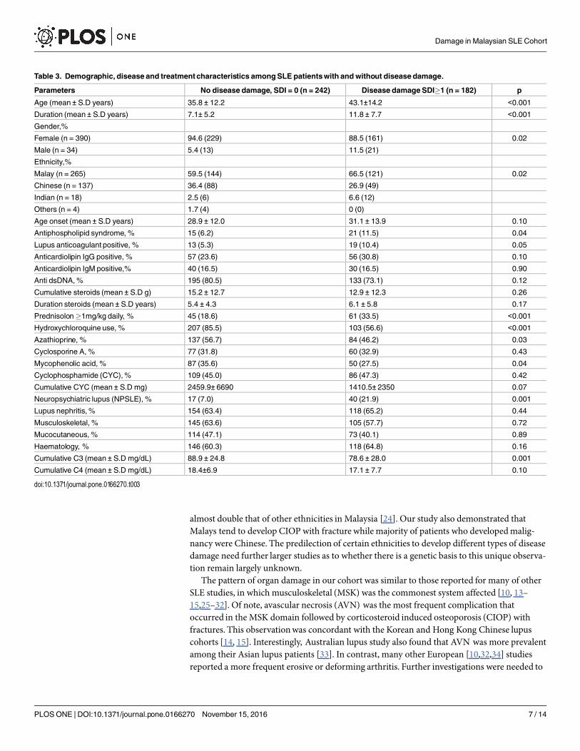
Factors associated with disease damage in SLEOn univariate analyses, a significantly higher proportion of male patients had disease damage(21/34, 61.8%) as compared to female patients (160/390, 41.0%), p = 0.02. On univariate analy-ses, presence of disease damage (SDI� 1)was significantly associated with older age, longerdisease duration, neuropsychiatry lupus (NPSLE), APLS, positive LA,use oral prednisolonemore than 1mg/kg daily over more than 2 weeks, and lower cumulative C3 levels (p<0.05).
Patients who were treated with hydroxychloroquine and received early hydroxychloroquine(HCQ) treatment (started less than 3 months after diagnosis)were less likely to develop diseasedamage (p<0.01). On the other hand, significantlymore patients who had ever received oralprednisolone of�1mg/kg daily over 2 weeks had disease damage. Table 3 summarized thedemographic and disease characteristics between SLE patients with and without diseasedamage.
Table 1. Type and frequency of system damage among the SLE cohort.
Organ/system damage Frequency (%)
Musculoskeletal 56 (13.2%)
• Avascular Necrosis (AVN) 28
• Osteoporosis 24
• Muscle weakness/atrophy 2
• Osteomyelitis/ septic arthritis 2
Renal 44 (10.4%)
• Chronic Kidney Disease (Glomerular filtration rate < 50%) 28
• End-StageRenal Disease 15
• Proteinuria 1
Steroid induced DiabetesMellitus (SDM) 34 (8.0%)
Ocular 26 (6.1%)
• Cataract 19
• Optic atrophy or retinal change 7
Neuropsychiatry 26 (6.1%)
• Cerebrovascular Accident (CVA) 18
• Cognitive Impairement/Major psychosis 3
• Seizures requiring therapy for 6 months 2
• Transverse myelitis 1
• Peripheral neuropathy 2
Cardiovascular 23 (5.4%)
• Angina/ Myocardial Infarction 16
• Valvular heart disease 6
• Cardiomyopathy 3
Pulmonary 19 (4.5%)
• Pulmonaryhypertension 4
• Pulmonaryfibrosis 15
Premature gonadal failure 5 (1.2%)
Peripheral Vascular 8 (1.9%)
• Significant tissue loss 4
• Thrombosis 4
Malignancy 7 (1.7%)
Skin 3 (0.7%)
Gastrointestinal resection 2 (0.4%)
doi:10.1371/journal.pone.0166270.t001
Damage in Malaysian SLE Cohort
PLOSONE | DOI:10.1371/journal.pone.0166270 November 15, 2016 5 / 14
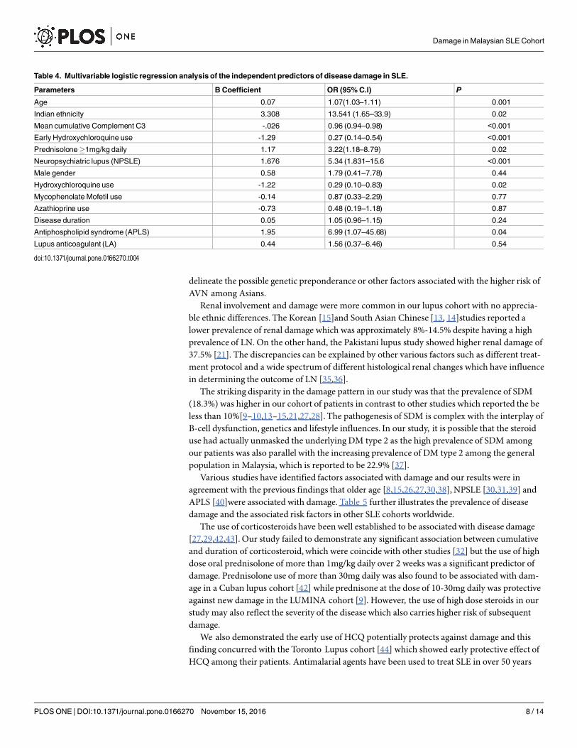
Onmultivariable logistic regression analysis, after all significant factors (p<0.05) wereincluded in the analysis, the independent predictors of disease damage among our SLE cohortwere age, Indian ethnicity, lower mean cumulative complement C3 level, NPSLE and APLS(allp<0.05). On the other hand, patients who had ever receivedHCQ and early HCQ treatmentwere protected against developing disease damage (Table 4).
DiscussionInformation on lupus disease progression and damage from South Asian region remains lim-ited and this study is attempted to fill the knowledge gap in this regard. We have presented theprevalence, type and the risk factors of damage in a large multi-ethnic SLE cohort from twoRheumatology centres in Malaysia. Although a direct comparisonmay not be accurate due todifferent population selection, disease characteristics (such as disease duration and severity)and study design, our patients demonstrated a comparatively similar prevalence of damagewith the two other Asian Chinese cohort [13,14]. In contrast, Pakistani patients were demon-strated to have a markedly high damage rate at 76% but they had longer disease duration of 15years [21].
The most important finding in our study was that despite Indians were rarely affected bySLE, they were more likely to develop disease damage as compared to other ethnicities. Indiansalso predominantly develop steroid-induced diabetes mellitus (SDM) complication and thisobservation can partly be explained by the genetic predisposition to develop insulin resistanceamong Indian ethnicity [22, 23]. In fact, the prevalence of DM Type 2 among Indians was
Table 2. Disease characteristics and damage scores by three major ethnic groups.
Variable Malay (n = 265) Chinese (n = 137) Indians (n = 18) p
Age (years) 37.7 ± 12.8 41.4 ± 15.1 40.1 ± 10.9 0.05
Gender
Female 91.3 (242) 93.4 (128) 88.9 (16) 0.68
Male 8.7 (23) 6.6 (9) 11.1 (2)
Disease duration 8.59 ± 6.7 10.0 ± 6.9 10.9 ± 5.7 0.08
SLE system involvement:
Musculoskeletal 165 (62.7) 71 (51.8) 13 (72.2) 0.06
Mucocutaneous 129 (49) 51 (37.2) 7 (38.9) 0.07
Haematological 166 (62.6) 84 (61.3) 11 (61.1) 0.96
Neuropsychiatric 41 (14.6) 16 (11.7) 0 (0) 0.13
Renal 162 (61.4) 97 (71.3) 10 (55.6) 0.11
Treatment
Cyclophosphamide, %(n) 43.8 (116) 49.6 (68) 44.4 (8) 0.53
Hydroxycholoroquine, %(n) 74.3 (197) 73.0 (100) 61.1 (11) 0.68
Ciclosporine A, %(n) 28.6 (76) 37.9 (52) 38.8 (7) 0.11
Mycophenolate Mofetil, %(n) 30.2 (80) 35.8 (49) 33.3 (6) 0.49
Azathioprine, % (n) 50.2 (133) 57.7 (79) 50.0 (9) 0.35
Hypertension, % (n) 43.0 (114) 29.2 (40) 33.3 (6) 0.89
Dyslipidaemia,% (n) 28.3 (75) 29.2 (40) 33.3 (6) 0.89
DiabetesMellitus 8.7 (23) 8.0 (11) 22.2 (4) 0.13
Disease damage 121 (45.7%) 49 (35.8%) 12 (66.7%) 0.02*
SDI scores 0.82 ± 1.0 0.56 ± 0.8 1.3±1.5 0.01*
*p<0.05 = statistically significant
doi:10.1371/journal.pone.0166270.t002
Damage in Malaysian SLE Cohort
PLOSONE | DOI:10.1371/journal.pone.0166270 November 15, 2016 6 / 14
almost double that of other ethnicities in Malaysia [24]. Our study also demonstrated thatMalays tend to develop CIOP with fracture while majority of patients who developedmalig-nancy were Chinese. The predilection of certain ethnicities to develop different types of diseasedamage need further larger studies as to whether there is a genetic basis to this unique observa-tion remain largely unknown.
The pattern of organ damage in our cohort was similar to those reported for many of otherSLE studies, in which musculoskeletal (MSK) was the commonest system affected [10, 13–15,25–32]. Of note, avascular necrosis (AVN) was the most frequent complication thatoccurred in the MSK domain followed by corticosteroid induced osteoporosis (CIOP) withfractures. This observationwas concordant with the Korean and Hong Kong Chinese lupuscohorts [14, 15]. Interestingly, Australian lupus study also found that AVN was more prevalentamong their Asian lupus patients [33]. In contrast, many other European [10,32,34] studiesreported a more frequent erosive or deforming arthritis. Further investigations were needed to
Table 3. Demographic, disease and treatment characteristics among SLE patients with and without disease damage.
Parameters No disease damage, SDI = 0 (n = 242) Disease damage SDI�1 (n = 182) p
Age (mean ± S.D years) 35.8 ± 12.2 43.1±14.2 <0.001Duration (mean ± S.D years) 7.1± 5.2 11.8 ± 7.7 <0.001Gender,%
Female (n = 390) 94.6 (229) 88.5 (161) 0.02
Male (n = 34) 5.4 (13) 11.5 (21)
Ethnicity,%
Malay (n = 265) 59.5 (144) 66.5 (121) 0.02
Chinese (n = 137) 36.4 (88) 26.9 (49)
Indian (n = 18) 2.5 (6) 6.6 (12)
Others (n = 4) 1.7 (4) 0 (0)
Age onset (mean ± S.D years) 28.9 ± 12.0 31.1 ± 13.9 0.10
Antiphospholipid syndrome, % 15 (6.2) 21 (11.5) 0.04
Lupus anticoagulant positive, % 13 (5.3) 19 (10.4) 0.05
Anticardiolipin IgG positive, % 57 (23.6) 56 (30.8) 0.10
Anticardiolipin IgM positive,% 40 (16.5) 30 (16.5) 0.90
Anti dsDNA,% 195 (80.5) 133 (73.1) 0.12
Cumulative steroids (mean ± S.D g) 15.2 ± 12.7 12.9 ± 12.3 0.26
Duration steroids (mean ± S.D years) 5.4 ± 4.3 6.1 ± 5.8 0.17
Prednisolon�1mg/kg daily, % 45 (18.6) 61 (33.5) <0.001Hydroxychloroquine use, % 207 (85.5) 103 (56.6) <0.001Azathioprine, % 137 (56.7) 84 (46.2) 0.03
Cyclosporine A, % 77 (31.8) 60 (32.9) 0.43
Mycophenolic acid, % 87 (35.6) 50 (27.5) 0.04
Cyclophosphamide (CYC),% 109 (45.0) 86 (47.3) 0.42
Cumulative CYC (mean ± S.D mg) 2459.9± 6690 1410.5± 2350 0.07
Neuropsychiatric lupus (NPSLE), % 17 (7.0) 40 (21.9) 0.001
Lupus nephritis,% 154 (63.4) 118 (65.2) 0.44
Musculoskeletal, % 145 (63.6) 105 (57.7) 0.72
Mucocutaneous, % 114 (47.1) 73 (40.1) 0.89
Haematology, % 146 (60.3) 118 (64.8) 0.16
Cumulative C3 (mean ± S.D mg/dL) 88.9 ± 24.8 78.6 ± 28.0 0.001
Cumulative C4 (mean ± S.D mg/dL) 18.4±6.9 17.1 ± 7.7 0.10
doi:10.1371/journal.pone.0166270.t003
Damage in Malaysian SLE Cohort
PLOSONE | DOI:10.1371/journal.pone.0166270 November 15, 2016 7 / 14
delineate the possible genetic preponderance or other factors associated with the higher risk ofAVN among Asians.
Renal involvement and damage were more common in our lupus cohort with no apprecia-ble ethnic differences. The Korean [15]and South Asian Chinese [13, 14]studies reported alower prevalence of renal damage which was approximately 8%-14.5% despite having a highprevalence of LN. On the other hand, the Pakistani lupus study showed higher renal damage of37.5% [21]. The discrepancies can be explained by other various factors such as different treat-ment protocol and a wide spectrumof different histological renal changes which have influencein determining the outcome of LN [35,36].
The striking disparity in the damage pattern in our study was that the prevalence of SDM(18.3%) was higher in our cohort of patients in contrast to other studies which reported the beless than 10%[9–10,13–15,21,27,28]. The pathogenesis of SDM is complex with the interplay ofB-cell dysfunction, genetics and lifestyle influences. In our study, it is possible that the steroiduse had actually unmasked the underlying DM type 2 as the high prevalence of SDM amongour patients was also parallel with the increasing prevalence of DM type 2 among the generalpopulation in Malaysia, which is reported to be 22.9% [37].
Various studies have identified factors associated with damage and our results were inagreement with the previous findings that older age [8,15,26,27,30,38], NPSLE [30,31,39] andAPLS [40]were associated with damage. Table 5 further illustrates the prevalence of diseasedamage and the associated risk factors in other SLE cohorts worldwide.
The use of corticosteroids have been well established to be associated with disease damage[27,29,42,43]. Our study failed to demonstrate any significant association between cumulativeand duration of corticosteroid, which were coincide with other studies [32] but the use of highdose oral prednisolone of more than 1mg/kg daily over 2 weeks was a significant predictor ofdamage. Prednisolone use of more than 30mg daily was also found to be associated with dam-age in a Cuban lupus cohort [42] while prednisone at the dose of 10-30mg daily was protectiveagainst new damage in the LUMINA cohort [9]. However, the use of high dose steroids in ourstudy may also reflect the severity of the disease which also carries higher risk of subsequentdamage.
We also demonstrated the early use of HCQ potentially protects against damage and thisfinding concurredwith the Toronto Lupus cohort [44] which showed early protective effect ofHCQ among their patients. Antimalarial agents have been used to treat SLE in over 50 years
Table 4. Multivariable logistic regression analysis of the independent predictors of disease damage in SLE.
Parameters B Coefficient OR (95% C.I) P
Age 0.07 1.07(1.03–1.11) 0.001
Indian ethnicity 3.308 13.541 (1.65–33.9) 0.02
Mean cumulative Complement C3 -.026 0.96 (0.94–0.98) <0.001Early Hydroxychloroquine use -1.29 0.27 (0.14–0.54) <0.001Prednisolone�1mg/kg daily 1.17 3.22(1.18–8.79) 0.02
Neuropsychiatric lupus (NPSLE) 1.676 5.34 (1.831–15.6 <0.001Male gender 0.58 1.79 (0.41–7.78) 0.44
Hydroxychloroquine use -1.22 0.29 (0.10–0.83) 0.02
Mycophenolate Mofetil use -0.14 0.87 (0.33–2.29) 0.77
Azathioprine use -0.73 0.48 (0.19–1.18) 0.87
Disease duration 0.05 1.05 (0.96–1.15) 0.24
Antiphospholipid syndrome (APLS) 1.95 6.99 (1.07–45.68) 0.04
Lupus anticoagulant (LA) 0.44 1.56 (0.37–6.46) 0.54
doi:10.1371/journal.pone.0166270.t004
Damage in Malaysian SLE Cohort
PLOSONE | DOI:10.1371/journal.pone.0166270 November 15, 2016 8 / 14
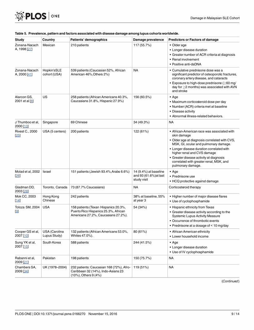
Table 5. Prevalence, pattern and factors associated with disease damage among lupus cohorts worldwide.
Study Country Patients’ demographics Damage prevalence Predictors or Factors of damage
Zonana-NacachA, 1998 [27]
Mexican 210 patients 117 (55.7%) • Older age• Longer disease duration• Greater number of ACR criteria at diagnosis• Renal involvement• Positive anti-dsDNA
Zonana-NacachA, 2000 [41]
Hopkin’sSLEcohort (USA)
539 patients (Caucasian 52%, AfricanAmerican 46%,Others 2%)
NA • Cumulative prednisone dose was asignificant predictor of osteoporotic fractures,coronaryarterydisease, and cataracts
• Exposure to high-dose prednisone (�60 mg/day for�2 months) was associated with AVNand stroke
AlarconGS,2001 et al [8]
US 258 patients (AfricanAmericans 40.3%,Caucasians 31.8%, Hispanic 27.9%)
156 (60.5%) • Age• Maximum corticosteroid dose per day• Number (ACR) criteriamet at baseline• Disease activity• Abnormal illness-related behaviors.
J Thumboo et al,2000 [13]
Singapore 69 Chinese 34 (49.3%) NA
Rivest C., 2000[25]
USA (5 centers) 200 patients 122 (61%) • African-American race was associated withskin damage
• Older age at diagnosis correlated with CVS,MSK, GI, ocular and pulmonary damage.
• Longer disease duration correlatedwithhigher renal and CVS damage
• Greater disease activity at diagnosiscorrelated with greater renal, MSK, andpulmonary damage.
Molad et al, 2002[26]
Israel 151 patients (Jewish 93.4%,Arabs 6.6%) 14 (9.4%) at baselineand 93 (61.6%)at laststudy visit
• Age• Prednisone use• HCQ protective against damage
GladmanDD,2003 [28]
Toronto, Canada 73 (87.7%Caucasians) NA Corticosteroid therapy
Mok CC, 2003[14]
Hong KongChinese
242 patients 38% at baseline, 55%at year 3
• Higher number of major disease flares• Use of cyclophosphamide
Toloza SM, 2004[9]
USA 158 patients (Texan Hispanics 20.3%,PuertoRico Hispanics 25.3%, AfricanAmericans 27.2%, Caucasians 27.2%).
54 (34%) • Hispanic ethnicity from Texas• Greater disease activity according to theSystemic Lupus Activity Measure
• Occurrence of thrombotic events• Prednisone at a dosage of < 10 mg/day
Cooper GS et al,2007 [10]
USA (CarolinaLupus Study)
132 patients (AfricanAmericans 53.0%,Whites 47.0%).
80 (61%) • AfricanAmerican ethnicity• Lower household income
Sung YK et al,2007 [15]
South Korea 588 patients 244 (41.5%) • Age• Longer disease duration• Use of IV cyclophosphamide
Rabanni et al,2009 [21]
Pakistan 198 patients 150 (75.7%) NA
Chambers SA,2009 [34]
UK (1978–2004) 232 patients: Caucasian 168 (72%), Afro-Caribbean 32 (14%), Indo-Asians 23(10%), Others 9 (4%)
119 (51%) NA
(Continued)
Damage in Malaysian SLE Cohort
PLOSONE | DOI:10.1371/journal.pone.0166270 November 15, 2016 9 / 14
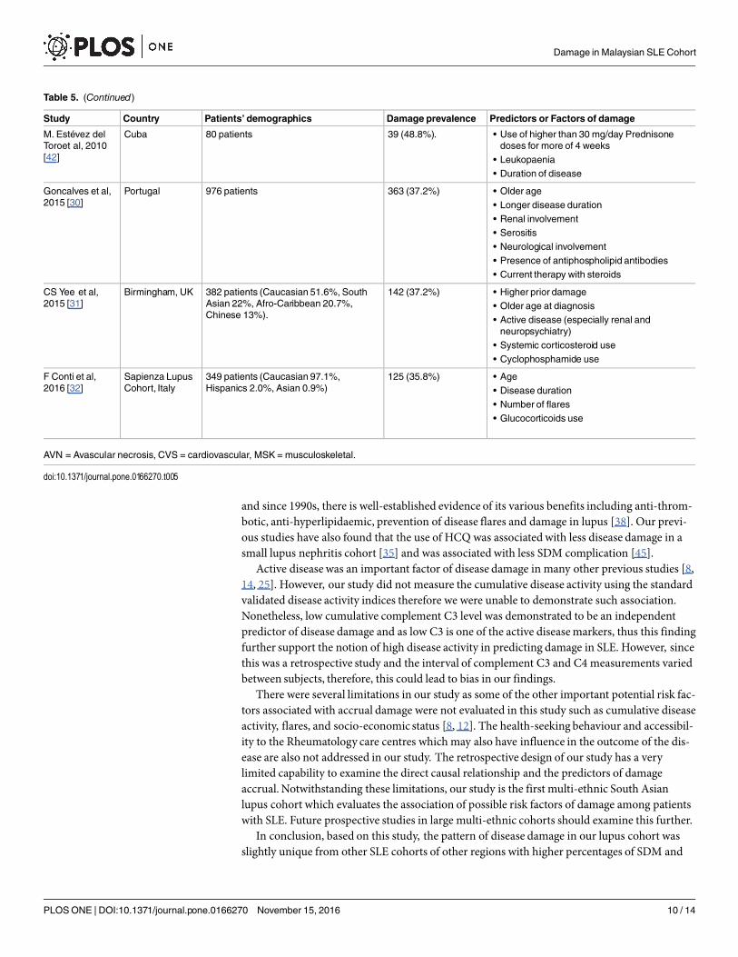
and since 1990s, there is well-established evidence of its various benefits including anti-throm-botic, anti-hyperlipidaemic, prevention of disease flares and damage in lupus [38]. Our previ-ous studies have also found that the use of HCQ was associated with less disease damage in asmall lupus nephritis cohort [35] and was associated with less SDM complication [45].
Active disease was an important factor of disease damage in many other previous studies [8,14, 25]. However, our study did not measure the cumulative disease activity using the standardvalidated disease activity indices therefore we were unable to demonstrate such association.Nonetheless, low cumulative complement C3 level was demonstrated to be an independentpredictor of disease damage and as low C3 is one of the active diseasemarkers, thus this findingfurther support the notion of high disease activity in predicting damage in SLE. However, sincethis was a retrospective study and the interval of complement C3 and C4 measurements variedbetween subjects, therefore, this could lead to bias in our findings.
There were several limitations in our study as some of the other important potential risk fac-tors associated with accrual damage were not evaluated in this study such as cumulative diseaseactivity, flares, and socio-economic status [8, 12]. The health-seeking behaviour and accessibil-ity to the Rheumatology care centres which may also have influence in the outcome of the dis-ease are also not addressed in our study. The retrospective design of our study has a verylimited capability to examine the direct causal relationship and the predictors of damageaccrual. Notwithstanding these limitations, our study is the first multi-ethnic South Asianlupus cohort which evaluates the association of possible risk factors of damage among patientswith SLE. Future prospective studies in large multi-ethnic cohorts should examine this further.
In conclusion, based on this study, the pattern of disease damage in our lupus cohort wasslightly unique from other SLE cohorts of other regions with higher percentages of SDM and
Table 5. (Continued)
Study Country Patients’ demographics Damage prevalence Predictors or Factors of damage
M. Estévez delToroet al, 2010[42]
Cuba 80 patients 39 (48.8%). • Use of higher than 30 mg/day Prednisonedoses for more of 4 weeks
• Leukopaenia• Duration of disease
Goncalves et al,2015 [30]
Portugal 976 patients 363 (37.2%) • Older age• Longer disease duration• Renal involvement• Serositis• Neurological involvement• Presence of antiphospholipid antibodies• Current therapy with steroids
CS Yee et al,2015 [31]
Birmingham, UK 382 patients (Caucasian 51.6%, SouthAsian 22%, Afro-Caribbean 20.7%,Chinese 13%).
142 (37.2%) • Higher prior damage• Older age at diagnosis• Active disease (especially renal andneuropsychiatry)
• Systemic corticosteroid use• Cyclophosphamide use
F Conti et al,2016 [32]
Sapienza LupusCohort, Italy
349 patients (Caucasian 97.1%,Hispanics 2.0%, Asian 0.9%)
125 (35.8%) • Age• Disease duration• Number of flares• Glucocorticoids use
AVN = Avascular necrosis, CVS = cardiovascular, MSK = musculoskeletal.
doi:10.1371/journal.pone.0166270.t005
Damage in Malaysian SLE Cohort
PLOSONE | DOI:10.1371/journal.pone.0166270 November 15, 2016 10 / 14
renal damage observed.The independent factors that were identified to be associated with thedisease damage in our SLE patients were age, Indian ethnicity, APLS, NPSLE and mean com-plement C3 levels. On the other hand, HCQ use and early treatment with HCQ significantlyreduces the risk of damage in lupus.
Supporting InformationS1 Appendix. Raw data.(SAV)
AcknowledgmentsThis study was approved by the National University of Malaysia [study code number:FF-2013-337] and Ministry of Health Malaysia [study code number: NMRR-14-386-19203]. We thankDr. HJ Ding for assistance in this study.
Author Contributions
Conceptualization: SSS.
Data curation: SSS.
Formal analysis: SSS.
Funding acquisition: SSS.
Investigation: SSS HH SR MSMS AHAG R.Mohd R.Mustafar.
Methodology:SSS HH.
Project administration: SSS.
Resources: SSS.
Software: SSS.
Supervision:HH.
Validation: SSS HH SRMSMS.
Visualization: SSS.
Writing – original draft: SSS HH SR.
Writing – review& editing: SSS HH SRMSMS AHAG R.Mohd R.Mustafar.
References1. KasitanonN, Magder LS, Petri M (2006) Predictors of survival in systemic lupus erythematosus. Medi-
cine (Baltimore) 85:147–56.
2. GladmanD, Ginzler E, Goldsmith C, FortinP, LiangM, UrowitzM, et al (1996) The development and ini-tial validation of the Systemic Lupus International Collaborating Clinics/American College of Rheuma-tology damage index for systemic lupus erythematosus. ArthritisRheum 39:363–9. PMID: 8607884
3. BernatskyS, Clarke A, Abrahamowicz M, Neville C, Karp I, PineauCA (2005) A comparison of pro-spective and retrospective evaluations of the Systemic Lupus International Collaborating Clinics/Ameri-can College of Rheumatology Damage Index for systemic lupus erythematosus. J Rheumatol 32:820–3. PMID: 15868615
4. GladmanDD, GoldsmithCH, Urowitz MB, Bacon P, Fortin P, Ginzler E, et al (2000) The SystemicLupus International Collaborating Clinics/AmericanCollege of Rheumatology (SLICC/ACR)Damage
Damage in Malaysian SLE Cohort
PLOSONE | DOI:10.1371/journal.pone.0166270 November 15, 2016 11 / 14
Index for Systemic Lupus Erythematosus InternationalComparison. J Rheumatol 27:373–6. PMID:10685799
5. Mok CC, Ho LY, Cheung MY, Yu KL, To CH (2009) Effect of disease activity and damage on quality oflife in patients with systemic lupus erythematosus: a 2-year prospective study. Scand J Rheumatol38:121–7. doi: 10.1080/03009740802415527 PMID: 18991189
6. Mok CC, Ho LY, Yu KL, To CH (2013)Relationship between individual organ damage andmortalityofsystemic lupus erythematosus (SLE): A prospective cohort study of 679 patients. Ann RheumDis71:545.
7. AlarconGS (2011)Multiethnic lupus cohorts:what have they taught us? Reumatol Clin 7:3–6. doi: 10.1016/j.reuma.2010.11.001PMID: 21794772
8. AlarconGS, McGwinG Jr, BartolucciAA, Roseman J, Lisse J, Fessler BJ, et al (2001) Systemic lupuserythematosus in three ethnic groups. IX. Differences in damage accrual. ArthritisRheum 44:2797–806. PMID: 11762940
9. Toloza SM, Roseman JM, AlarconGS, McGwinG Jr, Uribe AG, Fessler BJ, et al (2004). Systemiclupus erythematosus in a multiethnic US cohort (LUMINA): XXII. Predictors of time to the occurrence ofinitial damage. ArthritisRheum 50:3177–86. doi: 10.1002/art.20578PMID: 15476246
10. CooperGS, Treadwell EL, St Clair EW, Gilkeson GS, Dooley MA (2007) Sociodemographic associa-tions with early disease damage in patients with systemic lupus erythematosus. ArthritisRheum57:993–9. doi: 10.1002/art.22894 PMID: 17665464
11. Thumboo J, Wee H-L (2006) Systemic lupus erythematosus in Asia: is it more common andmoresevere? APLAR Journal of Rheumatology 9:320–6.
12. Pons-Estel BA, Catoggio LJ, Cardiel MH, SorianoER, Gentiletti S, Villa AR, et al (2004) TheGLADELmultinational Latin Americanprospective inception cohort of 1,214 patients with systemic lupus erythe-matosus: ethnic and disease heterogeneity among "Hispanics". Medicine (Baltimore) 83:1–17.
13. Thumboo J, Feng PH, Soh CH, Boey ML, Thio S, Fong KY (2000). Validation of a Chinese version ofthe Medical OutcomesStudy Family andMarital FunctioningMeasures in patients with SLE. Lupus9:702–7. PMID: 11199926
14. Mok CC, Ho CT, Wong RW, Lau CS (2003) Damage accrual in southernChinese patients with sys-temic lupus erythematosus. J Rheumatol 30:1513–9. PMID: 12858450
15. Sung YK, Hur NW, Sinskey JL, Park D, Bae SC (2007) Assessment of damage in Korean patients withsystemic lupus erythematosus. J Rheumatol 34:987–91. PMID: 17361986
16. CurrentPopulation Estimates, Malaysia, 2014–2016. In: Department of StatisticsMalaysia, Official Por-tal [Internet]. Malaysia 2016: [about 2 screens]. Available: https://www.statistics.gov.my/index.php?r=column/cthemeByCat&cat=155&bul_id=OWlxdEVoYlJCS0hUZzJyRUcvZEYxZz09&menu_id=L0pheU43NWJwRWVSZklWdzQ4TlhUUT09
17. Wang F, Wang CL, Tan CT, Manivasagar M (1997) Systemic lupus erythematosus in Malaysia: a studyof 539 patients and comparison of prevalence and disease expression in different racial and gendergroups. Lupus 6:248–53. PMID: 9104731
18. Jasmin R, Sockalingam S, Cheah T, Goh K (2013) Systemic lupus erythematosus in the multiethnicMalaysian population: disease expression and ethnic differences revisited. Lupus 22:967–71. doi: 10.1177/0961203313496299 PMID: 23846232
19. ShahrirM, ShahdanM, ShahidM, SulaimanW, Mokhtar AM, OthmanM, et al. Multicentre survey ofrheumatoidarthritispatients fromMinistry of Health Rheumatology Centers in Malaysia. Int J RheumDis. 2008; 11:287–92
20. HochbergMC (1997) Updating the American College of Rheumatology revised criteria for the classifi-cation of systemic lupus erythematosus. ArthritisRheum 40:1725.
21. Rabbani MA, Habib HB, IslamM, Ahmad B, Majid S, SaeedW, et al (2009) Survival analysis and prog-nostic indicators of systemic lupus erythematosus in Pakistani patients. Lupus 18:848–55. doi: 10.1177/0961203309103410 PMID: 19578112
22. Ramachandran A, SnehalathaC, Kapur A, Vijay V, MohanV, Das AK, et al (2001) High prevalence ofdiabetes and impaired glucose tolerance in India: National Urban Diabetes Survey. Diabetologia44:1094–101. doi: 10.1007/s001250100627 PMID: 11596662
23. Tan VM,Wu T, HenryCJ, Lee YS (2015)Glycaemic and insulin responses, glycaemic index and insuli-naemic index values of rice between three Asian ethnic groups. Br J Nutr 113:1228–36. doi: 10.1017/S0007114515000586PMID: 25789978
24. LetchumanGR,Wan NazaimoonWM,Wan MohamadWB, Chandran LR, Tee GH, Jamaiyah H, et al(2010) Prevalence of diabetes in theMalaysian National HealthMorbiditySurvey III 2006.Med J Malay-sia 65:180–6. PMID: 21939164
Damage in Malaysian SLE Cohort
PLOSONE | DOI:10.1371/journal.pone.0166270 November 15, 2016 12 / 14
25. Rivest C, Lew RA,Welsing PM, SanghaO, Wright EA, RobertsWN, et al (2000). Association betweenclinical factors, socioeconomic status, and organ damage in recent onset systemic lupus erythemato-sus. J Rheumatol 27:680–4. PMID: 10743808
26. Molad Y, GorshteinA, Wysenbeek AJ, Guedj D, MajadlaR,Weinberger A, et al (2002) Protective effectof hydroxychloroquine in systemic lupus erythematosus. Prospective long-termstudy of an Israelicohort. Lupus 11:356–61.PMID: 12139373
27. Zonana-Nacach A, Camargo-Coronel A, Yanez P, de Lourdes SanchezM, Jimenez-Balderas FJ,Aceves-Avila J, et al (1998)Measurementof damage in 210Mexican patients with systemic lupus ery-thematosus: relationshipwith disease duration. Lupus 7:119–23. PMID: 9580342
28. GladmanDD, Urowitz MB, RahmanP, Ibanez D, Tam LS (2003) Accrual of organ damage over time inpatients with systemic lupus erythematosus. J Rheumatol 30:1955–9. PMID: 12966597
29. Estevez Del Toro M, Chico Capote A, Hechavarria R, Jimenez Paneque R, Kokuina E (2010)Damagein cuban patients with systemic lupus erythematosus. Relation with disease features. Reumatol Clin6:11–5.
30. Goncalves MJ, Sousa S, Ines LS, DuarteC, Borges J, Silva C, et al (2015) Characterization of damagein Portuguese lupus patients: analysis of a national lupus registry. Lupus 24:256–62. doi: 10.1177/0961203314555172PMID: 25318970
31. Yee CS, Su L, Toescu V, Hickman R, Situnayake D, Bowman S, et al (2015) Birmingham SLE cohort:outcomes of a large inception cohort followed for up to 21 years. Rheumatology (Oxford) 54:836–43.
32. Conti F, Ceccarelli F, Perricone C, Leccese I, Massaro L, Pacucci VA, et al (2016) The chronic damagein systemic lupus erythematosus is driven by flares, glucocorticoids and antiphospholipid antibodies:results froma monocentric cohort. Lupus 25:719–26. doi: 10.1177/0961203315627199 PMID:26821965
33. Golder V, Connelly K, StaplesM, Morand E, Hoi A (2013) Association of Asian ethnicity with diseaseactivity in SLE: an observational study from theMonash Lupus Clinic. Lupus 22:1425–30. doi: 10.1177/0961203313500547 PMID: 23942610
34. ChambersSA, Allen E, RahmanA, IsenbergD (2009)Damage andmortality in a group of Britishpatients with systemic lupus erythematosus followed up for over 10 years. Rheumatology (Oxford)48:673–5.
35. Shaharir S, Ghafor AA, Said MM, Kong N (2014) A descriptive study of the factors associatedwith dam-age in Malaysian patients with lupus nephritis. Lupus 23:436–42. doi: 10.1177/0961203313518624PMID: 24399814
36. HiramatsuN, Kuroiwa T, Ikeuchi H, MaeshimaA, Kaneko Y, HiromuraK, et al (2008)Revised classifi-cation of lupus nephritis is valuable in predicting renal outcomewith an indication of the proportionofglomeruli affected by chronic lesions. Rheumatology (Oxford) 47:702–7.
37. Wan NazaimoonWM,Md Isa SH,Wan MohamadWB, Khir AS, Kamaruddin NA, Kamarul IM, et al(2013) Prevalence of diabetes in Malaysia and usefulness of HbA1c as a diagnostic criterion.DiabetMed 30:825–8. doi: 10.1111/dme.12161 PMID: 23413941
38. Bruce IN, O'Keeffe AG, Farewell V, Hanly JG, Manzi S, Su L, et al (2015) Factors associated with dam-age accrual in patients with systemic lupus erythematosus: results from the Systemic Lupus Interna-tional Collaborating Clinics (SLICC) InceptionCohort.Ann RheumDis 74:1706–13. doi: 10.1136/annrheumdis-2013-205171 PMID: 24834926
39. Jonsen A, BengtssonAA, Nived O, Ryberg B, Sturfelt G (2002)Outcomeof neuropsychiatric systemiclupus erythematosus within a defined Swedish population: increasedmorbidity but low mortality. Rheu-matology (Oxford) 41:1308–12.
40. Ruiz-Irastorza G, Egurbide M, Ugalde J, AguirreC (2004)High impact of antiphospholipid syndrome onirreversible organ damage and survival of patients with systemic lupus erythematosus. Arch InternMed164:77–82. doi: 10.1001/archinte.164.1.77 PMID: 14718326
41. Zonana-Nacach A, Barr SG, Magder LS, Petri M (2000) Damage in systemic lupus erythematosus andits associationwith corticosteroids. ArthritisRheum 43:1801–8. doi: 10.1002/1529-0131(200008)43:8<1801::AID-ANR16>3.0.CO;2-O PMID: 10943870
42. Estévez del Toro Miguel CC A, HechavarríaRafael, Jiménez Paneque Rosa and Kokuina Elena (2010)Damage in Cuban patients with systemic lupus erythematosus.Relationwith disease features. Reuma-tol Clin 6:11–5.
43. Petri M, Purvey S, Fang H, Magder LS (2012) Predictors of organ damage in systemic lupus erythe-matosus: the Hopkins Lupus Cohort.ArthritisRheum 64:4021–8. doi: 10.1002/art.34672PMID:22932985
Damage in Malaysian SLE Cohort
PLOSONE | DOI:10.1371/journal.pone.0166270 November 15, 2016 13 / 14
44. Akhavan PS, Su J, LouW, GladmanDD, Urowitz MB, Fortin PR (2013) The early protective effect ofhydroxychloroquine on the risk of cumulative damage in patients with systemic lupus erythematosus. JRheumatol 40:831–41. doi: 10.3899/jrheum.120572PMID: 23588942
45. Shaharir SS, Gafor AH, Said MS, Kong NC (2015) Steroid-induced diabetesmellitus in systemic lupuserythematosus patients: analysis from a Malaysian multi-ethnic lupus cohort. Int J RheumDis 18:541–7. doi: 10.1111/1756-185X.12474 PMID: 25294584
Damage in Malaysian SLE Cohort
PLOSONE | DOI:10.1371/journal.pone.0166270 November 15, 2016 14 / 14