biodegradation of coir waste by microfungi: an electron...
TRANSCRIPT
183
P.Y. Yau and R.J. MurphyJ. Trop. Agric. and Fd. Sc. 30(2)(2002): 183–188
Biodegradation of coir waste by microfungi: An electronmicroscopic study(Pereputan gabus habuk kelapa melalui kulat mikro: satu kajian menggunakanmikroskopi elektron)
P.Y. Yau* and R.J. Murphy**
Key words: coir waste, biodegradation, electron microscopy
AbstrakMikroskopi elektron jenis pengimbas (SEM) dan pemancar (TEM) telahdigunakan untuk mengkaji aspek morfologi ultrastruktur dinding sel ketikapereputan biologi gabus habuk kelapa. Mikroskopi SEM telah menunjukkanpenembusan meluas dan pengkolonian hifa, sama ada melalui muka selparenkima, lumen sel atau ruang antara sel dan runtuhnya dinding sel padaperingkat pereputan terakhir. Mikroskopi TEM turut membuktikan kewujudanaktiviti enzim kulat pada dinding sel ketika proses pereputan biologi gabus habukkelapa dan ini boleh dilihat dari bahan tumpat elektron dan lendir-bukan-sel yangdijumpai di sekeliling hifa. Reput dinding sel adalah seiras dengan rupa bentukreput lembut dengan terjadinya pembentukan rongga. Rongga ini banyak terdapatpada dinding sel sekunder dan pada reput peringkat akhir, dan juga terdapat padadinding sel dan penjuru sel.
AbstractScanning and transmission electron microscopy (SEM and TEM) were used tostudy morphological aspects of cell wall ultrastructure during the biodegradationof coir waste by microfungi. SEM microscopy revealed extensive hyphaepenetration and colonisation through either parenchyma cell surface, cell lumenaor intercellular spaces and collapse of cell walls at the advanced stage ofdegradation. TEM microscopy provided further details of the fungal enzymaticactivities in the cell walls during biodegradation of coir waste as seen from thepresence of electron dense and the extracellular mucilaginous materials foundaround the hyphae. The attack on the coir waste cell walls was typical of soft rotdecay with cavity formation evident. The cavities were mainly found in thesecondary cell walls and, at advanced stages of decay, were also found in themiddle lamella and the cell corner regions.
*Kluang MARDI Station, P.O. Box 525, 86007 Kluang, Johor, Malaysia**Department of Biology, Imperial College of Science, Technology and Medicine, London SW7 2AZ, U.K.Authors’ full names: Yau Peng Yam and Richard James MurphyE-mail: [email protected]©Malaysian Agricultural Research and Development Institute 2003
IntroductionBiodegraded coir waste was used as a plantgrowth substrate to reduce the risk of plantphytotoxicity (Verdonck et al. 1983; Yau andMurphy 1999, 2000). Tomato plants growing
on the biodegraded coir waste had betterroot and shoot development and also higherfruit yields compared with those grown inraw coir waste (Yau and Murphy 2000). Thebiodegradation process of coir waste could
184
Biodegradation of coir waste by microfungi
be enhanced by the addition of a nitrogenactivator and a group of wood decaymicrofungi (Yau and Murphy 1998, 2000).The added fungi primarily degraded part ofthe cellulose and hemicellulose contents ofthe cell walls of coir waste. Generally, thephytotoxicity decreased with the increasingperiods of decomposition (Patrick et al.1963; McCalla and Haskins 1964).
A light microscopic study on thebiodegraded coir waste has demonstratedaspects of the hyphal penetration,colonization and cell wall decay during thebiodegradation process (Yau and Murphy2001). The use of electron microscopy isalso an invaluable tool for studying theultrastructural aspects of lignocellulosedegradation by fungi and bacteria, and thishas been extensively reviewed by Daniel(1994). The soft rot fungi causingcharacteristic decay patterns in wood andother lignocellulosic materials were widelystudied by many workers using electronmicroscopy (Nilsson 1974; Murphy et al.1991; Sulaiman and Murphy 1994, 1995;Wong and Pearce 1997; Khalili 1999). Thispaper presents the results of studies on thecolonisation and decay of the cell walls ofcoir waste using both the scanning andtransmission electron microscopic studies tocomplement the earlier light microscopicstudy on the activities of the addedmicrofungi in coir waste (Yau and Murphy2001).
Materials and methodsCoir waste biodegradationSpore suspensions of individual funginamely the moulds Aspergillus niger,Penicillium citrinum, Trichoderma reesii andthe wood soft rot fungi Humicola grisea andChaetomium globosum and the mixedinoculum of all the five fungi were used toenhance the biodegradation of coir waste.The procedures for preparing the sporesuspensions and the biodegradation of coirwaste followed that described in theEuropean Prestandard For WoodPreservatives ENV 807 (Anon. 1993) and in
the earlier study on cell wall degradation ofcoir waste (Yau and Murphy 2001).
Scanning Electron Microscopy (SEM)Fixation The raw coir waste samples werefixed in 3% glutaraldehyde in 0.1 M sodiumcacodylate buffer (pH 7.2) for 12 h. Thiswas followed by two rinsings in sodiumcacodylate buffer and then post-fixed in 1%osmium tetroxide for 12 h. Subsequently, thesamples were dehydrated in an acetoneseries of 20, 35, 50, 70, 80, 90 and 100%for 15 min each before undergoing criticalpoint drying (CPD).
Critical point drying The samples in100% acetone were placed in a CPD boatand then carefully inserted into the CPDapparatus (Polaron Critical Point Drier,Model E3000). It was flushed with liquidCO2 for 3 min to replace the acetone. Thesamples were left to stand for an hour. Afterthat, the samples were flushed again withliquid CO2. The samples were allowed tostand for further one hour.
Mounting After CPD, the samples weremounted on aluminium SEM stubs withdouble-sided adhesive tape.
Sputter coating and SEM viewing Thesamples were sputter coated with goldpalladium in argon using a Polaron SputterCoater to give an ultra fine grain deposit ofgold (grain size of 10–15 Å). The sampleswere viewed at appropriate kV (12 kV to25 kV) to avoid beam damage of thespecimen using a Philips PSEM 500Scanning Electron Microscope.Photomicrographs of selected portions weretaken on Ilford Pan F film.
Transmission Electron Microscopy (TEM)Fixation The samples were fixed in 3%glutaraldehyde and subsequently in 1%osmium tetroxide as for the SEM. Thesamples were dehydrated in an ethanolseries of 20, 35, 50, 70, 80, 90 and 100%
185
P.Y. Yau and R.J. Murphy
for 15 min each and then washed twice inpropylene oxide for 10 min each.
Embedding For tissue embedding, Spurr’sresin of hard formulation was used (Spurr1969). To ensure proper infiltration, theembedding procedure used was propyleneoxide: Spurr’s resin ratio at 2:1 for 1 h, 1:1for 4 h and 1:2 for 4 h and then clearedtwice in Spurr’s resin. The tissue was thentransferred to a plastic mould, filled withpure Spurr’s resin and cured at 70 °C for 9 h.
Sectioning Sectioning was done using aReichert-Jung ultramicrotome using adiamond knife at an estimated thickness of0.075 µm. The sections were spread outcarefully, placed onto a copper grid andallowed to dry for about 30 min at roomtemperature.
Staining The sections were stained with2% aqueous uranyl acetate and a secondarystain of 2% lead citrate.
Viewing The ultrathin sections wereexamined under a Philip EM301Transmission Electron Microscope. Thebeam current was set at 17 pA, acceleratingvoltage at 80 kV, spot size at 200 nm, 20ºspecimen angle and magnifications variedfrom 2 800 to 22 000 times. Micrographs ofselected portions were taken on Agfa Scientafilms.
Results and discussionTwenty-five specimens of control andbiodegraded coir waste were examinedunder SEM and TEM. Numerousmicrographs were obtained and used in theinterpretation of characteristic features of thefungal colonisation and cell walldegradation. The followingdescription synthesises the principalmorphological features of note and presentsa range of micrographs of their typicalappearance.
Scanning Electron Microscopy (SEM)The SEM micrographs revealed severalinteresting features of the cell wall decay ofcoir waste. As biodegraded coir wastesamples were structurally loose, very minutein size (mostly <2 mm) and indefinite inshape as a result of fibre shredding. It wasvery difficult to obtain cross sectionalsurfaces of decayed material for SEMviewing. However, some sub-samples whichwere randomly selected from the largebiodegraded mass gave some reasonablesurfaces for observation of the decay causedby the various fungi.
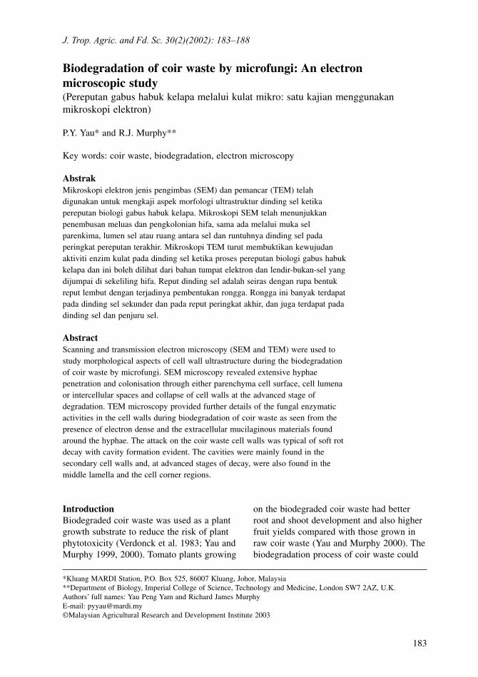
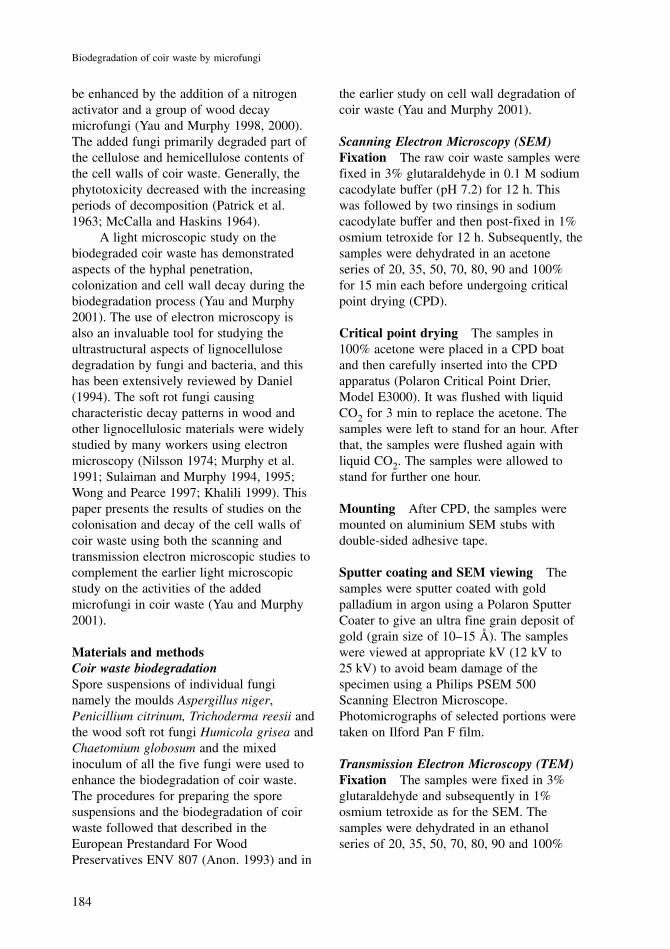
Unlike the light microscopy, the SEMmicrographs allowed inner surfaces ofbiodegraded coir waste to be revealed at ahigher magnification. For the controltreatment, the cell walls were relativelyclean without visible fungal attack eventhough the samples had been maintainedunder semi-sterile condition for 3 months.This suggested also that the startingmaterials were relatively clean (Plate 1).The treatments with the mould fungi allshowed extensive colonisation of hyphaeeither through parenchyma cell surfaces, celllumena or through intercellular spaces.Some areas of erosions were also found onthe surfaces of the parenchyma cells.Although there was extensive colonisationby the mould hyphae, cell wall decay wasnot severe (Plates 2 and 3). However, in thetreatments with the mixture of moulds andsoft rot fungi, the hyphae grew andpenetrated through the cell corner regionscausing the collapse of cell walls. Severecell wall decay was evident (Plate 4).
These SEM observations supportedearlier findings on the decay patterns of coirwaste using the light microscope (Yau andMurphy 2001).
Transmission Electron Microscopy (TEM)The noteworthy feature of TEM was that itrevealed the decaying pattern of microfungiinside the cell walls of coir waste.Generally, the soft rot hyphae formedcavities mainly in the secondary cell walls.
186
Biodegradation of coir waste by microfungi
Plate 4. Hyphal penetration through the celllumena and spaces between cell walls. The cellcorner regions show disintegration of cell wallsand separation of cells. Severe cell wall decaywas evident (arrow head) (bar in line =10 µm)
Plate 1. The ground tissue parenchyma cells andlumena of coir waste in the control treatmentwere relatively clean with no visible hyphae ordecay (bar in line = 10 µm)
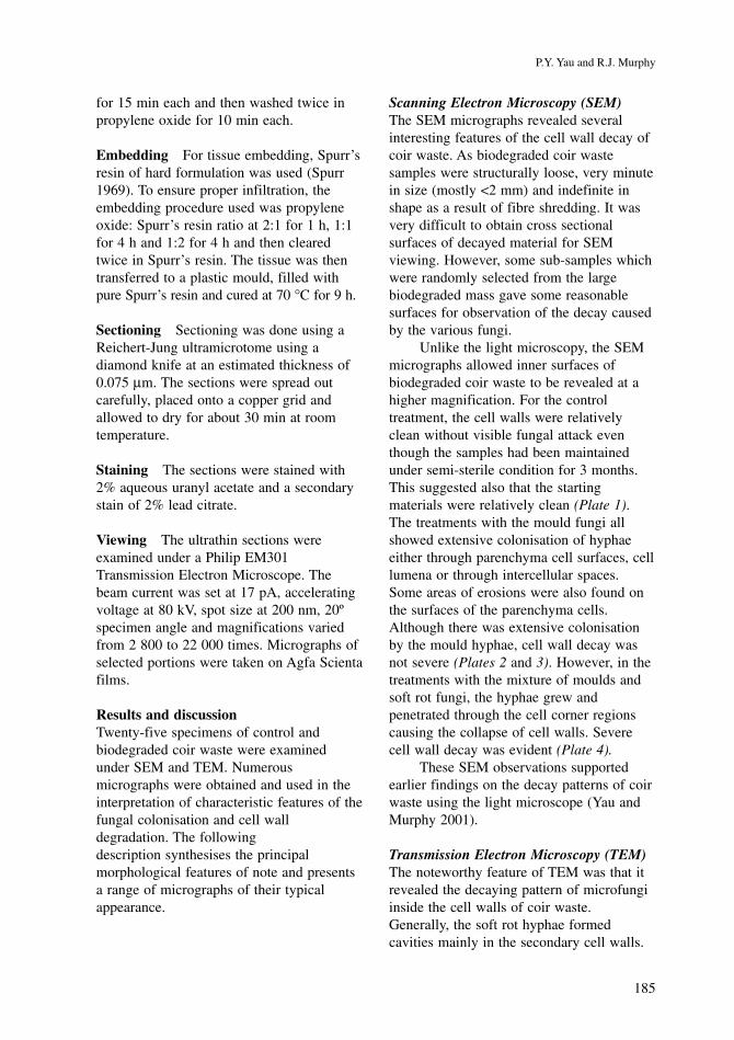
Plate 2. Penicillium citrinum hyphae growing onthe surface of the parenchyma cells. Slighterosion of cell wall (arrow) and pit (P) areindicated (bar in line = 10 µm)
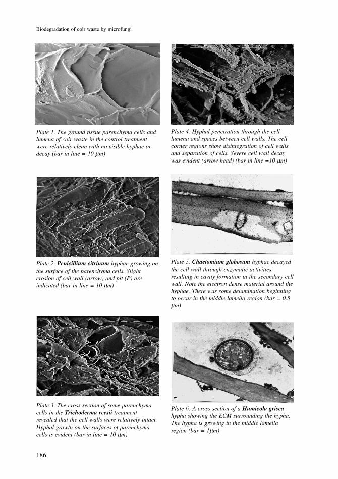
Plate 3. The cross section of some parenchymacells in the Trichoderma reesii treatmentrevealed that the cell walls were relatively intact.Hyphal growth on the surfaces of parenchymacells is evident (bar in line = 10 µm)
Plate 5. Chaetomium globosum hyphae decayedthe cell wall through enzymatic activitiesresulting in cavity formation in the secondary cellwall. Note the electron dense material around thehyphae. There was some delamination beginningto occur in the middle lamella region (bar = 0.5µm)
Plate 6: A cross section of a Humicola griseahypha showing the ECM surrounding the hypha.The hypha is growing in the middle lamellaregion (bar = 1µm)
187
P.Y. Yau and R.J. Murphy
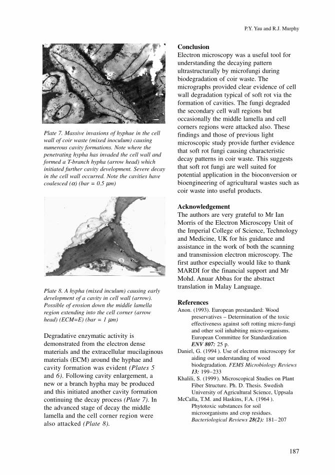
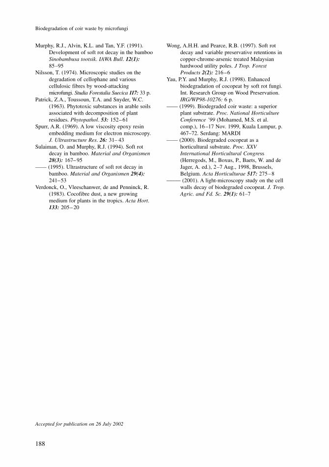
Degradative enzymatic activity isdemonstrated from the electron densematerials and the extracellular mucilaginousmaterials (ECM) around the hyphae andcavity formation was evident (Plates 5and 6). Following cavity enlargement, anew or a branch hypha may be producedand this initiated another cavity formationcontinuing the decay process (Plate 7). Inthe advanced stage of decay the middlelamella and the cell corner region werealso attacked (Plate 8).
Plate 8. A hypha (mixed inculum) causing earlydevelopment of a cavity in cell wall (arrow).Possible of erosion down the middle lamellaregion extending into the cell corner (arrowhead) (ECM=E) (bar = 1 µm)
Plate 7. Massive invasions of hyphae in the cellwall of coir waste (mixed inoculum) causingnumerous cavity formations. Note where thepenetrating hypha has invaded the cell wall andformed a T-branch hypha (arrow head) whichinitiated further cavity development. Severe decayin the cell wall occurred. Note the cavities havecoalesced (α) (bar = 0.5 µm)
ConclusionElectron microscopy was a useful tool forunderstanding the decaying patternultrastructurally by microfungi duringbiodegradation of coir waste. Themicrographs provided clear evidence of cellwall degradation typical of soft rot via theformation of cavities. The fungi degradedthe secondary cell wall regions butoccasionally the middle lamella and cellcorners regions were attacked also. Thesefindings and those of previous lightmicroscopic study provide further evidencethat soft rot fungi causing characteristicdecay patterns in coir waste. This suggeststhat soft rot fungi are well suited forpotential application in the bioconversion orbioengineering of agricultural wastes such ascoir waste into useful products.
AcknowledgementThe authors are very grateful to Mr IanMorris of the Electron Microscopy Unit ofthe Imperial College of Science, Technologyand Medicine, UK for his guidance andassistance in the work of both the scanningand transmission electron microscopy. Thefirst author especially would like to thankMARDI for the financial support and MrMohd. Anuar Abbas for the abstracttranslation in Malay Language.
ReferencesAnon. (1993). European prestandard: Wood
preservatives – Determination of the toxiceffectiveness against soft rotting micro-fungiand other soil inhabiting micro-organisms.European Committee for StandardizationENV 807: 25 p.
Daniel, G. (1994 ). Use of electron microscopy foraiding our understanding of woodbiodegradation. FEMS Microbiology Reviews13: 199–233
Khalili, S. (1999). Microscopical Studies on PlantFiber Structure. Ph. D. Thesis. SwedishUniversity of Agricultural Science, Uppsala
McCalla, T.M. and Haskins, F.A. (1964 ).Phytotoxic substances for soilmicroorganisms and crop residues.Bacteriological Reviews 28(2): 181– 207
188
Biodegradation of coir waste by microfungi
Murphy, R.J., Alvin, K.L. and Tan, Y.F. (1991).Development of soft rot decay in the bambooSinobambusa tootsik. IAWA Bull. 12(1):85–95
Nilsson, T. (1974). Microscopic studies on thedegradation of cellophane and variouscellulosic fibres by wood-attackingmicrofungi. Studia Forestalia Suecica 117: 33 p.
Patrick, Z.A., Toussoun, T.A. and Snyder, W.C.(1963). Phytotoxic substances in arable soilsassociated with decomposition of plantresidues. Phytopathol. 53: 152–61
Spurr, A.R. (1969). A low viscosity epoxy resinembedding medium for electron microscopy.J. Ultrastructure Res. 26: 31– 43
Sulaiman, O. and Murphy, R.J. (1994). Soft rotdecay in bamboo. Material and Organismen28(3): 167– 95
–––– (1995). Ultrastructure of soft rot decay inbamboo. Material and Organismen 29(4):241–53
Verdonck, O., Vleeschanwer, de and Penninck, R.(1983). Cocofibre dust, a new growingmedium for plants in the tropics. Acta Hort.133: 205– 20
Accepted for publication on 26 July 2002
Wong, A.H.H. and Pearce, R.B. (1997). Soft rotdecay and variable preservative retentions incopper-chrome-arsenic treated Malaysianhardwood utility poles. J Trop. ForestProducts 2(2): 216 –6
Yau, P.Y. and Murphy, R.J. (1998). Enhancedbiodegradation of cocopeat by soft rot fungi.Int. Research Group on Wood Preservation.IRG/WP98-10276: 6 p.
–––– (1999). Biodegraded coir waste: a superiorplant substrate. Proc. National HorticultureConference ’99 (Mohamed, M.S. et al.comp.), 16 –17 Nov. 1999, Kuala Lumpur, p.467–72. Serdang: MARDI
–––– (2000). Biodegraded cocopeat as ahorticultural substrate. Proc. XXVInternational Horticultural Congress(Herregods, M., Boxus, P., Baets, W. and deJager, A. ed.), 2–7 Aug., 1998, Brussels,Belgium. Acta Horticulturae 517: 275–8
––––– (2001). A light-microscopy study on the cellwalls decay of biodegraded cocopeat. J. Trop.Agric. and Fd. Sc. 29(1): 61–7