ankle injuries in sports: anatomical considerations and ... · ankle injuries in sports: anatomical...
TRANSCRIPT
Med & Health 2016; 11(2): 117-130
REVIEW ARTICLE
117
https://doi.org/10.17576/MH.2016.1102.02
Address for correspondence and reprint requests: Norzana Abd Ghafar, Department of Anatomy, Faculty of Medicine, Universiti Kebangsaan Malaysia Medical Centre, Jalan Yaacob Latif, Bandar Tun Razak, 56000 Cheras, Kuala Lumpur, Malaysia. Tel: +603-92898605 Fax: +603-91458607 E-mail: [email protected]
Ankle Injuries in Sports: Anatomical Considerations and Clinical Implications
SYARIFAH AISYAH SAH, ELVY SUHANA MR, SRIJIT D, NORZANA AG
Department of Anatomy, Faculty of Medicine, Universiti Kebangsaan Malaysia Medical
Centre, Jalan Yaacob Latif, Bandar Tun Razak, 56000 Cheras, Kuala Lumpur, Malaysia.
ABSTRAK
Kecederaan buku lali adalah kejadian yang biasa berlaku dalam mana-mana aktiviti sukan. Tujuan utama kajian adalah untuk menjelaskan anatomi buku lali, mekanisma kecederaan yang berkaitan dengan aktiviti sukan, keabnormalan secara kongenital atau variasi anatomi yang berkaitan dengan kecederaan buku lali serta perbincangan rawatan secara efektif. Suatu tinjauan perpustakaan telah dijalankan untuk mengetahui kecederaan buku lali yang berlaku akibat daripada pelbagai aktiviti sukan. Kami mendokumenkan semua sukan yang melibatkan kecederaan pada sendi buku lali. Anatomi pelbagai struktur bahagian tapak kaki dan keterlibatannya dalam kecederaan dibincangkan dengan teliti. Pengetahuan anatomi tentang kecederaan buku lali boleh memberi manfaat untuk diagnosis akan datang dan bagi tujuan rawatan.
Kata kunci: buku lali, kecederaan, ligament, sukan
ABSTRACT
Ankle injuries are commonly seen in various sports. The main aim of the present review was to highlight the normal anatomy of the ankle, mechanism of injuries related to sports, congenital abnormalities or anatomical variations related to ankle injury and discuss its effective management. A review of literature was done to determine the ankle injuries which occur as a result of various sports related activities. We documented all sports which involved injury to the ankle joint. The anatomy of various structures in the sole of foot and their involvement in injuries were discussed at length. The anatomical knowledge of ankle injury may be beneficial for future diagnosis and treatment purpose.
Keywords: ankle, injury, ligament, sports
118
Med & Health 2016;11(2): 117-130 Syarifah Aisyah S.A.H. et al.
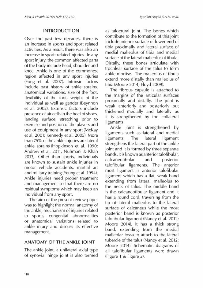
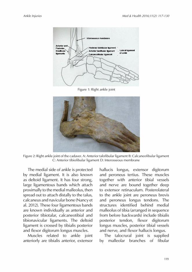
as talocrural joint. The bones which contribute to the formation of this joint include inferior surface of lower end of tibia proximally and lateral surface of medial malleolus of tibia and medial surface of the lateral malleolus of fibula. Distally, these bones articulate with trochlear surface of the talus to form ankle mortise. The malleolus of fibula extend more distally than malleolus of tibia (Moore 2014; Floyd 2009). The fibrous capsule is attached to the margins of the articular surfaces proximally and distally. The joint is weak anteriorly and posteriorly but thickened medially and laterally as it is strengthened by the collateral ligaments. Ankle joint is strengthened by ligaments such as lateral and medial ligaments. The lateral ligament strengthens the lateral part of the ankle joint and it is formed by three separate bands. It is known as anterior talofibular, calcaneofibular and posterior talofibular ligaments. The anterior most ligament is anterior talofibular ligament which has a flat, weak band extending from lateral malleolus to the neck of talus. The middle band is the calcaneofibular ligament and it has a round cord, traversing from the tip of lateral malleolus to the lateral surface of calcaneus while the most posterior band is known as posterior talofibular ligament (Nancy et al. 2012; Moore 2014). It has a thick strong band, extending from the medial malleolar fossa to attach to the lateral tubercle of the talus (Nancy et al. 2012; Moore 2014). Schematic diagrams of all talofibular ligaments were drawn (Figure 1 & Figure 2).
INTRODUCTION
Over the past few decades, there is an increase in sports and sport related activities. As a result, there was also an increase in sports related injuries. In any sport injury, the common affected parts of the body include head, shoulder and knee. Ankle is one of the commonest region affected in any sport injuries (Fong et al. 2007). Intrinsic factors include past history of ankle sprains, anatomical variations, size of the foot, flexibility of the foot, weight of the individual as well as gender (Beynnon et al. 2002). Extrinsic factors include presence of air cells in the heel of shoes, landing surface, stretching prior to exercise and position of the players and use of equipment in any sport (McKay et al. 2001; Kennedy et al. 2005). More than 75% of the ankle injuries are lateral ankle sprains (Hopkinson et al. 1990; Andrew et al. 2011; Nuhmani & Khan 2013). Other than sports, individuals are known to sustain ankle injuries in motor vehicle accidents, martial art and military training (Yeung et al. 1994). Ankle injuries need proper treatment and management so that there are no residual symptoms which may keep an individual from any sport. The aim of the present review paper was to highlight the normal anatomy of the ankle, mechanism of injuries related to sports, congenital abnormalities or anatomical variations related to ankle injury and discuss its effective management.
ANATOMY OF THE ANKLE JOINT
The ankle joint, a unilateral axial type of synovial hinge joint is also termed
119
Ankle Injuries Med & Health 2016;11(2): 117-130
The medial side of ankle is protected by medial ligament. It is also known as deltoid ligament. It has four strong, large ligamentous bands which attach proximally to the medial malleolus, then spread out to attach distally to the talus, calcaneus and navicular bone (Nancy et al. 2012). These four ligamentous bands are known individually as anterior and posterior tibiotalar, calcaneotibial and tibionavicular ligaments. The deltoid ligament is crossed by tibialis posterior and flexor digitorum longus muscles. Muscles related to ankle joint anteriorly are tibialis anterior, extensor
hallucis longus, extensor digitorum and peroneus tertius. These muscles together with anterior tibial vessels and nerve are bound together deep to extensor retinaculum. Posterolateral to the ankle joint are peroneus brevis and peroneus longus tendons. The structures identified behind medial malleolus of tibia (arranged in sequence from before backwards) include tibialis posterior tendon, flexor digitorum longus muscles, posterior tibial vessels and nerve, and flexor hallucis longus. The talocrural joint is supplied by malleolar branches of fibular
Figure 1: Right ankle joint
Figure 2: Right ankle joint of the cadaver. A: Anterior talofibular ligament B: Calcaneofibular ligament C: Anterior tibiofibular ligament D: Interosseous membrane
120
Med & Health 2016;11(2): 117-130 Syarifah Aisyah S.A.H. et al.
anterior and posterior tibial arteries while the lymphatics drain via veins accompanying the arteries and into long and short saphenous veins. It is innervated by the tibial nerve and deep fibular nerve (Moore 2014). There are two types of movements which occur at the ankle joint, i.e. dorsiflexion and plantarflexion. Inversion and eversion movements are commonly thought to be occurring at ankle joint but these movements occur at subtalar joint, not the ankle joint (Lin et al. 2006). Another joint located superior to the ankle joint, is the distal tibiofibular joint which is of syndesmosis type. The lower end of tibia and medial surface of lower end of fibula form this joint. These articulating bones are held together by interosseous membrane, anterior and posterior tibiofibular ligaments. It functions as stabilizer to prevent the separation of the distal end of tibia and fibula under the effect of body weight (Lin et al. 2006; Joshua et al. 2011). Anterior and posterior tibiofibular ligaments are considered as primary stabilizer for distal tibiofibular joint. The most common ligament which is affected during injury is the lateral ankle ligaments, especially the anterior talofibular ligament, followed by calcaneofibular ligament and posterior talofibular ligament. Deltoid ligament injury is rare in occurrence. Any tear or sprain is usually associated with a broken fibula (Nancy et al. 2012). Injury to the distal tibiofibular syndesmosis (less 18%) occurs less frequently than lateral ankle injury (Hopkinson et al. 1990; Norkus & Floyd 2001).
MECHANISM OF ANKLE INJURYMany sites in the body are affected in sports but the most common injury is in the ankle region (Fong et al. 2007). Ankle sprains are the commonest type of ankle injury followed by fracture, strain, abrasion, contusion, tendinitis, blister, impingement, cramp, bruise and laceration (Fong et al. 2007). Among all ankle sprains, 77% involve lateral ligament injuries (Gerber et al. 1998). Athletes sustain these injuries while running on uneven terrain, stepping on other athlete’s foot during play and improper landing from a jump. The most common reason for ankle injury is due to inversion with the foot in plantar flexed position (Baumhauer et al. 1995; Nuhmani & Khan 2013). Anatomically, lateral malleolus of fibula extend more distally and posterior compared to medial malleolus. This allows more inversion than eversion of the foot. Lateral ankle ligaments function to resist excessive inversion and plantar flexion of the ankle. Thus, lateral ligament complex are more susceptible for inversion injury. It usually happens during increase touchdown plantar flexion. Normal range for plantar flexion is 50 .̊ A cadaveric study showed lateral ankle ligament tear with inversion of talus more than 30˚ (Joshua et al. 2011). The severity of the injury occurs from anterior to posterior, involving first the anterior talofibular ligament, followed by calcaneofibular ligament and then the posterior talofibular ligament depending on the severity of the injury (Nuhmani & Khan 2013). Other structures that are injured during lateral ankle sprain include peroneal tendons,
121
Ankle Injuries Med & Health 2016;11(2): 117-130
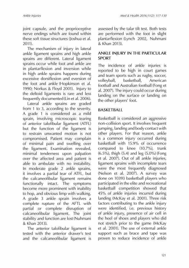
joint capsule, and the proprioceptive nerve endings which are found within these soft tissue structures (Joshua et al. 2011). The mechanism of injury in lateral ankle ligament sprains and high ankle sprains are different. Lateral ligament sprains occur while foot and ankle are in plantarflexion and inversion while in high ankle sprains happens during excessive dorsiflexion and eversion of the foot and ankle (Hopkinson et al. 1990; Norkus & Floyd 2001). Injury to the deltoid ligaments is rare and less frequently documented (Lin et al. 2006). Lateral ankle sprains are graded from 1 to 3, according to the severity. A grade 1 is considered as a mild sprain, involving microscopic tearing of anterior talofibular ligament (ATFL) but the function of the ligament is to restrain unwanted motion is not compromised. Patient may complain of minimal pain and swelling over the ligament. Examination revealed, minimal tenderness upon palpation over the affected area and patient is able to ambulate with no instability. In moderate grade 2 ankle sprains, it involves a partial tear of ATFL, but the calcaneofibular ligament remains functionally intact. The symptoms become more prominent with inability to hop, and obvious limp with walking. A grade 3 ankle sprain involves a complete rupture of the AFTL with partial or complete disruption of calcaneofibular ligament. The joint stability and function are lost (Nuhmani & Khan 2013). The anterior talofibular ligament is tested with the anterior drawer’s test and the calcaneofibular ligament is
assessed by the talar tilt test. Both tests are performed with the foot in slight plantarflexion (Lynch 2002, Nuhmani & Khan 2013).
ANKLE INJURY IN THE PARTICULAR SPORT
The incidence of ankle injuries is reported to be high in court games and team sports such as rugby, soccer, volleyball, basketball, American football and Australian football (Fong et al. 2007). The injury could occur during landing on the surface or landing on the other players’ foot.
BASKETBALL
Basketball is considered an aggressive non-collision sport. It involves frequent jumping, landing and body contact with other players. For that reason, ankle is a common injury occurred during basketball with 15.9% of occurrence compared to knee (10.7%), trunk (6.5%), thigh (5.4) and leg (5.0%) (Fong et al. 2007). Out of all ankle injuries, ligament sprains with incomplete tears were the most frequently diagnosed
(Nelson et al. 2007). A survey was done on 10393 basketball players who participated in the elite and recreational basketball competition showed that 45% of ankle injuries incurred during landing (McKay et al. 2001). Three risk factors contributing to the ankle injury were identified, i.e. previous history of ankle injury, presence of air cell in the heel of shoes and players who did not stretch prior to the game (McKay et al. 2001). The use of external ankle support such as brace and tape was proven to reduce incidence of ankle
122
Med & Health 2016;11(2): 117-130 Syarifah Aisyah S.A.H. et al.
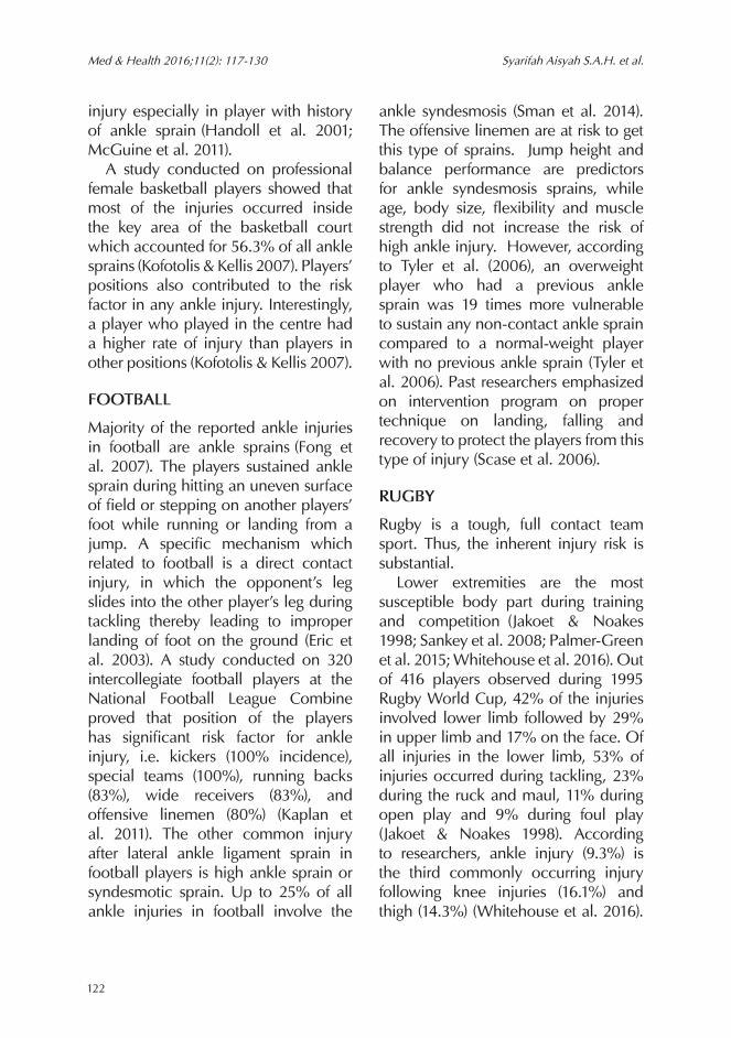
injury especially in player with history of ankle sprain (Handoll et al. 2001; McGuine et al. 2011). A study conducted on professional female basketball players showed that most of the injuries occurred inside the key area of the basketball court which accounted for 56.3% of all ankle sprains (Kofotolis & Kellis 2007). Players’ positions also contributed to the risk factor in any ankle injury. Interestingly, a player who played in the centre had a higher rate of injury than players in other positions (Kofotolis & Kellis 2007).
FOOTBALL
Majority of the reported ankle injuries in football are ankle sprains (Fong et al. 2007). The players sustained ankle sprain during hitting an uneven surface of field or stepping on another players’ foot while running or landing from a jump. A specific mechanism which related to football is a direct contact injury, in which the opponent’s leg slides into the other player’s leg during tackling thereby leading to improper landing of foot on the ground (Eric et al. 2003). A study conducted on 320 intercollegiate football players at the National Football League Combine proved that position of the players has significant risk factor for ankle injury, i.e. kickers (100% incidence), special teams (100%), running backs (83%), wide receivers (83%), and offensive linemen (80%) (Kaplan et al. 2011). The other common injury after lateral ankle ligament sprain in football players is high ankle sprain or syndesmotic sprain. Up to 25% of all ankle injuries in football involve the
ankle syndesmosis (Sman et al. 2014). The offensive linemen are at risk to get this type of sprains. Jump height and balance performance are predictors for ankle syndesmosis sprains, while age, body size, flexibility and muscle strength did not increase the risk of high ankle injury. However, according to Tyler et al. (2006), an overweight player who had a previous ankle sprain was 19 times more vulnerable to sustain any non-contact ankle sprain compared to a normal-weight player with no previous ankle sprain (Tyler et al. 2006). Past researchers emphasized on intervention program on proper technique on landing, falling and recovery to protect the players from this type of injury (Scase et al. 2006).
RUGBY
Rugby is a tough, full contact team sport. Thus, the inherent injury risk is substantial. Lower extremities are the most susceptible body part during training and competition (Jakoet & Noakes 1998; Sankey et al. 2008; Palmer-Green et al. 2015; Whitehouse et al. 2016). Out of 416 players observed during 1995 Rugby World Cup, 42% of the injuries involved lower limb followed by 29% in upper limb and 17% on the face. Of all injuries in the lower limb, 53% of injuries occurred during tackling, 23% during the ruck and maul, 11% during open play and 9% during foul play (Jakoet & Noakes 1998). According to researchers, ankle injury (9.3%) is the third commonly occurring injury following knee injuries (16.1%) and thigh (14.3%) (Whitehouse et al. 2016).
123
Ankle Injuries Med & Health 2016;11(2): 117-130
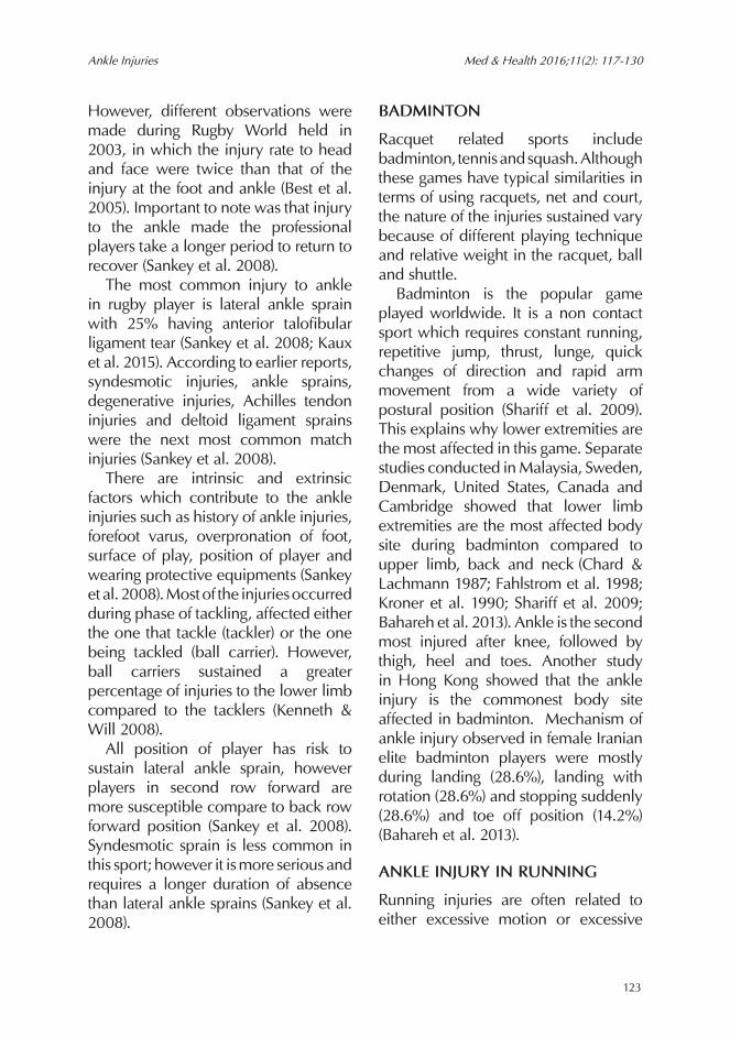
However, different observations were made during Rugby World held in 2003, in which the injury rate to head and face were twice than that of the injury at the foot and ankle (Best et al. 2005). Important to note was that injury to the ankle made the professional players take a longer period to return to recover (Sankey et al. 2008). The most common injury to ankle in rugby player is lateral ankle sprain with 25% having anterior talofibular ligament tear (Sankey et al. 2008; Kaux et al. 2015). According to earlier reports, syndesmotic injuries, ankle sprains, degenerative injuries, Achilles tendon injuries and deltoid ligament sprains were the next most common match injuries (Sankey et al. 2008). There are intrinsic and extrinsic factors which contribute to the ankle injuries such as history of ankle injuries, forefoot varus, overpronation of foot, surface of play, position of player and wearing protective equipments (Sankey et al. 2008). Most of the injuries occurred during phase of tackling, affected either the one that tackle (tackler) or the one being tackled (ball carrier). However, ball carriers sustained a greater percentage of injuries to the lower limb compared to the tacklers (Kenneth & Will 2008). All position of player has risk to sustain lateral ankle sprain, however players in second row forward are more susceptible compare to back row forward position (Sankey et al. 2008). Syndesmotic sprain is less common in this sport; however it is more serious and requires a longer duration of absence than lateral ankle sprains (Sankey et al. 2008).
BADMINTON
Racquet related sports include badminton, tennis and squash. Although these games have typical similarities in terms of using racquets, net and court, the nature of the injuries sustained vary because of different playing technique and relative weight in the racquet, ball and shuttle. Badminton is the popular game played worldwide. It is a non contact sport which requires constant running, repetitive jump, thrust, lunge, quick changes of direction and rapid arm movement from a wide variety of postural position (Shariff et al. 2009). This explains why lower extremities are the most affected in this game. Separate studies conducted in Malaysia, Sweden, Denmark, United States, Canada and Cambridge showed that lower limb extremities are the most affected body site during badminton compared to upper limb, back and neck (Chard & Lachmann 1987; Fahlstrom et al. 1998; Kroner et al. 1990; Shariff et al. 2009; Bahareh et al. 2013). Ankle is the second most injured after knee, followed by thigh, heel and toes. Another study in Hong Kong showed that the ankle injury is the commonest body site affected in badminton. Mechanism of ankle injury observed in female Iranian elite badminton players were mostly during landing (28.6%), landing with rotation (28.6%) and stopping suddenly (28.6%) and toe off position (14.2%) (Bahareh et al. 2013).
ANKLE INJURY IN RUNNING
Running injuries are often related to either excessive motion or excessive
124
Med & Health 2016;11(2): 117-130 Syarifah Aisyah S.A.H. et al.
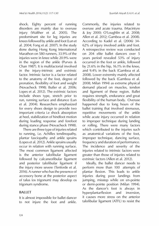
shock. Eighty percent of running disorders are mainly due to overuse injury (Walther et al. 2005). The predominant site for leg injuries are knees followed by ankle and foot (Lun et al. 2004; Fong et al. 2007). In the study done during Hong Kong International Marathon on 580 runners, 33.9% of the injuries were in knee while 20.9% were in the region of the ankle (Purves & Chan 1987). It is multifactorial involved in the injury-intrinsic and extrinsic factor. Intrinsic factor is a factor related to the anatomy of the foot, degree of pronation, flexibility of foot and weight (Novacheck 1998; Butler et al. 2006; Lopes et al. 2012). The extrinsic factors include shoes type, stretch prior to run, running surface and distance (Lun et al. 2004). Researchers emphasized for every shoes design to provide two important criteria i.e shock absorption at heel, stabilization of hindfoot motion during loading response and forefoot during stance phase (Novacheck 1998). There are three type of injuries related to running, i.e. Achilles tendinopathy, plantar fasciopathy and ankle sprains (Lopes et al. 2012). Ankle sprains usually occur in relation with running surface. The most common ligament affected is the anterior talofibular ligament followed by calcaneofibular ligament and posterior talofibular ligament if the injury more severe (Tenforde et al. 2016). A runner who has the presence of accessory bone at the posterior aspect of talus (os trigonum) may develop os trigonum syndrome.
BALLET
It is almost impossible for ballet dancer to not injure the foot and ankle.
Commonly, the injuries related to overuse and acute trauma. (Macintyre & Joy 2000; O’Loughlin et al. 2008; Allen et al. 2012; Gamboa et al. 2008). According to Kadel et al. (2006) 34-62% of injury involved ankle and foot. A retrospective review was conducted on 204 elite ballet dancers over 5 years period revealed 53% of injury occurred in the foot or ankle, followed by 21.6% in the hip, 16.1% in the knee, and 9.4% in the back (Gamboa et al. 2008). Lower extremity mainly affected followed by the back (Gamboa et al. 2008; Milan 1994) as extreme physical demand placed on muscles, tendon and ligament of these region. Ballet requires strength, endurance and great flexibility of the human body. Overuse happened due to long hours of the ballet training that involved strenuous, repetitive movements of the ankle while acute injury occurred in relation to improper technique during landing or rolling. There were many factors which contributed to the injuries such as anatomical variations of the foot, improper technique, dancing surface, frequency and duration of performance. The incidence and severity of the injuries related to intrinsic factors were greater than those of injuries related to extrinsic factors (Allen et al. 2012). Ideally, the ballet dancer needs to perform more than 100° attempts of plantar flexion. This leads to ankle injuries during poor landings from jumping, misstep while on en-pointe or demi-pointe position (Milan 1994). As the dancer’s foot is always in hyperplantarflexion and inversion, it causes more stress on the anterior talofibular ligament (ATFL) to resist the
125
Ankle Injuries Med & Health 2016;11(2): 117-130
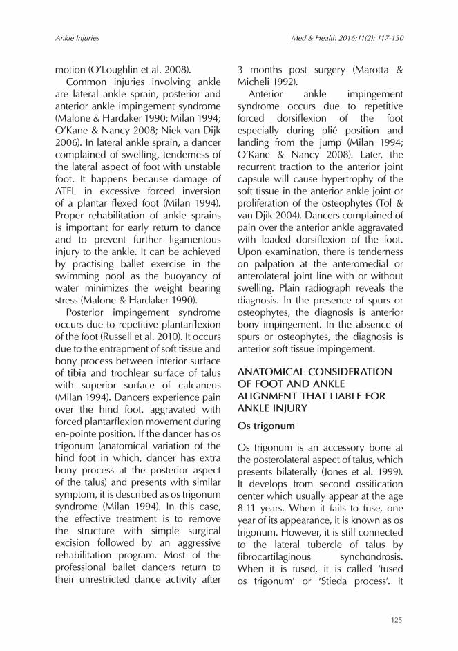
motion (O’Loughlin et al. 2008). Common injuries involving ankle are lateral ankle sprain, posterior and anterior ankle impingement syndrome (Malone & Hardaker 1990; Milan 1994; O’Kane & Nancy 2008; Niek van Dijk 2006). In lateral ankle sprain, a dancer complained of swelling, tenderness of the lateral aspect of foot with unstable foot. It happens because damage of ATFL in excessive forced inversion of a plantar flexed foot (Milan 1994). Proper rehabilitation of ankle sprains is important for early return to dance and to prevent further ligamentous injury to the ankle. It can be achieved by practising ballet exercise in the swimming pool as the buoyancy of water minimizes the weight bearing stress (Malone & Hardaker 1990). Posterior impingement syndrome occurs due to repetitive plantarflexion of the foot (Russell et al. 2010). It occurs due to the entrapment of soft tissue and bony process between inferior surface of tibia and trochlear surface of talus with superior surface of calcaneus (Milan 1994). Dancers experience pain over the hind foot, aggravated with forced plantarflexion movement during en-pointe position. If the dancer has os trigonum (anatomical variation of the hind foot in which, dancer has extra bony process at the posterior aspect of the talus) and presents with similar symptom, it is described as os trigonum syndrome (Milan 1994). In this case, the effective treatment is to remove the structure with simple surgical excision followed by an aggressive rehabilitation program. Most of the professional ballet dancers return to their unrestricted dance activity after
3 months post surgery (Marotta & Micheli 1992). Anterior ankle impingement syndrome occurs due to repetitive forced dorsiflexion of the foot especially during plié position and landing from the jump (Milan 1994; O’Kane & Nancy 2008). Later, the recurrent traction to the anterior joint capsule will cause hypertrophy of the soft tissue in the anterior ankle joint or proliferation of the osteophytes (Tol & van Djik 2004). Dancers complained of pain over the anterior ankle aggravated with loaded dorsiflexion of the foot. Upon examination, there is tenderness on palpation at the anteromedial or anterolateral joint line with or without swelling. Plain radiograph reveals the diagnosis. In the presence of spurs or osteophytes, the diagnosis is anterior bony impingement. In the absence of spurs or osteophytes, the diagnosis is anterior soft tissue impingement.
ANATOMICAL CONSIDERATION OF FOOT AND ANKLE ALIGNMENT THAT LIABLE FOR ANKLE INJURY
Os trigonum
Os trigonum is an accessory bone at the posterolateral aspect of talus, which presents bilaterally (Jones et al. 1999). It develops from second ossification center which usually appear at the age 8-11 years. When it fails to fuse, one year of its appearance, it is known as os trigonum. However, it is still connected to the lateral tubercle of talus by fibrocartilaginous synchondrosis. When it is fused, it is called ‘fused os trigonum’ or ‘Stieda process’. It
126
Med & Health 2016;11(2): 117-130 Syarifah Aisyah S.A.H. et al.
presents approximately in 5 to 15% of normal feet (Marotta & Micheli 1992). It is usually asymptomatic or noted incidentally by radiographic finding. However, it becomes symptomatic as a result of impingement of the soft tissue between the process and the posterior aspect of the tibia when the ankle is held in plantar flexion. It becomes obvious with strenuous physical activity
of the lower limb or during ankle injury, and termed as ‘os trigonum syndrome’. Patient presented with posterior ankle region pain occurred mostly when pushing off on the big toe (as in walking) or when pointing the toes downward (Marotta & Micheli 1992). It occurs due to the micro trauma from repetitive hyperplantarflexion or an episode of acute forced hyperplantarflexion which
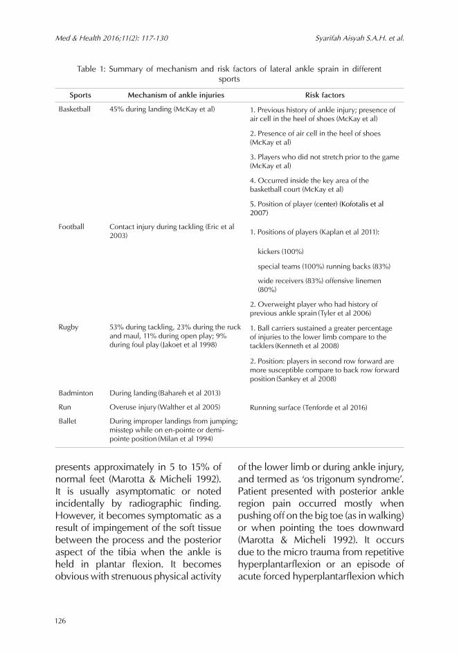
Table 1: Summary of mechanism and risk factors of lateral ankle sprain in different sports
Sports Mechanism of ankle injuries Risk factors
Basketball 45% during landing (McKay et al) 1. Previous history of ankle injury; presence of air cell in the heel of shoes (McKay et al)
2. Presence of air cell in the heel of shoes (McKay et al)
3. Players who did not stretch prior to the game (McKay et al)
4. Occurred inside the key area of the basketball court (McKay et al)
5. Position of player (center) (Kofotalis et al 2007)
Football Contact injury during tackling (Eric et al 2003) 1. Positions of players (Kaplan et al 2011):
kickers (100%)
special teams (100%) running backs (83%)
wide receivers (83%) offensive linemen (80%)
2. Overweight player who had history of previous ankle sprain (Tyler et al 2006)
Rugby 53% during tackling, 23% during the ruck and maul, 11% during open play; 9% during foul play (Jakoet et al 1998)
1. Ball carriers sustained a greater percentage of injuries to the lower limb compare to the tacklers (Kenneth et al 2008)
2. Position: players in second row forward are more susceptible compare to back row forward position (Sankey et al 2008)
Badminton During landing (Bahareh et al 2013)
Run Overuse injury (Walther et al 2005) Running surface (Tenforde et al 2016)
Ballet During improper landings from jumping; misstep while on en-pointe or demi-pointe position (Milan et al 1994)
127
Ankle Injuries Med & Health 2016;11(2): 117-130
compress the os trigonum between posterior malleolus of tibia and posterior process of calcaneus. This syndrome commonly occurs in ballet dancing, soccer, downhill running and volleyball.
Pes cavus
Pes cavus is also termed as high arched foot. It involves both the rear- and forefoot in which it is characterized by a high medial longitudinal arch, plantarflexed first ray, claw toes, and callous over the 5th metatarsal base. It is caused by bone malformations in the foot or nerve problems. One can develop this kind of foot if one continually wears shoes that have small toe box. Tight toe box force the toes to passively flex and raise the arch making the soft tissue structures to shorten and contract. This foot condition is very painful especially with motion (Linda 2009). This kind of foot is ideal for dancers as the requirement for plantar flexion is maximal. However, this position will limit dorsiflexion. Hence, dancers’ are more susceptible to anterior impingement syndrome.
Pes planus
Pes planus is also known as flat foot arch or flat foot. It can be structural which usually present from birth. It is normal for infant and young children to have flat feet but as they grow older, the normal arch forms. However, the normal arch does not form in the few children for unknown reasons. Pes planus may also be functional, caused by loose soft tissue structures of the
plantar portion of foot. An athlete with pes planus since birth, may not have any specific symptoms and signs of foot discomfort. However, they may have a tired feeling in the feet after prolonged standing, walking or running. The athlete, who has a foot injury, also may have pes planus (Linda 2009).
Management The principle management of acute ankle injury is R.I.C.E (rest, ice, compression and elevation). During acute situation, physical examination is not reliable as it is obscured with pain and swelling. Thus, ice therapy, non steroidal anti-inflammatory drugs, massage, compression bandage and leg elevation above the heart level are very important to ease the pain, reduce the swelling and inflammation (Chorley 2005; Joshua et al. 2011; Nuhmani & Khan 2013). Traditional Chinese medicine also been used widely in China to reduce pain and swelling and promote recovery of ankle function. It includes herb, acupuncture and massage (Koo et al. 2002; Joshua et al. 2011). Later, further treatment is given according to the diagnosis. The treatment for type 1 and 2 ankle sprains is mainly conservative. The use of Aircast ankle brace, elastic support bandage were recommended than immobilization. Immobilization may result in joint stiffness, loss of propioception and late return to sports (Nuhmani & Khan 2013). Aircast ankle brace appear to be more effective to restore ankle joint function compared to elastic bandage support bandage (Boyce et al. 2005).

128
Med & Health 2016;11(2): 117-130 Syarifah Aisyah S.A.H. et al.
If the leg’s swelling and pain is not reduced or becomes more severe after the initial treatment, there is highly suspicious of grade 3 lateral ankle sprain. Usually magnetic resonance imaging (MRI) study is conducted to assess the severity of the injury (Nuhmani & Khan 2013). Few surgeons opt for operative approach, but others prefer non-operative approach as the treatment. Players with syndesmotic ankle sprains are prescribed walking boots to avoid separation of the distal tibia and fibula. This allows healing of the interosseous ligaments within four to six weeks (Fong et al. 2009). Rehabilitation program post injury is very important and has been considered as a part of the management for ankle injury. The effectiveness of the program ensures the recovery of the ankle and the future performance of the athlete (Mattacola & Dwyer 2002). Inadequate management of ankle sprain injury may lead to residual symptoms and chronic problems to ankle such as chronic ankle instability and osteoarthritis. It was reported that 74% of players with ankle sprain complained of persisting symptoms up to 4 years following injury. Up to 20-40% of these patients develop chronic symptoms such as synovitis, tendinitis, swelling, pain, stiffness, weakness and crepitus (Fong et al. 2009; Valderrabano et al. 2007). Epidemiology study on Hong Kong Chinese athletes, reported that the athletes with less than 5 times history of ankle sprained complained of pain as their residual symptom, while athletes with more than 5 times history of ankle sprain suffered with instability (Yeung et al. 1994).
CONCLUSION
Prior anatomical knowledge of the lower limb, especially the ankle joint is important for ascertaining the exact nature of injury sustained in any sport. The present review was a humble attempt to discuss the ankle injuries related to sports and discuss the best available treatment outcomes.
REFERENCESAllen, N., Nevill, A., Brooks, J., Koutedakis, Y., Wyon,
M. 2012. Ballet injuries: injury incidence and severity over 1 year. J Orthop Sports Phys Ther 42(9): 781-90.
Andrew, F.M., Eanna, F., Paul, M., Peter, B., 2011.Clinical sport injury. Sydney: McGraw-Hill 307-346.
Bahareh, M., Golnaz, F., Mohammad, H.A., Nader, R. 2013. Common injuries in Iranian female elite badminton players. Global Journal of Science, Engineering and Technology 12(1): 24-8.
Baumhauer, J.F., Alosa, D.M., Renstrom, A.F., Trevino, S., Beynnon, B. 1995. A prospective study of ankle injury risk factors. Am J Sports Med 23(5): 564-70.
Best, J.P., McIntosh, A.S., Savage, T.N. 2005. Rugby World Cup 2003 injury surveillance project. Br J Sports Med 39(11): 812-7.
Beynnon, B.D., Murphy, D.F., Alosa, D.M 2002. Predictive Factors for Lateral Ankle Sprains: A Literature Review. J Athl Train 37(4): 376-80.
Boyce, S.H., Quigley, M.A, Campbell, S. 2005. Management of ankle sprains: a randomised controlled trial of the treatment of inversion injuries using an elastic support bandage or an Aircast ankle brace. Br J Sports Med 39(2): 91-6.
Butler, R.J., Davis, I.S, Hamill, J. 2006. Interaction of arch type and footwear on running mechanics. Am J Sports Med 34(12): 1998-2005.
Chard, M.D., Lachmann, S.M. 1987. Racquet sports--patterns of injury presenting to a sports injury clinic. Br J Sports Med 21(4): 150-3.
Chorley, J.N. 2005. Ankle sprain discharge instructions from the emergency department. Pediatr Emerg Care 21(8): 498-501.
Eric, G., Colin, F., Astrid, J., Jiri, D. 2003. Mechanisms of Foot and Ankle Injuries in Soccer. Am J Sports Med 31(4): 550-4.
Fahlstrom, M., Bjőrnstig, U., Lorentzon, R. 1998. Acute badminton injuries. Scand J Med Sci Sports 8(3): 145-8.
Floyd, R.T. 2009. Manual of structural kinesiology.
129
Ankle Injuries Med & Health 2016;11(2): 117-130
17th edition. New York: McGraw-Hill; 294-8.Fong, D.T., Chan, Y.Y., Mok, K.M., Yung, P.S.H,
Chan, K.M. 2009. Understanding acute ankle ligamentous sprain injury in sports. Sports Med Arthrosc Rehabil Ther Technol. 2009; 1: 14.
Fong, D.T., Hong, Y., Chan, L.K., Yung, P.S., Chan, K.M. 2007. A systematic review on ankle injury and ankle sprain in sports. Sports Med 37(1): 73-94.
Gamboa, J.M., Roberts, L.A., Maring, J., Fergus, A. 2008. Injury patterns in elite preprofessional ballet dancers and the utility of screening programs to identify risk characteristics. J Orthop Sports Phys Ther 38(30): 126-36.
Gerber, J.P., Williams, G.N., Scoville, C.R., Arciero, R.A., Taylor, D.C. 1998. Persistent disability associated with ankle sprains: a prospective examination of an athletic population. Foot Ankle Int 19(10): 653-60.
Handoll, H.H., Rowe, B.H., Quinn, K.M., de Bie, R. 2001. Interventions for preventing ankle ligaments injuries. Cochrane Database Syst Rev (3): CD000018.
Hopkinson, W.J., St Pierre, P., Ryan, J.B., Wheeler, J.H. 1990. Syndesmosis sprains of the ankle. Foot Ankle 10(6): 325-30.
Jakoet, I., Noakes, T.D.1998. A high rate of injury during the 1995 Rugby World Cup. S Afr Med J 88(1): 45-7.
Jones, D.M., Saltzman, C.L., El-Khoury, G. 1999. The diagnosis of the os trigonum syndrome with a fluoroscopically controlled injection of local anesthetic. Iowa Orthop J 19:122-6.
Joshua, C.D., Doug, C., Rebecca, I.M., Rachel, A.D., Ernest, F. 2011. Lateral and syndesmotic ankle sprain injuries: a narrative literature review. J Chiropr Med 10(3): 204-19.
Kadel, N.J. 2006. Foot and ankle injuries in dance. Phys Med Rehabil Clin N Am 17(4): 813-26.
Kaplan, L.D., Jost, P.W., Honkamp, N., Norwig, J., West, R., Bradley, J.P. 2011. Incidence and variance of foot and ankle injuries in elite college football players. Am J Orthop (Belle Mead NJ) 40(1): 40-4.
Kaux, J.F., Julia, M., Delvaux, F., Croisier, J.L., Forthomme, B., Monnot, D., Chupin, M., Crielaard, J.M., Goff, C.L., Durez, P., Ernst, P., Guns, S., Laly, A. 2015. Epidemiological Review of Injuries in Rugby Union. Sports 3: 21-9.
Kennedy, J.G., Knowles, B., Dolan, M., Bohne, W. 2005. Foot and ankle injuries in the adolescent runner. Curr Opin Pediatr 17(1): 34-42.
Kenneth, L.Q., Will, G.H. 2008. Tackle Injuries in Professional Rugby Union. The American Journal of Sports Medicine 36(39): 1705-16.
Kofotolis, N., Kellis, E. 2007. Ankle sprain injuries: a 2-year prospective cohort study in female Greek professional basketball players. J Athl
Train 42(3): 388-94.Koo, S.T., Park, Y.I., Lim, K.S., Chung, K., Chung,
J.M. 2002. Acupuncture analgesia in a new rat model of ankle sprain pain. Pain 99(3): 423-31.
Kroner, K., Schmidt, S.A., Nielsen, A.B., Yde, J. 1990. Badminton injuries. Br J Sport Med 24(2): 169-72.
Lin, C.F., Gross, M.L., Weinhold, P. 2006. Ankle syndesmosis injuries: anatomy, biomechanics, mechanism of injury, and clinical guidelines for diagnosis and intervention. J Orthop Sports Phys Ther 36(6): 372-84.
Linda, G., 2009. Survey of athletic injuries for exercise science. New York: McGraw-Hill; 271-2.
Lopes, A.D., Hespanhol Júnior, L.C., Yeung, S.S., Costa, L.O. 2012. What are the main running-related musculoskeletal injuries? A systematic review. Sports Med 42(10): 891-905.
Lun, V., Meeuwisse, W.H., Stergiou, P., Stefanyshyn, D. 2004. Relation between running injury and static lower limb alignment in recreational runners. Br J Sports Med 38(5): 576-80.
Lynch, S.A. 2002. Assesment of the injured ankle in the athlete. J Athl Train 37(4): 406-12.
Macintyre, J., Joy, E. 2000. Foot and ankle injuries in dance. Clin Sports Med 19(2): 351-68.
Malone, T.R., Hardaker, W.T. 1990. Rehabilitation of foot and ankle injuries in ballet dancers. J Orthop Sports Phys Ther 11(8): 355-61.
Marotta, J.J, Micheli, L.J. 1992. Os trigonum impingement in dancers. Am J Sports Med 20(5): 533-6.
Mattacola, C.G., Dwyer, M.K. 2002. Rehabilitation of the ankle after acute ankle sprain or chronic instability. J Athl Train 37(4) :413-29.
McGuine, T.A., Brooks, A., Hetzel, S. 2011. The effect of lace-up ankle braces on injury rates in high school basketball players. Am J Sports Med 39(9): 1840-8.
McKay, G.D., Goldie, P.A., Payne, W.R., Oakes, B.W. 2001. Ankle injuries in basketball: injury rate and risk factors. Br J of Sports 35(2): 103-108.
Milan, K.R. 1994. Injury in ballet: A review of relevant topics for the physical therapist. J Orthop Sports Phys Ther 19(2): 121-9.
Moore, K.L., Dalley, A.F., Agur, A.M.R. 2014. Clinically oriented anatomy. 7th edition. Philadelphia: Lippincott Willian & Wilkin; 647-49
Nancy, H., Wendi, W., Kathryn, L. 2012. Kinesiology-Scientigic basis of human motion 12th edition. New York: McGraw-Hill.
Nelson, A.J., Collins, C.L., Yard, E.E., Fields, S.K., Comstock, R.D. 2007. Ankle injuries among United States high school sports athletes, 2005-2006. J Athl Train 42(3): 381-7.
Niek van Dijk, C. 2006. Anterior and posterior ankle impingement. Foot Ankle Clin 11(3): 663-83.
130
Med & Health 2016;11(2): 117-130 Syarifah Aisyah S.A.H. et al.
Norkus, S.A., Floyd, R.T. 2001. The anatomy and mechanisms of syndesmotic ankle sprains. J Athl Train 36(1): 68-73.
Novacheck, T.F. 1998. The biomechanics of running. Gait Posture 7(1): 77-95.
Nuhmani, S., Khan, M.H. 2013. Lateral ankle sprain – an update. Journal of musculoskeletal research 16(4): 1330003.
O’Kane, J.W., Nancy, K. 2008. Anterior impingement syndrome in dancers. Curr Rev Musculoskeletal Med 1(1): 12-6.
O’Loughlin, P.F., Hodgkins, C.W., Kennedy, J.G. 2008. Ankle sprains and instability in dancers. Clin Sports Med 27(2): 247-62.
Palmer-Green, D.S., Stokes, K.A., Fuller, C.W., England, M., Kemp, S.P., Trewartha G. 2015. Training activities and injuries in English youth academy and schools rugby union. Am J Sports Med 43(2): 475-81.
Purves, C. Chan, K.M. 1987. Injury profile of runners in the 1987. The Journal of the Hong Kong Physiotherapy Association 9: 24-8.
Russell, J.A., Kruse, D.W., Koutedakis, Y., McEwan, I.M., Wyon, M.A. 2010. Pathoanatomy of posterior ankle impingement in ballet dancers. Clin Anat 23(6): 613-21.
Sankey, R.A., Brooks, J.H., Kemp, S.P., Haddad, F.S. 2008. The epidemiology of ankle injuries in professional rugby union players. Am J Sports Med 36(12): 2415-24.
Scase, E., Cook, J. Makdissi, M., Gabbe, B., Shuck, L. 2006. Teaching landing skills in elite junior Australian football: evaluation of an injury prevention strategy. Br J Sports Med 40(10): 834-8.
Sman, A.D., Hiller, C.E., Rae, K., Linklater, J., Morellato, J., Trist, N., Nicholson, L.L., Black, D.A., Refshauge, K.M. 2014. Predictive factors for ankle syndesmosis injury in football players: a prospective study. J Sci Med Sport 17(6): 586-90.
Shariff, A.H., George, J. Ramlan, A.A. 2009. Musculoskeletal injuries among Malaysian badminton players. Singapore Med J 50(11): 1095-7.
Tenforde, A.S., Yin, A., Hunt, K.J. 2016. Foot and ankle injuries in runners. Phys Med Rehabil Clin N Am 27(1): 121-37.
Tol, J.L., van Djik, C.N. 2004. Etiology of the anterior ankle impingement syndrome: a descriptive anatomical study. Foot Ankle Int 25(6): 382-6.
Tyler, T.F., McHugh, M.P., Mirabella, M.R., Mullaney, M.J., Nicholas, S.J. 2006. Risk factors for noncontact ankle sprains in high school football players: the role of previous ankle sprains and body mass index. Am J Sports Med 34(3): 471-5.
Valderrabano, V., Wiewiorski, M., Frigg, A., Hintermann, B., Leumann, A. 2007. Chronic ankle instability. Unfallchirurg 110(8): 691-9.
Walther, M., Reuter, I., Leonhard, T. Engelhardt, M. 2005. Injuries and response to overload stress in running. Orthopäde 34(5): 399-404.
Whitehouse, T., Orr, R., Fitzgerald, E., Harries, S., McLellan, C.P. 2016. The epidemiology of injuries in Australian Professional Rugby Union 2014 Super Rugby Competition. Orthop J Sports Med 4(3): 2325967116634075.
Yeung, M.S., Chan. K.M., So, C.H., Yuan, W.Y. 1994. An epidemiological survey on ankle sprain. Br J Sports Med 28(2): 112-6.