l6 the lymphatic system blok bms.pptx
TRANSCRIPT
THE LYMPHATIC SYSTEM
Capaian pembelajaran :
Setelah mengikuti perkuliahan mahasiswa diharapkan mampu :
1. Menjelaskan organ sistem limfatik2. Menjelaskan fungsi sistem limfatik3. Menjelaskan mekanisme kerja sistem
limfatik
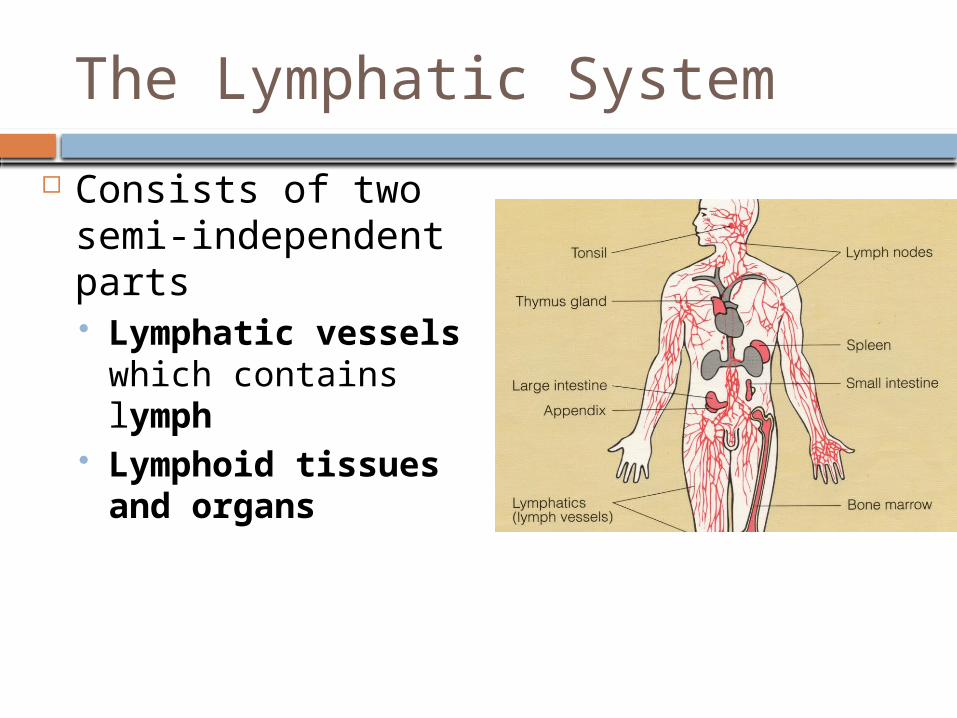
The Lymphatic System
Consists of two semi-independent parts Lymphatic vessels
which contains lymph
Lymphoid tissues and organs
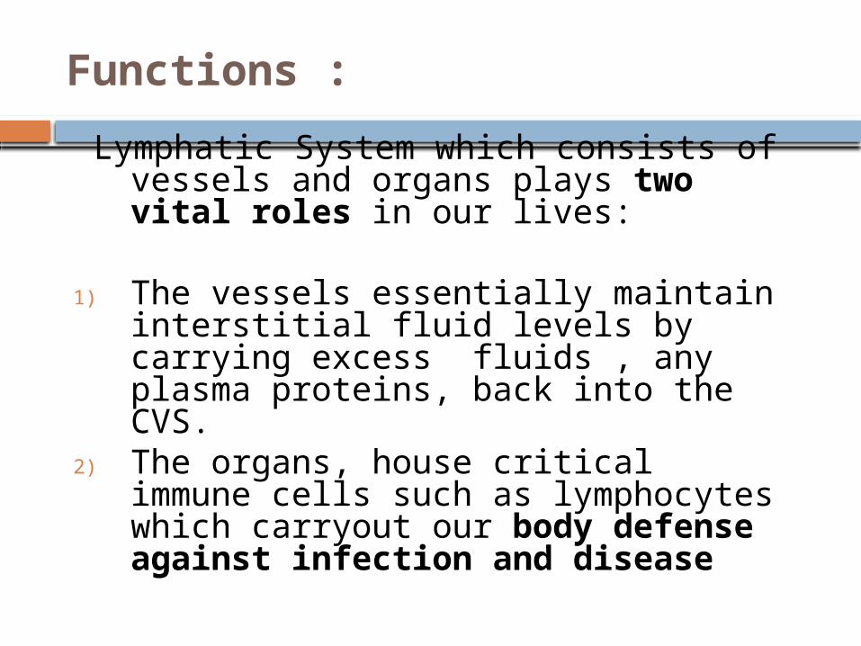
Functions :
Lymphatic System which consists of vessels and organs plays two vital roles in our lives:
1) The vessels essentially maintain interstitial fluid levels by carrying excess fluids , any plasma proteins, back into the CVS.
2) The organs, house critical immune cells such as lymphocytes which carryout our body defense against infection and disease
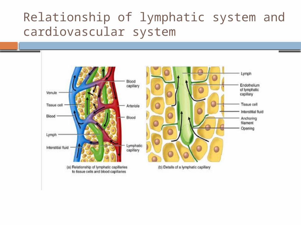
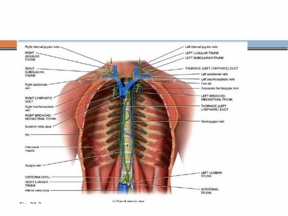
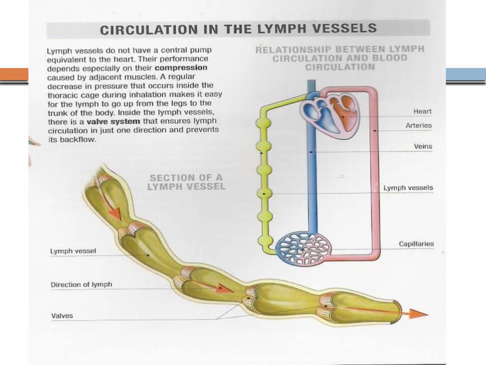
Relationship of lymphatic system and cardiovascular system
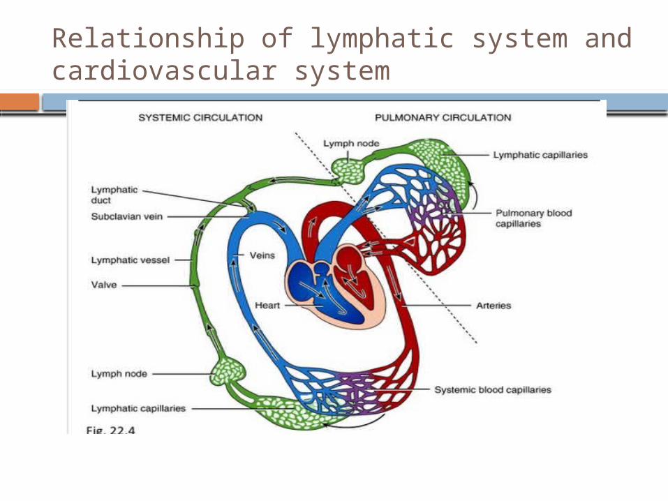
Relationship of lymphatic system and cardiovascular system
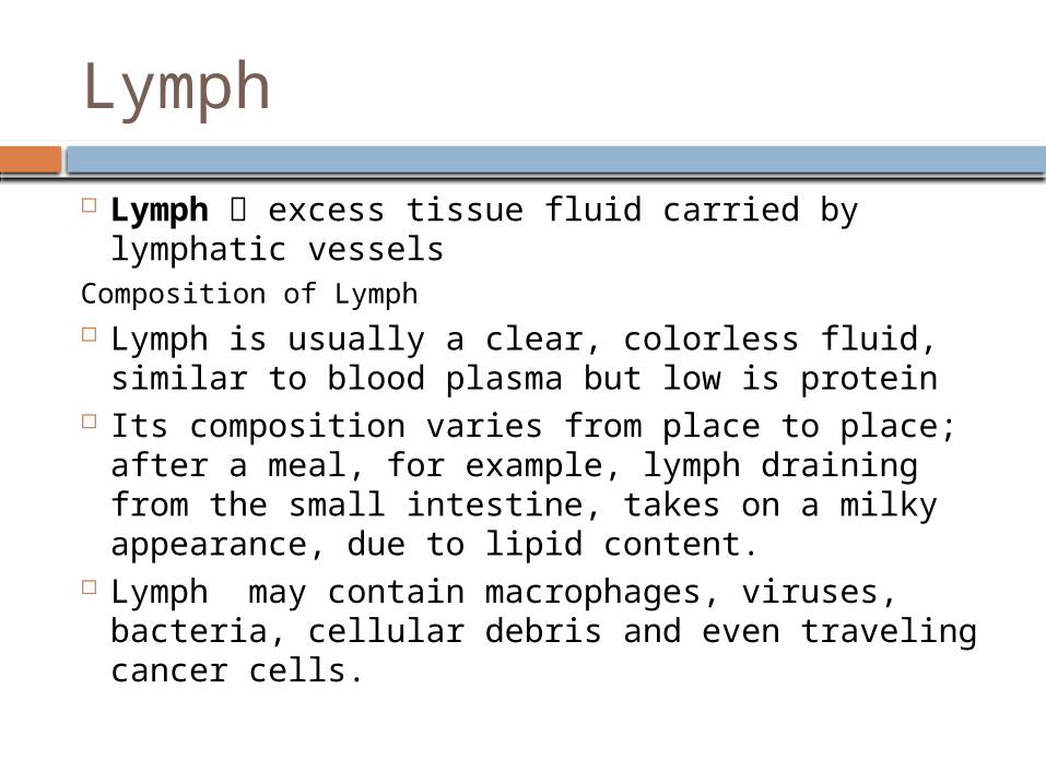
Lymph
Lymph excess tissue fluid carried by lymphatic vessels
Composition of Lymph Lymph is usually a clear, colorless fluid, similar to
blood plasma but low is protein Its composition varies from place to place; after a
meal, for example, lymph draining from the small intestine, takes on a milky appearance, due to lipid content.
Lymph may contain macrophages, viruses, bacteria, cellular debris and even traveling cancer cells.
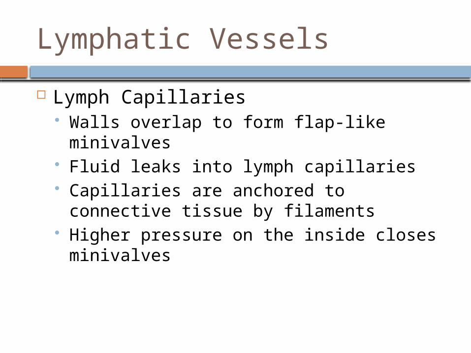
Lymphatic Vessels
Lymph Capillaries Walls overlap to form flap-like minivalves Fluid leaks into lymph capillaries Capillaries are anchored to connective
tissue by filaments Higher pressure on the inside closes
minivalves
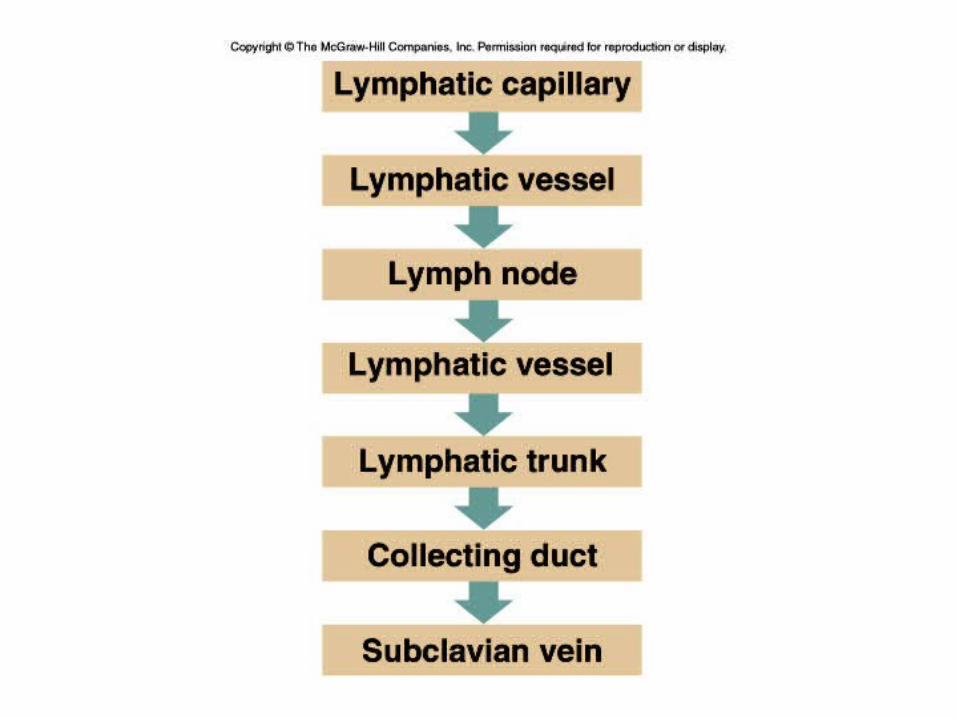
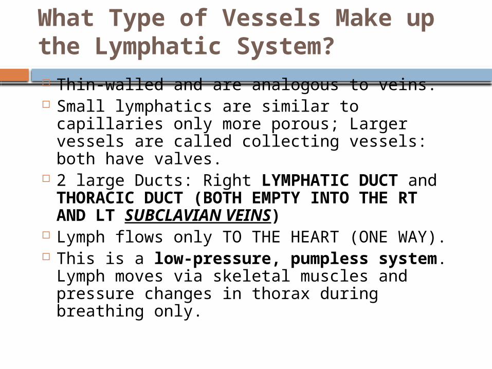
What Type of Vessels Make up the Lymphatic System? Thin-walled and are analogous to veins. Small lymphatics are similar to capillaries
only more porous; Larger vessels are called collecting vessels: both have valves.
2 large Ducts: Right LYMPHATIC DUCT and THORACIC DUCT (BOTH EMPTY INTO THE RT AND LT SUBCLAVIAN VEINS)
Lymph flows only TO THE HEART (ONE WAY). This is a low-pressure, pumpless
system. Lymph moves via skeletal muscles and pressure changes in thorax during breathing only.
Lymphatic Organs:
A Lymph Node. Spleen Thymus Gland Tonsils MALT Peyer’s patch
MALT
The collection of lymphoid tissues linked with the digestive system is called the mucosa-associated lymphoid tissue (MALT).
Clusters of lymphoid nodules deep to the epithelial lining of the intestine are known as aggregate lymphoid nodules, or Peyer’s patches
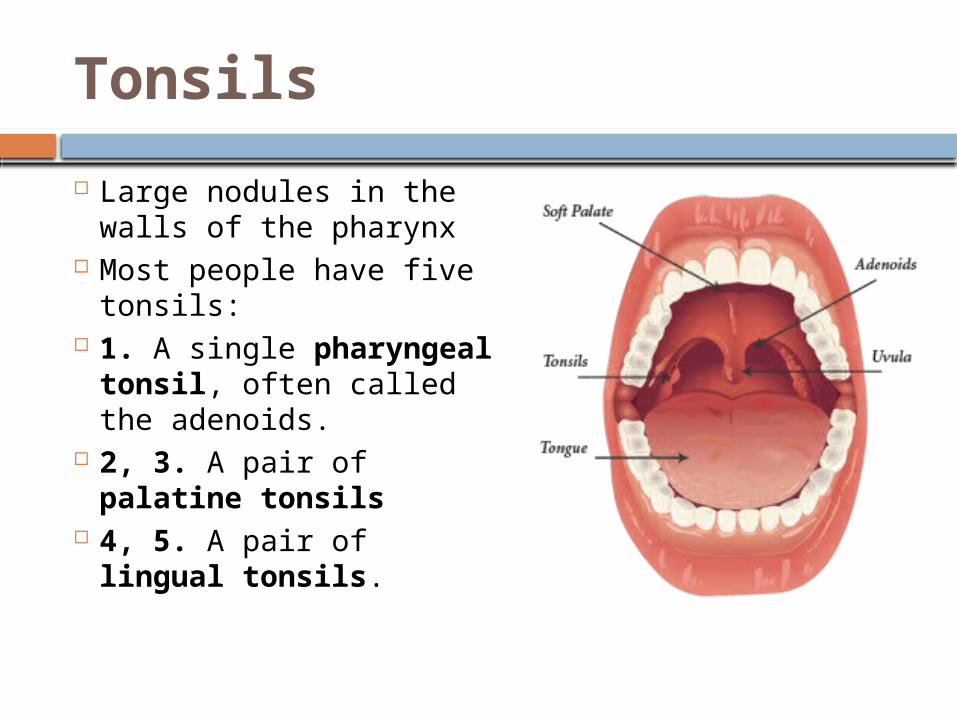
Tonsils
Large nodules in the walls of the pharynx
Most people have five tonsils:
1. A single pharyngeal tonsil, often called the adenoids.
2, 3. A pair of palatine tonsils
4, 5. A pair of lingual tonsils.
The Thymus
The thymus is located in the mediastinum, generally just posterior to the sternum.
In newborn infants and young children, the thymus is relatively large.
The thymus reaches its greatest size (relative to body size) in the first year or two after birth. The thymus reaches its maximum absolute size just before puberty. After pubertyinvolution
The thymus produces Thymosin that promotes the development and maturation of lymphocytes
Lymph Nodes
Lymph Nodes take the germ-filled lymph and Filter lymph before it is returned to the blood
Defense cells within lymph nodes Macrophages – engulf and destroy foreign
substances Lymphocytes – provide immune response to
antigens
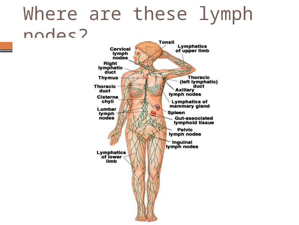
Where are these lymph nodes?
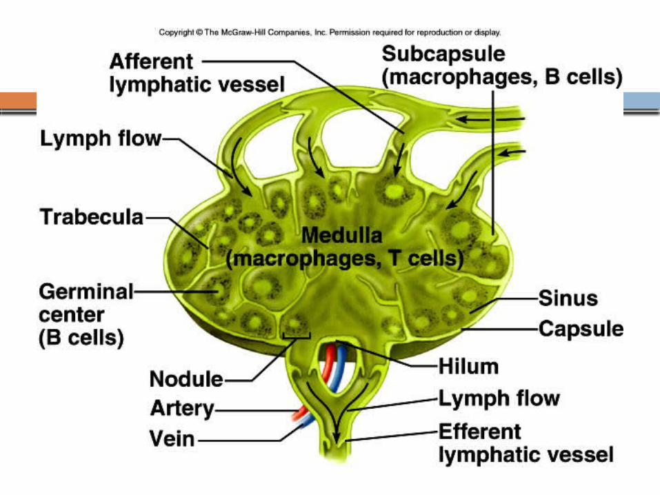
Lymph Node Structure
Most are kidney-shaped, less than 1 inch long
Cortex Outer part Contains follicles – collections of
lymphocytes Medulla
Inner part Contains phagocytic macrophages
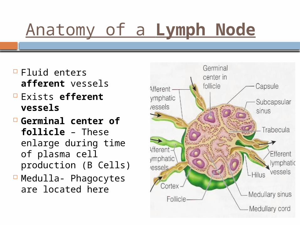
Anatomy of a Lymph Node
Fluid enters afferent vessels
Exists efferent vessels
Germinal center of follicle – These enlarge during time of plasma cell production (B Cells)
Medulla- Phagocytes are located here
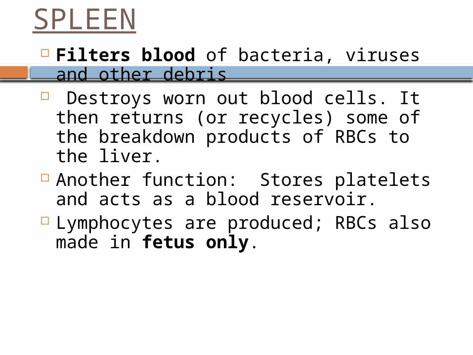
SPLEEN Filters blood of bacteria, viruses and
other debris Destroys worn out blood cells. It then
returns (or recycles) some of the breakdown products of RBCs to the liver.
Another function: Stores platelets and acts as a blood reservoir.
Lymphocytes are produced; RBCs also made in fetus only.
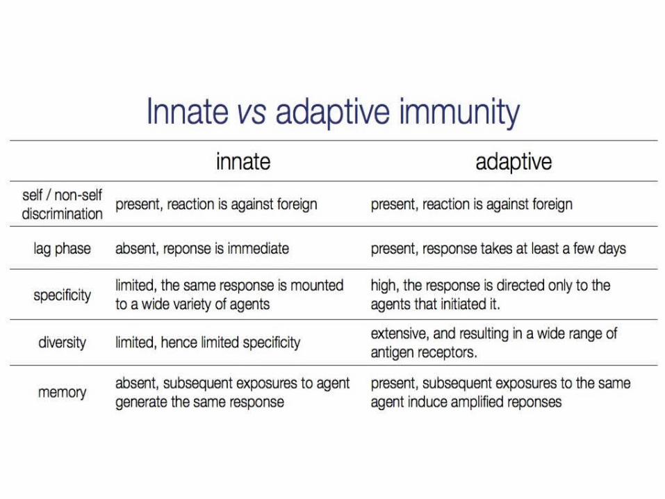
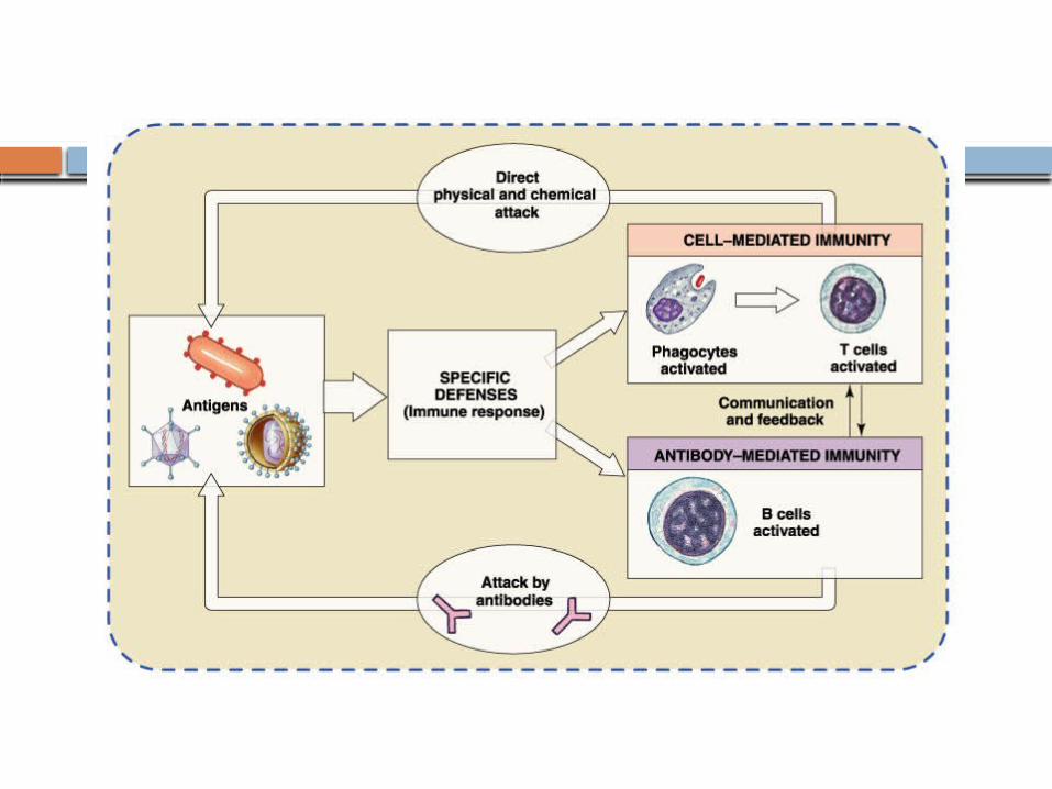
Lymphatic system and Body’s Defense System
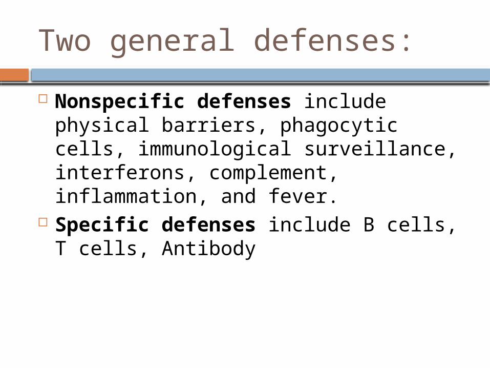
Two general defenses:
Nonspecific defenses include physical barriers, phagocytic cells, immunological surveillance, interferons, complement, inflammation, and fever.
Specific defenses include B cells, T cells, Antibody
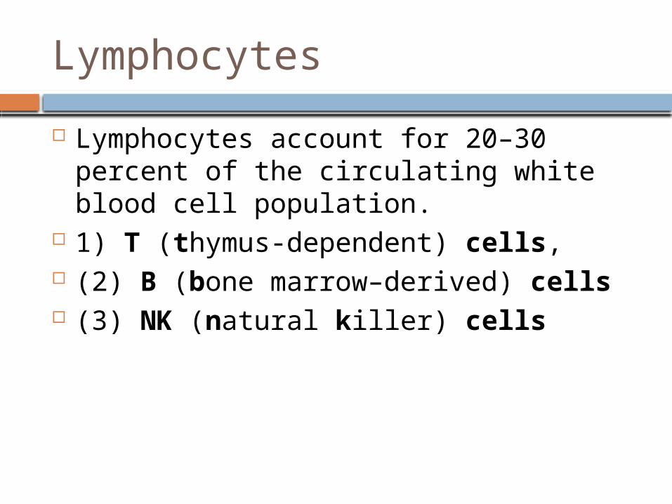
Lymphocytes
Lymphocytes account for 20–30 percent of the circulating white blood cell population.
1) T (thymus-dependent) cells, (2) B (bone marrow–derived) cells (3) NK (natural killer) cells
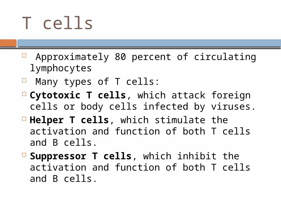
T cells
Approximately 80 percent of circulating lymphocytes
Many types of T cells: Cytotoxic T cells, which attack foreign cells
or body cells infected by viruses. Helper T cells, which stimulate the activation
and function of both T cells and B cells. Suppressor T cells, which inhibit the
activation and function of both T cells and B cells.
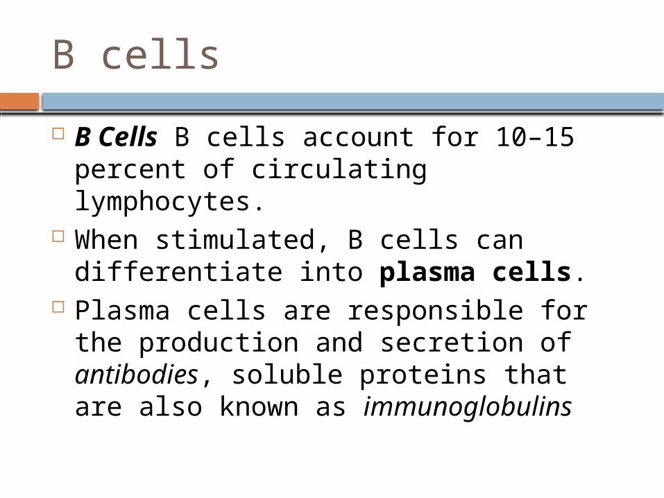
B cells
B Cells B cells account for 10–15 percent of circulating lymphocytes.
When stimulated, B cells can differentiate into plasma cells.
Plasma cells are responsible for the production and secretion of antibodies, soluble proteins that are also known as immunoglobulins
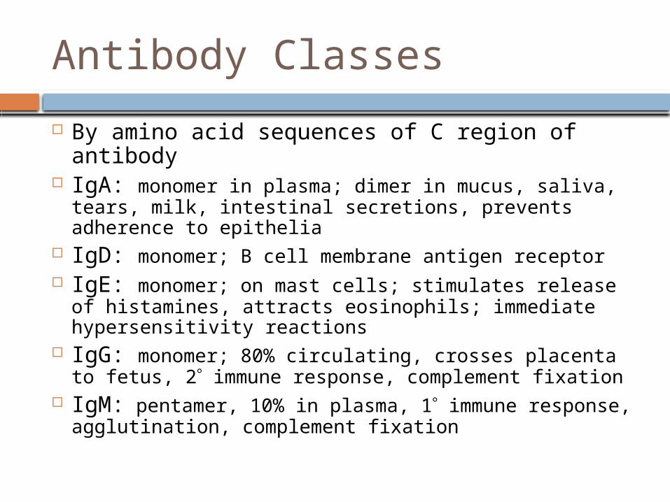
Antibody Classes
By amino acid sequences of C region of antibody
IgA: monomer in plasma; dimer in mucus, saliva, tears, milk, intestinal secretions, prevents adherence to epithelia
IgD: monomer; B cell membrane antigen receptor IgE: monomer; on mast cells; stimulates release of
histamines, attracts eosinophils; immediate hypersensitivity reactions
IgG: monomer; 80% circulating, crosses placenta to fetus, 2 immune response, complement fixation
IgM: pentamer, 10% in plasma, 1 immune response, agglutination, complement fixation
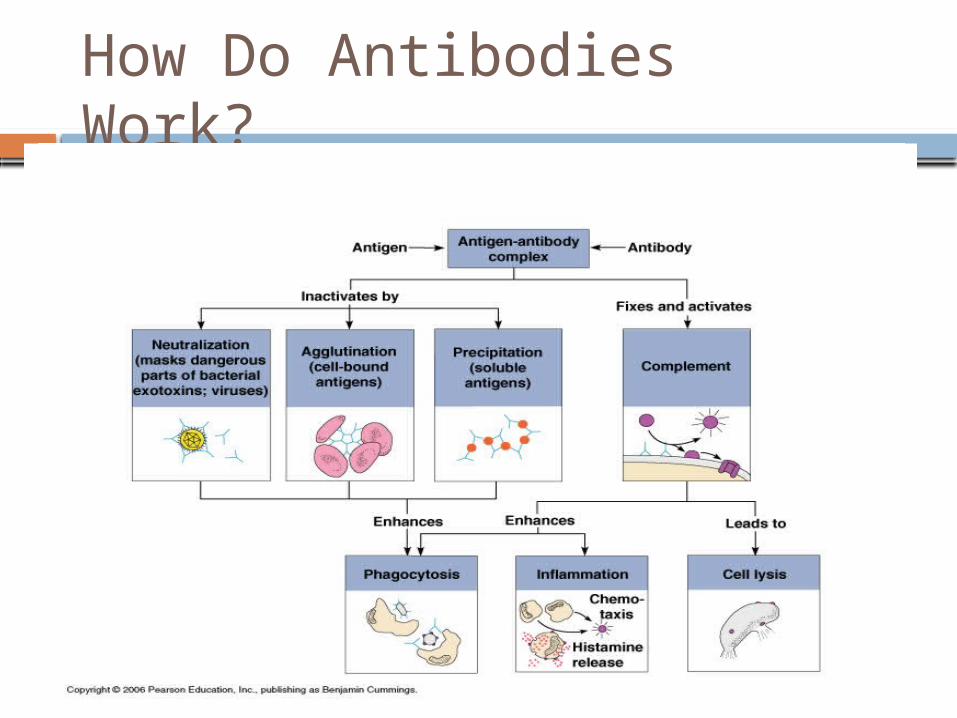
How Do Antibodies Work?
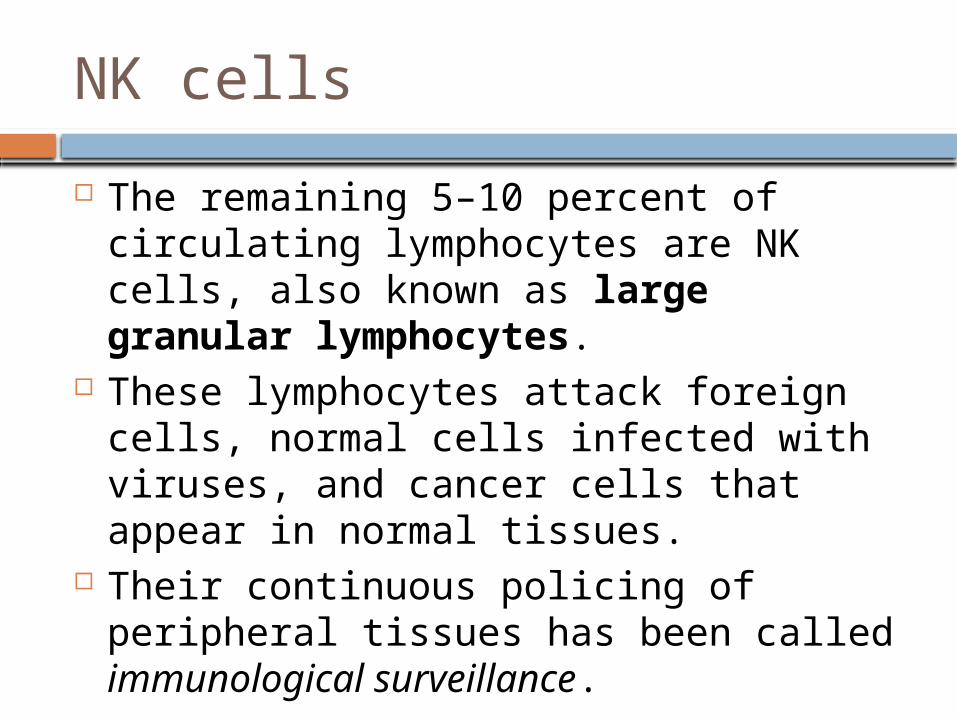
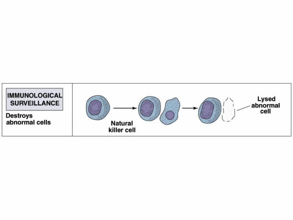
NK cells
The remaining 5–10 percent of circulating lymphocytes are NK cells, also known as large granular lymphocytes.
These lymphocytes attack foreign cells, normal cells infected with viruses, and cancer cells that appear in normal tissues.
Their continuous policing of peripheral tissues has been called immunological surveillance.
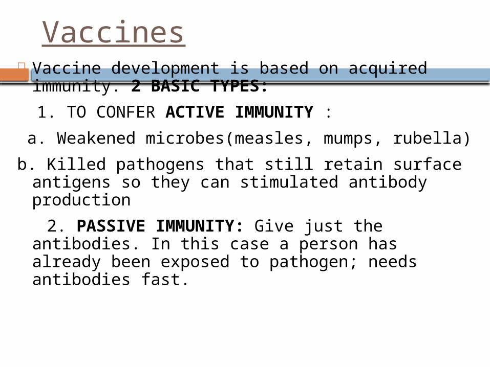
Vaccines Vaccine development is based on acquired
immunity. 2 BASIC TYPES:
1. TO CONFER ACTIVE IMMUNITY :
a. Weakened microbes(measles, mumps, rubella)
b. Killed pathogens that still retain surface antigens so they can stimulated antibody production
2. PASSIVE IMMUNITY: Give just the antibodies. In this case a person has already been exposed to pathogen; needs antibodies fast.
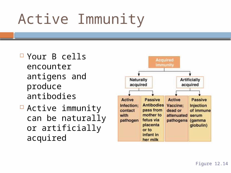
Active Immunity
Your B cells encounter antigens and produce antibodies
Active immunity can be naturally or artificially acquired
Figure 12.14

TERIMA KASIH