asuhankeperawatan kebumen ...elib.stikesmuhgombong.ac.id/135/1/hening muji raharjo nim...iv...
TRANSCRIPT
i
ASUHAN KEPERAWATAN
PEMENUHAN KEBUTUHAN RASA AMAN DAN NYAMAN
PADA NY.W DIRUANG TERATAI RUMAH SAKIT DR SOEDIRMAN
KEBUMEN
Diajukan Untuk Memenuhi Tugas Akhir Ujian Komprehensif
Jenjang Pendidikan Diploma III Keperawatan
Pendidikan Ahli Madya Keperawatan
Disusun oleh
Hening Muji Raharjo
A01301760
SEKOLAH TINGGI ILMU KESEHATAN MUHAMMADIYAH GOMBONG
PROGRAM STUDI DIII KEPERAWATAN
2016
ii
iii
iv
Program Studi DIII KeperawatanSekolah Tinggi Ilmu Kesehatan Muhammadiyah GombongKTI, Juli 2016Hening Muji Raharjo1, Irmawan Andri Nugroho2.
ABSTRAK
ASUHAN KEPERAWATAN PEMENUHAN KEBUTUHAN RASA AMANDAN NYAMAN PADA NY. W DI RUANG TERATAI RS DR SOEDIRMAN
KEBUMEN
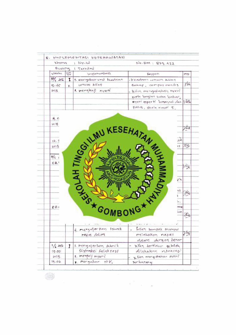
Latar belakang: Komplikasi yang paling umum terjadi pada luka bakar adalahkerusakan integritas kulit. Hal ini menyebabkan pemenuhan kebutuhan rasa amandan nyaman terganggu yaitu nyeri.Tujuan: Memaparkan asuhan keperawatan pemenuhan kebutuhan rasa aman dannyaman nyeri akut pada luka bakar.Analisa data: Dari pengkajian diperoleh masalah nyeri akut b.d agen cedera fisik.Intervensi: Kaji nyeri, ajarkan teknik nafas dalam, teknik distraksi dan relaksasi.Implementasi: Mengkaji nyeri, Mengajarkan teknik distraksi dan relaksasi.Evaluasi: Masalah nyeri akut belum teratasi, skala nyeri berkurang dari skala 5menjadi skala 3, klien mengerang kesakitan bila luka di pegang atau tersentuh.
Kata kunci: aman dan nyaman, asuhan keperawatan, nyeri.
v
Diploma III of Nursing ProgramMuhammadiyah Health Sciences Institut of GombongNursing Care Report, July 2016Hening Muji Raharjo1, Irmawan Andri Nugroho2.
ABSTRACT
NURSING CARE OF FULFILLMENT SAFETY AND COMFORTABLEIN NY. W THE TERATAI ROOM DR SOEDIRMAN HOSPITAL
KEBUMEN
Background: The most common Complications occur at the burn is damage tothe integrity of the skin. This led to the fulfillment of the needs of the sense ofsecurity and comfort are compromised that is painful.Goal: Expose the nursing care of the fulfillment of the needs of safe andcomfortable sense of acute pain in Burns.Data analysis: From the study of acute pain problems obtained a.w. agentphysical injury.Intervention: Observation pains, teaches the techniques of breath in, distractionand relaxation techniques.Implementation: Examining pain, distraction and relaxation techniques Taught.Evaluation of acute pain: The problem is not resolved, the pain scale wasreduced from 5 scale be scale 3, clients moaned in pain when it cuts on hold oruntouched.
Keywords: comfortable and safe, nursing care, pain.
1. University Student Diploma III of Nursing, Muhammadiyah Health SciencesInstitut of Gombong.
2. Lecturer Doploma III of Nursing, Muhammadiyah Health Sciences Institut ofGombong.
vi
KATA PENGANTAR
Assalamu’alaikum Wr. Wb
Puji syukur kehadirat Allah SWT, atas berkat dan rahmat-Nya penulis
dapat menyelesaikan penyusunan Karya Tulis Ilmiah ini dengan judul “Asuhan
Keperawatan Pemenuhan Kebutuhan Rasa Aman dan Nyaman pada Ny.W di
Ruang Teratai RSUD Dr. Soedirman Kebumen”.
Adapun maksud dan tujuan penulis membuat Karya Tulis Ilmiah ini
adalah untuk menerapkan hasil ujian komperhensif dalam tahap akhir jenjang
pendidikan Diploma III Keperawatan di Sekolah Tinggi Ilmu Kesehatan
Muhammadiyah Gombong.
Terwujudnya Karya Tulis Ilmiah ini tidak lepas dari bantuan dari
bimbingan dari berbagai pihak. Untuk itu dalam kesempatan yang baik ini penulis
menyampaikan penghargaan dan ucapan terimakasih yang tulus kepada yang
terhormat :
1. Bapak M. Madkhan Anis, M. Kep., Ns selaku Ketua Sekolah Tinggi Ilmu
Kesehatan Muhammadiyah Gombong.
2. Bapak Sawiji, S.Kep., Ns., M.Sc selaku Ketua Program Studi DIII
Keperawatan Sekolah Tinggi Ilmu Kesehatan Muhammadiyah Gombong.
3. Pembimbing Akademik DIII Keperawatan IIIB Ibu Ery Purwanti, M.Sc.
4. Bapak Irmawan Andri Nugroho, S.Kep, Ns., M.Kep selaku Penguji
akademik Ujian Akhir Program (UAP) dan dosen pembimbing Karya
Tulis Ilmiah (KTI).
5. Ibu Dwi Andah S.Kep, Ns selaku Penguji lahan Ujian Akhir Program
(UAP).
6. Segenap dosen dan Karyawan-Karyawati Sekolah Tinggi Ilmu Kesehatan
Muhammadiyah Gombong.
7. Segenap perawat dan Staf RS Dr. Soedirman Kebumen terutama Ruang
Teratai.
vii
8. Pasien Ibu.W beserta keluarga sebagai klien dalam Ujian Akhir Program
(UAP) dan penyusunan Laporan Kasus pada Karya Tulis Ilmiah (KTI).
9. Ayahku Suripto dan Ibuku Sutiyah yang selalu memberikan doa dan
motivasi, dukungan moral dan material untuk segera menyelesaikan
Karya Tulis Ilmiah (KTI) ini.
10. Saudaraku Andri Nurdiansyah S.T., M.M, Gangsar Swa Purba, dan
Kautsar Ramadhan Mubarok yang selalu memberikan doa dan motivasi
untuk segera menyelesaikan Karya Tulis Ilmiah (KTI) ini.
11. Teman-teman di kelas III B yang telah sama-sama berjuang dalam
menyelesaikan Karya Tulis Ilmiah (KTI).
12. Sahabat-sahabat di luar Institusi Aldino Oktavian, Diego Julio Zenza,
Diyan Prakoso S.E, Fx Septian, Rian Bagus, dan Yusup Nur Cahyo S.T
yang selalu memberikan doa, motivasi, dan dukungan moral untuk
menyelesaikan Karya Tulis Ilmiah (KTI) ini.
13. Seseorang yang spesial bagi penulis yang selalu memberikan doa,
semangat serta dunkungan untuk menyelesaikan Karya Tulis Ilmiah (KTI)
ini.
14. Semua pihak yang tidak dapat penulis sebutkan satu per satu yang telah
membantu dalam penyusunan Laporan Kasus ini.
Penulis sangat mengharapkan partisipasi dari pembaca untuk memberikan
saran dan kritik yang sifatnya membangun untuk perbaikan dikemudian hari.
Akhir kata penulis berharap agar apa yang telah tertulis dalam Karya Tulis Ilmiah
ini dapat bermanfaat bagi para pembaca.
Wassalamu’alaikum Wr Wb.
Gombong, 05 Agustus 2016
Penulis,
viii
DAFTAR ISI
COVER................................................................................................................ i
LEMBAR PENGESAHAN PEMBIMBING................................................... ii
LEMBAR DEWAN PENGUJI......................................................................... iii
ABSTRAK.......................................................................................................... iv
KATA PENGANTAR........................................................................................ vi
DAFTAR ISI....................................................................................................... viii
BAB I PENDAHULUAN.................................................................................. 1
A. Latar Belakang...................................................................................... 1
B. Tujuan Penulisan.................................................................................. 5
C. Manfaat Penulisan................................................................................ 6
BAB II TINJAUAN PUSTAKA....................................................................... 7
A. Kebutuhan Rasa Aman dan Nyaman.................................................. 7
B. Gangguan Rasa Aman dan Nyaman Nyeri......................................... 8
1. Definisi Nyeri................................................................................. 8
2. Penyebab Nyeri.............................................................................. 8
3. Macam-macam Nyeri.................................................................... 9
C. Nyeri Pada Pasien Dengan Luka Bakar.............................................. 11
1. Fisiologi Nyeri............................................................................... 11
2. Mekanisme Nyeri.......................................................................... 12
3. Faktor yang Mempengaruhi Nyeri............................................... 15
4. Respons Nyeri................................................................................ 15
5. Penatalaksanaan Nyeri.................................................................. 17
6. Pengkajian Nyeri........................................................................... 19
D. Tindakan Keperawatan Untuk Mengurangi Nyeri
Pada PasienLuka Bakar........................................................................ 22
1. Distraksi.......................................................................................... 22
2. Relaksasi Otot Progresif................................................................ 24
ix
E. Prosedur Pelaksanaan Distraksi dan Relaksasi Otot Progresif......... 25
1. Pengertian....................................................................................... 25
2. Tujuan............................................................................................. 25
3. Prosedur Pelaksanaan.................................................................... 26
F. Umpan Balik Tubuh............................................................................. 27
G. Sentuhan Terapeutik............................................................................. 28
BAB III RESUME KEPERAWATAN............................................................. 29
A. Pengkajian............................................................................................. 29
1. Identitas Klien................................................................................ 29
2. Riwayat Kesehatan........................................................................ 29
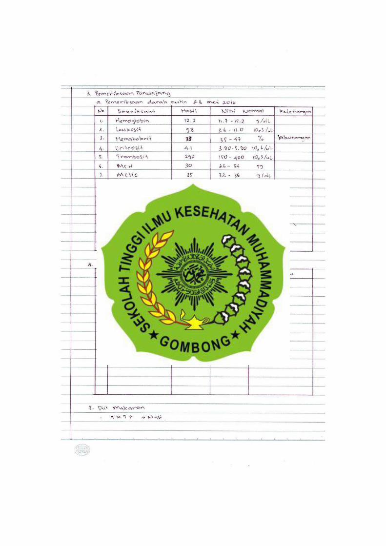
3. Pengkajian Fokus........................................................................... 30
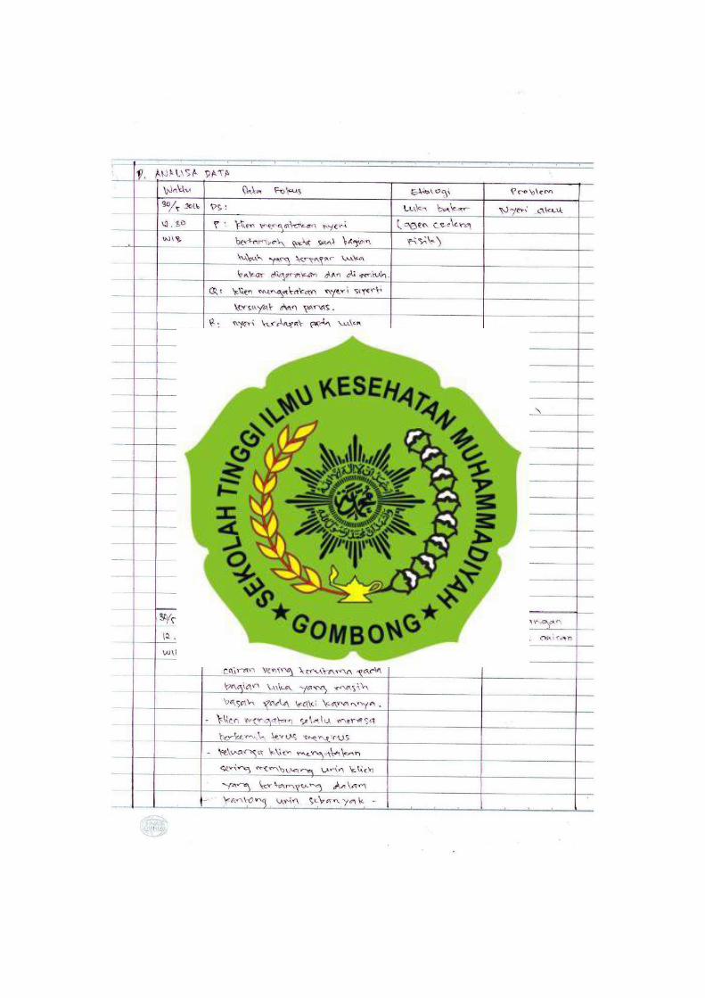
B. Analisa Data.......................................................................................... 33
C, Intervensi, Impementasi, dan Evaluasi............................................... 34
1. Diagnosa Keperawatan Nyeri Akut Berhubungan
DenganAgen Cedera Fisik............................................................ 34
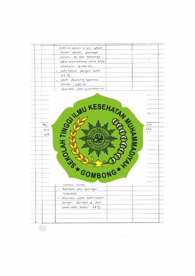
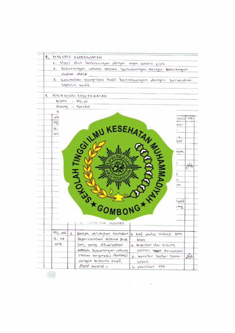
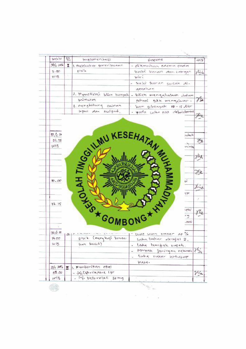
2. Diagnosa Keperawatan Kekurangan Volume Cairan
Berhubungan dengan Kehilangan Cairan Aktif.......................... 36
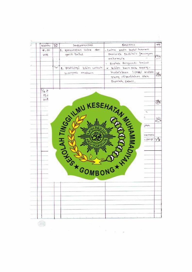
3. Diagnosa Keperawatan Kerusakan Integritas Kulit
Berhubungan dengan Kerusakan Lapisan Kulit.......................... 38
BAB IV PEMBAHASAN.................................................................................. 40
A. Diagnosa Keperawatan......................................................................... 40
1. Nyeri Akut Berhubungan Dengan Agen Cedera Fisik............... 41
2. Kekurangan Volume Cairan Berhubungan Dengan
Kehilangan Cairan Aktif............................................................... 47
3. Kerusakan Integritas Kulit Berhubungan Dengan
Kerusakan Kerusakan Lapisan Kulit............................................ 52
B. Analisis Inovasi Tindakan Keperawatan Relaksasi Otot
Progresif................................................................................................ 57
1. Prosedur Tindakan Inovasi........................................................... 57
2. Kekurangan Atas Tindakan Inovasi............................................. 60
3. Hasil Penelitian Tentang Inovasi Tindakan Keperawatan......... 61
x
BAB V PENUTUP............................................................................................. 62
A. Kesimpulan........................................................................................... 62
B. Saran .................................................................................................. 64
1. Bagi Klien dan Keluarga............................................................... 64
2. Bagi Rumah Sakit.......................................................................... 65
3. Bagi Institusi.................................................................................. 65
DAFTAR PUSTAKA......................................................................................... 66
DAFTAR LAMPIRAN...................................................................................... 68
1
BAB I
PENDAHULUAN
A. Latar Belakang
Keamanan adalah suatu keadaan yang terbebas dari cedera fisik dan
gangguan psikologis. Keamanan adalah salah satu kebutuhan dasar manusia yang
harus terpenuhi (Potter & Perry, 2006). Menurut Kolcaba, (1992) dalam Potter &
Perry (2006) kenyamanan adalah keadaan telah terpenuhinya kebutuhan dasar
manusia. Kebutuhan ini meliputi kebutuhan akan ketentraman (suatu rasa
kepuasan terhadap penampilan sehari hari), kegiatan (kebutuhan telah terpenuhi),
dan transenden (keadaan yang melebihi suatu masalah atau nyeri).
Salah satu bentuk gangguan kenyamanan adalah nyeri. Nyeri adalah
sensasi ketidaknyamanan yang dimanifestasikan sebagai penderitaan yang
diakibatkan oleh persepsi jiwa terhadap suatu keadaan yang nyata, ancaman, dan
fantasi luka (Kozier & Erb, 1983 dalam Tamsuri, 2007). Menurut teori dari
Asosiasi Nyeri Internasional, pemahaman tentang nyeri lebih untuk
penatalaksanaan nyeri yang menitikberatkan pada tindakan fisik atau
menghilangkan kausa fisik. Namun menurut Kozier dan Erb, (1983) dalam
Tamsuri (2007) nyeri diperkenalkan sebagai suatu pengalaman emosional yang
penatalaksanaannya tidak hanya pada pengelolaan fisik saja, namun penting juga
untuk melakukan tindakan psikologis untuk mengatasi nyeri.
Nyeri dapat terjadi karena akibat kerusakan jaringan aktual atau potensial
yang digambarkan sebagai kerusakan atau faktor yang berhubungan dengan
cedera biologis, cedera fisik, cedera kimiawi. Luka bakar merupakan salah satu
contoh cedera fisik yang dapat menimbulkan nyeri (Arif & Kumala, 2013).
Berdasarkan data dari Riskesdas (Riset Kesehatan Dasar) provinsi Jawa
Tengah (2007) menyatakan jumlah kasus luka bakar di Jawa Tengah adalah 2,1%
dari keseluruhan cedera, usia terbanyak yang mengalami cedera luka bakar yaitu
23,3% pada usia < 1 tahun, jenis kelamin perempuan lebih banyak dibandingkan
2
dengan jenis kelamin pria dengan 2,5 %, sedangkan untuk penderita terbanyak
berdasarkan tingkat pendidikan adalah 2,8% pada lulusan SMA.
Berdasarkan data dari rekam medik di RS Dr Soedirman Kebumen pada
tahun 2014, terdapat 10 angka kejadian cidera luka bakar. Sedangkan di RS Dr
Soetomo Surabaya terdapat 106 kasus luka bakar di instalasi rawat inap pada
tahun 2000, dengan angka kematian 26,41%. Selanjutnya di RS Pusat Pertamina
pada tahun 2007 terdapat pasien luka bakar rata-rata sebanyak 40 penderita
pertahun yang dirawat di unit luka bakar, dari jumlah tersebut yang masuk dalam
kategori luka bakar sekitar 21% dengan angka kematian 40-50% (Majid &
Prayogi, 2013).
Luka bakar merupakan cedera yang sering dihadapi oleh dokter. Jenis
yang berat memperlihatkan derajat cacat yang relatif tinggi dibanding dengan
cedera oleh sebab yang lainya. Penyebab luka bakar selain terbakar api, juga tak
langsung, juga paparan suhu yang tinggi dari cahaya matahari, listrik dan bahan
kimia. Luka bakar karena api atau akibat tak langsung dari api misalnya tersiram
air panas, tersiram minyak goreng panas, dan banyak terjadi pada kecelakaan
rumah tangga. Berat ringannya luka bakar tergantung pada keadaan jaringan yang
terbakar dan intensitas trauma panas. Kulit yang tebal, berpigmen dan banyak
mengandung kelenjar sebasea akan lebih tahan terhadap trauma panas dibanding
dengan kulit yang tipis dan kering (Nugroho, 2012).
Jaringan dibawah kulit trauma panas tersebut akan menerima rambatan
panas yang serupa. Kandungan air dalam jaringan dan banyak tidak nya jaringan
aliran darah merupakan faktor yang sangat penting. Gawat darurat dan
penatalaksanaan pertama pada cedera luka bakar merupakan bagian terpenting
dari perawatan secara keseluruhan, terutama bila lukanya luas dan kemungkinan
melibatkan beberapa pembedahan serta upaya yang besar oleh kalangan medik,
perawat dan paramedik. Untuk tujuan ini, mutlak bahwa perawatan gawat darurat
dipikirkan mencakup 5 hari pengobatan pertama (Nugroho, 2012).
Kulit terdiri atas tiga lapisan, yang masing-masing memiliki jenis sel dan
memiliki fungsi yang bermacam-macam. Ketiga lapisan tersebut adalah epidermis,
dermis, dan subkutis. Epidermis merupakan struktur jaringan lapisan kulit terluar,
3
sel-sel epidermis terus-menerus mengalami mitosis, dan berganti dengan sel yang
baru sekitar 30 hari. Epidermis mengandung reseptor-reseptor sensorik untuk
sentuhan, suhu, getaran, dan nyeri. Dermis merupakan lapisan kulit bawah
epidermis atau disebut juga dengan kutan yang membentuk sebagian terbesar kulit
dengan memberikan kekuatan serta struktur pada kulit. Dermis mengandung
sel-sel fibroblas yang dapat menghasilkan suatu bentuk kolagen, yaitu komponen
dari jaringan ikat. Dermis tersusun dari pembuluh darah, limfe, serabut saraf,
kelenjar sebasea, dan akar rambut. Subkutis merupakan lapisan kulit yang terletak
dibawah lapisan dermis. Subkutis terdiri dari lemak dan jaringan ikat bawah yang
berfungsi memberikan bantalan antara lapisan kulit dan struktur internal, struktur
internal yaitu otot dan tulang, serta sebagai peredam kejut dan insulator panas
(Arif & Kumala, 2013).
Derajat pada luka bakar dibagi menjadi tiga meliputi pada luka bakar
derajat I, luka hanya mengenai lapisan epidermis, luka tampak berwarna merah
muda sampai merah dan terasa nyeri/hyperethetic, dan nyeri berkurang dengan
pendinginan. Pada luka bakar derajat II, kedalaman pada lapisan superfisial dalam,
warna berbintik-bintik, putih, coklat, merah muda, atau merah coklat. Timbul
gelembung berisi cairan berwarna jernih tetapi kental, rasa nyeri atau sakit sangat
mengganggu/sangat nyeri. Dan luka bakar derajat III, yaitu luka yang mengenai
semua lapisan kulit, lemak, dan dapat juga mengenai permukaan jaringan otot,
persyarafan dan pembuluh darah serta tulang. Luka bakar tampak kering serta
kulit mengelupas tekstur keras dan kasar, warna putih, merah sampai hitam dan
terdapat edema. Tidak terasa nyeri atau sedikit nyeri karena serabut saraf telah
rusak (Majid & Prayogi, 2013).
Menurut Tamsuri (2007), managemen nyeri dibedakan menjadi dua
kelompok utama, yaitu tindakan pengobatan (farmakologis) dan tindakan tanpa
pengobatan (non-farmakologis). Penatalaksanaan nyeri secara farmakologis
meliputi pengguanaan opioid (narkotik), nonopioid/NSAIDs (Non-Steroid
Anti-Inflamation Drugs), dan adjuvan, serta ko-analgesik. Sedangkan penanganan
nyeri tanpa pengobatan (non-farmakologis) dibagi menjadi dua yaitu penanganan
berdasarkan stimulasi fisik dan perilaku kognitif. Penanganan fisik meliputi
4
stimulasi atau sentuhan pada kulit, stimulasi elektrik saraf kulit stranskutan
(TENS, Transcutaneous Electrical Nerve Stimulation), akupuntur, dan pemberian
plasebo. Sedangkan intervensi dengan perilaku kognitif meliputi tindakan
distraksi, teknik nrelaksasi, imajinasi terbimbing, umpan balik biologis, hipnosis,
dan sentuhan terapeutik.
Distraksi adalah sistem aktivasi retikular menghambat stimulus yang
terasa sakit pada seseorang jika menerima masukan sensori yang cukup atau
berlebihan. Stimulus yang menyenangkan menyebabkan pelepasan endofrin
(endofrin yaitu zat kimia dalam otak yang menyebabkan perasaan bahagia).
Distraksi mengalihkan perhatian klien ke hal lain dengan demikian menurunkan
kewaspadaan terhadap rasa nyeri, bahkan meningkatkan toleransi terhadap rasa
nyeri. Namun ada kerugian, distraksi hanya bekerja paling baik untuk jangka
waktu yang singkat (Potter & Perry, 2006).
Sedangkan relaksasi adalah kebebasan mental dan fisik dari ketegangan
dan keadaan stress. Teknik relaksasi memberikan individu kontrol diri ketika
terjadi rasa yang tidak nyaman atau nyeri, stress fisik dan emosi pada nyeri.
Teknik relaksasi merupakan upaya pencegahan membantu tubuh agar segar
kembali dan beregenerasi setiap hari dan merupakan langkah alternatif terhadap
konsumsi alkohol, merokok, atau makan berlebihan (Potter & Perry, 2006).
Karakteristik nyeri berdasarkan skala dibagi menjadi empat yaitu skala
penilaian numeric (Numeric Respons Scale) yang digunakan dengan cara klien
mendeskripsikan nyeri dengan menilai nyeri mengguanakan skala 1-10,
menanyakan respon klien (Verbal Respons Scale) yang digunakan dengan cara
menanyakan respon klien terhadap nyeri secara verbal dengan memberikan lima
pilihan rentang nyeri, skala analog visual (Visual Analogue Scale) yang digunakan
dengan cara memberikan pasien kebebasan dalam mengidentifikasi pengukuran
keparahan nyeri, dan gambaran ekspresi wajah (Face Pain Scale) yang digunakan
dengan cara mengukur ekspresi wajah dengan tujuh macam ekspresi wajah (Potter
& Perry, 2006).
5
Menurut Price & Wilson (2006), menyatakan dalam penatalaksanaan
nyeri memerlukan penilaian dan usaha yang cermat atau disebut dengan
pengkajian nyeri, untuk memahami pengalaman nyeri dan mengidentifikasi kausa,
sehingga kausa tersebut dapat dihilangkan. Perawat pertama-tama harus
melakukan anamnesa yang teliti, yang harus mencakup data mengenai nyeri.
Melihat bahwa nyeri pada luka bakar akan memberikan dampak terhadap
kualitas sumber daya manusia dan kesehatan yang cukup besar, maka penulis
berkeinginan untuk mempelajari lebih lanjut tentang nyeri pada luka bakar serta
ingin mengaplikasikan ilmu yang telah dipelajari dalam menangani pasien dengan
nyeri pada luka bakar dalam bentuk asuhan keperawatan yang komprehensif.
B. Tujuan Penulisan
1. Tujuan Umum
Tujuan dari penulisan karya tulis ilmiah ini adalah memaparkan
asuhan keperawatan pemenuhan kebutuhan rasa aman dan nyaman nyeri
pada luka bakar.
2. Tujuan Khusus
a. Memaparkan hasil pengkajian pada pasien dengan gangguan
pemenuhan kebutuhan rasa aman dan nyaman (nyeri akut pada luka
bakar).
b. Memaparkan hasil analisa data berdasarkan hasil pengkajian pada
pasien dengan gangguan pemenuhan kebutuhan rasa aman dan
nyaman (nyeri akut pada luka bakar).
c. Memaparkan perencanaan atau intervensi tindakan pada pasien
gangguan pemenuhan kebutuhan rasa aman dan nyaman (nyeri akut
pada luka bakar).
d. Memaparkan hasil evaluasi tindakan keperawatan dengan gangguan
pemenuhan kebutuhan rasa aman dan nyaman (nyeri akut pada luka
bakar).
6
C. Manfaat Penulisan
1. Bagi Institusi Pendidikan
Karya tulis ilmiah ini diharapkan menjadi referensi bagi institusi
keperawatan dalam mengembangkan asuhan keperawatan agar setiap
mahasiswa yang membaca karya tulis ini dapat mengetahui asuhan
keperawatan tentang gangguan kebutuhuan rasa aman dan nyaman yaitu
nyeri.
2. Bagi Rumah Sakit
Karyta tulis ilmiah ini diharapkan dapat memberikan manfaat praktis
dalam keperawatan yang ada dalam rumah sakit yaitu sebagai panduan
perawat dalam pengelolaan asuhan keperawatan nyeri pada klien dengan
gangguan kebutuhan rasa aman dan nyaman.
3. Bagi Klien dan Keluarga
Karya tulis ilmiah ini dapat memberikan informasi bagi klien dan
keluarga mengenai penatalaksanaan atau tindakan keperawatan yang
dapat dilakukan untuk mengatasi nyeri pada klien.
66
DAFTAR PUSTAKA
Arif, Muttaqin & Kumala, Sari. (2013). Asuhan Keperawatan Gangguan Sistem
Integumen, Jakarta : Salemba Medika.
Asmadi. (2008). Teknik Prosedural Konsep & Aplikasi Kebutuhan Dasar Klien,
Jakarta : Salemba Medika.
Depkes RI, (2009). Laporan Hasil Riset Kesehatan Dasar (Riskesdas) Provinsi
Jawa Tengah Tahun 2007. Jakarta : Badan Litbangkes Depkes RI.
Eni, K., et al. (2012). Keterampilan & Prosedur Laboratorium Keperawatan
Dasar Edisi 2, Jakarta : EGC.
Herdman, T.H. (2015). NANDA Internasional Inc. Diagnosis Keperawatan :
Definisi & Klasifikasi 2015-2017. Jakarta : EGC.
Hoffman, H.G., et al. (2011). Virtual Reality as an Adjunctive Non-pharmacologic
Analgesic for Acute Burn Pain During Medical Procedure. Annals of
Behavioral Medicine.
Ikhrima, Rahmasari. (2015). Relaksasi Otot Progresif Dapat Menurunkan Nyeri
Kepala. Jurnal Ilmiah Kesehatan Keperawatan, Volume 2, Nomor 2.
Majid, Abdul & Prayogi, A.S. (2013). Perawatan Pasien Luka Bakar.
Yogyakarta : Goysen Publishing.
Nugroho, Taufan. (2012). Luka Bakar dan Artritis Reumatoid. Yogyakarta : Nuha
Medika.
Nurarif, Amin. H & Kusuma, Hardi. (2013). Aplikasi Asuhan Keperawatan
Berdasarkan Diagnosa Medis & NANDA NIC NOC Jilid 2, Jakarta :
EGC.
Nurhayati., et al. (2011). Pengaruh Teknik Distraksi Relaksasi Terhadap
Penurunan Nyeri Pada Pasien Post Operasi Laparatomi di PKU
Muhammadiyah Gombong. Jurnal Ilmiah Kesehatan Keperawatan,
Volume 7, Nomor 1.
67
Patasic, C.K., et al. (2013). Efektifitas Teknik Relaksasi Nafas Dalam dan Guided
Imagery Terhadap Penurunan Nyeri Pada Paien Post Operasi Sectio
Caesarea Di Irina D Blu RSUP Prof. Dr. R. D. Kandou Manado. Ejurnal
Keperawatan (e-Kp) Volume 1, Nomor 1.
Potter, P.A. & Perry, A.G. (2006). Buku Ajar Fundamental Keperawatan Konsep
& Praktik Volume 2 Edisi Keempat. Jakarta : EGC.
Price, A.S. & Wilson, M.L. (2006). Patofisiologi Konsep Klinis Proses-Proses
Penyakit. Jakarta : EGC
Tamsuri, Anas. (2007). Konsep dan Penatalaksanaan Nyeri. Jakarta : EGC.
Wijaya, A.S. & Putri, Y.M. (2013). Keperawatan Medikal Bedah 2: Keperawatan
Dewasa Teori dan Contoh Askep. Yogyakarta : Nuda Medika.
ejurnal keperawatan (e-Kp) Volume 1. Nomor 1. Agustus 2013
1
EFEKTIFITAS TEKNIK RELAKSASI NAFAS DALAM DAN GUIDED IMAGERY TERHADAP PENURUNAN NYERI PADA PASIEN POST
OPERASI SECTIO CAESARE DI IRINA D BLU RSUP Prof. Dr. R. D. KANDOU
MANADO
Chandra Kristianto Patasik Jon Tangka Julia Rottie
Program Studi Ilmu Keperawatan Fakultas Kedokteran
Universitas Sam Ratulangi Manado E-mail:[email protected]
ABSTRACT: Pain is an unpleasant symptom that felt by patients post sectio caesarea. Alternatives to reduce pain with deep breathing relaxation techniques and guided imagery not definitely known how its effectiveness. The purpose of this research was to determine the effectivity of deep breathing relaxation techniques and guided imagery to reduction pain in post surgery patients sectio caesarea. This research is an analytical research with quasi-experimental research methods. The research design was a single group pre-post test without control group. The sampling technique used is accidental sampling where found samples as many as 20 people. The data analysis using paired sample t-test in SPSS 17 programe at 95% significance level (α=0.05). The results showed that deep breathing relaxation techniques and guided imagery proved to be effective in reducing pain intensity in post surgery patients sectio caesarea in the Irina D Department of RSUP Prof. Dr. R. D. Kandou Manado (p value=0.000; α 0.05) which means that the hypothesis is accepted. Conclusion, deep breathing relaxation techniques and guided imagery can reduce pain intensity in post surgery patients sectio caesarea in the Irina D Department of RSUP Prof. Dr. R. D. Kandou Manado. Implementation of deep breathing relaxation techniques and guided imagery to reduce pain in post surgery patients sectio caesarea can be be done according to the SOP so not just focus on the pharmacological treatment only. Keywords: Deep Breathing Relaxation Techniques, Guided Imagery, Pain, Sectio Caesarea.
ABSTRAK: Nyeri merupakan suatu gejala yang tidak menyenangkan yang dialami oleh pasien post operasi sectio caesarea. Alternatif untuk menurunkan nyeri yaitu dengan menggunakan teknik relaksasi nafas dalam dan guided imagery bagaimana efektifitasnya belum jelas diketahui. Tujuan penelitian ini adalah untuk mengetahui efektifitas teknik relaksasi nafas dalam dan guided imagery terhadap penurunan nyeri pada pasien post operasi sectio caesarea. Penelitian ini merupakan penelitian analitik dengan metode penelitian kuasi eksperimen.Desain penelitian adalah satu kelompok pre-post tes tanpa kelompok kontrol.Teknik pengambilan sampel yaitu Accidental Sampling dimana didapatkan sampel sebanyak 20 responden. Teknik analisa data menggunakan uji paired sampel t-tes program SPSS 17 pada tingkat kemaknaan 95% (α=0,05). Hasil penelitian menunjukkan bahwa teknik relaksasi nafas dalam dan guided imagery terbukti efektif dalam menurunkan intensitas nyeri pada pasien post operasi sectio caesarea di Irina D BLU RSUP Prof. Dr. R. D. Kandou Manado (nilai p=0,000; α 0,05) yang berarti hipotesis diterima. Kesimpulan, teknik relaksasi nafas dalam dan guided imagery mampu menurunkan intensitas nyeri pada pasien post operasi sectio caesarea di Irina D BLU RSUP Prof. Dr. R.D.
ejurnal keperawatan (e-Kp) Volume 1. Nomor 1. Agustus 2013
2
Kandou Manado. Penerapan teknik relaksasi nafas dalam dan guided imagery untuk menurunkan nyeri pada pasien post operasi sectio caesarea dapat dilakukan sesuai dengan SOP agar tidak hanya terpaku pada penaganan secara farmakologis saja. Kata kunci : Teknik Relaksasi Nafas Dalam, Guided Imagery, Nyeri, Sectio Caesarea.
PENDAHULUAN Dewasa ini, ilmu pengetahuan dan teknologi telah meningkat pesat. Kemajuan teknologi dan ilmu dibidang kesehatan membawa manfaat yang besar bagi manusia, termasuk pada penatalaksanaan sectio caesarea.Sectio caesarea merupakan salah satu cara yang digunakan dibidang kesehatan untuk membantu persalinan ketika ada masalah tak terduga terjadi selama persalinan, seperti faktor dari ibu yaitu panggul yang sempit, faktor dari janin yang letaknya lintang, tidak cukup ruang bagi janin untuk melalui vagina, dan kelainan pada janin seperti berat badan janin melebihi 4000 gram (National Institute of Health, 2012).Sectio caesarea adalah operasi bedah dimana dokter kandungan membuat sebuah lubang di perut dan rahim ibu kemudian mengeluarkan janin dari lubang tersebut (National Institute for Clinical Excellence, 2004).
Berdasarkan data yang ditemukan di Irina D BLU RSUP Prof. Dr. R. D. Kandou Manado terdapat 1.504 operasi sectio caesarea pada tahun 2011 dan 1.511 operasi sectio caesarea pada tahun 2012. Ini menunjukkan bahwa banyaknya operasi sectio caesarea pada saat ini yang dilakukan di BLU RSUP Prof. Dr. R. D. Kandou Manado, walaupun operasi ini sangat beresiko.
Respon nyeri yang dirasakan oleh pasien merupakan efek samping yang timbul setelah menjalani suatu operasi.Nyeri yang disebabkan oleh operasi biasanya membuat pasien merasa sangat kesakitan.Ketidaknyamanan atau nyeri bagaimanapun keadaannya harus diatasi dengan manajemen nyeri, karena kenyamanan merupakan kebutuhan dasar manusia.
Nyeri merupakan sensasi yang sangat tidak menyenangkan dan bervariasi pada tiap individu.Nyeri dapat mempengaruhi seluruh pikiran seseorang, mengatur aktivitasnya, dan mengubah kehidupan orang tersebut.Nyeri merupakan faktor psikososial yang perlu diungkap lewat komunikasi terapeutik, karena seorang perawat perlu mendapatkan data baik secara subjektif maupun objektif untuk menilai seberapa besar pengaruh nyeri tersebut pada pasien (Berman, Snyder, Kozier, & Erb, 2003).
Penanganan nyeri dengan melakukan teknik relaksasi merupakan tindakan keperawatan yang dilakukan untuk mengurangi nyeri. Penanganan nyeri dengan tindakan relaksasi mencakup teknik relaksasi nafas dalam dan guided imagery.Beberapa penelitian telah menunjukkan bahwa relaksasi nafas dalam sangat efektif dalam menurunkan nyeri pasca operasi (Sehono, 2010). Relaksasi adalah sebuah keadaan dimana seseorang terbebas dari tekanan dan kecemasan atau kembalinya keseimbangan (equilibrium) setelah terjadinya gangguan.Tujuan dari teknik relaksasi adalah mencapai keadaan relaksasi menyeluruh, mencakup keadaan relaksasi secara fisiologis, secara kognitif, dan secara behavioral. Secara fisiologis, keadaan relaksasi ditandai dengan penurunan kadar epinefrin dan non epinefrin dalam darah, penurunan frekuensi denyut jantung (sampai mencapai 24 kali per menit), penurunan tekanan darah, penurunan frekuensi nafas (sampai 4-6 kali per menit), penurunan ketegangan otot, metabolisme menurun, vasodilatasi dan peningkatan temperatur pada extermitas (Rahmayati, 2010).
ejurnal keperawatan (e-Kp) Volume 1. Nomor 1. Agustus 2013
3
Teknik relaksasi nafas dalam akan lebih efektif bila dikombinasikan dengan beberapa teknik lainnya, seperti guided imagery. Guided imagery merupakan teknik yang menggunakan imajinasi seseorang untuk mencapai efek positif tertentu (Smeltzer, Bare, Hinkle, & Cheever, 2010).Teknik ini dimulai dengan proses relaksasi pada umumnya yaitu meminta kepada klien untuk perlahan-lahan menutup matanya dan fokus pada nafas mereka, klien didorong untuk relaksasi mengosongkan pikiran dan memenuhi pikiran dengan bayangan untuk membuat damai dan tenang (Rahmayati, 2010).
Penelitian yang dilakukan oleh Syahriyani (2010), tentang pengaruh teknik relaksasi terhadap perubahan intensitas nyeri pada pasien post operasi apendiktomi di ruang perawatan bedah RSU TK II Pelamonia Makassar, menunjukkan bahwa intensitas nyeri responden sebelum dan sesudah pemberian teknik relaksasi mengalami peningkatan penurunan nyeri dari nyeri ringan 20,00% ke 66,67%, nyeri sedang 53,33% ke 20,00%, dan nyeri berat 26,67% ke 13,33%. Uji lebih lanjut membuktikan ada pengaruh pemberian teknik relaksasi terhadap perubahan intensitas nyeri pada pasien post operasi apendiktomi di ruang perawatan bedah RSU TK II Pelamonia Makassar. Hasil survei sementara di Irina D BLU RSUP Prof. Dr. R. D. Kandou Manado pada salah seorang perawat senior menyatakan umumnya perawat jarang bahkan tidak melakukan teknik relaksasi nafas dalam dan guided imagery karena penanganan nyeri berfokus hanya pada penanganan secara farmakologis saja. Berdasarkan uraian tersebut di atas maka penulis merasa tertarik untuk mengetahui lebih lanjut mengenai efektifitas teknik relaksasi nafas dalam dan guided imagery terhadap penurunan nyeri abdomen pada pasien post operasi sectio caesareadi
Irina D BLU RSUP Prof. Dr. R. D. Kandou Manado.
METODE PENELITIAN
Jenis penelitian ini adalah analitik dengan metode penelitian kuasi eksperimen.Desain penelitian adalah satu kelompok pre-post tes tanpa kelompok kontrol. Penelitian dilaksanakan di Irina D BLU RSUP Prof. Dr. R. D. Kandou Manado pada tanggal 7 Juni – 15 Juni 2013. Populasi dalam penelitian ini adalah semua pasien yang telah menjalani operasi sectio caesarea di Irina D BLU RSUP Prof. Dr. R. D. Kandou Manado dengan teknik pengambilan sampel Accidental Sampling. Kriteria inklusi dalam penelitian ini adalah pasien post sectio caesareahari pertama, pasien yang bersedia menjadi responden, pasien yang reaksi analgetiknya telah hilang atau 6 jam setelah pemberian analgetik dan belum mendapatkan analgetik lagi. Sedangkan kriteria eksklusi dalam penelitian ini adalah pasien yang telah diberikan teknik relaksasi lainnya, pasien yang telah melakukan operasi sectio caesarea sebelumnya, pasien sectio caesarea yang mengalami komplikasi. Instrumen yang digunakan dalam penelitian ini berupa lembar observasi yang berisi tentang data umum responden, dan lembar isian nyeri yang terdiri dari nomor, inisial, umur, jam post operasi sectio caesarea, jam terjadinya nyeri, hasil skala nyeri sebelum dilakukan tindakan, lama dan jumlah tindakan, dan hasil skala nyeri sesudah dilakukan tindakan serta lembar intensitas nyeri berupa skala intensitas nyeri 10 poin dengan kata-kata penjelas dan prosedur teknik relaksasi nafas dalam dan guided imagery. Data primer diambil langsung dari responden dengan cara penggunaan lembar observasi dan data sekunder terdiri dari gambaran umum lokasi penelitian dan data
ejurnal keperawatan (e-Kp) Volume 1. Nomor 1. Agustus 2013
4
pasien yang telah menjalani operasi sectio caesarea. Prosedur teknik relaksasi nafas dalam dan guided imagery yaitu ciptakan lingkungan yang tenang, jaga privasi pasien, usahakan tangan dan kaki pasien alam keadaan rileks, minta pasien untuk memejamkan mata dan usahakan agar pasien berkonsentrasi, minta pasien menarik nafas melalui hidung secara perlahan-lahan sambil menghitung dalam hati “hirup, dua, tiga”, selama pasien memejamkan mata kemudian minta pasien untuk membayangkan hal-hal yang menyenangkan atau keindahan, minta pasien untuk menghembuskan udara melalui mulut dan membuka mata secara perlahan-lahan sambil menghitung dalam hati “hembuskan, dua, tiga”, minta pasien untuk mengulangi lagi sama seperti prosedur sebelumnya sebanyak tiga kali selama lima menit. Teknik pengolahan data pada penelitian ini terdiri dari koding, editing, tabulasi data, dan entri data.sedangkan analisa data dilakukan dengan pengujian analisa univariat dan bivariat. Menurut Yurisa 2008, etika penelitian memiliki berbagai macam prinsip, namun terdapat empat prinsip utama yang perlu dipahami, yaitu menghormati harkat dan martabat manusia (respect for human dignity), menghormati privasi dan kerahasiaan subyek penelitian (respect for privacy and confidentiality), keadilan dan inklusivitas (respect for justice and inclusiveness), memperhitungkan manfaat dan kerugian yang ditimbulkan (balancing harms and benefits).
HASIL dan PEMBAHASAN Tabel 1. Distribusi menurut umur di Irina D BLU RSUP Prof. Dr. R. D. Kandou Manado (n=20 orang)
Umur Banyak Responden
n %
10 – 20 Tahun 2 10,0
21 – 30 Tahun 10 50,0
31 – 40 Tahun 6 30,0
>40 Tahun 2 10,0
Total 20 100,0
Sumber : Data primer
Tabel 2. Distribusi menurut tingkat pendidikan di Irina D BLU RSUP Prof. Dr. R. D. Kandou Manado (n=20 orang)
Tingkat Pendidikan Banyak Responden
n %
SD 1 5,0
SMP 1 5,0
SMA 18 90,0
Total 20 100,0
Sumber : Data primer
Tabel 3. Distribusi menurut pekerjaan di Irina D BLU RSUP Prof. Dr. R. D. Kandou Manado (n=20 orang)
Pekerjaan Banyak Responden
n %
Ibu Rumah Tangga 15 75,0
Wiraswasta 5 25,0
Total 20 100,0
Sumber : Data primer
ejurnal keperawatan (e-Kp) Volume 1. Nomor 1. Agustus 2013
5
Tabel 4. Distribusi menurut intensitas nyeri sebelum dilakukan teknik relaksasi nafas dalam dan guided imagery di Irina D BLU RSUP Prof. Dr. R. D. Kandou Manado (n=20 orang)
Intensitas Nyeri Banyak Responden
n % Tidak Nyeri 0 0,0 Nyeri Ringan 0 0,0
Nyeri Sedang 5 25,0
Nyeri Hebat 12 60,0
Nyeri Sangat Hebat
3 15,0
Nyeri Terhebat 0 0,0
Total 20 100,0
Sumber : Data primer Tabel 5. Distribusi menurut intensitas nyeri sesudah dilakukan teknik relaksasi nafas dalam dan guided imagery di Irina D BLU RSUP Prof. Dr. R. D. Kandou Manado (n=20 orang)
Intensitas Nyeri Banyak Responden
n %
Tidak Nyeri 0 0,0
Nyeri Ringan 13 65,0
Nyeri Sedang 7 35,0
Nyeri Hebat 0 0,0
Nyeri Sangat Hebat
0 0,0
Nyeri Terhebat 0 0,0
Total 20 100,0
Sumber : Data primer
Tabel 6. Hasil analisis perbandingan rata-rata intensitas nyeri sebelum dilakukan teknik relaksasi nafas dalam dan guided imagery dan intensitas nyeri sesudah dilakukan teknik relaksasi nafas dalam dan guided imagery di Irina D BLU RSUP Prof. Dr. R. D. Kandou Manado (n=20 orang)
Intensitas
Nyeri
Mean
Std. Deviati
on
Std. erro
r mea
n
t p value N
Sebelum
6,15 1,089 0,24 10,10
0 0,00
0 20
Sesudah
3,05 0,945 0,21
1 Sumber : Data primer
Penelitian yang dilakukan di Irina D BLU RSUP Prof. Dr. R. D. Kandou Manado ini menggunakan 20 orang sampel. Berdasarkan umur responden paling banyak berumur 21-30 tahun yaitu 10 orang (50,0%). Umur responden adalah variabel penting yang akan mempengaruhi reaksi maupun ekspresi responden terhadap rasa nyeri. Semakin meningkatnya umur, semakin tinggi reaksi maupun respon terhadap nyeri yang dirasakan. Berdasarkan tingkat pendidikan, responden paling banyak berpendidikan SMA yaitu 18 orang (90,0%). Tingkat pendidikan seseorang sangat mempengaruhi pengetahuan seseorang.Semakin tinggi tingkat pengetahuan seseorang, semakin tinggi tingkat pencegahan berbagai jenis penyakit maupun kelainan-kelainan yang dapat mengakibatkan operasi. Menurut Nikita 2012, semakin tinggi tingkat pendidikan seseorang maka akan semakin tinggi kemampuan seseorang untuk menyerap informasi dan mengimplementasikannya ke dalam perilaku dan gaya hidup sehari-hari, khususnya dalam kepeduliannya pada tingkat kebutuhan kesehatan. Berdasarkan pekerjaan, responden paling banyak hanya bekerja sebagai ibu rumah tangga yaitu 15 orang (75,0%) sementara yang lainnya
ejurnal keperawatan (e-Kp) Volume 1. Nomor 1. Agustus 2013
6
sebagai wiraswasta. Pekerjaan memiliki peran penting dalam tingkat kesehatan seseorang.Beban berat yang dilakukan oleh seseorang sesuai dengan pekerjaannya dapat menyebabkan timbulnya berbagai penyakit maupun kelainan-kelainan. Nikita berpendapat bahwa pekerjaan dan aktifitas seseorang menuntut beban kerja berat yang semakin beresiko untuk mengalami penyakit dan pekerjaan seseorang yang kurang memperhatikan kehati-hatian akan beresiko untuk mengalami cedera. Cedera yang dialami seseorang kadang-kadang menyebabkan seseorang harus menjalani operasi. Frekuensi pasien yang datang untuk melakukan persalinan di BLU RSUP Prof. Dr. R. D. Kandou Manado cukup banyak karena rumah sakit ini merupakan rumah sakit rujukan. Tidak semua pasien yang datang dapat melakukan persalinan secara normal karena mengalami beberapa hambatan seperti faktor dari ibu yaitu panggul yang sempit, faktor dari janin yang letaknya lintang, tidak cukup ruang bagi janin untuk melalui vagina, dan kelainan pada janin seperti berat badan janin melebihi 4000 gram, atau secara faktor sosial yaitu keinginan orang tua untuk melahirkan sesuai dengan waktu yang telah ditentukan, sehingga harus dilakukan suatu operasi yaitu sectio caesarea. Selama periode penelitian pada 7 Juni sampai 15 Juni 2013 didapatkan 20 responden yang menjalani operasi sectio caesarea, dan 60,0% dari responden tersebut mengalami nyeri hebat bahkan nyeri sangat hebat (15,0%) dan yang lainnya mengalami nyeri sedang (25,0%). Intensitas nyeri ini diukur menggunakan skala intensitas nyeri 10 poin dengan kata-kata penjelas.Nyeri merupakan efek samping yang dialami pasien setelah menjalani suatu operasi.Nyeri yang dirasakan oleh pasien diakibatkan oleh terputusnya kontinuitas jaringan karena insisi.Jenis operasi juga mempengaruhi respon nyeri seseorang terhadap nyeri.Ada
dua jenis operasi yang dilakukan berdasarkan keadaan pasien yaitu Cito dan elektif.Operasi Cito merupakan operasi yang harus segera dilakukan karena darurat.Pasien yang menjalani operasi Cito tidak dapat mempersiapkan diri untuk menghadapi kemungkinan-kemungkinan yang terjadi setelah operasi seperti nyeri.Berbeda dengan operasi elektif, pasien sudah dipersiapkan sejak lama sesuai jadwal sehingga pasien dapat mempersiapkan diri untuk menghadapi nyeri setelah selesai operasi. Dalam penelitian ini, responden yang menjalani operasi Cito lebih banyak yaitu 17 responden (85,0%) sedangkan yang lainnya menjalani operasi elektif. Respon nyeri yang dirasakan oleh setiap pasien berbeda-beda sehingga perlu dilakukan eksplorasi untuk menentukan nilai nyeri tersebut. Menurut Syahriyani (2010), perbedaan tingkat nyeri yang dipersepsikan oleh pasien disebabkan oleh kemampuan sikap individu dalam merespon dan mempersepsikan nyeri yang dialami. Kemampuan mempersepsikan nyeri dipengaruhi oleh beberapa faktor dan berbeda diantara individu. Tidak semua orang terpajan terhadap stimulus yang sama mengalami intensitas nyeri yang sama. Sensasi yang sangat nyeri bagi seseorang mungkin hampir tidak terasa bagi orang lain. Salah satu upaya untuk menurunkan nyeri adalah dengan menggunakan teknik farmakologis dan teknik nonfarmakologis. Teknik farmakologis yaitu dengan menggunakan obat-obatan sedangkan teknik nonfarmakologis salah satunya yaitu dengan relaksasi nafas dalam dan guided imagery. Pada penelitian ini, sesudah dilakukan teknik relaksasi nafas dalam dan guided imagery terjadi perubahanintensitas nyeri, dimana responden hanya mengalami dua tingkat nyeri yaitu nyeri sedang (35,0%) dan nyeri ringan (65,0%). Tidak ada lagi yang mengalami nyeri hebat dan sangat hebat.
ejurnal keperawatan (e-Kp) Volume 1. Nomor 1. Agustus 2013
7
Efek relaksasi nafas dalam dan guided imagery membuat responden merasa rileks dan tenang. Responden menjadi rileks dan tenang saat mengambil oksigen di udara melalui hidung, oksigen masuk kedalam tubuh sehingga aliran darah menjadi lancar serta dikombinasikan dengan guided imagery menyebabkan pasien mengalihkan perhatiannya pada nyeri ke hal-hal yang membuatnya senang dan bahagia sehingga melupakan nyeri yang sedang dialaminya. Inilah yang menyebabkan intensitas nyeri yang dirasakan pasien post operasi sectio caesareaberkurang setelah dilakukan teknik relaksasi nafas dalam dan guided imagery. Adanya perubahan intensitas nyeri sebelum dan sesudah dilakukan teknik relaksasi nafas dalam dan guided imagery juga dapatdiketahui setelah dilakukan uji statistik menggunakan uji paired sample t-testpada tingkat kemaknaan 95% (α = 0,05). Nilai mean sebelum dilakukan teknik relaksasi nafas dalam dan guided imagery yaitu 6,15 sedangkan sesudah dilakukan teknik relaksasi nafas dalam dan guided imagery yaitu 3,05. Hasil analisi diperoleh nilai p=0,000 dengan kata lain p<0,05. Oleh karena itu maka hipotesis diterima. Jadi, hasil penelitian ini menunjukkan bahwa teknik relaksasi nafas dalam dan guided imagery terbukti efektif dalam menurunkan intensitas nyeri pada pasien post operasi sectio caesarea. Hasil ini mendukung penelitian sebelumnya yang dilakukan oleh Nikita (2012) yang meneliti tentang pengaruh teknik relaksasi terhadap perubahan intensitas nyeri pada pasien post operasi apendektomi, dengan hasil yaitu ada pengaruh yang signifikan pada intensitas nyeri pasien post operasi apendektomi sesudah dilakukan teknik relaksasi, dari 4 orang yang mengalami nyeri hebat (40,0%) sesudah dilakukan teknik relaksasi menjadi 2 orang (20,0%), nyeri sedang 5 orang (50,0%) menjadi 2 orang (20,0%), dan tidak
nyeri yang semula 1 orang (10,0%) menjadi 6 orang (60,0%). Namun yang membedakan penelitian ini dan penelitian yang dilakukan oleh Nikita (2012) adalah tempat penelitian, jumlah responden, tindakan relaksasi, dan operasi yang dialami responden. Nikita (2012) melakukan penelitian di Irina A pada 10 responden post operasi apendektomi dan hanya menggunakan teknik relaksasi nafas dalam saja, sedangkan penelitian ini dilakukan di Irina D pada 20 responden post operasi sectio caesarea dan menggunakan teknik relaksasi nafas dalam yang dikombinasikan dengan guided imagery. SIMPULAN Kesimpulan dari penelitian ini yaitu tingkat nyeri pada pasien post operasi sectio caesarea sebelum dilakukan teknik relaksasi nafas dalam dan guided imagery di Irina D BLU RSUP Prof. Dr. R. D. Kandou Manado sebagian besar mengalami nyeri hebat sampai sangat hebat, tingkat nyeri pada pasien post operasi sectio caesarea sesudah dilakukan teknik relaksasi nafas dalam dan guided imagery di Irina D BLU RSUP Prof. Dr. R. D. Kandou Manado sebagian besar mengalami penurunan ke kategori nyeri ringan selebihnya ke kategori nyeri sedang, dan teknik relaksasi nafas dalam dan guided imagery efektifterhadap penurunan nyeri pada pasien post operasi sectio caesarea. DAFTAR PUSTAKA A service of the U.S. National Library of
Medicine, 2012.Cesarean Section.http://www.nlm.nih.gov/medlineplus/cesareansection.html diakses pada tanggal 01 April 2013 pukul 18. 30 WITA
Berman A. Snyder S. Kozier B & Erb G. 2003.Buku Ajar Praktik Keperawatan KlinisKozier & Erb edisi 5. Jakarta: Penerbit Buku Kedokteran EGC
Hastono, Sutanto Priyo. 2007. Analisis Data Kesehatan.Depok: SPH
ejurnal keperawatan (e-Kp) Volume 1. Nomor 1. Agustus 2013
8
Jayanthi, Niken. 2010. Analisa Data. http://rentalhikari.wordpress.com/2010/ 03/23/analisa-data/ diakses pada tanggal 15 Mei 2013 pukul 13.20 WITA
Jayanthi, Niken. 2010. Teknik Relaksasi Nafas Dalam. http://rentalhikari. wordpress.com/2010/03/23/teknik-relaksasi-nafas-dalam/ diakses pada tanggal 01 April 2013 pukul 17.40 WITA
Mawei, Nikita Mayumi. 2012. Pengaruh Teknik Relaksasi Terhadap Perubahan Intensitas Nyeri Pada Pasien Post Operasi Apendektomi.
National Institute for Clinical Excellence, 2004.Caesarean Section.http:// www.nice.org.uk/nicemedia/pdf/CG013publicinfoenglish.pdf didownloa d pada tanggal 05 April 2013 pukul 21.00 WITA
Oxorn H & Forle W. 2010.Ilmu Kebidanan: Patologi & Fisiologi Persalinan. Yogyakarta: Yayasan Essentia Medika
Rahmayati, Yeni Nur. 2010. Pengaruh Guided Imagery Terhadap Tingkat Kecemasan Pada Pasien Skizoafektif Di RSJD SURAKARTA. http://etd.eprints.ums.ac.id/9482/1/J210060060.pdf didownload pada tanggal : 04 Mei 2013 pukul 17.00 WITA
Sehono, Endrayani. 2010. Pengaruh Teknik Relaksasi Guided Imagery Terhadap Penurunan Nyeri Pada Pasien Pasca Operasi Fraktur Di RSUD DR. Moewardi. http://etd.eprints.ums.ac.id/10412/ di download pada tanggal 03 Mei 2013 pukul 12.40 WITA
Sidauruk P. 2011.Mekanisme Terjadinya Nyeri. http://www.forexsid.com/ 2011/03/mekanisme-terjadinya-nyeri.html diakses pada tanggal 04 April 2013 pukul 20.30 WITA
Smeltzer. Suzanne C. Bare Brenda G. Hinkle Janice L & Cheever Kerry H.
2010. Brunner & Suddarth’s Textbook of Medical-Surgical Nursing edisi 12. Philadelphia: Wolters Kluwer Health
Suyanto. 2011. Metodologi dan Aplikasi Penelitian Keperawatan. Yogyakarta: Nuha Medika
Syahriyani ST. 2010. Pengaruh Teknik Relaksasi Terhadap Perubahan Intensitas Nyeri Pada Pasien Post Operasi Apendiktomi di Ruang Perawatan Bedah RSU TK II Pelamonia Makassar.
https://www.box.com/s/d306231b8d03f80cf358 didownload pada tanggal 10 Mei 2013 pukul 19.30 WITA
Vaandda, Vhadh. 2012. Pengertian Sectio Caesaria. http://www.scribd.com/ doc/77221559/Pengertian-Sectio-Caesaria diakses pada tanggal 04 Mei 2013 pukul 13.30 WITA
Yurisa, Wella. 2008. Etika Penelitian Kesehatan. http://yayanakhyar. files.wordpress.com/2009/01/etika-penelitian-kesehatan_files-of-drsmed.pdf didownload pada tanggal 15 Mei 2013 pukul 14.30 WITA
Zhukmana, Aulia Dwi. 2009. LP Sectio caesarea. http://www.scribd.com/ doc/16308854/LP-Sectio-Caesarea diakses pada tanggal 04 April 2013 pukul 17.40 WITA
65ISSN 2443-1249 (Print) 2356-1313 (Online) - ijmsbm.org
IJMS - Indonesian Journal On Medical Science - Volume 2 No 2 - Juli 2015
Di RSUD Dr. Moewardi Surakarta
Kata Kunci:
66 ISSN 2443-1249 (Print) 2356-1313 (Online) - ijmsbm.org
IJMS - Indonesian Journal On Medical Science - Volume 2 No 2 - Juli 2015
I. PENDAHULUAN
2009).II. METODE PENELITIAN
ISSN 2443-1249 (Print) 2356-1313 (Online) - ijmsbm.org
IJMS - Indonesian Journal On Medical Science - Volume 2 No 2 - Juli 2015
III. HASIL PENELITIAN
Penelitian Kelompok Perlakuan dan Kontrol
2. Analisis Univariat
Tabel 2 Penurunan Intensitas Nyeri Pada Pasien Nyeri Kepala (Kelompok Perlakuan dan Kelompok Kontrol) )
ISSN 2443-1249 (Print) 2356-1313 (Online) - ijmsbm.org
IJMS - Indonesian Journal On Medical Science - Volume 2 No 2 - Juli 2015
Kelompok Perlakuan
N rerata55
55
Kelompok Kontrol
N rerata55
55
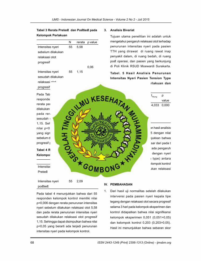
3. Analisis Bivariat
Tabel. 5 Hasil Analisis Penurunan Intensitas Nyeri Pasien Tension Type Headache Kelompok Perlakuan dan Kelompok Kontrol
t
Intensitas
IV. PEMBAHASAN
69ISSN 2443-1249 (Print) 2356-1313 (Online) - ijmsbm.org
IJMS - Indonesian Journal On Medical Science - Volume 2 No 2 - Juli 2015
atan teratur.
V. SIMPULAN
ISSN 2443-1249 (Print) 2356-1313 (Online) - ijmsbm.org
IJMS - Indonesian Journal On Medical Science - Volume 2 No 2 - Juli 2015
2005).
DAFTAR PUSTAKA
Disease.
Patients.
Fisiologi Manusia.
About
ORIGINAL ARTICLE
Virtual Reality as an Adjunctive Non-pharmacologicAnalgesic for Acute Burn Pain During Medical Procedures
Hunter G. Hoffman, PhD & Gloria T. Chambers, RN & Walter J. Meyer III, MD, PhD &
Lisa L. Arceneaux, PhD & William J. Russell, MS & Eric J. Seibel, PhD &
Todd L. Richards, PhD & Sam R. Sharar, MD & David R. Patterson, PhD
# The Society of Behavioral Medicine 2011
AbstractIntroduction Excessive pain during medical proceduresis a widespread problem but is especially problematicduring daily wound care of patients with severe burninjuries.Methods Burn patients report 35–50% reductions in pro-cedural pain while in a distracting immersive virtual reality,and fMRI brain scans show associated reductions in pain-related brain activity during VR. VR distraction appears tobe most effective for patients with the highest pain intensitylevels. VR is thought to reduce pain by directing patients’attention into the virtual world, leaving less attentionavailable to process incoming neural signals from painreceptors.
Conclusions We review evidence from clinical and labora-tory research studies exploring Virtual Reality analgesia,concentrating primarily on the work ongoing within ourgroup. We briefly describe how VR pain distraction systemshave been tailored to the unique needs of burn patients todate, and speculate about how VR systems could be tailoredto the needs of other patient populations in the future.
Keywords Virtual reality . Pain distraction . Analgesia
Introduction
The Problem: Uncontrolled Pain
The treatment of severely burn-injured patients is one of themost painful processes in medicine. Few injuries involvemore painful and numerous procedures than severe burns.In the USA, each year, an estimated 700,000 people visitthe emergency room for treatment of burns. Of these,45,000 have burns significant enough to require inpatienthospitalization [1]. In order to prevent infection andpromote healing, patients with severe burns typically musthave their bandages removed and have their woundscleaned daily for weeks or even months. During cleaning/debridement, foreign materials and dead tissue are removedfrom the open wound, antiseptic ointments are applied, andthe wound is re-dressed/re-bandaged. These wound caresessions allow caregivers to look at the wound and monitorhealing progress. Surgeons may need to surgically removedamaged skin and transplant fresh skin from another part ofthe body, e.g., the patient’s own unburned thigh to theirburned hands, or in some cases, with donated skin from acadaver. Once the graft takes hold on the burn site, staplesor other adhesive devices that have been temporarily
Electronic supplementary material The online version of this article(doi:10.1007/s12160-010-9248-7) contains supplementary material,which is available to authorized users.
H. G. Hoffman :G. T. Chambers : E. J. Seibel : T. L. Richards :S. R. Sharar :D. R. PattersonUniversity of Washington,Seattle, WA, USA
W. J. Meyer III : L. L. Arceneaux :W. J. RussellUniversity of Texas Medical Branch and Shriners Children’sHospital Galveston TX,Galveston, TX, USA
H. G. Hoffman (*)Human Interface Technology Laboratory, Human Photonics Lab,and Department of Mechanical Engineering,University of Washington,Seattle, WA, USAe-mail: [email protected]
H. G. Hoffmane-mail: [email protected]: www.vrpain.com
ann. behav. med.DOI 10.1007/s12160-010-9248-7
holding the transplanted skin in place must be removed.Wound care sessions involving staple removal from healingskin grafts are often especially painful. Furthermore, thesite where the healthy skin was “harvested” from a non-joint area is now an additional painful raw wound that mustalso be kept clean. While most patients report only mildpain when lying still (termed “resting pain”), most patientswith burn injuries report severe pain during burn woundcare [2–4].
Under-medication contributes to severe pain [5]. Repeat-ed administration of opioids often results in graduallyreduced analgesic effects, a phenomenon known as toler-ance. With frequent medications over days, weeks ormonths, escalating doses of opioid analgesics are neededto achieve the same analgesic effect. Over time, daily use ofopioids is frequently accompanied by physical dependence,the need for continued drug use to prevent physical andemotional withdrawal symptoms [6]. Even maximal opioiddoses often fail to control all pain [7, 8]. Opioid side effectscan include nausea, excessive sedation, cognitive dysfunc-tion, constipation, and other concerns, and become increas-ingly problematic with higher dose levels [7], limiting whatdose is considered appropriate.
In addition to numerous wound-cleaning procedures,burn patients must also endure weeks or months of dailyphysical therapy exercises both as inpatients and afterdischarge as outpatients. Hand burns are very common.After healing, patients who sustain burns in vulnerablejoints such as fingers may find it challenging to move theirfingers enough to grasp objects or type on a computer. Tocounteract the tendency of healing burned skin to harden,contract, and lose its elasticity, frequent physical therapy isconducted to help retain full use of their injured limbs. Thisis especially important for burn wounds that cross jointssuch as fingers, elbows, shoulders, and knees. Physicaltherapy is essential for maximizing functionality and canalso help minimize the number of skin grafts needed tosurgically release skin that has contracted during healing.But pain can interfere with compliance [9]. Adjunctive non-pharmacologic techniques, including use of hypnosis [10–13] and related cognitive behavioral approaches may beused in addition to traditional pain medications to helpreduce severe procedural pain. There are numerous studiesreporting evidence that conventional distraction such asmusic can help reduce pain [14, 15]. However, according toa recent systematic Cochrane review meta-analysis, listen-ing to music only reduced pain intensity levels by one halfof one point on a ten-point rating scale and only slightlyreduced opioid analgesic use. According to Cepeda et al.[16], “the magnitude of these benefits is small, and,therefore, its clinical importance unclear” (p. 1). A muchstronger, more robust adjunctive non-pharmacologic anal-gesic is needed.
Immersive Virtual Reality Pain Distraction
Interdisciplinary research teams are exploring ways to useemerging computer technologies to help address thisimportant medical problem of how to better control acuteprocedural pain. Immersive virtual reality (VR) visuallyisolates patients from the “real world.” The helmet typicallyused to deliver VR, blocks the patients’ view of the hospitalroom and substitutes computer-generated images via smallcomputer screens and lenses positioned near the patient’seyes. Noise canceling earphones block/replace hospitalnoises with sound effects and relaxing background musicfrom the virtual world. The goal of immersive VR is to givepatients the illusion they are inside the 3D computer-generated world, as if the virtual world is a place they arevisiting. In theory, while health care professionals areconducting invasive procedures, instead of cognitivelyremaining in the painful real world, the patient is allowedto perceptually escape into a pleasant alternative 3D virtualworld.
The logic for how VR works is as follows. Pain requiresattention [17, 18]. Humans have limited attentional capacity[19]. Interacting with virtual reality uses a substantialamount of the patient’s limited controlled attentionalresources. For example, VR has been found to reduceperformance on a divided attention task [20]. Consequently,when in VR, the patient has less attention available toprocess incoming signals from pain receptors. As a result,patients report less pain while in VR, they spend less timethinking about their pain during VR, and often reporthaving more fun during wound care while in VR comparedwith wound care with no VR [2, 21, 22].
The first immersive VR software designed for paincontrol was named SnowWorld (www.vrpain.com)1. InSnowWorld, patients interact with snowmen, igloos, pen-guins, woolly mammoths, and flying fish by throwingsnowballs. Patients aim with a computer mouse (orsometimes via head tracking) and left click the mouse tothrow snowballs. The virtual objects respond in variousways when hit by snowballs (e.g., snowmen shatter in 3Dwith sound effects and mammoths trumpet angrily, withPaul Simon songs from the album Graceland playing in thebackground).
In the series of preliminary studies with patients undergo-ing painful medical procedures, patients report feeling 35–50% less pain while in VR with immersive VR (standardmedications+VR) compared with treatment as usual (stan-dard medications alone+no VR). VR analgesia has been
1 SnowWorld is made available free of charge to eligible medicalcenters by the Hoffman and Patterson via [email protected]. Themost recent build of SnowWorld 2006 was designed by our UW teamwith creative input and worldbuilding by Firsthand Inc, Seattle.
ann. behav. med.
demonstrated in burn patients both during wound care [2, 22–24] and during physical therapy [25–29].
Is VR Analgesia Effective for Patients ExperiencingSeverely Intense Pain?
Previous pain researchers have theorized that distractionwill be less effective at reducing severe pain intensitylevels compared with reducing mild to moderate painintensities. For example, McCaul and Malott [30]proposed that “stimulus intensity is an important deter-minant of whether and when a distraction will occur. Inother words, as a painful stimulus reaches some intenselevel, it will begin to attract attention and impede theeffectiveness of the distraction” (p. 518). Other research-ers have argued that distraction will be less effective ifthe pain is perceived as very threatening (affectivefactors), for instance in high pain catastrophizers whohave trouble disengaging their attention from pain [31].According to these researchers, distraction should be-come less effective during severe and higher painintensity. In other words, McCaul and Mallot, and otherspredict that distraction will fail exactly when an effectivetreatment is needed the most.
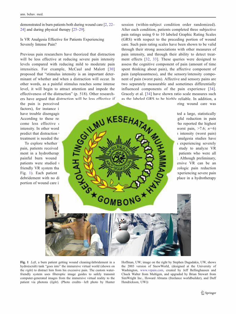
To explore whether VR can reduce severe and higherpain, patients received VR during burn wound debride-ment in a hydrotherapy tank, where some of the mostpainful burn wound care is conducted [2]. Elevenpatients were studied using a custom fiberoptic water-friendly VR system that can safely be used in water (seeFig. 1). Each patient spent a portion of their wounddebridement with no distraction and spent an equivalentportion of wound care in VR during the same wound care
session (within-subject condition order randomized).After each condition, patients completed three subjectivepain ratings using 0 to 10 labeled Graphic Rating Scales(GRS) with respect to the preceding portion of woundcare. Such pain rating scales have been shown to be validthrough their strong associations with other measures ofpain intensity, and through their ability to detect treat-ment effects [32, 33]. These queries were designed toassess the cognitive component of pain (amount of timespent thinking about pain), the affective component ofpain (unpleasantness), and the sensory/intensity compo-nent of pain (worst pain). Affective and sensory pains aretwo separately measurable and sometimes differentiallyinfluenced components of the pain experience [34].Gracely et al. [34] have shown ratio scale measures suchas the labeled GRS to be highly reliable. In addition, asingle GRS rating of “fun” during wound care wasmeasured.
Overall, patients (n=11) reported a large, statisticallysignificant and clinically meaningful reduction in painduring VR [2]. The six patients who reported the highestpain intensity during “no VR” (worst pain, >7.6; n=6)reported a 41% reduction in pain intensity (worst pain)during VR. Although other VR analgesia studies havecommonly included burn patients experiencing severelyintense pain, this was the first study to analyze VRanalgesia in a subgroup of burn patients who were allexperiencing severe pain intensity. Although preliminary,these results suggest that immersive VR can be aneffective adjunctive non-pharmacologic pain reductiontechnique, even for burn patients experiencing severe painduring wound care that is taking place in a hydrotherapytank.
Fig. 1 Left, a burn patient getting wound cleaning/debridement in ahydro(scrub) tank “goes into” the immersive virtual world (shown onthe right) to distract him from his excessive pain. The custom water-friendly system uses fiberoptic image guides to safely transmitcomputer-generated images from the immersive virtual reality to thepatient via photons (light). (Photo credits—left photo by Hunter
Hoffman, UW; image on the right by Stephen Dagadakis, UW, showsthe 2003 version of SnowWorld, (designed at the University ofWashington, www.vrpain.com, created by Jeff Bellinghausen andChuck Walter from Multigen, and upgraded by Brian Stewart fromSimWright Inc., Howard Abrams (freelance worldbuilder), and DuffHendrickson, UW))
ann. behav. med.
The Relationship Between the Immersiveness of the VRSystem and Analgesic Effectiveness
Using the concept of immersion as a theoretical framework,researchers have begun to analyze what makes VR effectivefor reducing pain. Slater and Wilbur define immersion as anobjective, quantifiable description of what a particular VRsystem can provide to a participant. Immersion is differentfrom the subjective psychological illusion of going into thevirtual world, known as presence. According to Slater andWilbur [35], presence is a psychological state of conscious-ness and is reliant on subjective measures (asking users torate on a scale from 1 to 10 how much they felt like theywent into the computer-generated world as if it is a placethey visited). In contrast, immersiveness is objectivelymeasurable (e.g., using trigonometry to calculate the “fieldof view” or amount of peripheral vision stimulated by a VRhelmet’s displays).
In several laboratory studies exploring the relationshipbetween immersion and analgesic effectiveness, healthyvolunteers received brief thermal pain stimuli at carefullycontrolled temperatures and rated how painful they foundthe stimuli. These studies found that highly immersive VRsystems are more effective at reducing pain than less-immersive VR systems [36–38] and as described next, thedifference in amount of analgesia achieved with a highlyimmersive VR system can be considerable.
In one laboratory study, high-technology VR gogglesincreased the patient’s peripheral vision in the virtual world,increasing how much VR reduced pain [36]. Researchers[36] randomly assigned participants (healthy volunteers) toeither a low-technology VR helmet group (n=28), a high-technology VR helmet group (n=26) or to a no VR group(n=23). To help minimize demand characteristics, both thesubjects and the research assistant collecting the experi-mental pain ratings remained unaware that helmet qualitywas being manipulated. Compared with the group thatreceived the low-technology VR helmet (35° field of viewdiagonal), the high-technology VR goggles group (60° fieldof view diagonal) reported 34% more reduction in worstpain, 46% more reduction in pain unpleasantness, 29%more reduction in time spent thinking about pain, and 32%more fun during the pain stimulus during VR. Sixty-fivepercent of participants in the high-technology VR gogglesgroup showed a clinically meaningful reduction in painintensity during virtual reality, compared with only 29% forthe low-technology VR helmet group. These results suggestthat helmet quality (i.e., goggle size/field of view/amount ofperipheral vision looking into VR) is an especiallyimportant factor for achieving clinically meaningful reduc-tions in pain intensity, and the study design helps reduce thelikelihood that VR analgesia is due to an artifact such asdemand characteristics.
In a related study [38], instead of manipulating helmetquality, the objective immersiveness of the VR system wasmanipulated via interactivity, i.e., whether participantsinteracted with the virtual world or not. Twenty-oneparticipants (healthy volunteers) were randomly assignedto one of two treatment groups. All participants individuallyglided through the virtual world SnowWorld, but one groupcould look around and interact with the virtual world via atrackball, and the other group could not interact with thevirtual world (no trackball). Afterwards, each participantprovided subjective 0–10 pain ratings.
The more-immersive VR group who interacted with thevirtual world via a trackball showed significantly more painreduction than the less-immersive VR group who receivednon-interactive VR with no track ball [38, see also 39].Compared with the non-interactive VR group, participantsin the interactive VR group showed 75% more reduction inpain unpleasantness (p<.005) and 74% more reduction inworst pain (p<.005) and 32% more reduction in time spentthinking about pain (p=.01). Interactivity increased theobjective immersiveness of the VR system, and aspredicted, increased the analgesic effectiveness. In summa-ry, so far, high-technology VR helmet quality (wide field ofview goggles), and interactivity (playing with a mouse-liketrackball or other input device) have been isolated asespecially important factors contributing to VR analgesia.
Using fMRI Brain Scans to Measure Pain-Related BrainActivity
What is going on in people’s brains when they feel pain,and how are those patterns of brain activity altered (if at all)when participants go into immersive virtual reality andexperience large reductions in how much pain they feel? Toexplore these topics, Hoffman and colleagues [40, 41]measured the objective physiological neural correlates ofVR analgesia. Custom magnet-friendly VR goggles [42]were designed and built that allowed participants toexperience the illusion of going inside the computer-generated world while simultaneously assessing neuralactivity using fMRI brain scans. A thermal pain stimulatorwas attached to the healthy volunteer’s foot. Participantsreceived 30 s of thermal stimulation at a painful buttolerable temperature pre-approved by each participant,then 30 s with lukewarm temperature, and this cycle of“pain on/pain off” was repeated three times over a 6-minfMRI brain scan.
During half of the brain scan, the control condition,participants looked at a fixation cross and saw no VR, andheard no music and no VR sound effects. During the otherhalf of their fMRI brain scan they went into the 3Dcomputer-generated world, and interacted with the virtual
ann. behav. med.
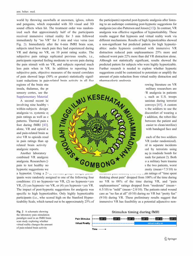
world by throwing snowballs at snowmen, igloos, robotsand penguins, which responded with 3D visual and 3Dsound effects when hit. The treatment order was random-ized such that approximately half of the participantsreceived immersive virtual reality for 3 min followedimmediately by “no VR” for 3 min and vice versa (seeFig. 2). Immediately after the 6-min fMRI brain scan,subjects rated how much pain they had experienced duringVR and during no VR, on 10 point rating scales. Thesubjective pain ratings replicated previous results, i.e.,participants reported feeling moderate to severe pain duringthe pain stimuli with no VR, and subjects reported muchless pain when in VR. In addition to reporting lesssubjective pain, objective measures of the neural correlatesof pain showed large (50% or greater) statistically signif-icant reductions in pain-related brain activity in all fiveregions of the brain studied (the anterior cingulate cortex,insula, thalamus, the primary and the secondary somato-sensory cortex, see the figure provided in the ElectronicSupplementary Material).
A second recent laboratory fMRI brain scan studyinvolving nine healthy volunteer participants (also using awithin-subjects design) compared and contrasted VRanalgesia vs. systemic opioid analgesia, both via subjectivepain ratings as well as objective measures of brain activitypatterns. Thermal pain stimuli were applied to the patient’sfoot during fMRI [43]. Results showed that when usedalone, VR and opioid analgesia each reduced pain ratingsand pain-related brain activity. Furthermore, adding immer-sive VR to opioids resulted in significantly more reductionin pain ratings than opioids alone, and patterns of pain-related brain activity were consistent with subjectiveanalgesic reports.
Another laboratory study compared, contrasted, andcombined VR analgesia with conventional post-hypnoticanalgesia. Researchers [44] experimentally induced thermalpain to test healthy normal volunteer participants. Post-hypnotic suggestions were administered via an audiotape ofa hypnotist. Using a 2×2, between-groups design, partic-ipants were randomly assigned to one of the following fourconditions: (1) no hypnosis+no VR, (2) no hypnosis+yesVR, (3) yes hypnosis+no VR, or (4) yes hypnosis+yes VR.The impact of post-hypnotic suggestions for analgesia wasspecific to high hypnotizables. Only highly hypnotizableparticipants (i.e., who scored high on the Stanford Hypno-tizability Scale, which turned out to be approximately 25% of
the participants) reported post-hypnotic analgesia after listen-ing to an audiotape containing post-hypnotic suggestions foranalgesia (see also Patterson and Jensen [13]). In contrast, VRanalgesia was effective regardless of hypnotizability. Theseresults suggest that hypnosis and virtual reality work viadifferent mechanisms. Results of high hypnotizables showeda non-significant but predicted pattern for high hypnotiz-ables: audio hypnosis combined with immersive VRdistraction reduced pain unpleasantness 25% more andreduced worst pain 22% more than did VR distraction alone.Although not statistically significant, results showed thepredicted pattern for subjects who were highly hypnotizable.Further research is needed to explore whether hypnoticsuggestions could be customized to potentiate or amplify theamount of pain reduction from virtual reality distraction andpharmacologic analgesia.
Encouraged by the small but growing literature on VRanalgesia in civilian burn patients, military researchers arebeginning to explore the use of VR analgesia in patientswith combat-related burn injuries, such as U.S. troopsseverely burned in Iraq and Afghanistan during terroristroadside bomb attacks on humvee convoys [45]. A custom“robot-like” arm (see Fig. 3) allows the soldiers to use theimmersive VR world without the discomfort of wearing a1.75 lb VR helmet on their head. In addition, the robot-likearm reduces or eliminates contact between the patient andthe equipment (making the goggles easier to clean/sterilize)and makes VR available to patients with bandaged face andhead burns.
Using graphic pain rating scales, each of the two soldiersrated their pain during VR vs. no VR (order randomized).Both patients were severely burned in separate incidentswhen their humvees were attacked by terrorists usingimprovised explosive devices in Iraq (a roadside bomb forpatient 1 and a rocket propelled grenade for patient 2). Bothpatients were evacuated from Iraq to a military burn traumacenter in the USA. Averaged across the two patients, worstpain dropped from severe pain intensity (mean=7.5/10) tomoderate pain intensity (4.5/10). Pain ratings of “time spentthinking about pain” dropped from 100% of the time duringno VR to 08% of the time during VR, and “painunpleasantness” ratings dropped from “moderate” (mean=6.5/10) to “mild” (mean=2.0/10). The patients rated woundcare as “no fun at all” (0/10) during no VR but “pretty fun”(9/10) during VR. These preliminary results suggest thatimmersive VR has feasibility as a potential adjunctive non-
Fig. 2 A schematic showingthe laboratory pain stimulationparadigm used in an fMRI brainscan study exploring whethervirtual reality changes the amountof pain-related brain activity
ann. behav. med.
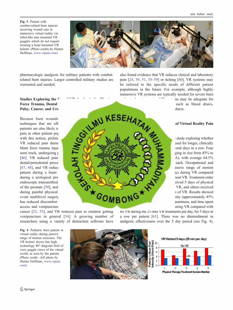
pharmacologic analgesic for military patients with combat-related burn injuries. Larger controlled military studies arewarranted and needed.
Studies Exploring the Use of VR Analgesia for BluntForce Trauma, Dental Fears, Claustrophobia, CerebralPalsy, Cancer, and Urological Endoscopy Patients
Because burn wounds are unusually painful injuries,techniques that are effective for reducing pain in burnpatients are also likely to be effective in treating proceduralpain in other patient populations besides burns. Consistentwith this notion, preliminary case studies have found thatVR reduced pain during physical therapy in a non-burnblunt force trauma injury (a pedestrian who was hit by asemi truck, undergoing physical therapy in the trauma unit)[46]. VR reduced pain and fear in two patients duringdental/periodontal procedures in patients with dental fears[47, 48], and VR reduced fear/anxiety in a claustrophobicpatient during a brain scan [49]. VR has reduced painduring a urological procedure in an older man receivingendoscopic transurethral microwave thermotherapy ablationof the prostate [50], and in pediatric cerebral palsy patientsduring painful physical therapy rehabilitation after singleevent multilevel surgery to increase ambulation [51]. VRhas reduced discomfort during subcutaneous vascular portaccess and venipuncture in children and adolescents withcancer [52, 53], and VR reduced pain in children gettingvenipuncture in general [54]. A growing number ofresearchers using a variety of distraction software have
also found evidence that VR reduces clinical and laboratorypain [24, 39, 51, 55–59] or itching [60]. VR systems maybe tailored to the specific needs of different patientpopulations in the future. For example, although highlyimmersive VR systems are typically needed for severe burnpatients, less-immersive VR systems may be adequate forsome other medical procedures such as blood draws,cannula implants, and dental procedures.
Future Directions: Repeated Use of Virtual Reality PainDistraction
Researchers conducted a preliminary study exploring whetherVR continues to be effective when used for longer, clinicallyrelevant treatment durations, for several days in a row. Fourchildren with large severe burns ranging in size from 45% to82% total body surface area (TBSA), with average 64.5%TBSA, were studied for 10 days each. Occupational andphysical therapists orchestrated passive range of motionexercises with each patient for 5 days during VR comparedwith similar treatment for 5 days without VR. Treatment orderwas randomized. Some patients received 5 days of physicaltherapy with VR vs. 5 days with no VR, and others received5 days of no VR followed by 5 days of VR. Results showedlarge reductions in worst pain intensity (approximately 45%reduction in worst pain), pain unpleasantness, and time spentthinking about pain, and more fun during VR compared withno VR during the 25-min VR treatments per day, for 5 days ina row per patient [61]. There was no diminishment inanalgesic effectiveness over the 5 day period (see Fig. 4),
Fig. 3 Patient withcombat-related burn injuriesreceiving wound care inimmersive virtual reality viarobot-like arm mounted VRgoggles which do not requirewearing a head mounted VRhelmet. (Photo credits by HunterHoffman, www.vrpain.com)
Fig. 4 Pediatric burn patient invirtual reality during passiverange of motion exercises. TheVR helmet shown has high-technology 80° diagonal field ofview goggle views of the virtualworld, as seen by the patient.(Photo credit—left photo byHunter Hoffman, www.vrpain.com)
ann. behav. med.
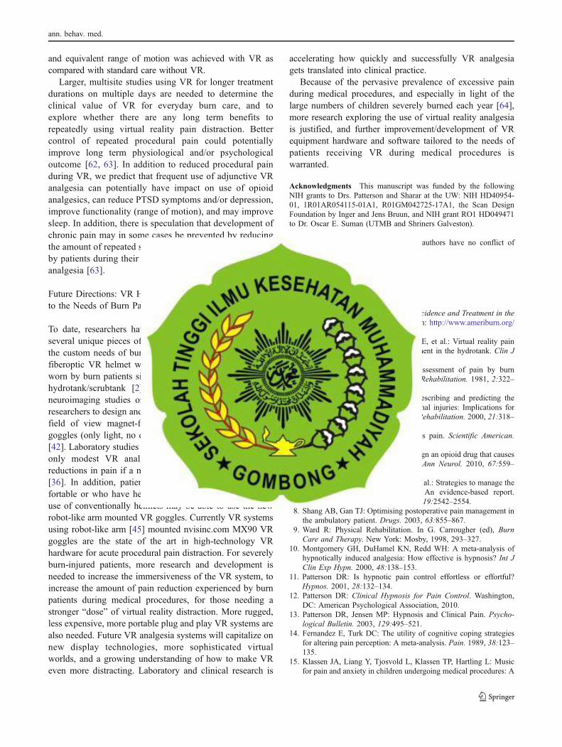
and equivalent range of motion was achieved with VR ascompared with standard care without VR.
Larger, multisite studies using VR for longer treatmentdurations on multiple days are needed to determine theclinical value of VR for everyday burn care, and toexplore whether there are any long term benefits torepeatedly using virtual reality pain distraction. Bettercontrol of repeated procedural pain could potentiallyimprove long term physiological and/or psychologicaloutcome [62, 63]. In addition to reduced procedural painduring VR, we predict that frequent use of adjunctive VRanalgesia can potentially have impact on use of opioidanalgesics, can reduce PTSD symptoms and/or depression,improve functionality (range of motion), and may improvesleep. In addition, there is speculation that development ofchronic pain may in some cases be prevented by reducingthe amount of repeated severe procedural pain experiencedby patients during their hospital stay, i.e., via preventativeanalgesia [63].
Future Directions: VR Hardware and Software Tailoredto the Needs of Burn Patients
To date, researchers have been able to design and buildseveral unique pieces of equipment specifically tailored tothe custom needs of burn patients. For example, a customfiberoptic VR helmet was developed that could be safelyworn by burn patients sitting in a tub of water known as ahydrotank/scrubtank [2]. Similarly, the first two fMRIneuroimaging studies on VR analgesia [40, 43] requiredresearchers to design and build the first pair of custom widefield of view magnet-friendly fiberoptic “photonic” VRgoggles (only light, no electricity, reaches the participants)[42]. Laboratory studies suggest that participants who showonly modest VR analgesia are likely to show largerreductions in pain if a more-immersive VR system is used[36]. In addition, patients who find VR helmets uncom-fortable or who have head or face burns that preclude theuse of conventionally helmets may be able to use the newrobot-like arm mounted VR goggles. Currently VR systemsusing robot-like arm [45] mounted nvisinc.com MX90 VRgoggles are the state of the art in high-technology VRhardware for acute procedural pain distraction. For severelyburn-injured patients, more research and development isneeded to increase the immersiveness of the VR system, toincrease the amount of pain reduction experienced by burnpatients during medical procedures, for those needing astronger “dose” of virtual reality distraction. More rugged,less expensive, more portable plug and play VR systems arealso needed. Future VR analgesia systems will capitalize onnew display technologies, more sophisticated virtualworlds, and a growing understanding of how to make VReven more distracting. Laboratory and clinical research is
accelerating how quickly and successfully VR analgesiagets translated into clinical practice.
Because of the pervasive prevalence of excessive painduring medical procedures, and especially in light of thelarge numbers of children severely burned each year [64],more research exploring the use of virtual reality analgesiais justified, and further improvement/development of VRequipment hardware and software tailored to the needs ofpatients receiving VR during medical procedures iswarranted.
Acknowledgments This manuscript was funded by the followingNIH grants to Drs. Patterson and Sharar at the UW: NIH HD40954-01, 1R01AR054115-01A1, R01GM042725-17A1, the Scan DesignFoundation by Inger and Jens Bruun, and NIH grant RO1 HD049471to Dr. Oscar E. Suman (UTMB and Shriners Galveston).
Conflicts of Interest Statement The authors have no conflict ofinterest to disclose.
References
1. American Burn Association: Burn Incidence and Treatment in theUS: 2007 Fact Sheet. Available from: http://www.ameriburn.org/resources_factsheet.php
2. Hoffman HG, Patterson DR, Seibel E, et al.: Virtual reality paincontrol during burn wound debridement in the hydrotank. Clin JPain. 2008, 24:299–304.
3. Perry S, Heidrich G, Ramos E: Assessment of pain by burnpatients. Journal of Burn Care and Rehabilitation. 1981, 2:322–326.
4. Ptacek J, Patterson D, Doctor J: Describing and predicting thenature of procedural pain after thermal injuries: Implications forresearch. Journal of Burn Care and Rehabilitation. 2000, 21:318–326.
5. Melzack R: The tragedy of needless pain. Scientific American.1990, 262:27–33.
6. Berger AC, Whistler JL: How to design an opioid drug that causesreduced tolerance and dependence. Ann Neurol. 2010, 67:559–569.
7. Cherny N, Ripamonti C, Pereira J, et al.: Strategies to manage theadverse effects of oral morphine: An evidence-based report.Journal of Clinical Oncology. 2001, 19:2542–2554.
8. Shang AB, Gan TJ: Optimising postoperative pain management inthe ambulatory patient. Drugs. 2003, 63:855–867.
9. Ward R: Physical Rehabilitation. In G. Carrougher (ed), BurnCare and Therapy. New York: Mosby, 1998, 293–327.
10. Montgomery GH, DuHamel KN, Redd WH: A meta-analysis ofhypnotically induced analgesia: How effective is hypnosis? Int JClin Exp Hypn. 2000, 48:138–153.
11. Patterson DR: Is hypnotic pain control effortless or effortful?Hypnos. 2001, 28:132–134.
12. Patterson DR: Clinical Hypnosis for Pain Control. Washington,DC: American Psychological Association, 2010.
13. Patterson DR, Jensen MP: Hypnosis and Clinical Pain. Psycho-logical Bulletin. 2003, 129:495–521.
14. Fernandez E, Turk DC: The utility of cognitive coping strategiesfor altering pain perception: A meta-analysis. Pain. 1989, 38:123–135.
15. Klassen JA, Liang Y, Tjosvold L, Klassen TP, Hartling L: Musicfor pain and anxiety in children undergoing medical procedures: A
ann. behav. med.
systematic review of randomized controlled trials. Ambul Pediatr.2008, 8:117–128.
16. Cepeda MS, Carr DB, Lau J, Alvarez H: Music for pain relief.Cochrane Database of Systematic Reviews (Vol. Issue 2), 2006.
17. Eccleston C: Role of psychology in pain management. Br JAnaesth. 2001, 87:144–152.
18. Eccleston C, Crombez G: Pain demands attention: A cognitive-affective model of the interruptive function of pain. PsychologicalBulletin. 1999, 125:356–366.
19. Kahneman D: Attention and effort. Englewood Cliffs, NJ:Prentice-Hall, 1973.
20. Hoffman HG, Garcia-Palacios A, Kapa VA, Beecher J, Sharar SR:Immersive virtual reality for reducing experimental ischemic pain.International Journal of Human-Computer Interaction. 2003,15:469–486.
21. Hoffman HG: Virtual Reality Therapy. Scientific American. 2004,291:58–65.
22. Hoffman HG, Doctor JN, Patterson DR, Carrougher GJ, FurnessTA, 3rd: Use of virtual reality as an adjunctive treatment ofadolescent burn pain during wound care: A case report. Pain.2000, 85:305–309.
23. Hoffman HG, Patterson DR, Magula J, et al.: Water-friendlyvirtual reality pain control during wound care. Journal of ClinicalPsychology. 2004, 60:189–195.
24. van Twillert B, Bremer M, Faber AW: Computer-generated virtualreality to control pain and anxiety in pediatric and adult burnpatients during wound dressing changes. J Burn Care Res. 2007,28:694–702.
25. Carrougher GJ, Hoffman HG, Nakamura D, et al.: The effect ofvirtual reality on pain and range of motion in adults with burninjuries. J Burn Care Res. 2009, 30:785–791.
26. Hoffman HG, Patterson DR, Carrougher GJ: Use of virtual realityfor adjunctive treatment of adult burn pain during physicaltherapy: A controlled study. Clin J Pain. 2000, 16:244–250.
27. Hoffman HG, Patterson DR, Carrougher GJ, Sharar SR: Effec-tiveness of virtual reality-based pain control with multiple treat-ments. Clin J Pain. 2001, 17:229–235.
28. Schmitt YS, Hoffman HG, Blough DK, et al.: A randomized,controlled trial of immersive virtual reality analgesia, duringphysical therapy for pediatric burns. Burns. 2010.
29. Sharar SR, Carrougher GJ, Nakamura D, et al.: Factors influenc-ing the efficacy of virtual reality distraction analgesia duringpostburn physical therapy: Preliminary results from 3 ongoingstudies. Arch Phys Med Rehabil. 2007, 88:S43–49.
30. McCaul KD, Malott JM: Distraction and coping with pain.Psychological Bulletin. 1984, 95:516–533.
31. Crombez G, Eccleston C, Baeyens F, Eelen P: When somaticinformation threatens, catastrophic thinking enhances attentionalinterference. Pain. 1998, 75:187–198.
32. Jensen MP: The validity and reliability of pain measures in adultswith cancer. Journal of Pain. 2003, 4:2–21.
33. Jensen MP, Karoly P: Self-report scales and procedures forassessing pain in adults. In D. C. Turk and R. Melzack (eds),Handbook of pain assessment. New York: Guilford Publications,2001, 15–34.
34. Gracely RH, McGrath P, Dubner R: Ratio scales of sensory andaffective verbal pain descriptors. Pain. 1978, 5:5–18.
35. SlaterM,Wilbur S: A framework for immersive virtual environments(FIVE): speculations on the role of presence in virtual environments.Presence Teleoper Virtual Environ. 1997, 6:603–616.
36. Hoffman HG, Seibel EJ, Richards TL, et al.: Virtual reality helmetdisplay quality influences the magnitude of virtual realityanalgesia. J Pain. 2006, 7:843–850.
37. Hoffman HG, Sharar SR, Coda B, et al.: Manipulating presenceinfluences the magnitude of virtual reality analgesia. Pain. 2004,111:162–168.
38. Wender R, Hoffman HG, Hunner HH, et al.: InteractivityInfluences the Magnitude of Virtual Reality Analgesia. J CyberTher Rehabil. 2009, 2:27–33.
39. Dahlquist LM, McKenna KD, Jones KK, et al.: Active andpassive distraction using a head-mounted display helmet: Effectson cold pressor pain in children. Health Psychol. 2007, 26:794–801.
40. Hoffman HG, Richards TL, Coda B, et al.: Modulation of thermalpain-related brain activity with virtual reality: Evidence fromfMRI. Neuroreport. 2004, 15:1245–1248.
41. Hoffman HG, Richards TL, Bills AR, et al.: Using fMRI to studythe neural correlates of virtual reality analgesia. CNS Spectrums.2006, 11:45–51.
42. Hoffman HG, Richards TL, Magula J, et al.: A magnet-friendlyvirtual reality fiberoptic image delivery system. Cyberpsychol.Behav. 2003, 6:645–648.
43. Hoffman HG, Richards TL, Van Oostrom T, et al.: The analgesiceffects of opioids and immersive virtual reality distraction:Evidence from subjective and functional brain imaging assess-ments. Anesth Analg. 2007, 105:1776–1783, table of contents.
44. Patterson DR, Hoffman HG, Palacios AG, Jensen MP: Analgesiceffects of posthypnotic suggestions and virtual reality distractionon thermal pain. J Abnorm Psychol. 2006, 115:834–841.
45. Maani C, Hoffman HG, DeSocio PA, et al.: Pain control duringwound care for combat-related burn injuries using customarticulated arm mounted virtual reality goggles. Journal ofCyberTherapy and Rehabilitation. 2008, 1:193–198.
46. Hoffman HG, Patterson DR, Soltani M, et al.: Virtual reality paincontrol during physical therapy range of motion exercises for apatient with multiple blunt force trauma injuries. CyberpsycholBehav. 2008, 19:47–49.
47. Furman E, Jasinevicius TR, Bissada NF, et al.: Virtual realitydistraction for pain control during periodontal scaling and rootplaning procedures. J Am Dent Assoc. 2009, 140:1508–1516.
48. Hoffman HG, Garcia-Palacios A, Patterson DR, Jensen MP,Furness III TA: The effectiveness of virtual reality for dentalpain control: A case study. Cyberpsychol Behav. 2001, 4:527–535.
49. Garcia-Palacios A, Hoffman HG, Richards TR, Seibel EJ, ShararSR: Use of virtual reality distraction to reduce claustrophobiasymptoms during a mock magnetic resonance imaging brain scan:a case report. Cyberpsychol Behav. 2007, 10:485–488.
50. Wright JL, Hoffman HG, Sweet RM: Virtual reality as anadjunctive pain control during transurethral microwave thermo-therapy. Urology. 2005, 66:1320.
51. Steele E, Grimmer K, Thomas B, et al.: Virtual reality as apediatric pain modulation technique: A case study. CyberpsycholBehav. 2003, 6:633–638.
52. Gershon J, Zimand E, Pickering M, Rothbaum BO, Hodges L: Apilot and feasibility study of virtual reality as a distraction forchildren with cancer. J Am Acad Child Adolesc Psychiatry. 2004,43:1243–1249.
53. Windich-Biermeier A, Sjoberg I, Dale JC, Eshelman D, GuzzettaCE: Effects of distraction on pain, fear, and distress during venousport access and venipuncture in children and adolescents withcancer. J Pediatr Oncol Nurs. 2007, 24:8–19.
54. Gold JI, Kim SH, Kant AJ, Joseph MH, Rizzo AS: Effectivenessof virtual reality for pediatric pain distraction during i.v.placement. Cyberpsychol Behav. 2006, 9:207–212.
55. Chan EA, Chung JW, Wong TK, Lien AS, Yang JY: Applicationof a virtual reality prototype for pain relief of pediatric burn inTaiwan. J Clin Nurs. 2007, 16:786–793.
56. Das D, Grimmer K, Sparnon A, McRae S, Thomas B: Theefficacy of playing a virtual reality game in modulating pain forchildren with acute burn injuries: A randomized controlled trial.BMC Pediatric. 2005, 5:1.
ann. behav. med.
57. Malloy KM, Milling LS: The effectiveness of virtual realitydistraction for pain reduction: A systematic review. Clin PsycholRev. 2010, 30:1011–1018.
58. Morris LD, Louw QA, Grimmer-Somers K: The effectiveness ofvirtual reality on reducing pain and anxiety in burn injury patients:A systematic review. Clin J Pain. 2009, 25:815–826.
59. Schneider SM, Prince-Paul M, Allen MJ, Silverman P, Talaba D:Virtual reality as a distraction intervention for women receivingchemotherapy. Oncol Nurs Forum. 2004, 31:81–88.
60. Liebovici V, Magora F, Cohen S, Ingber A: Effects of virtualreality immersion and audiovisual distraction techniques forpatients with pruritus. Pain Res Manag. 2009, 14:283–286.
61. FloresA, HoffmanHG, RussellW, et al.: Longer, multiple virtual realitypain distraction treatments of Hispanic and Caucasian children withlarge severe burns. CyberTherapy Conference. San Diego, CA, 2008.
62. Patterson DR, Sharar SR: Burn pain. In J. D. Loeser (ed), Bonica’sManagement of Pain. Philadelphia, PA: Lippincott, Williams &Wilkins, 2001, 780–787.
63. Malchow RJ, Black IH: The evolution of pain management in thecritically ill trauma patient: Emerging concepts from the globalwar on terrorism. Crit Care Med. 2008, 36:S346–357.
64. D’Souza AL, Nelson NG, McKenzie LB: Pediatric burn injuriestreated in US emergency departments between 1990 and 2006.Pediatrics. 2009, 124:1424–1430.
ann. behav. med.