universiti putra malaysia degreening … · suatu kajian dijalankan ke atas perubahan struktur sel,...
TRANSCRIPT
UNIVERSITI PUTRA MALAYSIA
DEGREENING CHARACTERISTIC OF MUSA AAA 'BERANGAN' AND "WILLIAM CAVENDISH' BANANAS
PHEBE DING.
FP 2004 22
DEGREENING CHARACTERISTICS OF MUSA AAA 'BERANGAN' AND 'WILLIAM CAVENDISH' BANANAS
PHEBE DING
DOCTOR OF PHILOSOPHY UNIVERSITI PUTRA MALAYSIA
DEGREENING CHARACTERISTICS OF MUSA AAA 'BERANGAN' AND 'WILLIAM CAVENDISH' BANANAS
BY
PHEBE DING
Thesis Submitted to the School of Graduate Studies, Universiti Putra Malaysia, in Fulfilment of the Requirements for the
Degree of Doctor of Philosophy
March 2004
Abstract of thesis presented to the Senate of Universiti Putra Malaysia in fulfilment of the requirements for the degree of Doctor of Philosophy
DEGREENING CHARACTERISTICS OF MUSA AAA 'BERANGAN' AND 'WILLIAM CAVENDISH' BANANAS
BY
PHEBE DING
March 2004
Chairman: Associate Professor Siti Hajar Ahmad, Ph.D.
Faculty: Agriculture
A study was conducted on the changes in cellular structure, physiology and physio-
chemical of Musa AAA 'Berangan' and 'Cavendish' as the ripening progressed at
18+2 and 2722 OC. Mature green (ripening stage (RS) 1) bananas were initiated to
ripen using 0.02% acetylene from calcium carbide (CaC2) source (with an equivalent
of 1 g cac2.kg-' fruit) for 24 h. A hand of Berangan and Cavendish fmit was split
into 2 clusters. One of the clusters was initiated and ripened at 18t2 OC RH 90-
94%, while the other was initiated and ripened at 2722 OC RH 75-80%. The
experiment was conducted using randomized complete block design with four
replications. Five fruits per replicate were used. The various ripening stages were
evaluated with the aid of FAMA visual colour score until the fruit turned intosdl?
yellow at RS 6. For the Cavendish ripened at 27+2 OC (C27) where the fruit failed
to degreen, the evaluation was done daily until senescence, when brown specks
appeared on the peel. Data from measurements of ripening duration, L*, C* and h0
values, chlorophyll a, b and total chlorophylls, water loss, stomatal density, stomatal
length and opening, peel thickness, peel and pulp fresh and dry weight, cell length
and width of photosynthetic, epidermal, crystalliferous, tanniferous and starch
granules, pulp firmness, soluble solids concentration (SSC), titratable acidity and pH
were analysed using analysis of variance and differences between means were
determined by Duncan Multiple Range Test. Data from starch iodine test, cellular
structure and ultrastructure were documented as photographs or micrographs.
Berangan degreened naturally under tropical ripening temperature of 27+2 OC, and a
golden yellow fruit of RS 6 was obtained within 4 d of ripening. In contrast,
Cavendish failed to degreen at 27+2 OC even though the pulp had softened. By day 5
after the acetylene treatment brown specks started to appear on the fruit surface
indicating senescence had commenced. Cavendish could only degreen when ripened
at 1822 OC, and a yellow fruit of RS 6 was obtained after 9 d of ripening. On the
contrary, Berangan could not degreen and ripen under 1822 OC thus the fruit was
discarded. TEM revealed that at RS 6 the grana-thylakoid membrane of chromoplast
Berangan ripened at 2722 OC (B27) and Cavendish ripened at 1852 OC (C18) had
lysed. Besides, plastoglobuli increased in number, and types of staining density and
vesicles increased in number and size. However at day 5 of C27, the grana-thylakoid
membrane retained and this was in concurrent with the high retention of chlorophyll
content in fruit peel. The total chlorophyll retained in C27 was 57%, while only 25
and 40% of total chlorophyll was retained in B27 and C18 respectively. The high
retention of chlorophyll content in C27 had caused it to correlate significantly with
L*, C* and h0 values. Among B27, C18 and C27, C27 encountered the most water
loss. However, there was no correlation between water loss and stomata1 density and
opening. The existence of cracks and pores on the banana peel surface could be the
iii
passage for water loss. The severe water loss caused the peel thickness of C27 to be
the thinnest among the bananas studied although initially its peel was thicker than
B27. The fruit pulp and peel behaved differently towards ripening temperature. The
moisture content in pulp increased, while no moisture content in peel decreased as
ripening progressed. This led to increase of pulp to peel fresh weight ratio and C27
had the highest ratio as compared to C18 and B27. The softening of the banana fruit
was due to starch degradation and dissolution of middle lamellae. SEM revealed
that the pulp starch granules decreased in size and density as ripening progressed.
The blue-black stained area of starch-iodine complex cleared from the central core
of fmit towards the periphery of peel in all the bananas studied. The clearing pattern
was most rapid in C27. The hydrolysed starch increased the SSC of all the bananas
studied but the most significant increase was in C27. The titratable acidity of the
three bananas studied increased then decreased as ripening progressed. In contrast,
the pH decreased then increased as contrary to the trend of titratable acidity. The
tropical temperature of 2722 OC, besides failing to degreen, had caused poor keeping
and eating quality of Cavendish.
Abstrak tesis yang dikemukakan kepada Senat Universiti Putra Malaysia sebagai memenuhi keperluan untuk ijazah Doktor Falsafah
PENCIRIAN PENYAHHIJAUAN PADA PISANG, MUSA AAA 'BERANGAN' DAN 'WILLIAM CAVENDISH'
Oleh
PHEBE DING
Mac 2004
Pengerusi: Profesor Madya Siti Hajar Ahmad, Ph,D.
Fakulti: Pertanian
Suatu kajian dijalankan ke atas perubahan struktur sel, fisiologi dan fiziko-kimia
semasa proses kemasakan pada Musa AAA 'Berangan' dan 'Cavendish' yang
diranurnkan pada suhu 18+2 dan 27+2 OC. Pisang yang hijau matang (Peringkat
Kematangan (PK) 1) diperam dengan menggunakan 0.02% asetilena yang
dibebaskan dari surnber kalsium karbida (CaC2) (bersarnaan dengan 1 g cac2.kge1
buah) selama 24 j. Sesikat Berangan dan Cavendish dipisahkan kepada dua
bahagian. Salah satu bahagian diperam dan diranum pada suhu 18+2 OC dengan
kelembapan relatif (RH) 90-94%. Manakala bahagian yang lain diperam dan
diranwn pada suhu 27+2 OC RH 75-80%. Ujikaji dijalankan dengan menggunakan
rekabentuk blok rawak lengkap dengan empat replikasi. Sebanyak lima biji
buahheplikasi digunakan. Penilaian dibuat berdasarkan peringkat kemasakan
dengan Panduan Carta Warna FAMA sehingga buah mencapai kuning sepenuhnya
pada PK 6 . Untuk buah Cavendish yang diranum pada suhu 27+2 OC (C27) di mana
buah gaga1 menyahhijau, penilaian dijalankan setiap hari sehingga senesens di mana
bintik perang kelihatan pada kulit buah. Data-data masa peranuman, nilai-nilai L*,
C* dan hO, klorofil a, b dan jumlah klorofil, kehilangan air, ketumpatan stomata,
panjang dan lebar stomata, ketebalan kulit, jisim segar dan kering kulit dan isi,
panjang dan lebar untuk sel-sel fotosintetik, epidermis, 'crystalliferous' dan
'tanniferous' dan butiran kanji, kekerasan isi, jumlah kandungan pepejal terlarut,
asid tertitrat dan pH dianalisa dengan menggunakan kaedah analisis varian dan
perbezaan antara setiap min ditentukan dengan menggunakan kaedah 'Duncan
Multiple Range Test'. Data ujian iodin terhadap kanji, struktw dan ultrastruktw sel
dicatatkan sebagai gambar foto atau mikro. Berangan dapat menyahhijau secara
semula jadi di bawah suhu tropikal pada 27+2 OC dan buah kuning keemasan
diperolehi selepas 4 hari peranuman. Sebaliknya Cavendish tidak dapat
menyahhijau pada suhu 2722 OC sungguhpun isinya sudah lembut. Pada hari ke-5
selepas dirawat dengan dengan asetilena, bintik perang mulai kelihatan pada kulit
buah yang menandakan bermulanya senesens. Cavendish hanya boleh dinyahhijau
apabila diranum pada suhu 1852 OC dan buah benvarna kuning penuh pada PK 6
selepas 9 hari diranum Sebaliknya Berangan tidak dapat dinyahhijau dan masak
pada suhu 1822 OC. Penemuan melalui TEM mendapati pada PK 6, rnembran
grana-tilakoid pada kromoplast Berangan yang diranum pada 2722 OC (B27) dan
Cavendish yang diranum pada suhu 1852 OC (C18) sudah lisis. Selain itu, bilangan
dan jenis darjah kepekatan pewarnaan elektron plastoglobuli bertambah dan
bilangan dan saiz vesikel juga bertambah. Tetapi, pada hari ke-5, membran grana-
tilakoid C27 masih kelihatan dan ini adalah sejajar dengan pengekalan kandungan
klorofil yang tinggi pada kulit buah. Jumlah klorofil yang masih wujud pada C27
adalah 57% sementara hanya 25 dan 40% jumlah klorofil yang tinggal pada B27 dan
C18 masing-masing. Pengekalan kandungan klorofil yang tinggi di dalam C27 telah
menyebabkan kandungan klorofil berkolerasi bererti dengan nilai-nilai L*, C* d m
hO. Di kalangan B27, C18 dan C27, C27 mengalami kehilangan air yang paling
banyak, tetapi tiada kolerasi di antara kehilangan air dengan ketumpatan dan
pembukaan stoma pada kesemua pisang yang dikaji. Kewujudan retak dan lubang
pada permukaan kulit pisang mungkin merupakan laluan untuk kehilangan air.
Kehilangan air yang teruk mengakibatkan ketebalan kulit C27 menjadi paling nipis
di kalangan pisang yang dikaji walaupun ketebalan kulitnya pada peringkat
permulaan adalah lebih tebal daripada B27. Kelakuan isi dan kulit buah terhadap
suhu peranuman adalah berlainan. Kelembapan kandungan pada isi bertambah,
manakala kelembapan kandungan kulit berkurangan semasa proses peranuman. Ini
mengakibatkan nisbah jisim segar isi kepada kulit bertambah dan C27 mempunyai
nisbah yang paling tinggi jika berbanding dengan C18 dan B27. Kelembutan isi
buah pisang disebabkan oleh penguraian kanji dan lamela tengah. Penemuan dari
SEM mendapati saiz dan ketumpatan butiran kanji pada isi berkurangan semasa
proses peranuman. Kawasan biru kehitaman yang terhasil dari kompleks kanji-iodin
luntur dari bahagian tengah buah dan berlanjutan sehingga periferi buah di kulit
pada kesemua jenis pisang yang dikaji. Corak pelunturan adalah paling cepat pada
C27. Kanji yang telah dihidrolisis meningkatkan kandungan jumlah pepejal terlarut
dengan pertambahan yang paling bererti pada buah C27. Ketiga-tiga jenis pisang
menunjukkan pertarnbahan asid tertirat, diikuti dengan pengurangan semasa proses
peranurnan. Sebaliknya, pH meningkat, kemudian meunurun di mana pola
perubahannya adalah berlawanan dengan asid tertirat. Akhir kata, suhu peranuman
vii
tropikal pada 2722 O C selain menyebabkan kegagalan menyahhijau, ia juga telah
menurunkan kualiti penyimpanan dan nilai pemakanan pada Cavendish.
viii
ACKNOWLEDGEMENTS
I wish to express my sincere thanks and gratitude to my supervisors, Associate
Professor Dr. Siti Hajar Ahmad, a Postharvest Physiologists, Professor Dr. Abdul
Rahrnan Abdul Razak, a Cellular Developmental Biologist, Associate Professor Dr.
Nazamid Saari, an Enzymologist and Associate Professor Dr. Tengku Mahrnud
Tengku Moharned, a Postharvest Physiologists for their invaluable advice, guidance,
ideas, encouragement and patience throughout the course of this study.
Thanks are also due to the staff and friends of the Postharvest Laboratory, Electron
Microscopy Unit, Histology Laboratory and Enzyme Laboratory who had helped me
in completing this experiment successfully.
Finally, my sincere appreciation to my dearest dad, step-mother, sister, brother and
friends for their understanding, patience and moral support throughout my study.
I certify that the Examination Committee have met on 25" March, 2004 to conduct the final examination of Phebe Ding on her Doctor of Philosophy thesis entitled "Degreening Characteristics of Musa AAA 'Berangan' and 'William Cavendish' Bananas" in accordance with Universiti Pertanian Malaysia (Higher Degree) Act 1980 and Universiti Pertanian Malaysia (Higher Degree) Regulations 198 1. The Committee recommended that the candidate be awarded the relevant degree. Members of the Examination Committee are as follows:
Gulam Rusul Rahmat Ali, Ph.D. Professor Faculty of Food Science and Biotechnology Universiti Putra Malaysia (C hainnan)
Siti Hajar Ahmad, Ph.D. Associate Professor Faculty of Agriculture Universiti Putra Malaysia (Member)
Abdul Rahman Abdul Razak, Ph.D. Professor Faculty of Agriculture Universiti Putra Malaysia (Member)
Nazamid Saari, Ph.D. Associate Professor Faculty of Food Science and Biotechnology Universiti Putra Malaysia (Member)
Mahmud Tengku Muda Tengku Mohamed, Ph.D. Associate Professor Faculty of Agriculture Universiti Putra Malaysia (Member)
~rofessor /~e~ut) '~ean School of Graduate Studies Universiti Putra Malaysia
Date: 3 0 JUN 20M
This thesis submitted to the Senate of Universiti Putra Malaysia and has been accepted as fulfilment of the requirements for the degree of Doctor of Philosophy. The members of the Supervisory Committee are as follows:
Siti Hajar Ahmad, Ph.D. Associate Professor Faculty of Agriculture Universiti Putra Malaysia (Member)
Abdul Rahman Abdul Razak, Ph.D. Professor Faculty of Agriculture Universiti Putra Malaysia (Member)
Nazamid Saari, Ph.D. Associate Professor Faculty of Food Science and Biotechnology Universiti Putra Malaysia (Member)
Mahmud Tengku Muda Tengku Mohamed, Ph.D. Associate Professor Faculty of Agriculture Universiti Putra Malaysia (Member)
AINI IDERIS, Ph.D. ProfessorIDean School of Graduate Studies Universiti Putra Malaysia
Date: 0 9 JUL 2004
DECLARATION
I hereby declare that the dissertation based on my original work except for quotations and citations which have been duly acknowledged. I also declare that it has not been submitted for any other degree at UPM or other institutions.
Date: I&/ C/O 4
xii
TABLE OF CONTENTS
ABSTRACT ABSTRAK ACKNOWLEDGEMENTS APPROVAL DECLARATION LIST OF TABLES LIST OF FIGURES
CHAPTER
1
2
INTRODUCTION
LITERATURE REVIEW 2.1 Banana 2.2 Fruit Ripening 2.3 Peel Degreening of Banana
2.3.1 Structure of Chloroplast 2.3.2 Ultrastructural Changes of Chloroplast during
Degreening 2.3.3 Chlorophyll
2.4 Water Loss and Peel Surface Morphology
CHANGES IN PEEL COLOUR OF MUSA AAA 'BERANGAN' AND 'WILLIAM CAVENDISH' DURING RIPENING 3.1 Introduction 3.2 Materials and Methods
3.2.1 Plant Materials 3.2.2 Ripening Duration Determination 3.2.3 Peel Colour Determination 3.2.4 Chlorophyll Determination 3.2.5 Statistical Analysis
3.3 Results and Discussion 3.3.1 Ripening Duration 3.3.2 Peel Colour 3.3.3 Chlorophyll Content
3.4 Conclusion
CHANGES IN PEEL PLASTID OF MUSA AAA 'BERANGAN' AND 'WILLIAM CAVENDISH' DURING DEGREENING 4.1 Introduction 4.2 Materials and Methods
Page . . 11
v ix X
xii xvi xix
. . . X l l l
4.2.1 Plant Materials 4.2.2 Plastid Ultrastructural Studies 4.2.3 Statistical Analysis
4.3 Results and Discussion 4.3.1 Plastid Structural Changes 4.3.2 Granal-thylakoid Lamellae Changes 4.3.3 Plastoglobuli Number and Density and Vesicle
Number Changes 4.4 Conclusion
FRUIT WATER LOSS IN RELATION TO PEEL SURFACE MORPHOLOGY AND PHYSICAL PROPERTIES OF MUSA AAA 'BERANGAN' AND 'WILLIAM CAVENDISH' DURING DEGREENING 5.1 Introduction 5.2 Materials and Methods
5.2.1 Plant Materials 5.2.2 Water Loss Determination 5.2.3 Peel Surface Area to Fruit Volume Ratio
Determination 5.2.4 Stornatal Density Determination 5.2.5 Stornatal Length and Opening Determination 5.2.6 Photosynthetic Cells Size Determination 5.2.7 Peel Thickness Determination 5.2.8 Statistical Analysis
5.3 Results and Discussion 5.3.1 Water Loss 5.3.2 Stornatal Density 5.3.3 Stornatal Length and Opening 5.3.4 Peel Thickness 5.3.5 Photosynthetic Cells Length and Width
5.4 Conclusion
RELATION BETWEEN PULP AND PEEL OF MUSA AAA 'BERANGAN' AND 'WILLIAM CAVENDISH' DURING RIPENING 6.1 Introduction 6.2 Materials and Methods
6.2.1 Plant Materials 6.2.2 Pulp and Peel Content Determination 6.2.3 Pulp to Peel Fresh Weight Ratio
Determination 6.2.4 Pulp and Peel Moisture Content Determination 6.2.5 Pulp and Peel Dry Matter Determination 6.2.6 Cell Macrostructural Studies
6.2.6.1 Tissue Preparation for SEM Studies 6.2.6.2 Tissue Preparation for Light
Microscopic Studies
xiv

6.2.7 Statistical Analysis 6.3 Results and Discussion
6.3.1 Pulp and Peel Content 6.3.2 Pulp to Peel Fresh Weight Ratio 6.3.3 Pulp and Peel Moisture Content 6.3.4 Pulp and Peel Dry Matter 6.3.5 Cell Macrostructural Changes
6.3.5.1 Epidermal Layer of Peel Region 6.3.5.2 Peel-pulp Transition Region 6.3.5.3 PulpRegion
6.4 Conclusion
PHYSICO-CHEMICAL CHANGES IN MUSA AAA 'BERANGAN' AND 'WILLIAM CAVENDISH' DURING RIPENING 7.1 Introduction 7.2 Materials and Methods
7.2.1 Plant Materials 7.2.2 Pulp Firmness Determination 7.2.3 Peel Ultrastructural Studies 7.2.4 Starch Iodine Test 7.2.5 Pulp Starch Granules Size Determination 7.2.6 Soluble Solids Concentration Determination 7.2.7 Titratable Acidity Determination 7.2.8 pH Determination 7.2.9 Statistical Analysis
7.3 Results and Discussion 7.3.1 Pulp Firmness 7.3.2 Peel Ultrastructural Changes During Ripening
7.3.2.1 Peel Ultrastructural Changes of Berangan Ripened at 27+2 OC
7.3.2.2 Peel Ultrastructural Changes of Cavendish Ripened at 18+2 OC
7.3.2.3 Peel Ultrastructural Changes of Cavendish Ripened at 2752 OC
7.3.3 Pulp Starch Granules 7.3.4 Starch Pattern 7.3.5 Soluble Solids Concentration 7.3.6 Titratable Acidity 7.3.7 pH
7.4 Conclusion
CONCLUSION
BIBLIOGRAPHY BIODATA OF THE AUTHOR
LIST OF TABLES
Page Table
1 Duration for each stage of ripening of Berangan ripened at 2722 OC and Cavendish ripened at 1822 OC. 3.7
Effects of ripening stage or days after acetylene treatment on peel colours (L*, C* and hO) of Berangan ripened at 2722 OC and Cavendish ripened at 1822 and 2722 OC.
Effects of ripening stage or days after acetylene treatment on chlorophyll a (Chl a) and b (Chl b), total chlorophyll (Total chl) and chlorophyll d b ratio (Chl d b ratio) of Berangan ripened at 2722 OC and Cavendish ripened at 1822 and 2722 OC. 3.14
4 Correlation coefficients (r) for chlorophyll a (Chl a) and b (Chl b), total chlorophyll (T chl), chlorophyll d b ratio (Chl db), L*, C* and h0 of Berangan ripened at 2722 OC and Cavendish ripened at 1822 3.27 and 2722 OC.
Effects of ripening stage (RS) or days after acetylene treatment (DAAT) on length and width 5 SD in ten chloroplasts of Berangan ripened at 2722 OC and Cavendish ripened at1 822 and 2722 OC. 4.8
Effects of ripening stage (RS) or days after acetylene treatment (DAAT) on granum length and number of thylakoid per stack of granum in ten chloroplasts of Berangan ripened at 27+2 OC and Cavendish ripened at 1822 and 2722 OC.
Effects of ripening stage (RS) or days after acetylene treatment (DAAT) on diameter 2 SD of five differential electron staining densities of plastoglobuli and vesicles of Berangan ripened at 2722 OC and Cavendish ripened at 1822 and 2722 OC. 4.20
Effects of ripening stage or days after acetylene treatment on water loss and water loss rate of Berangan ripened at 2722 OC and Cavendish ripened at 1852 and 2722 OC.
Main and interaction effects of six ripening stages (RS) or days after acetylene treatment (D), five faces (F) and three regions (R) of Berangan ripened at 2722 OC and Cavendish ripened at 1822 and 2 7 9 OC on stornatal density per rnrn2. 5.12
Correlation coefficients (r) for water loss (WL), stomatal density (SD), stornatal opening (SO), peel thickness (Thick) and photosynthetic cell width (Width) of the first five-layer peel cells in Berangan and Cavendish during ripening.
xvi
Effects of ripening stage or days after acetylene treatment on stomata1 length and opening of Berangan ripened at 27+2 OC and Cavendish ripened at 1822 and 2722 OC. 5.22
12 Effects of ripening stage or days after acetylene treatment on peel thickness of Berangan ripened at 2722 OC and Cavendish ripened at l8+2 and 2722 OC.
13 Effects of ripening stage or days after acetylene treatment on photosynthetic cell length and width of the first five layers of cells in peel of Berangan ripened at 2752 OC and Cavendish ripened at 5.30 1 822 and 2722 OC.
Effects of ripening stage or days after acetylene treatment on the percentage of pulp and peel content and pu1p:peel fiesh weight (Plp) of Berangan ripened at 27+2 OC and Cavendish ripened at 18+2 and 6.7 2752 OC.
Effects of ripening stage or days after acetylene treatment on the percentage of pulp and peel moisture content of Berangan ripened at 2722 OC and Cavendish ripened at 1822 and 2722 OC. 6.1 1
Effects of ripening stage or days after acetylene treatment on the percentage of pulp and peel dry matter of Berangan ripened at 2722 OC and Cavendish ripened at 1822 and 2722 OC. 6.14
Correlation coefficients (r) for pulp to peel fresh weight ratio (pip), pulp content, peel content, pulp moisture content (Pulp MC), peel moisture content (Peel MC), pulp dry matter (Pulp DM), peel dry matter (Peel DM), peel thickness and water loss of Berangan 6.17 ripened at 2722 OC.
Correlation coefficients (r) for pulp to peel fresh weight ratio (pip), pulp content, peel content, pulp moisture content (pulp MC), peel moisture content (peel MC), pulp dry matter (pulp DM), peel dry matter (peel DM), peel thickness (Thick), and water loss of Cavendish ripened at 18+2 OC.
CorreIation coefficients (r) for pulp to peel fresh weight ratio (p/p), pulp content, peel content, pulp moisture content (pulp MC), peel moisture content (peel MC), pulp dry matter (pulp DM), peel dry matter (peel DM), peel thickness (Thick), and water loss of Cavendish ripened at 27+2 OC. 6.19
Effects of ripening stage or days after acetylene treatment (DAAT)' on epidermal cell length and width of Berangan ripened at 2752 OC (B27) and Cavendish ripened at 1852 (C 18) and 27+2 OC (C27).
xvii
Effects of ripening stage or days after acetylene treatment (DAAT)' on crystalliferous idioblast cell length, width and frequency at peel region of Berangan ripened at 2722 OC (B27) and Cavendish ripened at 1822 (C18) and 27+2 OC (C27). 6.38
Effects of ripening stage or days after acetylene treatment (DAAT)' on tanniferous cells length, width and frequency at peel region of Berangan ripened at 2722 OC (B27), Cavendish ripened at 1822 (C 1 8) and 2722 OC (C27). 6.42
Effects of ripening stage or days after acetylene treatment (DAAT)' on crystalliferous idioblast cell number, length and width at peel- pulp transition region of Berangan ripened at 2722 OC (B27) and Cavendish ripened at 1 8 2 (C 18) and 2722 OC (C27). 6.56
Effects of ripening stage or days after acetylene treatment (DAAT)' on bead-like structure of coagulated latex cell length and width at peel-pulp transition region of Berangan ripened at 2722 OC (B27) and Cavendish ripened at 1822 (C 18) and 2722 OC (C27). 6.57
Effects of ripening stage or days after acetylene treatment (DAAT)' on tanniferous cell frequency, length and width at peel-pulp transition region of Berangan ripened at 2722 OC (B27) and Cavendish ripened at 1822 (C 18) and 2722 OC (C27). 6.59
Effects of ripening stage or days after acetylene treatment on pulp firmness, SSC, titratable acidity and pH of Berangan ripened at 2722 OC and Cavendish ripened at 1822 and 2722 OC. 7.7
Correlation coefficients (r) for pulp firmness (Firm), soluble solids concentration (SSC), titratable acidity (TA), pH and peel colours (L*, C* and hO) of Berangan ripened at 2722 OC and Cavendish ripened at 18% and 2722 OC. 7.13
Effects of ripening stage or days after acetylene treatment on pulp starch granules length and width of Berangan ripened at 2722 OC and Cavendish ripened at 1822 and 2722 OC. 7.35
xviii
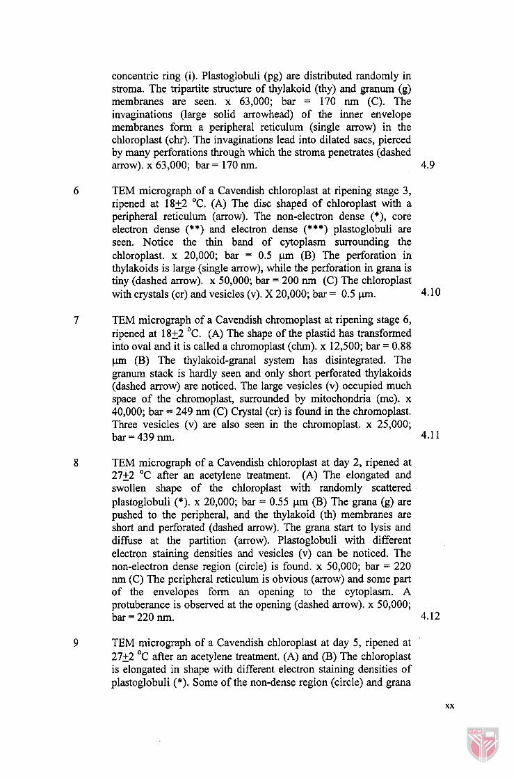
concentric ring (i). Plastoglobuli (pg) are distributed randomly in stroma. The tripartite structure of thylakoid (thy) and granurn (g) membranes are seen. x 63,000; bar = 170 nm (C). The invaginations (large solid arrowhead) of the inner envelope membranes form a peripheral reticulum (single arrow) in the chloroplast (chr). The invaginations lead into dilated sacs, pierced by many perforations through which the stroma penetrates (dashed arrow). x 63,000; bar = 170 nm. 4.9
TEM micrograph of a Cavendish chloroplast at ripening stage 3, ripened at 1822 OC. (A) The disc shaped of chloroplast with a peripheral reticulum (arrow). The non-electron dense (*), core electron dense (**) and electron dense (***) plastoglobuli are seen. Notice the thin band of cytoplasm surrounding the chloroplast. x 20,000; bar = 0.5 pm (B) The perforation in thylakoids is large (single arrow), while the perforation in grana is tiny (dashed arrow). x 50,000; bar = 200 nm (C) The chloroplast with crystals (cr) and vesicles (v). X 20,000; bar = 0.5 p. 4.10
TEM micrograph of a Cavendish chromoplast at ripening stage 6, ripened at 18+2 OC. (A) The shape of the plastid has transformed into oval and it is called a chromoplast (chm). x 12,500; bar = 0.88 pm (B) The thylakoid-granal system has disintegrated. The granum stack is hardly seen and only short perforated thylakoids (dashed arrow) are noticed. The large vesicles (v) occupied much space of the chromoplast, surrounded by mitochondria (mc). x 40,000; bar = 249 nrn (C) Crystal (cr) is found in the chromoplast. Three vesicles (v) are also seen in the chromoplast. x 25,000; bar = 439 nm. 4.1 1
TEM micrograph of a Cavendish chloroplast at day 2, ripened at 27+2 OC after an acetylene treatment. (A) The elongated and swollen shape of the chloroplast with randomly scattered plastoglobuli (*). x 20,000; bar = 0.55 pm (B) The grana (g) are pushed to the peripheral, and the thylakoid (th) membranes are short and perforated (dashed arrow). The grana start to lysis and diffuse at the partition (arrow). Plastoglobuli with different electron staining densities and vesicles (v) can be noticed. The non-electron dense region (circle) is found. x 50,000; bar = 220 nm (C) The peripheral reticulum is obvious (arrow) and some part of the envelopes form an opening to the cytoplasm. A protuberance is observed at the opening (dashed arrow). x 50,000; bar = 220 nm.
TEM micrograph of a Cavendish chloroplast at day 5, ripened at 2722 OC after an acetylene treatment. (A) and (B) The chloroplast is elongated in shape with different electron staining densities of plastoglobuli (*). Some of the non-dense region (circle) and grana
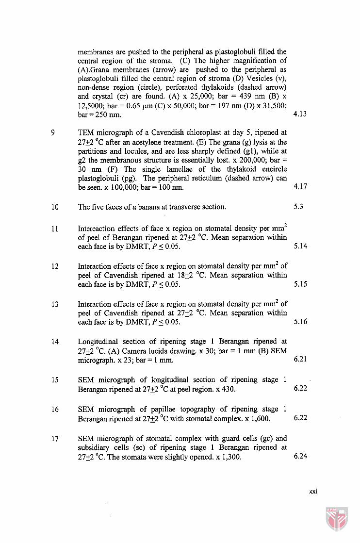
membranes are pushed to the peripheral as plastoglobuli filled the central region of the stroma. (C) The higher magnification of (A).Grana membranes (arrow) are pushed to the peripheral as plastoglobuli filled the central region of stroma (D) Vesicles (v), non-dense region (circle), perforated thylakoids (dashed arrow) and crystal (cr) are found. (A) x 25,000; bar = 439 nm (B) x 12,5000; bar = 0.65 pm (C) x 50,000; bar = 197 nm (D) x 3 1,500; bar = 250 nrn. 4.13
TEM micrograph of a Cavendish chloroplast at day 5, ripened at 2722 OC after an acetylene treatment. (E) The grana (g) lysis at the partitions and locules, and are less sharply defined (gl), while at g2 the membranous structure is essentially lost. x 200,000; bar =
30 nm (F) The single lamellae of the thylakoid encircle plastoglobuli (pg). The peripheral reticulum (dashed arrow) can be seen. x 100,000; bar = 100 nm. 4.17
The five faces of a banana at transverse section.
Intereaction effects of face x region on stomatal density per mm2 of peel of Berangan ripened at 2722 OC. Mean separation within each face is by DMRT, P 5 0.05. 5.14
Interaction effects of face x region on stomatal density per mm2 of peel of Cavendish ripened at 1822 OC. Mean separation within each face is by DMRT, P 5 0.05. 5.15
Interaction effects of face x region on stomatal density per mm2 of peel of Cavendish ripened at 2722 OC. Mean separation within each face is by DMRT, P 5 0.05.
Longitudinal section of ripening stage 1 Berangan ripened at 2722 OC. (A) Camera lucida drawing. x 30; bar = 1 mm (B) SEM micrograph. x 23; bar = 1 rnm. 6.2 1
SEM micrograph of longitudinal section of ripening stage 1 Berangan ripened at 2722 OC at peel region. x 430. 6.22
SEM micrograph of papillae topography of ripening stage 1 Berangan ripened at 2722 OC with stomata1 complex. x 1,600. 6.22
SEM micrograph of stomatal complex with guard cells (gc) and subsidiary cells (sc) of ripening stage 1 Berangan ripened at 2722 OC. The stomata were slightly opened. x 1,300. 6.24
xxi
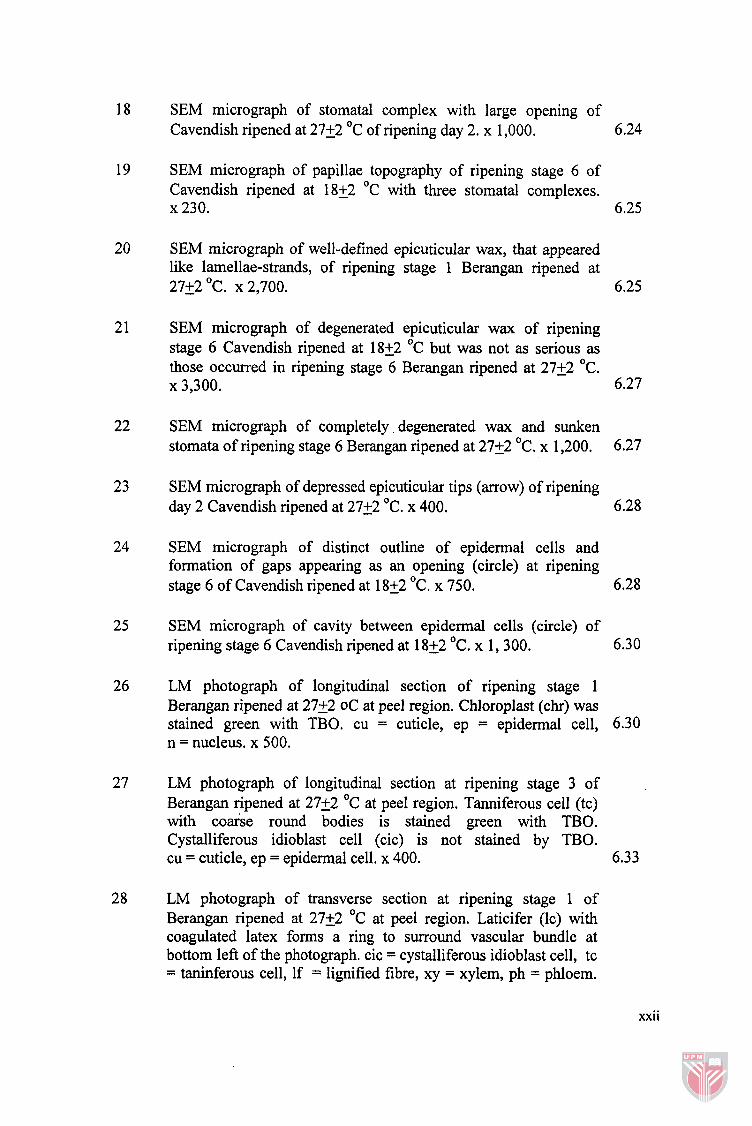
SEM micrograph of stomatal complex with large opening of Cavendish ripened at 2722 OC of ripening day 2. x 1,000.
SEM micrograph of papillae topography of ripening stage 6 of Cavendish ripened at 18+2 OC with three stomatal complexes. x 230. 6.25
SEM micrograph of well-defined epicuticular wax, that appeared like lamellae-strands, of ripening stage 1 Berangan ripened at 2722 OC. x 2,700. 6.25
SEM micrograph of degenerated epicuticular wax of ripening stage 6 Cavendish ripened at 1822 OC but was not as serious as those occurred in ripening stage 6 Berangan ripened at 2722 OC.
x 3,300. 6.27
SEM micrograph of completely degenerated wax and sunken stomata of ripening stage 6 Berangan ripened at 2722 OC. x 1,200. 6.27
SEM micrograph of depressed epicuticular tips (arrow) of ripening day 2 Cavendish ripened at 2722 OC. x 400. 6.28
SEM micrograph of distinct outline of epidermal cells and formation of gaps appearing as an opening (circle) at ripening stage 6 of Cavendish ripened at 1822 OC. x 750. 6.28
SEM micrograph of cavity between epidermal cells (circle) of ripening stage 6 Cavendish ripened at 1822 OC. x 1,300.
LM photograph of longitudinal section of ripening stage 1 Berangan ripened at 2722 oC at peel region. Chloroplast (chr) was stained green with TBO. cu = cuticle, ep = epidermal cell, 6.30 n = nucleus. x 500.
LM photograph of longitudinal section at ripening stage 3 of Berangan ripened at 2722 OC at peel region. Tanniferous cell (tc) with coarse round bodies is stained green with TBO. Cystalliferous idioblast cell (cic) is not stained by TBO. cu = cuticle, ep = epidermal cell. x 400. 6.33
LM photograph of transverse section at ripening stage 1 of Berangan ripened at 27+2 OC at peel region. Laticifer (lc) with coagulated latex forms a ring to surround vascular bundle at bottom left of the photograph. cic = cystalliferous idioblast cell, tc = taninferous cell, If = lignified fibre, xy = xylem, ph = phloem.
xxii
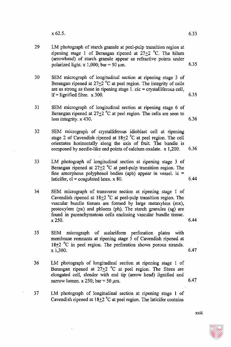
LM photograph of starch granule at peel-pulp transition region at ripening stage 1 of Berangan ripened at 2722 OC. The hilum (arrowhead) of starch granule appear as refractive points under polarized light. x 1,000; bar = 50 pm. 6.35
SEM micrograph of longitudinal section at ripening stage 3 of Berangan ripened at 27+2 OC at peel region. The integrity of cells are as strong as those in ripening stage 1. cic = crystalliferous cell, If = lignified fibre. x 300. 6.35
SEM micrograph of longitudinal section at ripening stage 6 of Berangan ripened at 2722 OC at peel region. The cells are seen to loss integrity. x 430. 6.36
SEM micrograph of crystalliferous idioblast cell at ripening stage 2 of Cavendish ripened at 1822 OC at peel region. The cell orientates horizontally along the axis of h i t . The bundle is composed by needle-like end points of calcium oxalate. x 1,200. 6.36
LM photograph of longitudinal section at ripening stage 3 of Berangan ripened at 27+2 OC at peel-pulp transition region. The fine amorphous polyphenol bodies (apb) appear in vessel. lc = laticifer, cl = coagulated latex. x 80. 6.44
SEM micrograph of transverse section at ripening stage 1 of Cavendish ripened at 1852 OC at peel-pulp transition region. The vascular bundle tissues are formed by large metaxylem (mx), protoxylem (px) and phloem (ph). The starch granules (sg) are found in parenchymatous cells enclosing vascular bundle tissue. x 250. 6.44
SEM micrograph of scalariform perforation plates with membrane remnants at ripening stage 5 of Cavendish ripened at 1822 OC in peel region. The perforation shows porous strands. x 1,300. 6.47
LM photograph of longitudinal section at ripening stage 1 of Berangan ripened at 2722 OC at peel region. The fibres are elongated cell, slender with end tip (arrow head) lignified and narrow lumen. x 250; bar = 50 p.m. 6.47
LM photograph of longitudinal section at ripening stage 1 of Cavendish ripened at 1822 OC at peel region. The laticifer contains
xxiii