respiratory diphtheria in three paediatric patients · diphtheria in three paediatric j k f...
TRANSCRIPT
Respiratory Patients
Diphtheria in Three Paediatric
J K F Lee*MRCP, S Huda*, M.Med, A Francis**, BSc, Aisai***, M. Med, M Jmioh****, MPH, *Paediatrics Unit, "*Microbiology Un.it, ***Anaesthesioiogy Unit, Hm;pitai Kuala Terengganu ****Pejabat Kesihatan Daerah, Kuala Terengganu
Diphtheria has been known to affect human beings for more than a hundred years. The introduction of diphtheria vaccination in the past fifty years has greatly reduced its incidence. Nevertheless, the disease has reemerged recently as epidemics in the independent states offormer Russia] from 1990 to 1995 and in Thailand2 in 1994. Vaccination against diphtheria was introduced in Malaysia in the 1960's. The last significant diphtheria outbreaks in Malaysia3 occurred in isolated unimmunised communities in Klang and Kuala Terengganu in 1988 and 1989. The incidence rate of reported diphtheria cases in Malaysia was less than 0.2 per 100,000 population since 1992. There were two last reported cases in Terengganu in 1990 and no reported case of diphtheria in Peninsular Malaysia from 1994 to
Med J Malaysia Vol 54 No 3 Sepf 1999
1997. However, we have recently managed three children with diphtheria who required artificial ventilation. Steps must be taken to contain the spread of this infection through the intensification of vaccination.
Pa~ient Rep©d
C{lI$1i'l N@, 1
Patient A.H. was a 4-year 9-month old boy who presented in August 1998 with a history of noisy breathing of two days' duration and severe breathlessness on the day of admission. His fever was said to be mild. He was not immunised for diphtheria before. On admission, he was drowsy but was still able to obey simple commands. Central cyanosis was present. His respiratory rate was 50 per minute with presence of
377
CASE REPORT
intercostal and subcostal recession. Air-entry over both lungs was markedly reduced. His perfusion was normal. The provisional diagnosis was acute epiglottitis. Laryngoscopic examination under general anaesthesia done three hours after admission revealed white plagues over both tonsils and an inflammed pharygeal wall. The epiglottis, supraglottis and vocal cords were normal. He required mechanical ventilation and was started on intravenous ampicillin. Chest radiograph revealed pneumonic opacities in right upper and left mid-zones.
On the fourth day of admission, the tonsil swab taken at admission grew Corynebacterium diphtheriae. A reexamination of his throat showed white membrane over both tonsils. His antibiotic was changed to intravenous penicillin and intravenous diphtheria antitoxin was given. His progress was satisfactory and he was ventilated for a total of eight days. Post-extubation chest radiograph showed mild secondary pneumonia.
With regards to his heart, his heart rate increased to 160 per minute on third day of admission. He was afebrile then. The serum lactate dehydrogenase LDH peaked at 1169u/L (normal range 240 to 480u/L) on third day of admission. He had muscle weakness and was only able to sit up at day 11 and walked at day 14 of admission. EMG and nerve conduction tests were not done. He was discharged home on day 22 of admission.
The throat swabs taken on admission and three days later grew Corynebacterium diphtheriae (identified by morphology and carbohydrate fermentation tests). This was confirmed and biotyped as mitis by Microbiology Division of Institute Medical Research, Kuala Lumpur and was toxigenic by the Elek test (toxin-antitoxin precipitation assay). Throat swabs were taken from 624 I
contacts and the results were as shown in Table 11. The positive cultures were biotypes mitis and gravis. The age range of these 13 positive-culture contacts was 10
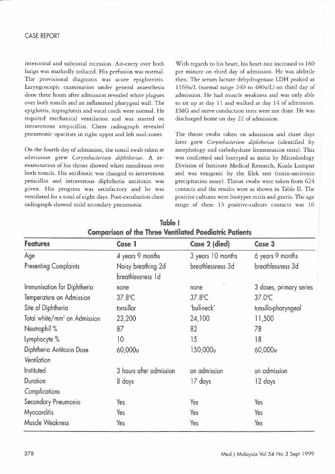
Table I Comparison of the Three Ventilated Paediatric Patients
Features Case 1 Case 2 (died) Case 3
Age 4 years 9 months 3 years 1 0 months 6 years 9 months Presenting Complaints Noisy breathing 2d breathlessness 3d breathlessness 3d
breathlessness 1 d Immunisation for Diphtheria none none 3 doses, primary series Temperature on Admission 37.8°C 37.8°C 37.0°C Site of Diphtheria tonsillar 'bull-neck' tons i lIo-pharyngea I Total white/mm3 on Admission 23,200 24,100 11,500 Neutrophil % 87 82 78 Lymphocyte % 10 15 18 Diphtheria Antitoxin Dose 60,000u 150,000u 60,000u Ventilation Instituted 3 hours after admission on admission on admission Duration 8 days 17 days 12 days Complications Secondary Pneumonia Yes Yes Yes Myocarditis Yes Yes Yes Muscle Weakness Yes Yes - Yes
378 Med J Malaysia Vol 54 No 3 Sept 1 999

months to 15 years and the average age was 7.2 years. All culture positive contacts were asymptomatic and they were isolated and given ten days of oral erythromycin together with DPT vaccination. They were discharged home when two throat cultures were negative for diphtheria.
Patient A.A. was a 3-year 10-month old girl who presented in October 1998 with a history of fever and breathlessness for three days. There was hoarseness of voice for two days and poor oral intake. She did not receIve any diphtheria-pertussis-tetanus DPT vaccination before. On admission, she was in severe respiratory distress, pale, cyanosed and restless. The respiratory rate was 40 per minute. Stridor was present with intercostal and subcostal recession. There was absent breath sounds bilaterally. The heart rate was 160 per minute and capillary refill was delayed more than two seconds. During intubation, white membrane was seen at the post-pharyngeal wall and soft palate. There was significant bilateral cervical lymphadenopathy resembling 'bull-neck diphtheria'. Chest radiograph showed collapse consolidation of the left lung and right Llpper lobe (Fig. 1). She was started on intravenous )enicillin and diphtheria antitoxin.
VIed J Malaysia Vol 54 No 3 Sept 1 999
RESPIRATORY DIPHTHERIA
There was evidence of myocarditis with persistent tachycardia of 150 to 160 per minute upto fifth day of admission with a peak serum LDH of 2047u/L on day 14 of admission. Pancuronium and midazolam infusion were stopped on 14th and 15th day of admission respectively. She was on endotracheal CPAP (continuous positive airway pressure) on the 18th day of admission. She looked alert and could follow simple instructions. She could open her mouth slightly and moved her fingers on command. However, there was no movement of her shoulders, elbows and whole of lower limbs and there was hardly any chest rise on inspiration. Her reflexes were diminished with down-going Babinski and these signs were suggestive of lower motor neuron weakness. This was followed by desaturation and cardiorespiratory failure 10 hours later and she was put back on positive pressure ventilation. The repeat chest radiograph showed severe lung infection. She finally succumbed to cardiogenic shock with third degree heart block on day 19 of admission.
The slough from membrane taken on admission grew Corynebacterium diphtheriae and this was confirmed as toxigenic biotype mitis. Throat swabs of 122 contacts were taken and the results are as shown in Table n. The two positive-culture contacts were 4 and 11 years old and their management was as described in case 1.
Patient W.A. was a 6-year 9-month old boy who presented in November 1998 with breathlessness, cough and poor oral intake for three days. He had noisy breathing and poor oral intake one day prior to admission. He had completed his primary series of three DPT vaccination in infancy. On examination, he was in respiratory distress with intercostal and subcostal recession. Air-entry on both lungs was diminished. He had normal perfusion. White membrane was present in posterior pharyngeal wall with no membrane over the enlarged tonsils. The impression was upper airway obstruction due to diphtheria. He was ventilated within one hour of admission. Chest radiograph showed mild haziness over left lower zone. He was started on intravenous penicillin and diphtheria antitoxin.
There was evidence of myocarditis on day 7 of admission. He developed pulseless supraventricular tachycardia requiring four doses of synchronised
379

CASE REPORT
cardioversion and finally responded to intravenous adenosine. His highest serum LDH was 1228uIL on the fifth day of admission.
He had a generalised tonic-clonic convulsion of uncertain origin on the day 11 of admission. He selfextubated while on low ventilator settings on the following day. Chest radiograph revealed secondary pneumonic patches and cefotaxime was continued. He was able to walk unaided twenty days after admission and was discharged home.
Culture from the slough of his throat membrane on admission grew Corynebacterium diphtheriae which was confirmed as toxigenic biotype mitis. Contact tracing was done and all nineteen throat cultures was negative for diphtheria.
Discussion
During the 4-month period from August to November 1998, three very ill patients presented with upper airway obstruction due to respiratory diphtheria. They required mechanical ventilation almost immediately after admission. The tonsillar and pharyngeal membrane due to diphtheria was characteristic although I the diagnosis of the first patient was delayed for 4 days due to lack of awareness of the disease. One death was due to cardiogenic shock with secondary pneumonia. The pathogen responsible for all three cases was toxigenic Corynebacterium diphtheriae biotype mitis. A summary of the clinical presentation of the three cases is shown in Table I. A previous lack of awareness of this disease
might have resulted in 'missed' cases in the past presenting as pneumonia or even sequelae of motor weakness or cardiomyopathy.
It was noted that the three cases were from different areas in the District of Kuala Terengganu. The staff from the Health Department of Kuala Terengganu District had done contact tracing for each of the three index cases. A total of fifteen throat cultures were positive for Corynebacterium diphtheriae out of a total of 765 throat cultures taken from the contacts. Thirteen of these positive cultures were from contacts of case one alone. Out of the fifteen positive cultures, two were toxigenic and three non-toxigenic biotype mitis and ten nontoxigenic biotype gravis. A summary of results is shown in Table n. Toxigenic and non-toxigenic Corynebacterium diphtheriae has been isolated regularly from patients in endemic areas4,5. However, it seems reassuring that in one study of 41 respiratory cases over a 16-year period\ there were no identified cases resulting from secondary transmission.
The possible causes of the occurrence,,2,4,6,7 of these three
serious cases included the importation of cases due to population movement across national boundaries. Secondly, the coverage of immunisation of children in the community may not be adequate. The third reason could be the lack of immunity in the adult population as the second and final booster of diphtheria vaccination in Malaysia is given at 7 years of age. This could result in the occurrence of asymptomatic carriers who are important in disease transmission to susceptible unimmunised children.
Table 11 Summary of Positive Cultures from Contacts of the Three Index Cases with
Toxigenic Corynebacterium Diphtheriae Biotype Mitis
Index Number of POSITIVE CULTURES Case Contacts Toxigenic Non-toxigenic Non-toxigenic Total Number Swabbed Mitis Mitis Gravis 1 624 1 3 9 13 2 122 1 0 1 2 3 19 0 0 0 0
Total 765 2 3 W 15
380 Med J Malaysia Vol 54 No 3 Sept 1999
In the outbreak in Saraburi, Thailand' in 1994 it was found that the carriage rates among household and school contacts were 4% and 8% respectively. Carriers may harbour the organism on the throat as well as on skin sores4•5•6. A cross-sectional prevalence study of throat and skin sores cultures for Corynebacterium diphtheriae could be done to determine the carrier rate. Toxigenic and non-toxigenic strains of diphtheria of biotypes mitis and gravis are possibly endemic in Kuala Terengganu district. If this is so, then booster doses of vaccination with a lower dose of diphtheria toxoid (as in the Td preparation) should perhaps be recommended every ten years to the adult population4•7 . Even though immunisation does not necessarily prevent asymptomatic carriage of the organism6 , it does protect against both severe disease and death4• Control strategies recommended by WHO in countries with high incidence rates included mass immunisation rates7 to achieve an uptake of95% in children and 90% in adults.
Even if the children of a country are highly immunised, the population could still be at risk of carriage and infection7 with non-toxigenic Corynebacterium diphtheriae from imported cases or carriers. This is the result of previous success in eradicating the organism from the community through high coverage vaccination during childhood. Thus the large majority of adult population would be unprotected in the absence of regular booster doses of diphtheria vaccination.
Therefore eradicating the disease from the community depends on improving the socio-economic status of the
Med J Malaysia Vol 54 No 3 Sept 1 999
RESPIRATORY DIPHTHERIA
community, high coverage of immunisation during childhood, and most probably regular booster doses for the adult population.
Three cases of respiratory diphtheria occurred over a 4-month period in 1998 in Kuala Terengganu District. They presented with upper airway obstruction and secondary pneumonia requiring artificial ventilation. Two of them did not have vaccination against diphtheria while the remaining one completed only the primary series. Cultures taken from the tonsillo-pharyngeal membrane of the three cases grew toxigenic Corynebacterium diphtheriae biotype mitis. Throat culture of contacts revealed a mixture of biotypes mitis and gravis. Reasons for this outbreak included the strong possibility of endemicity of diphtheria, inadequate coverage of immunisation in children and lack of immunity in the adult population. Immediate actions need to be taken including the intensification of vaccination in children and adult population in order to stop the spread of this disease.
We wish to thank the Director-General of Health for permission to publish this paper. Special thanks to Director of Health, Terengganu and the Microbiology Division ofInstitute of Medical Research, Kuala Lumpur.
381
CASE REPORT
1. Hardy, RB lan, Dittmann, Siehart, Sutter, W Roland. Current situation and control strategies for resurgence of diphtheria in newly independent states of the former Soviet Union. Lancet 1996; 347(9017): 1739-44.
2. Diphtheria outbreak - Saraburi Province, Thailand, 1994. MMWR 1996; 45: 271-73.
3. Disease Control Division, Department of Public Health, Ministry of Health, Malaysia. Incidence of Diphtheria in Malaysia from 1987 to 1997.
4. Bisgard, Kristine, Hardy, RB lain, Popovic, Tanja.
382
Respiratory diphteria in the United States, 1980 through 1995. Am] Public Health 1998; 88(5): 787-91.
5.
6.
7.
M Patel, F Morey, A Butcher, C Moore, R Brennan, L Mollison. The Frequent isolation of toxigenic and nontoxigenic Corynebacterium diphtheriae at Alice Springs Hospital. Communicable Diseases Intelligence 1994; 18(13): 310-11.
N Begg, V Balraj. Diphtheria: are we ready for it? Arch Dis Child 1995; 73: 568-72.
Gilbert, Kilman, Benn. diphtheriae changing
Infections with Cmynebacterittm epidemiology and clinical
manifestations; Diphtheria - the Australian perspective; National Health and Medical Research Council recommendations on diphtheria vaccination. Communicable Diseases Intelligence 1997; 21 (12): 161-65.
Med J Malaysia Vol 54 No 3 Sept 1999