norhidayu binti sahimin - core.ac.uk · daripada tiga kaedah tersebut, tindak balas polimer...
TRANSCRIPT
PARASITIC INFECTIONS AMONGST MIGRANT WORKERS IN MALAYSIA
NORHIDAYU BINTI SAHIMIN
FACULTY OF SCIENCE
UNIVERSITY OF MALAYA KUALA LUMPUR
2017
PARASITIC INFECTIONS AMONGST MIGRANT
WORKERS IN MALAYSIA
NORHIDAYU BINTI SAHIMIN
THESIS SUBMITTED IN FULFILMENT OF THE
REQUIREMENTS FOR THE DEGREE OF
DOCTOR OF PHILOSOPHY
FACULTY OF SCIENCE
UNIVERSITY OF MALAYA
KUALA LUMPUR
2017
ii
UNIVERSITY OF MALAYA
ORIGINAL LITERARY WORK DECLARATION
Name of Candidate: NORHIDAYU BINTI SAHIMIN (I.C No: )
Matric No: SHC130051
Name of Degree: DOCTOR OF PHILOSOPHY
Title of Project Paper/Research Report/Dissertation/Thesis (―this Work‖):
PARASITIC INFECTIONS AMONGST MIGRANT WORKERS IN MALAYSIA
Field of Study: PARASITOLOGY
I do solemnly and sincerely declare that:
(1) I am the sole author/writer of this Work;
(2) This Work is original;
(3) Any use of any work in which copyright exists was done by way of fair
dealing and for permitted purposes and any excerpt or extract from, or
reference to or reproduction of any copyright work has been disclosed
expressly and sufficiently and the title of the Work and its authorship have
been acknowledged in this Work;
(4) I do not have any actual knowledge nor do I ought reasonably to know that
the making of this work constitutes an infringement of any copyright work;
(5) I hereby assign all and every rights in the copyright to this Work to the
University of Malaya (―UM‖), who henceforth shall be owner of the
copyright in this Work and that any reproduction or use in any form or by any
means whatsoever is prohibited without the written consent of UM having
been first had and obtained;
(6) I am fully aware that if in the course of making this Work I have infringed
any copyright whether intentionally or otherwise, I may be subject to legal
action or any other action as may be determined by UM.
Candidate‘s Signature Date:
Subscribed and solemnly declared before,
Witness‘s Signature Date:
Name:
Designation:
iii
ABSTRACT
Sociodemographic background of 610 migrant workers employed in Malaysia was
collected via questionnaire to determine their parasitic health status. Six nationalities
were recruited with most workers from Indonesia (49.5%), followed by Bangladesh
(19.2%), Nepal (16.4%), India (10.5%), Myanmar (4.3%) and Vietnam (0.2%) and
employed in five working sectors namely; domestic service (24.3%), construction
(22.8%), food service (21.0%), plantation (16.7%) and manufacturing (15.2%). A total
of 388 individuals provided faecal samples for parasitic screening via microscopy. Four
nematode species (Ascaris lumbricoides, Trichuris trichiura Enterobius vermicularis,
and hookworms), one cestode (Hymenolepis nana) and three protozoan species
(Entamoeba histolytica/dispar, Giardia spp. and Cryptosporidium spp.) were recovered.
High prevalence of infections with Ascaris lumbricoides (43.3%) was recorded
followed by hookworms (13.1%) and E. histolytica/dispar (11.6%) with infections
significantly influenced by nationality, years of residence in Malaysia, employment
sector and education level. Toxoplasma gondii infections were screened serologically
from 484 workers with more than half of the workers were seropositive (57.4%) with
52.9% seropositive for anti-Toxoplasma IgG only, 0.8% seropositive for anti-
Toxoplasma IgM only and 3.7% seropositive with both IgG and IgM antibodies.
Samples positive for both IgG and IgM antibodies were further tested for IgG avidity
showed high avidity suggesting latent infection in 18 workers. Four significant factors
recorded namely; age, nationality, employment sector and length of residence in
Malaysia. Three diagnostic methods were tested and compared to detect Strongyloides
stercoralis infections in 306 migrant workers with 37.6% were seropositive. Subsequent
confirmation using a nested PCR showed successful amplification from three males
iv
(2.6%) with target amplicon of approximately 680bp. For the three methods, nested
PCR was the most sensitivity method in the detection for strongyloidiasis and should be
applied in future studies. PCR method was also applied to determine the species level
for four parasite‘s genus recovered in the population. Internal transcribed spacer 2 and
28S ribosomal RNA region of N. americanus and Ancylostoma spp. was successfully
amplified and resulted in A. duodenale reported for the first time in Malaysia. Nested
PCR targeting 16S-like ribosomal RNA gene successfully recovered E. dispar as the
most dominant infection among workers. Despite the low presence of E. histolytica in
the population, it still carries a public health risk. Amplification of the triosephosphate
isomerase (TPI) gene from G. duodenalis isolates successfully obtained the presence of
assemblage B and sub-assemblage AII suggesting the mode of transmission was human-
to-human. Based on the SSU rRNA gene, the C. parvum amplicons were successfully
detected in 9 human isolates.

v
ABSTRAK
Latar belakang sosiodemografi 610 pekerja asing yang bekerja di Malaysia telah
dikumpul melalui soal selidik untuk menentukan tahap kesihatan parasitik mereka.
Enam warganegara telah direkrut dengan majoriti pekerja dari Indonesia (49.5%),
diikuti oleh Bangladesh (19.2%), Nepal (16.4%), India (10.5%), Myanmar (4.3%) dan
Vietnam (0.2%) dan bekerja dalam lima sektor iaitu; perkhidmatan domestik (24.3%),
pembinaan (22.8%), perkhidmatan makanan (21.0%), perladangan (16.7%) dan
pembuatan (15.2%). Seramai 388 individu memulangkan sampel najis untuk
pemeriksaan parasit melalui mikroskop. Empat spesies nematoda (Ascaris lumbricoides,
Trichuris trichiura, Enterobius vermicularis dan cacing tambang), satu cestoda
(Hymenolepis nana) dan tiga spesies protozoa (Entamoeba histolytica / dispar, Giardia
spp. dan Cryptosporidium spp.) ditemui. Jangkitan parasit tertinggi dicatatkan oleh
Ascaris lumbricoides (43.3%), diikuti oleh cacing tambang (13.1%) dan E. histolytica /
dispar (11.6%) dan faktor jangkitan dipengaruhi oleh kewarganegaraan, jangka masa
menetap di Malaysia, sektor pekerjaan dan tahap pendidikan. Jangkitan Toxoplasma
gondii telah disaring secara serologi dari 484 pekerja dan lebih daripada separuh pekerja
adalah seropositif (57.4%) dengan 52.9% seropositif untuk anti-Toxoplasma IgG sahaja,
0.8% seropositif untuk anti-Toxoplasma IgM sahaja dan 3.7% seropositif dengan kedua-
dua antibodi IgG dan IgM. Sampel positif untuk kedua-dua antibodi IgG dan IgM
kemudiannya diuji dengan ujian IgG aviditi dan keputusan menunjukan aviditi tinggi
yang mencadangkan jangkitan terpendam dari 18 pekerja tersebut. Empat faktor penting
didapati iaitu; umur, kewarganegaraan, sektor pekerjaan dan jangka masa menetap di
Malaysia. Tiga kaedah diagnostik telah diuji dan dibandingkan untuk mengesan
jangkitan Strongyloides stercoralis di kalangan 306 pekerja asing dengan 37.6% adalah
vi
seropositif. Pengesahan berikutnya menggunakan tindak balas polimer berantai (PCR)
menunjukkan tiga lelaki (2.6%) dijangkiti parasit ini dengan sasaran amplicon kira-kira
680bp. Daripada tiga kaedah tersebut, tindak balas polimer berantai (PCR) adalah
kaedah paling sensitiviti dalam pengesanan jangkitan strongyloidiasis dan disyorkan
diguna pakai dalam kajian di masa hadapan. Tindak balas polimer berantai (PCR) juga
digunakan bagi menentukan spesies daripada empat genus parasit yang dijumpai di
dalam populasi. Internal transcribed spacer 2 dan 28S ribosomal RNA daripada N.
americanus dan Ancylostoma spp. telah berjaya dikesan dan jangkitan A. duodenale
dilaporkan buat kali pertama di Malaysia. Tindak balas polimer berantai menyasarkan
gen 16S-like ribosomal RNA berjaya mendapati E. dispar sebagai jangkitan yang paling
dominan di kalangan pekerja. Walaupun penemuan E. histolytica yang rendah di dalam
populasi, ia masih boleh membawa risiko kesihatan awam. Penguatan gen
triosephosphate isomerase (TPI) G. duodenalis berjaya menemui kehadiran himpunan B
dan sub-himpunan AII yang mencadangkan jangkitan dari manusia ke manusia.
Berdasarkan gen SSU rRNA, amplicon C. parvum telah berjaya dikesan daripada 9
pekerja asing.
vii
ACKNOWLEDGEMENTS
In the name of Allah, Most Gracious, Most Merciful. Without Him, nothing is
possible.
I am deeply grateful to both of my supervisors, Associate Professor Dr. Siti
Nursheena Mohd Zain from Institute of Biological Science, Faculty of Science,
University of Malaya and Professor Dr. Yvonne Lim Ai Lian from Department of
Parasitology, Faculty of Medicine, University of Malaya for their support, guidance and
encouragement throughout this project.
I am indebted to Professor John Lewis from Royal Holloway, University of London,
Professor Jerzy Behnke from School of Biology, University of Nottingham, Professor
Datuk Dr. Khairul Anuar Abdullah from Mahsa University, Professor Dr Rahmah
Noordin from Institute for Research in Molecular Medicine, University of Science
Malaysia and Dr Farnaza Ariffin from Faculty of Medicine, MARA Technology
University for their guidance and support.
Special thanks to all nurses and medical officer from University Malaya Medical
Centre (UMMC) and Hospital Universiti Kebangsaan Malaysia (HUKM) for providing
help and technical assistant, the Ministry of Health, Malaysia and all collaborators from
companies and recruiting agencies in Malaysia for their support to this study.
Last but not least, I thank my family especially my husband, beloved daughter and
son, my parents, siblings and friends for their support and encouragement. I would not
be able to complete this journey without all of you.
viii
TABLE OF CONTENTS
ABSTRACT………………………………………………………………….........
ABSTRAK………………………………………………………………………...
ACKNOWLEDGEMENTS……………………………………………………….
TABLE OF CONTENTS………………………………………………………….
LIST OF FIGURES……………………………………………………………….
LIST OF TABLES………………………………………………………………...
LIST OF SYMBOLS AND ABBREVIATIONS…………………………………
LIST OF APPENDICES…………………………………………………………..
CHAPTER 1: GENERAL INTRODUCTION
1.1 Malaysia……………………………………………………………………….
1.1.1 Economy status of the ASEAN region………………………………….
1.2 Migrant workers in Malaysia………………………………………………….
1.2.1 Migrant workers…………………………………………………………
1.2.2 Migrant workers in Malaysia……………………………………………
1.2.3 Statistic of migrant workers……………………………………………..
1.3 Health status of migrant workers……………………………………………...
1.3.1 Medical procedure of workers upon entry………………………………
1.3.2 Common health problems……………………………………………….
1.4 Common parasitic infections in migrant workers……………………………..
1.4.1 Helminthes………………………………………………………………
1.4.1.1 Ascaris lumbricoides………………………………………………
1.4.1.2 Hookworm………………………………………………………...
1.4.1.3 Trichuris trichiura………………………………………………...
iii
v
vii
viii
xiv
xviii
xxiv
xxvi
1
1
2
7
7
7
9
13
13
16
19
20
21
23
25
ix
1.4.1.4 Strongyloides stercoralis………………………………………….
1.4.1.5 Enterobius vermicularis…………………………………………...
1.4.1.6 Hymenolepis nana…………………………………………………
1.4.2 Protozoa…………………………………………………………………
1.4.2.1 Entamoeba spp. …………………………………………………...
1.4.2.2 Giardia sp. ………………………………………………………..
1.4.2.3 Cryptosporidium spp………………………………………………
1.4.2.4 Toxoplasma gondii………………………………………………...
1.5 Studies on the status of parasitic infections amongst migrant workers……….
1.5.1 Studies in Asia ………………………………………………………….
1.5.2 Migrant health status studies in Malaysia……………………………….
1.6 Justification of the study………………………………………………………
1.7 Objectives……………………………………………………………………..
CHAPTER 2: MIGRANT WORKERS IN MALAYSIA: SOCIO-
DEMOGRAPHY BACKGROUND
2.1 Introduction……………………………………………………………………..
2.2 Materials and methods………………………………………………………….
2.2.1 Subjects…………………………………………………………………...
2.2.2 Questionnaire……………………………………………………………..
2.2.3 Ethical considerations…………………………………………………….
2.2.4 Data analysis……………………………………………………………...
2.3 Results…………………………………………………………………………..
2.3.1 Socio-demographic profile………………………………………………...
2.3.2 Migration history…………………………………………………………..
2.3.3 Environmental health………………………………………………………
27
29
31
33
34
36
38
40
43
43
46
47
48
49
49
51
51
51
52
52
53
53
56
58
x
2.3.4 Life-style habits……………………………………………………………
2.3.5 Medical history and recent illness…………………………………………
2.3.6 Occupational Health and Safety…………………………………………...
2.4 Discussion………………………………………………………………………
2.5 Conclusion………………………………………………………………………
CHAPTER 3: CURRENT IMPLICATIONS OF SOCIO-DEMOGRAPHIC
AND ENVIRONMENTAL CHARACTERISTICS IN THE
TRANSMISSION OF INTESTINAL PARASITIC INFECTIONS (IPIs)
3.1 Introduction……………………………………………………………………..
3.2 Materials and Methods………………………………………………………….
3.2.1 Subjects and questionnaire………………………………………………...
3.2.2 Collection and analysis of faecal samples…………………………………
3.2.3 Statistical analysis………………………………………………………….
3.3 Results…………………………………………………………………………..
3.3.1 Socio-demographic characteristics………………………………………...
3.3.2 Prevalence of intestinal parasitic infections (IPIs)………………………...
3.3.3 Intrinsic effects on prevalence of IPIs……………………………………..
3.3.3.1 Higher taxa………………………………………………………….
3.3.3.2 Individual helminth species…………………………………………
3.3.3.3 Individual protozoan species………………………………………..
3.3.4 Extrinsic (environmental) effects on intestinal parasitic infections……….
3.3.4.1 Higher taxa………………………………………………………….
3.3.4.2 Individual helminth species…………………………………………
3.3.4.3 Individual protozoan species………………………………………..
3.4 Discussion………………………………………………………………………
59
60
62
63
67
68
68
71
71
71
72
74
74
74
75
75
76
78
78
78
81
82
93
xi
3.5 Conclusion………………………………………………………………………
CHAPTER 4: SEROPREVALENCE OF Toxoplasma gondii INFECTIONS
AMONG MIGRANT WORKERS IN MALAYSIA
4.1 Introduction……………………………………………………………………..
4.2 Materials and methods………………………………………………………….
4.2.1 Study population and sample collection…………………………………...
4.2.2 Detection of immunoglobulin G and M antibodies to T. gondii…………..
4.2.3 Statistical analysis…………………………………………………………
4.3 Results………………………………………………………………………….
4.3.1 Sociodemographic characteristics…………………………………………
4.3.2 Seroprevalence of T. gondii………………………………………………..
4.3.2.1 Intrinsic effects on seroprevalence of IgG and IgM antibodies to
T. gondii infections……………………………………………….
4.3.2.2 Extrinsic effects on seroprevalence of IgG and IgM antibodies to
T. gondii infections………………………………………………
4.4 Discussion………………………………………………………………………
4.5 Conclusion………………………………………………………………………
CHAPTER 5: SEROPREVALENCE OF Strongyloides stercoralis
INFECTIONS AMONG MIGRANT WORKERS IN MALAYSIA
5.1 Introduction……………………………………………………………………..
5.2 Materials and methods………………………………………………………….
5.2.1 Study population and sample collection…………………………………...
5.2.2 Detection of immunoglobulin G to Strongyloides stercoralis infection…..
5.2.3 Statistical Analysis………………………………………………………...
97
98
98
101
101
101
104
106
106
106
107
108
111
115
116
116
119
119
119
121
xii
5.3 Results…………………………………………………………………………..
5.3.1 Socio-demographic characteristics………………………………………...
5.3.2 Seroprevalence of strongyloidiasis and seropositivity of S. stercoralis
infection……………………………………………………………………
5.4 Discussion………………………………………………………………………
5.5 Conclusion………………………………………………………………………
CHAPTER 6: MOLECULAR CHARACTERIZATION OF HUMAN
INTESTINAL PARASITE INFECTIONS
6.1 Introduction……………………………………………………………………..
6.2 Materials and methods…………………………………………………………
6.2.1 Samples collection…………………………………………………………
6.2.2 Extraction of genomic DNA……………………………………………….
6.2.3 Nested polymerase chain reaction (nested PCR)………………………….
6.2.3.1 Strongyloides stercoralis……………………………………………
6.2.3.2 Hookworm…………………………………………………………..
6.2.3.3 Entamoeba spp. …………………………………………………….
6.2.3.4 Giardia spp. ………………………………………………………...
6.2.3.5 Cryptosporidium spp………………………………………………..
6.2.4 Purification of PCR product……………………………………………….
6.2.5 DNA Sequencing…………………………………………………………..
6.2.5.1 Strongyloides stercoralis……………………………………………
6.2.5.2 Hookworm…………………………………………………………..
6.2.5.3 Entamoeba spp. …………………………………………………….
6.2.5.4 Giardia spp. ………………………………………………………...
6.2.6 Sequencing analysis……………………………………………………….
122
122
122
124
129
130
130
133
133
133
135
135
136
137
138
139
139
140
140
141
141
141
142
xiii
6.2.7 RFLP (Restriction Fragment Length Polymorphism)……………………..
6.2.8 Summary of methodology…………………………………………………
6.3 Results…………………………………………………………………………..
6.3.1 Strongyloides stercoralis…………………………………………………..
6.3.2 Hookworm…………………………………………………………………
6.3.3 Entamoeba spp…………………………………………………………….
6.3.4 Giardia spp………………………………………………………………...
6.3.5 Cryptosporidium spp. …………………………………………………….
6.4 Discussion………………………………………………………………………
6.4.1 Strongyloides stercoralis…………………………………………………..
6.4.2 Hookworm…………………………………………………………………
6.4.3 Entamoeba spp…………………………………………………………….
6.4.4 Giardia spp………………………………………………………………...
6.4.5 Cryptosporidium spp. ……………………………………………………..
6.5 Conclusion………………………………………………………………………
CHAPTER 7: GENERAL DISCUSSION AND CONCLUSION
7.1 General discussion……………………………………………………………...
7.2 Conclusion………………………………………………………………………
REFERENCES……………………………………………………………………..
LIST OF PUBLICATIONS AND PAPERS PRESENTED………………………..
APPENDICES………………………………………………………………………
142
143
144
144
145
149
151
154
156
156
157
159
161
163
166
168
168
179
183
222
224
xiv
LIST OF FIGURES
Figure 1.1 : Malaysia and the neighbouring countries (Southeast Asia and
South Asia countries; Thailand, Indonesia, The Philippines,
Vietnam, Laos, Myanmar, India, Nepal, and Bangladesh)
(Source: WorldAtlas.com, 2016)……………...
Figure 1.2 : Medical screening processes of foreign workers in Malaysia.
Source: FOMEMA (2015)…………………………………..
Figure 1.3 : Prevalence of communicable and non-communicable
diseases among foreign workers in 2012. Source: Disease
Control Division, Ministry of Health (2012)………………..
Figure 1.4 : Five most common communicable diseases among foreign
workers in 2012. Source: Disease Control Division,
Ministry of Health (2012)…………………………………...
Figure 1.5 : Five most common non-communicable diseases among
foreign workers in 2012. Source: Disease Control Division,
Ministry of Health (2012)…………………………………...
Figure 1.6 : Worldwide distribution of soil-transmitted helminth survey
data. Source: Global Atlas of Helminth Infections (2016)….
Figure 1.7 : Life cycle of Ascaris lumbricoides (Source: DPDx, 2013)….
2
14
17
18
18
21
23
xv
Figure 1.8 : Life cycle of the hookworms (Source: DPDx, 2013)……….
Figure 1.9 : Life cycle of Trichuris trichiura (Source: DPDx, 2013)……
Figure 1.10: Life cycle of Strongyloides stercoralis (Source: DPDx, 2015)
Figure 1.11: Life cycle of Enterobius vermicularis (Source: DPDx, 2013)
Figure 1.12: Life cycle of Hymenolepis nana (Source: DPDx, 2013)….
Figure 1.13: Life cycle of Entamoeba spp. (Source: DPDx, 2013)……….
Figure 1.14: Life cycle of Giardia sp. (Source: DPDx, 2013)……………
Figure 1.15: Life cycle of Cryptosporodium spp. (Source: DPDx, 2013)...
Figure 1.16: Life cycle of Toxoplasma gondii. (Source: DPDx, 2015)…...
Figure 2.1 : Education profile of migrant workers according to working
sectors……………………………………………………......
Figure 2.2 : Worker‘s country origin profile in relation to working sectors
Figure 2.3 : Home setting distributions in country origin of migrant
workers………………………………………………………
25
26
28
30
33
36
38
40
42
55
55
57
xvi
Figure 2.4 : Distribution of migrant workers according to length of stay
in Malaysia ………………………………………………….
Figure 2.5 : Percentage of usage of personal protective equipment (PPE)
among migrant workers……………………………………...
Figure 3.1 : Ascaris lumbricoides………………………………………...
Figure 3.2 : Hookworm………………………………………………......
Figure 3.3 : Trichuris trichiura…………………………………………...
Figure 3.4 : Enterobius vermicularis……………………………………..
Figure 3.5 : Hymenolepis nana…………………………………………...
Figure 3.6 : Entamoeba sp………………………………………………..
Figure 3.7 : Giardia sp…………………………………………………
Figure 3.8 : Cryptosporidium sp…………………………………….....
Figure 3.9 : Prevalence of combined protozoan infections in the host
population in relation to levels of education and types of
residences……………………………………………………
57
62
84
84
84
84
85
85
85
85
89
xvii
Figure 3.10: Prevalence of Entamoeba in relation to the host-sex and
levels of education…………………………………………..
Figure 6.1 : Summary of molecular characterization procedure of S.
stercoralis, hookworm, Entamoeba spp., Giardia sp. and
Cryptosporidium spp………………………………………..
Figure 6.2 : A phylogenetic tree based on partial ITS2 sequences of
hookworm species constructed using MEGA6 program……
Figure 6.3 : Phylogenetic relationship of Giardia sp. by neighbor-joining
analysis of the triosephosphate isomerase (tpi) nucleotide
sequences………………………………………....................
Figure 6.4 : Nested PCR product of Cryptosporidium spp………………
Figure 6.5 : PCR-RFLP of Cryptosporidium SSUrRNA gene. Secondary
PCR product was digested by VSPI restriction enzyme……..
89
143
147
152
155
155
xviii
LIST OF TABLES
Table 1.1 : Socio-demographic and economic status between Malaysia
and neighbouring countries (Indonesia, India, Bangladesh,
Nepal, Myanmar and Vietnam). Source: The World
Factbook – Central Intelligence Agency (2016)…………….
Table 1.2 : Number of migrant workers employed in Malaysia
according to country of origin (2000-2014). Source:
Temporary Work Visit Pass (PLKS), Immigration
Department: Ministry of Home Affairs (Ministry of Human
Resources, 2015)…………………………………………….
Table 1.3 : Employment distributions of migrant workers according to
working sectors (2000-2014). Source: Temporary Work
Visit Pass (PLKS), Immigration Department: Ministry of
Home Affairs (Ministry of Human Resources, 2015)............
Table 1.4 : Regulatory agencies of migrant workers according to
working sectors in Malaysia. Source: Ministry of Human
Resources (2015)……………………………………………
Table 1.5 : Categories of medical examination as stipulated by Ministry
of Health. Source: FOMEMA (2015)……………………….
4
10
11
12
15
xix
Table 1.6 : Number of workers went for FOMEMA screening
according to country of origin in 2012. Source: Disease
Control Division, Ministry of Health (2012)………………..
Table 1.7 : Geographic distributions of common human intestinal
parasitic infections. Source: Godue & Gyorkos, 1990;
Stauffer et al., 2002; Cook & Zumla, 2003……………........
Table 1.8 : Scientific classification of Ascaris lumbricoides……………
Table 1.9 : Scientific classification of the hookworm…………………...
Table 1.10: Scientific classification of Trichuris trichiura………………
Table 1.11: Scientific classification of Strongyloides stercoralis………..
Table 1.12: Scientific classification of Enterobius vermicularis…...........
Table 1.13: Scientific classification of Hymenolepis nana………………
Table 1.14: Scientific classification of Entamoeba spp………………….
Table 1.15: Scientific classification of Giardia sp……………………….
Table 1.16: Scientific classification of Cryptosporidium spp…………….
17
19
22
24
26
28
30
32
35
37
39
xx
Table 1.17: Scientific classification of Toxoplasma gondii………………
Table 2.1 : Demographic profile of migrant workers according to sex,
age, working sector, nationality, educational level, religion
and marital status. (N=610)…………………………………
Table 2.2 : Types of accommodation provided and living companion of
migrant workers in Malaysia………………………………...
Table 2.3 : Risk behavior lifestyle profiles of migrant workers……….
Table 2.4 : Migrant workers access to medical treatment and mode of
payment………………………………………………….......
Table 2.5 : Migrant workers with symptoms of parasitic infection in the
past year……………………………………………………..
Table 2.6 : Number of migrant worker had given occupational health &
safety briefing and provision with personal protective
equipment (PPE)……………………………………………
Table 3.1 : Species of intestinal parasitic infections recovered from
migrant workers in Peninsular Malaysia…………………….
41
54
59
60
61
61
62
86
xxi
Table 3.2 : Multiplicity of intestinal parasitic infections amongst
migrant workers infected………………………………….....
Table 3.3 : Prevalence of intestinal parasitic infections amongst migrant
workers according to nationality, employment sector,
education, accommodation type and years of residence in
Malaysia……………………………………………………..
Table 3.4 : Prevalence of individual helminth species amongst migrant
workers according to nationality, employment sector,
education, accommodation type and years of residence in
Malaysia…………………………………………………......
Table 3.5 : Prevalence of individual protozoan species amongst migrant
workers according to nationality, employment sector,
education, accommodation type and years of residence in
Malaysia……………………………………………………
Table 4.1 : Seroprevalence of IgG and IgM antibodies to T. gondii
among 484 migrant workers using ELISA: CI=95%-
confidence intervals……………………………………........
Table 4.2 : Seroprevalence of IgG and IgM antibodies to T. gondii
infections among migrant workers in Malaysia according to
sex, age, sex, nationality, employment sector, years of
residence, accommodation and education…………………..
87
90
91
92
108
109
xxii
Table 5.1 : Prevalence of S. stercoralis infection in relation to socio-
demographic characteristics (sex, age, nationality,
employment sector, years of residence, accommodation and
education)……………………………………………………
Table 6.1 : Detection of Strongyloides stercoralis by microscopy
examination, ELISA and nested PCR. (N=306)…………….
Table 6.2 : The prevalence of N. americanus and A. duodenale
infections among migrant workers in Malaysia relative to
factors such as sex, age, country of origin, working sector
and years of residence in Malaysia………………………….
Table 6.3 : The prevalence of Entamoeba dispar and E. histolytica
among migrant workers in Malaysia relative to factors such
as sex, age, country of origin, working sector and years of
residence in Malaysia. ………………………………………
Table 6.4 : The infections of G. duodenalis assemblages among migrant
workers in Malaysia relative to factors such as sex, age,
country of origin, working sector and years of residence in
Malaysia……………………………………………………
Table 7.1 : Historical timeline of parasitic infections studies among
migrants in Malaysia……………………………………….
123
144
148
150
153
173
xxiii
Table 7.2 : Participants employment according to employment sectors
in Malaysia……………………………………………..........
Table 7.3 : Prevalence of helminth infection among migrant workers in
Malaysia in relation to factors; sex, age, nationality,
employment sector and years of residence…………………
Table 7.4 : Prevalence of protozoan infection among migrant workers
in Malaysia relation to factors; sex, age, nationality,
employment sector and years of residence……………….....
174
175
177
xxiv
LIST OF SYMBOLS AND ABBREVIATIONS
n : sample size
% : percentage
µg/µl : microgram per microliter
µl : microliter
°C : degree Celsius
bp : base pair
CI : confidence interval
CL : confidence limit
cm : centimeter
dH2O : distilled water
DNA : deoxyribonucleic acid
dNTP : deoxyribonucleoside triphosphate
EDTA : ethylenediaminetetraacetic acid
g : gram
GE : gel extraction
IgG : immunoglobulin G
IgM : immunoglobulin M
ITS : Internal Transcribed Spacer
IPIs : Intestinal parasitic infections
min : minute
mL : milliliter
mM : miliMolar
mm : millimeter
ng : nanogram
xxv
nm : nanometer
PCR : Polymerase Chain Reaction
rDNA : ribosomal DNA
RFLP : Restriction Fragment Length Polymorphism
Taq : Thermus aquaticus
TAE : Tris Acetic acid EDTA
U : Unit
UV : Ultraviolet
V : Volt
w/v : weight/volume
x : Times
Χ2
:
chi-square
xxvi
LIST OF APPENDICES
APPENDIX A: Foreign worker‘s medical examination registration form
APPENDIX B: Consent form…………………………………………...
APPENDIX C: Questionnaires………………………………………….
APPENDIX D: Published Paper I: Sahimin, N., Yvonne A.L. Lim,
Ariffin, F., Behnke, J.M., Lewis, J.W. & Mohd Zain,
S.N. (2016). Migrant Workers in Malaysia: Current
Implications of Sociodemographic and Environmental
Characteristics in the Transmission of Intestinal
Parasitic Infections. PLoS Neglected Tropical Diseases,
10(11): e0005110. doi:10.1371/journal.pntd.0005110.
(ISI-Cited Publication)…………………………………
224
225
229
245
1
CHAPTER 1: GENERAL INTRODUCTION
1.1 Malaysia
Malaysia comprises of Peninsular Malaysia, Sabah and Sarawak. It is situated in
Southeastern Asia, with the peninsula bordering Thailand in the north and Singapore in
the south, and one-third of the island of Borneo, which borders Indonesia and Brunei.
The location is very strategic along the Strait of Malacca and southern South China Sea.
The total population is approximately 30.5 million with Malays (50.1%) as the
dominant ethnic group, followed by Chinese (22.6%), indigenous (11.8%), Indian
(6.7%) and others (0.7%) (Central Intelligence Agency, 2016).
The standard of living in this country is better compared the neighbouring countries
in the region. A total of 74.7% of the population has undergone urbanization with a
change rate of 2.66% annually (2010 – 2015) (Central Intelligence Agency, 2016). The
population is made up in major urban cities such as the capital, Kuala Lumpur and Johor
Bahru is approximately 6.8 million and 912,000, respectively with a majority having
access to good sanitation (96%) and clean drinking water (98.2%).
2
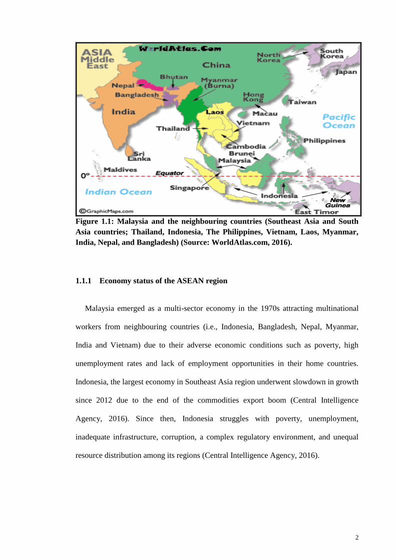
Figure 1.1: Malaysia and the neighbouring countries (Southeast Asia and South
Asia countries; Thailand, Indonesia, The Philippines, Vietnam, Laos, Myanmar,
India, Nepal, and Bangladesh) (Source: WorldAtlas.com, 2016).
1.1.1 Economy status of the ASEAN region
Malaysia emerged as a multi-sector economy in the 1970s attracting multinational
workers from neighbouring countries (i.e., Indonesia, Bangladesh, Nepal, Myanmar,
India and Vietnam) due to their adverse economic conditions such as poverty, high
unemployment rates and lack of employment opportunities in their home countries.
Indonesia, the largest economy in Southeast Asia region underwent slowdown in growth
since 2012 due to the end of the commodities export boom (Central Intelligence
Agency, 2016). Since then, Indonesia struggles with poverty, unemployment,
inadequate infrastructure, corruption, a complex regulatory environment, and unequal
resource distribution among its regions (Central Intelligence Agency, 2016).
3
Bangladesh is an underdeveloped country however, since 1996, the economy grew
roughly 6% per year despite political instability, poor infrastructure, corruption,
insufficient power supplies, slow implementation of economic reforms, global financial
crisis and recession to becoming a country showing improvements in development
(Central Intelligence Agency, 2016).
Nepal is among the poorest and least developed country in the world, with about one-
quarter of its population living below the poverty line. Agriculture is the mainstay of the
economy, providing a livelihood for almost 70% of the population and accounting for
about one-third of the gross domestic product (GDP). Industrial activity mainly involves
the processing of agricultural products, including pulses, jute, sugarcane, tobacco and
grain (Central Intelligence Agency, 2016).
Socio and economic status between Malaysia and the neighbouring countries is
described in Table 1.1. The data records Malaysia as one of the highest standard of
living and stable economy compared with other neighbouring countries.
4
Table 1.1: Socio-demographic and economic status between Malaysia and neighbouring countries (Indonesia, India, Bangladesh, Nepal,
Myanmar and Vietnam).
Malaysia Indonesia India Bangladesh Nepal Myanmar Vietnam
Population
(July 2015 est.)
30,513,848 255,993,674 1,251,695,584 168,957,745 31,551,305 56,320,206 94,348,835
Population
growth rate
(2015 est.)
1.44% 0.92% 1.22% 1.6% 1.79% 1.01% 0.97%
Net migration
rate (2015 est.)
-0.33
migrant(s)/1,000
population
-1.16
migrant(s)/1,000
population
-0.04
migrant(s)/1,000
population
0.46
migrant(s)/1,000
population
3.86
migrant(s)/1,000
population
-0.28
migrant(s)/1,000
population
-0.3
migrant(s)/1,000
population
Urbanization
74.7% 53.7% 32.7% 34.3% 18.6% 34.1% 33.6%
Sanitation
facility access
96% 60.8% 39.6% 60.6% 45.8% 77.4% 78%
Drinking water
resources
98.2% 87.4% 94.1% 86.9% 91.6% 80.6% 97.6%
GDP – real
growth rate
(2015 est.)
4.7% 4.7% 7.3% 6.5% 3.4% 8.5% 6.5%
GDP by sector
(2015 est.)
Agric.: 8.9%
industry: 35%
services:56.1%
Agric.: 13.6%
industry: 42.8%
services: 43.6%
Agric.: 16.1%
industry: 29.5%
services: 54.4%
Agric.: 16%
industry: 30.4%
services: 53.6%
Agric.: 31.7%
industry: 15.1%
services: 53.2%
Agric.: 36.1%
industry: 22.3%
services: 41.6%
Agric.: 17.4%
industry: 38.8%
services: 43.7%
5
Malaysia Indonesia India Bangladesh Nepal Myanmar Vietnam
Industrial
production
growth rate
(2015 est.)
5.5% 4.5% 2.8% 9.4% 2.6% 12.2% 7.5%
Labor force
(2015 est.)
14.3 million 122.4 million 502.1 million 81.95 million 15.2 million 36.18 million 54.93 million
Labor force by
sector
Agric.: 11%
industry: 36%
services: 53%
(2012 est.)
Agric.: 38.9%
industry: 13.2%
services: 47.9%
(2012 est.)
Agric.: 49%
industry: 20%
services: 31%
(2012 est.)
Agric.: 47%
industry: 13%
services: 40%
(2010 est.)
Agric.: 69%
industry: 12%
services: 19%
(2014 est.)
Agric.: 70%
industry: 7%
services: 23%
(2001 est.)
Agric.: 48%
industry: 21%
services: 31%
(2012 est.)
Unemployment
rate (2015 est.)
2.7% 5.5% 7.1% 4.9% 46%
(2008 est.)
5% 3%
Population
below poverty
line
3.8%
(2009 est.)
11.3%
(2014 est.)
29.8%
(2010 est.)
31.5%
(2010 est.)
25.2%
(2011 est.)
32.7%
(2007 est.)
11.3%
(2012 est.)
Household
income:
Lowest 10%
Highest 10%
1.8%
34.7%
(2009 est.)
3.4%
28.2%
(2010 est.)
3.6%
31.1%
(2005 est.)
4%
27%
(2010 est.)
3.2%
29.5%
(2011 est.)
2.8%
32.4%
(1998 est.)
3.2%
30.2%
(2008 est.)
Inflation rate
(2015 est.)
2.1% 6.7% 5.6% 5.7% 7.2% 9.2% 0.9%
6
Malaysia Indonesia India Bangladesh Nepal Myanmar Vietnam
Industries Rubber & oil
palm processing
& manufacturing,
petroleum &
natural gas, light
manufacturing,
pharmaceutical,
medical
technology,
electronics &
semiconductor,
timber
processing;
logging and
agriculture
processing.
Petroleum &
natural gas,
textiles,
automotive,
electrical
appliances,
apparel,
footwear,
mining, cement,
medical
instruments &
appliances,
handicrafts,
chemical
fertilizers,
plywood,
rubber,
processed food,
jewelry and
tourism
Textiles,
chemicals, food
processing, steel,
transportation
equipment,
cement, mining,
petroleum,
machinery,
software and
pharmaceuticals.
Jute, cotton,
garments, paper,
leather, fertilizer,
iron & steel,
cement, petroleum
products, tobacco,
pharmaceuticals,
ceramics, tea, salt,
sugar, edible oils,
soap & detergent,
fabricated metal
products,
electricity and
natural gas.
Tourism,
carpets, textiles,
small rice, jute,
sugar, oil seed
mills, cigarettes,
cement and brick
production.
Agricultural
processing; wood &
wood products,
copper, tin, tungsten,
iron, cement,
construction
materials,
pharmaceuticals,
fertilizer, oil &
natural gas,
garments, jade and
gems.
Food
processing,
garments, shoes,
machine-
building, mining,
coal, steel,
cement, chemical
fertilizer, glass,
tires, oil and
mobile phones.
Source: The World Factbook – Central Intelligence Agency (2016).
7
1.2 Migrant workers in Malaysia
1.2.1 Migrant workers
According to the International Convention on the Rights of Migrants Workers and
Members of Their Families 1990, "migrant worker" refers to a person engaged, or
previously engaged in a remunerated activity in a State of which he or she is not a
national. The latest statistic shows an estimated 232 million migrant workers around the
world. According to International Labour Organization (ILO, 2015), factors such as
globalization, demographic shifts, conflicts, income inequalities and climate change are
the push factor for workers and their families to cross borders in the search of
employment and security. Migrant workers contribute to growth and development in the
country of destination, while the country origin greatly benefit from their remittances
and skills acquired during employment.
1.2.2 Migrant workers in Malaysia
The robust growth of Malaysia‘s economy led to the high demand of workforce from
small to large-scale enterprises. This in turn, created an influx for workers to fulfill the
work demand. It is a practice in this country that potential employers are obligated to
advertise work opportunities to all potential registered job seekers at the Labour
Department through the registration in Jobs Malaysia with priorities given to the locals.
A foreign worker is defined as non-citizen or Permanent Residence (PR) but is
permitted for employment and temporary stay on Visit Pass (Temporary Employment)
or Pas Lawatan (Kerja Sementara) (PLKS). Department of Labour Peninsular Malaysia
8
is the body given the mandate to process employment of foreign workers for them to
enjoy protection and benefits prescribed by the labour law. However, Malaysia
Employers Federation (MEF) (Bardan, 2014) denotes four categories of migrant
workers; legal workers, expatriates, illegal workers and refugees.
Expatriates are those who are issued with and Employment Pass and largely
professionals and highly skilled workers. There were about 80,000 expatriates in 1980s
however, the numbers have dropped almost half to 44,938 in 2013 due to
competitiveness and innovative capabilities of the local workforce. Presently, the
majority of the expatriates are employed under the service (n=15,746; 35%), petroleum
(n=8,654; 19.3%) and information technology (8,410; 18.7%) industries (Bardan,
2014).
The illegal or undocumented workers enter Malaysia illegally or subsequently failed
to renew the work permit and work in the breach of the immigration laws. Therefore,
employment is without protection and vulnerable to abuse and exploitation. The final
category is the refugees. Asylum seeking refugees are issued with an identification card
(ID) by the United Nations High Commissions for Refugees (UNHCR) for resettlement
to a third country. Reports by MEF in March 2014, records a total of 143, 435 refugees
and asylum-seekers registered with UNHCR in Malaysia (Bardan, 2014). Refugees are
normally not allowed to work and are not issued with employment pass. However, they
may seek employment normally in the informal sector until the process of resettlement
is finalized.
9
1.2.3 Statistic of migrant workers
Malaysia experienced an economic bloom in the early 1970s, that led to high
demand for low skilled and semi-skilled workforce from neighboring countries
including from the South East Asian nations (e.g., Indonesia, Cambodia, Vietnam, the
Philippines and Myanmar) and South Asian countries (e.g., Nepal, India and
Bangladesh) particularly in five sectors including manufacturing, services, agriculture
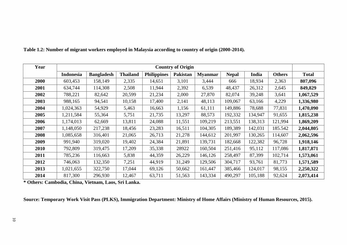
and plantation, construction and domestic. Statistics from 2000 to 2014 showed half of
the workers were majority from Indonesia (Table 1.2).
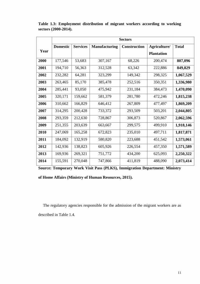
Table 1.3 shows the employment breakdown of migrant workers in the different
working sectors. Employment of migrant workers was primarily in the manufacturing
sector followed by agriculture and plantation, construction, services and domestic
works.
10
Table 1.2: Number of migrant workers employed in Malaysia according to country of origin (2000-2014).
Year Country of Origin
Indonesia Bangladesh Thailand Philippines Pakistan Myanmar Nepal India Others Total
2000 603,453 158,149 2,335 14,651 3,101 3,444 666 18,934 2,363 807,096
2001 634,744 114,308 2,508 11,944 2,392 6,539 48,437 26,312 2,645 849,829
2002 788,221 82,642 20,599 21,234 2,000 27,870 82,074 39,248 3,641 1,067,529
2003 988,165 94,541 10,158 17,400 2,141 48,113 109,067 63,166 4,229 1,336,980
2004 1,024,363 54,929 5,463 16,663 1,156 61,111 149,886 78,688 77,831 1,470,090
2005 1,211,584 55,364 5,751 21,735 13,297 88,573 192,332 134,947 91,655 1,815,238
2006 1,174,013 62,669 13,811 24,088 11,551 109,219 213,551 138,313 121,994 1,869,209
2007 1,148,050 217,238 18,456 23,283 16,511 104,305 189,389 142,031 185.542 2,044,805
2008 1,085,658 316,401 21,065 26,713 21,278 144,612 201,997 130,265 114,607 2,062,596
2009 991,940 319,020 19,402 24,384 21,891 139,731 182,668 122,382 96,728 1,918,146
2010 792,809 319,475 17,209 35,338 28922 160,504 251,416 95,112 117,086 1,817,871
2011 785,236 116,663 5,838 44,359 26,229 146,126 258,497 87,399 102,714 1,573,061
2012 746,063 132,350 7,251 44,919 31,249 129,506 304,717 93,761 81,773 1,571,589
2013 1,021,655 322,750 17,044 69,126 50,662 161,447 385,466 124,017 98,155 2,250,322
2014 817,300 296,930 12,467 63,711 51,563 143,334 490,297 105,188 92,624 2,073,414
* Others: Cambodia, China, Vietnam, Laos, Sri Lanka.
Source: Temporary Work Visit Pass (PLKS), Immigration Department: Ministry of Home Affairs (Ministry of Human Resources, 2015).
11
Table 1.3: Employment distribution of migrant workers according to working
sectors (2000-2014).
Year
Sectors
Domestic Services Manufacturing Construction Agriculture/
Plantation
Total
2000 177,546 53,683 307,167 68,226 200,474 807,096
2001 194,710 56,363 312,528 63,342 222,886 849,829
2002 232,282 64,281 323,299 149,342 298,325 1,067,529
2003 263,465 85,170 385,478 252,516 350,351 1,336,980
2004 285,441 93,050 475,942 231,184 384,473 1,470,090
2005 320,171 159,662 581,379 281,780 472,246 1,815,238
2006 310,662 166,829 646,412 267,809 477,497 1,869,209
2007 314,295 200,428 733,372 293,509 503,201 2,044,805
2008 293,359 212,630 728,867 306,873 520,867 2,062,596
2009 251,355 203,639 663,667 299,575 499,910 1,918,146
2010 247,069 165,258 672,823 235,010 497,711 1,817,871
2011 184,092 132,919 580,820 223,688 451,542 1,573,061
2012 142,936 138,823 605,926 226,554 457,350 1,571,589
2013 169,936 269,321 751,772 434,200 625,093 2,250,322
2014 155,591 270,048 747,866 411,819 488,090 2,073,414
Source: Temporary Work Visit Pass (PLKS), Immigration Department: Ministry
of Home Affairs (Ministry of Human Resources, 2015).
The regulatory agencies responsible for the admission of the migrant workers are as
described in Table 1.4.
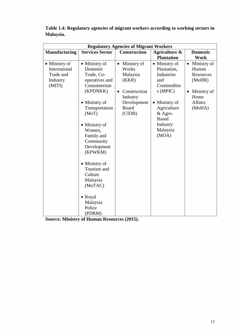
12
Table 1.4: Regulatory agencies of migrant workers according to working sectors in
Malaysia.
Regulatory Agencies of Migrant Workers
Manufacturing Services Sector Construction Agriculture &
Plantation
Domestic
Work
Ministry of
Internatonal
Trade and
Industry
(MITI)
Ministry of
Domestic
Trade, Co-
operatives and
Consumerism
(KPDNKK)
Ministry of
Transportation
(MoT)
Ministry of
Women,
Family and
Community
Development
(KPWKM)
Ministry of
Tourism and
Culture
Malaysia
(MoTAC)
Royal
Malaysia
Police
(PDRM)
Ministry of
Works
Malaysia
(KKR)
Construction
Industry
Development
Board
(CIDB)
Ministry of
Plantation,
Industries
and
Commoditie
s (MPIC)
Ministry of
Agriculture
& Agro-
Based
Industry
Malaysia
(MOA)
Ministry of
Human
Resources
(MoHR)
Ministry of
Home
Affairs
(MoHA)
Source: Ministry of Human Resources (2015).
13
1.3 Health status of migrant workers
1.3.1 Heath screening of workers upon entry
Each worker is obligatory to undergo medical screening upon entry to Malaysia and
the subsequent year up to the third year of service (under the same employer). Unitab
Medic Sdn. Bhd. through FOMEMA is an agency involved in the implementation,
management and supervision of a nationwide mandatory health screening programme
for all legal migrant workers in Malaysia. FOMEMA ensures that the health status of
each migrant worker is free from communicable diseases and promotes the well being
of the society by safeguarding the health of the general community living.
The medical screening process is designed and managed by a group of medical
professionals in public health, occupational health, radiology, laboratory services and
other related specialties (Figure 1.2). All registration and payment is centralized with
standardized fee. Employers are given the option a list of doctors in their registry to run
the medical screening for their workers. Standardized medical examination is carried
out stipulated by Ministry of Health, which is monitored and supervised through IT
surveillance and inspectorate activities. Medical reports, X-ray and laboratory results
are submitted independently and electronically to FOMEMA and to Immigration
Department Headquarters to facilitate issuance of work pass or deportation. The results
are obtained via online and those who failed the screening are certified ‗UNFIT‘ and
sent back to their country, with or without appeal to FOMEMA. Only ‗FIT‘ workers are
allowed to continue with their employment in Malaysia.

14
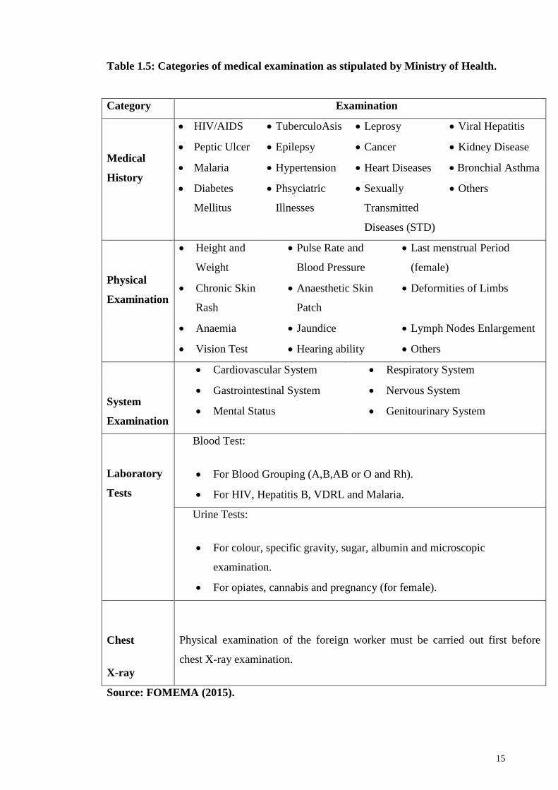
The medical examination is normally carried out within 30 days from the date of
registration. Details of medical examination covered under the system stipulated by the
Ministry of Health includes medical history, physical examination, system examination
laboratory test and x-ray examination (Table 1.5).
Benefits of this system include the assurance of a healthier workforce, prevention of
the spread of identified communicable diseases to other workers or the public at large,
increased productivity through reduction in absenteeism due to illness, in addition to
lower incidence of imported diseases, morbidity and mortality. As a result, an overall
reduction in healthcare cost to employers, taxpayers and the Government and better use
of local public health facilities for the citizens.
Figure 1.2: Medical screening process of foreign workers in Malaysia. Source:
FOMEMA (2015).
15
Table 1.5: Categories of medical examination as stipulated by Ministry of Health.
Category Examination
Medical
History
HIV/AIDS TuberculoAsis Leprosy Viral Hepatitis
Peptic Ulcer Epilepsy Cancer Kidney Disease
Malaria Hypertension Heart Diseases Bronchial Asthma
Diabetes
Mellitus
Phsyciatric
Illnesses
Sexually
Transmitted
Diseases (STD)
Others
Physical
Examination
Height and
Weight
Pulse Rate and
Blood Pressure
Last menstrual Period
(female)
Chronic Skin
Rash
Anaesthetic Skin
Patch
Deformities of Limbs
Anaemia Jaundice Lymph Nodes Enlargement
Vision Test Hearing ability Others
System
Examination
Cardiovascular System Respiratory System
Gastrointestinal System Nervous System
Mental Status Genitourinary System
Laboratory
Tests
Blood Test:
For Blood Grouping (A,B,AB or O and Rh).
For HIV, Hepatitis B, VDRL and Malaria.
Urine Tests:
For colour, specific gravity, sugar, albumin and microscopic
examination.
For opiates, cannabis and pregnancy (for female).
Chest
X-ray
Physical examination of the foreign worker must be carried out first before
chest X-ray examination.
Source: FOMEMA (2015).
16
Medical results are transmitted independently, thus averting physical handling or
report tampering by employers or agents to ensure the integrity of the health-screening
system, as well as to facilitate the employers' application or permit renewal in a timely
manner and also plays a role as an access for the Government authorities to a
centralized database. The system also provides timely information and vital statistics
relating to communicable diseases and to facilitate immediate counter-action and
preventive measures.
1.3.2 Common health problems
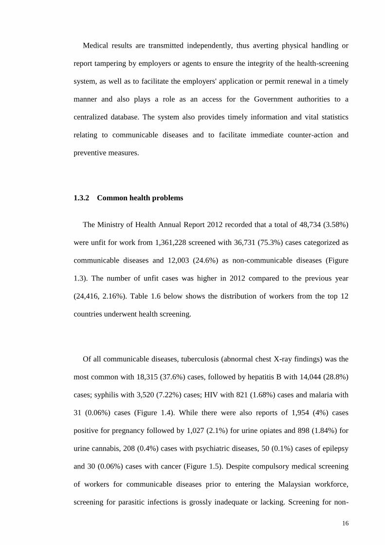
The Ministry of Health Annual Report 2012 recorded that a total of 48,734 (3.58%)
were unfit for work from 1,361,228 screened with 36,731 (75.3%) cases categorized as
communicable diseases and 12,003 (24.6%) as non-communicable diseases (Figure
1.3). The number of unfit cases was higher in 2012 compared to the previous year
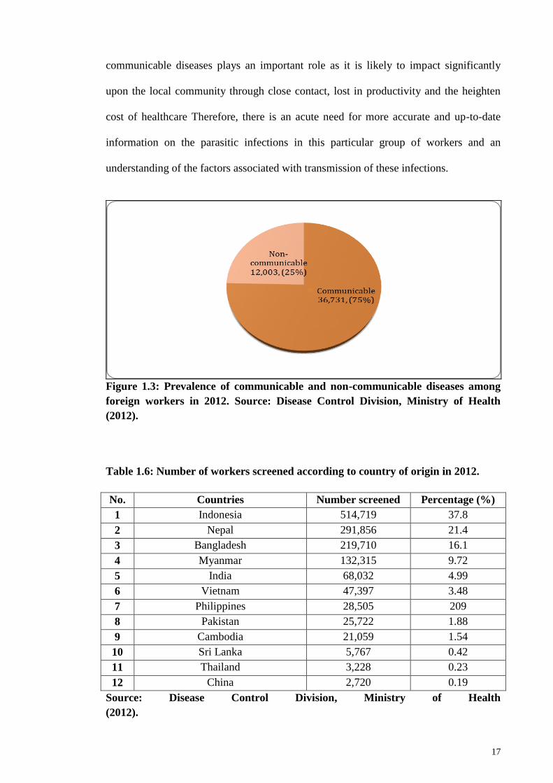
(24,416, 2.16%). Table 1.6 below shows the distribution of workers from the top 12
countries underwent health screening.
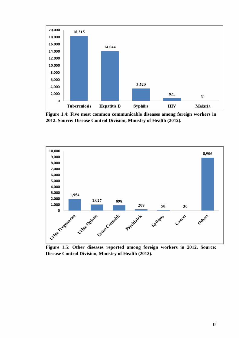
Of all communicable diseases, tuberculosis (abnormal chest X-ray findings) was the
most common with 18,315 (37.6%) cases, followed by hepatitis B with 14,044 (28.8%)
cases; syphilis with 3,520 (7.22%) cases; HIV with 821 (1.68%) cases and malaria with
31 (0.06%) cases (Figure 1.4). While there were also reports of 1,954 (4%) cases
positive for pregnancy followed by 1,027 (2.1%) for urine opiates and 898 (1.84%) for
urine cannabis, 208 (0.4%) cases with psychiatric diseases, 50 (0.1%) cases of epilepsy
and 30 (0.06%) cases with cancer (Figure 1.5). Despite compulsory medical screening
of workers for communicable diseases prior to entering the Malaysian workforce,
screening for parasitic infections is grossly inadequate or lacking. Screening for non-
17
communicable diseases plays an important role as it is likely to impact significantly
upon the local community through close contact, lost in productivity and the heighten
cost of healthcare Therefore, there is an acute need for more accurate and up-to-date
information on the parasitic infections in this particular group of workers and an
understanding of the factors associated with transmission of these infections.
Figure 1.3: Prevalence of communicable and non-communicable diseases among
foreign workers in 2012. Source: Disease Control Division, Ministry of Health
(2012).
Table 1.6: Number of workers screened according to country of origin in 2012.
No. Countries Number screened Percentage (%)
1 Indonesia 514,719 37.8
2 Nepal 291,856 21.4
3 Bangladesh 219,710 16.1
4 Myanmar 132,315 9.72
5 India 68,032 4.99
6 Vietnam 47,397 3.48
7 Philippines 28,505 209
8 Pakistan 25,722 1.88
9 Cambodia 21,059 1.54
10 Sri Lanka 5,767 0.42
11 Thailand 3,228 0.23
12 China 2,720 0.19
Source: Disease Control Division, Ministry of Health
(2012).
18
Figure 1.4: Five most common communicable diseases among foreign workers in
2012. Source: Disease Control Division, Ministry of Health (2012).
Figure 1.5: Other diseases reported among foreign workers in 2012. Source:
Disease Control Division, Ministry of Health (2012).
19
1.4 Common parasitic infections in migrant workers
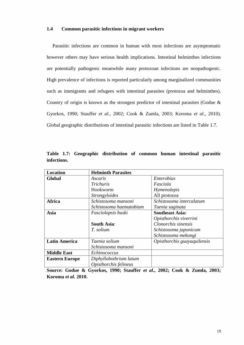
Parasitic infections are common in human with most infections are asymptomatic
however others may have serious health implications. Intestinal helminthes infections
are potentially pathogenic meanwhile many protozoan infections are nonpathogenic.
High prevalence of infections is reported particularly among marginalized communities
such as immigrants and refugees with intestinal parasites (protozoa and helminthes).
Country of origin is known as the strongest predictor of intestinal parasites (Godue &
Gyorkos, 1990; Stauffer et al., 2002; Cook & Zumla, 2003; Koroma et al., 2010).
Global geographic distributions of intestinal parasitic infections are listed in Table 1.7.
Table 1.7: Geographic distribution of common human intestinal parasitic
infections.
Location Helminth Parasites
Global Ascaris
Trichuris
Hookworm
Strongyloides
Enterobius
Fasciola
Hymenolepis
All protozoa
Africa Schistosoma mansoni
Schistosoma haematobium
Schistosoma intercalatum
Taenia saginata
Asia Fasciolopsis buski
South Asia:
T. solium
Southeast Asia:
Opisthorchis viverrini
Clonorchis sinensis
Schistosoma japonicum
Schistosoma mekongi
Latin America Taenia solium
Schistosoma mansoni
Opisthorchis guayaquilensis
Middle East Echinococcus
Eastern Europe Diphyllobothrium latum
Opisthorchis felineus
Source: Godue & Gyorkos, 1990; Stauffer et al., 2002; Cook & Zumla, 2003;
Koroma et al. 2010.
20
1.4.1 Helminthes
The neglected intestinal parasitic infections (IPIs) such as soil-transmitted helminth
(STH), is recognized as one of the most significant causes of illnesses and diseases
especially among disadvantaged communities. World Health Organization (WHO)
categorizes STH as one of the 17 neglected tropical diseases in the world population.
More than 1.5 billion people, or 24% of the world‘s population are infected with single
or multiple infections of common helminth such as roundworm (Ascaris lumbricoides),
whipworm (Trichuris trichiura) and hookworms (Necator americanus and Ancylostoma
duodenale). Other helminth species include Enterobius vermicularis and Hymenolepis
nana.
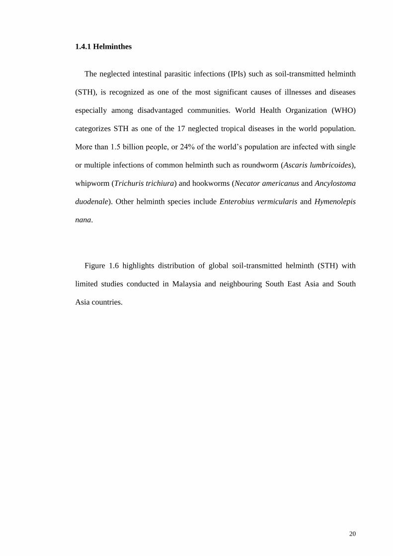
Figure 1.6 highlights distribution of global soil-transmitted helminth (STH) with
limited studies conducted in Malaysia and neighbouring South East Asia and South
Asia countries.
21
Figure 1.6: Worldwide distribution of soil-transmitted helminth. Source: Global
Atlas of Helminth Infections (2016).
1.4.1.1 Ascaris lumbricoides
Ascaris lumbricoides (Table 1.8) is the most prevalent human parasitic infections
and the largest nematode parasitizing human intestine infecting >1 billion persons
globally (Bethony et al., 2006). The adult length for female ranges from 20 to 35cm,
while the adult male from 15 to 30cm.
The adult worm establishes in the lumen of human intestine after ingestion of the
infective eggs where a female can produce as many as 200,000 eggs per day. Eggs
become infective after 18 days to several weeks dependent on the optimum moist, warm
and soil. After ingestion, the eggs hatch into larvae and transported via the portal to the
lungs and mature (10 to 14 days). The larvae then penetrate the alveolar walls to the
throat before being swallowed. Upon reaching the small intestine, the worm matures
22
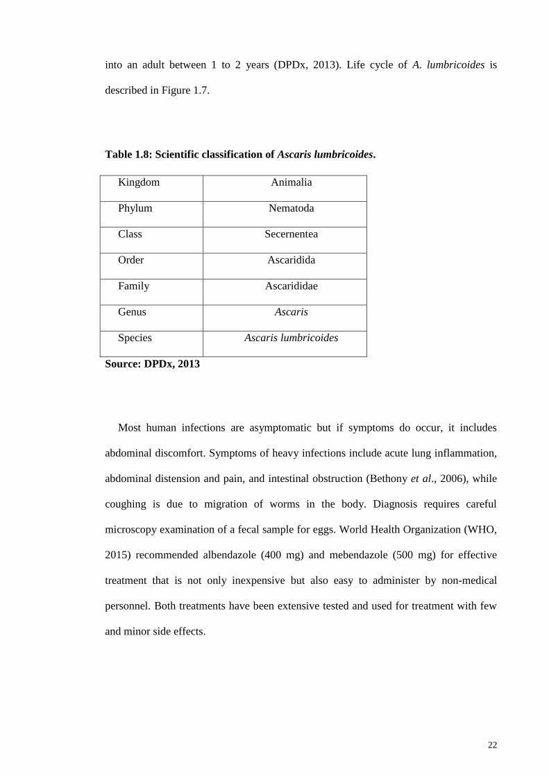
into an adult between 1 to 2 years (DPDx, 2013). Life cycle of A. lumbricoides is
described in Figure 1.7.
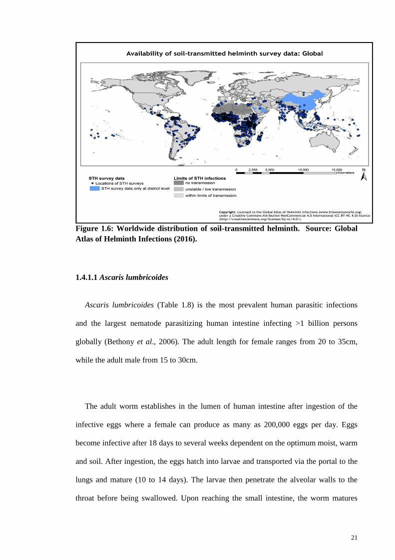
Table 1.8: Scientific classification of Ascaris lumbricoides.
Kingdom Animalia
Phylum Nematoda
Class Secernentea
Order Ascaridida
Family Ascarididae
Genus Ascaris
Species Ascaris lumbricoides
Source: DPDx, 2013
Most human infections are asymptomatic but if symptoms do occur, it includes
abdominal discomfort. Symptoms of heavy infections include acute lung inflammation,
abdominal distension and pain, and intestinal obstruction (Bethony et al., 2006), while
coughing is due to migration of worms in the body. Diagnosis requires careful
microscopy examination of a fecal sample for eggs. World Health Organization (WHO,
2015) recommended albendazole (400 mg) and mebendazole (500 mg) for effective
treatment that is not only inexpensive but also easy to administer by non-medical
personnel. Both treatments have been extensive tested and used for treatment with few
and minor side effects.
23
Figure 1.7: Life cycle of Ascaris lumbricoides (Source: DPDx, 2013)
1.4.1.2 Hookworm
Another important species of soil-transmitted helminthes is the hookworm (Table
1.9). Two species (Ancylostoma duodenale and Necator americanus) infect an
estimated of 600 million people globally (Hotez, 2009). N. americanus infection is
more common worldwide, while A. duodenale is more geographically restricted.
Unlike Ascaris, hookworm eggs are not infective. Eggs are passed in the stool of an
infected person and the larvae hatch under favorable conditions (moisture, warmth, and
shade) between 1 to 2 days. The released first stage (rhabditiform) larvae grow in the
feces and/or the soil, and after 5 to 10 days develop into infective third stage (filariform)
larvae which can survive 3 to 4 weeks in favorable environmental conditions. Upon
contact with the human skin the larvae is transported via blood vessels to the heart and
then lungs and penetrate the pulmonary alveoli before ascending to the bronchial tree
24
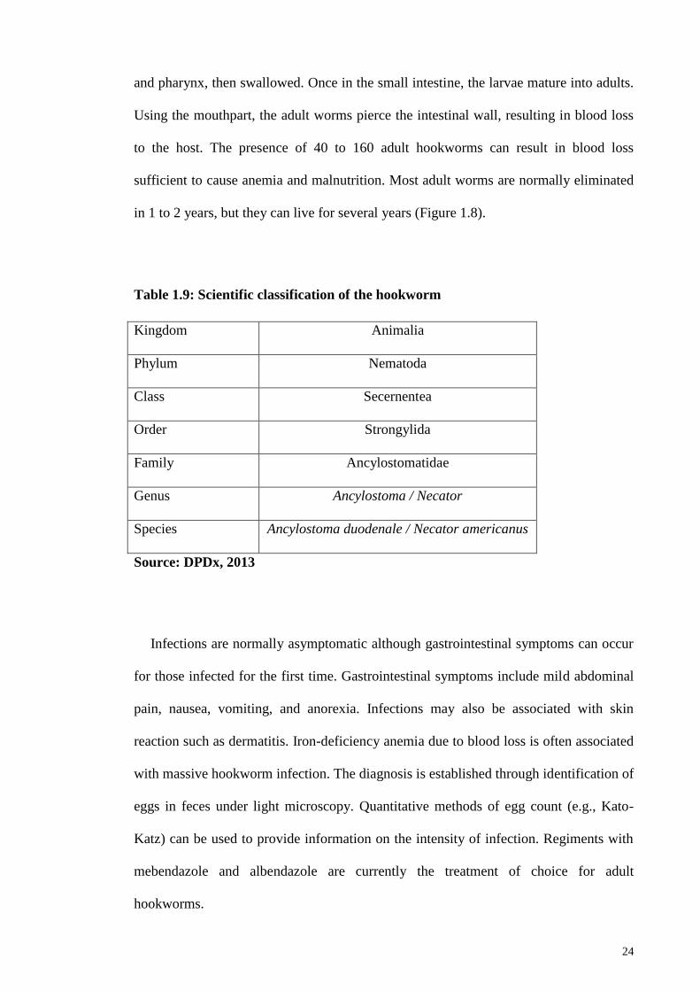
and pharynx, then swallowed. Once in the small intestine, the larvae mature into adults.
Using the mouthpart, the adult worms pierce the intestinal wall, resulting in blood loss
to the host. The presence of 40 to 160 adult hookworms can result in blood loss
sufficient to cause anemia and malnutrition. Most adult worms are normally eliminated
in 1 to 2 years, but they can live for several years (Figure 1.8).
Table 1.9: Scientific classification of the hookworm
Kingdom Animalia
Phylum Nematoda
Class Secernentea
Order Strongylida
Family Ancylostomatidae
Genus Ancylostoma / Necator
Species Ancylostoma duodenale / Necator americanus
Source: DPDx, 2013
Infections are normally asymptomatic although gastrointestinal symptoms can occur
for those infected for the first time. Gastrointestinal symptoms include mild abdominal
pain, nausea, vomiting, and anorexia. Infections may also be associated with skin
reaction such as dermatitis. Iron-deficiency anemia due to blood loss is often associated
with massive hookworm infection. The diagnosis is established through identification of
eggs in feces under light microscopy. Quantitative methods of egg count (e.g., Kato-
Katz) can be used to provide information on the intensity of infection. Regiments with
mebendazole and albendazole are currently the treatment of choice for adult
hookworms.
25
Figure 1.8: Life cycle of the hookworms (Source: DPDx, 2013).
1.4.1.3 Trichuris trichiura
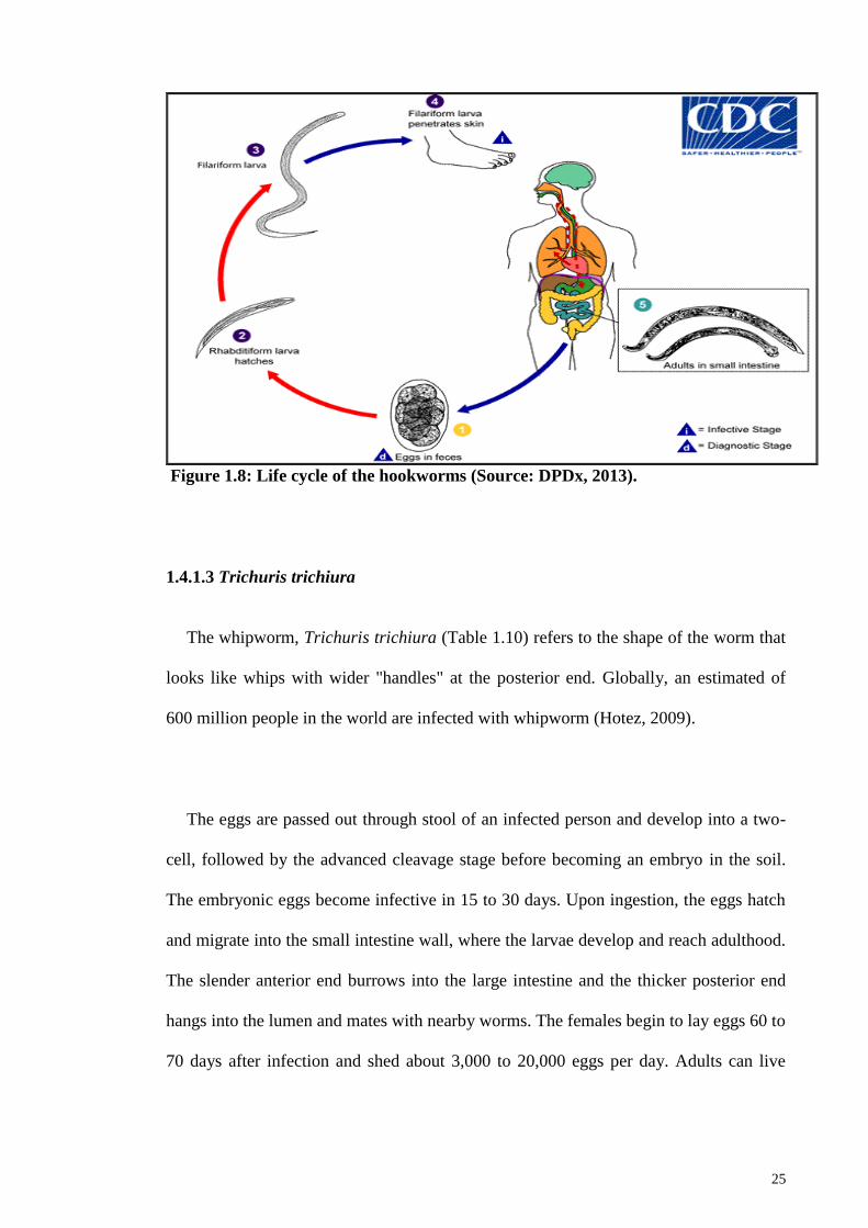
The whipworm, Trichuris trichiura (Table 1.10) refers to the shape of the worm that
looks like whips with wider "handles" at the posterior end. Globally, an estimated of
600 million people in the world are infected with whipworm (Hotez, 2009).
The eggs are passed out through stool of an infected person and develop into a two-
cell, followed by the advanced cleavage stage before becoming an embryo in the soil.
The embryonic eggs become infective in 15 to 30 days. Upon ingestion, the eggs hatch
and migrate into the small intestine wall, where the larvae develop and reach adulthood.
The slender anterior end burrows into the large intestine and the thicker posterior end
hangs into the lumen and mates with nearby worms. The females begin to lay eggs 60 to
70 days after infection and shed about 3,000 to 20,000 eggs per day. Adults can live
26
about 1 to 3 years, and females can grow up to 50 mm (2 inches) long (Figure 1.9)
(DPDx, 2013).
Table 1.10: Scientific classification of Trichuris trichiura.
Kingdom Animalia
Phylum Nematoda
Class Enoplea
Order Trichocephalida
Family Trichuridae
Genus Trichuris
Species Trichuris trichiura
Source: DPDx, 2013
Figure 1.9: Life cycle of Trichuris trichiura (Source: DPDx, 2013).
27
Trichuriasis is common in tropical countries and poor sanitation practices. Light
infections are normally asymptomatic while heavier infections include frequent, painful
passage of stool that contains a mixture of mucus, water, and blood. In children, heavy
infections can lead to growth retardation. Similarly, diagnosis is through microscopic
examination for the feces for eggs. Mebendazole and albendazole are currently the
drugs of choice for treatment of adult worms.
1.4.1.4 Strongyloides stercoralis
Strongyloides stercoralis (Table 1.11) is a threadworm and known to exist in all
continents except for Antarctica, but is most common in the tropics, subtropics, and in
warm temperate regions. The estimate global prevalence of strongyloidiasis is between
30–100 million worldwide (Olsen et al., 2009).
The life cycle involves a free-living cycle and parasitic cycle. The free-living
rhabditiform larva passed out through the stool can either become the infective
filariform larvae (direct development) or free-living adult male or female that mates and
produces eggs. The filariform larvae penetrate the human skin and migrate into the
small intestine to initiate the parasitic cycle. The L3 larvae are transported via the
bloodstream to the lungs, where they are eventually coughed up and swallowed. In the
small intestine, the larvae molt twice before becoming adult female worms. The females
live threaded in the epithelium of the small intestine and through parthenogenesis
produce eggs, which yield rhabditiform larvae. The rhabditiform larvae can either be
passed in the stool or can cause autoinfection. In autoinfection, the rhabditiform larvae
become infective and penetrate either the intestinal mucosa (internal autoinfection) or
28
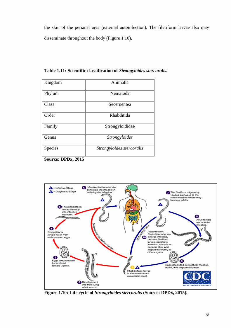
the skin of the perianal area (external autoinfection). The filariform larvae also may
disseminate throughout the body (Figure 1.10).
Table 1.11: Scientific classification of Strongyloides stercoralis.
Kingdom Animalia
Phylum Nematoda
Class Secernentea
Order Rhabditida
Family Strongyloididae
Genus Strongyloides
Species Strongyloides stercoralis
Source: DPDx, 2015
Figure 1.10: Life cycle of Strongyloides stercoralis (Source: DPDx, 2015).
29
Those infected normally showed no symptom however, in severe infections,
infected persons may experience stomach ache, bloating, heartburn, intermittent
episodes of diarrhea and constipation, nausea and loss of appetite, dry cough, throat
irritation, itch, red rash that occurs when the worm enter the skin and recurrent raised
red rash typically along the thighs and buttocks. Results of treatment for
strongyloidiasis with albendazole and mebendazole vary while, ivermectin has been
shown to be more effective than albendazole.
1.4.1.5 Enterobius vermicularis
Enterobius vermicularis (Table 1.12) or also known as pinworm is a common human
parasite. The worms are small, white, and thread-like, with larger females ranging
between 8-13 mm x 0.3-0.5 mm and males ranging between 2-5 mm x 0.1-0.2 mm in
length. Females also possess a long, pin-shaped at the posterior end from which the
parasite's name is derived. Pinworm infection occurs worldwide and affects all ages and
socioeconomic background especially school-aged children and household members or
caretakers with pinworm infection.
The worm is spread via fecal-oral route i.e.by the transfer of infective pinworm eggs
from the anus to someone‘s mouth, either directly by hand or indirectly through
contaminated clothing, bedding, food or other articles. Following ingestion of infective
eggs, the larvae hatches in the small intestine before maturing as adults in the colon.
The time interval from ingestion to oviposition of an adult female is about 1 month. The
life span of the adults is about 2 months. Gravid females migrate nocturnally outside the
anus and oviposit on the skin of the perianal area. The larvae contained inside the eggs
30
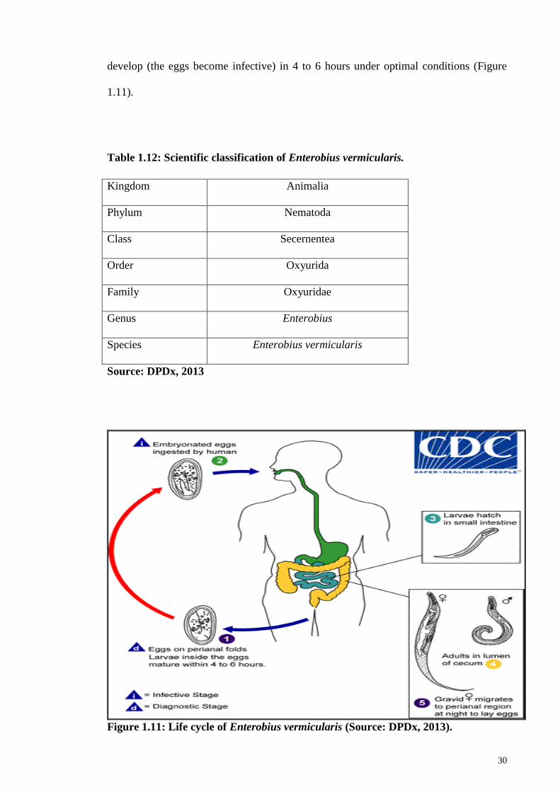
develop (the eggs become infective) in 4 to 6 hours under optimal conditions (Figure
1.11).
Table 1.12: Scientific classification of Enterobius vermicularis.
Kingdom Animalia
Phylum Nematoda
Class Secernentea
Order Oxyurida
Family Oxyuridae
Genus Enterobius
Species Enterobius vermicularis
Source: DPDx, 2013
Figure 1.11: Life cycle of Enterobius vermicularis (Source: DPDx, 2013).
31
The most common method in diagnosing pinworm infection is via the ―Scotch tape‖
test, where a clear adhesive cellulose tape is applied to the anal area early in the
morning before bathing or defecation. This is then observed under a microscope for the
presence of pinworm eggs. Treatment of pinworm includes mebendazole, pyrantel
pamoate and albendazole. In all cases, treatment of the entire household is strongly
recommended, with or without the presence of symptoms due to the fact that pinworms
are easily transmitted among members of the household.
1.4.1.6 Hymenolepis nana
Hymenolepis nana (Table 1.13) is a small worm (adults are only 15–40 mm long)
found worldwide especially in children, in persons living in institutional settings and
areas where sanitation and personal hygiene is inadequate.
Infection is through accidental ingestion of contaminated foods or water, by touching
mouth with contaminated fingers, ingesting contaminated soil or ingestion of an
infected arthropod (intermediate host, such as a small beetle or mealworm). Eggs
become infective immediately after passing out through the stool and cannot survive
more than 10 days in the external environment. Upon ingestion by an intermediate
arthropod host, the eggs develop into cysticercoids. Humans or rodents become infected
upon ingestion of the arthropod host and develop into adults in the small intestine. Upon
ingestion of eggs (in contaminated food or water or from hands contaminated with
feces), the oncospheres contained in the eggs are released. The oncospheres (hexacanth
larvae) penetrate the intestinal villus and develop into cysticercoid larvae. Upon rupture
of the villus, the cysticercoids return to the intestinal lumen, evaginate their scoleces,
attach to the intestinal mucosa and develop into adults in the ileal of the small intestine
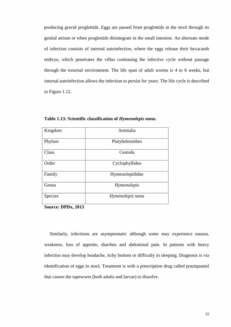
32
producing gravid proglottids. Eggs are passed from proglottids in the stool through its
genital atrium or when proglottids disintegrate in the small intestine. An alternate mode
of infection consists of internal autoinfection, where the eggs release their hexacanth
embryo, which penetrates the villus continuing the infective cycle without passage
through the external environment. The life span of adult worms is 4 to 6 weeks, but
internal autoinfection allows the infection to persist for years. The life cycle is described
in Figure 1.12.
Table 1.13: Scientific classification of Hymenolepis nana.
Kingdom Animalia
Phylum Platyhelminthes
Class Cestoda
Order Cyclophyllidea
Family Hymenolepididae
Genus Hymenolepis
Species Hymenolepis nana
Source: DPDx, 2013
Similarly, infections are asymptomatic although some may experience nausea,
weakness, loss of appetite, diarrhea and abdominal pain. In patients with heavy
infection may develop headache, itchy bottom or difficulty in sleeping. Diagnosis is via
identification of eggs in stool. Treatment is with a prescription drug called praziquantel
that causes the tapeworm (both adults and larvae) to dissolve.
33
Figure 1.12: Life cycle of Hymenolepis nana (Source: DPDx, 2013).
1.4.2 Protozoa
Most protozoan species are free living and but the small numbers of parasitic species
infect higher animals with one or more species. Protozoa are microscopic unicellular
eukaryotes with a relatively complex internal structure and carry out complex metabolic
activities. Some have structures for propulsion or other modes for movement (Yaeger,
1996). Infection ranges from asymptomatic to life threatening and dependent on the
species, strain and resistance of the host.
The pathogenic species include the amoeba, Entamoeba histolytica and the
flagellate, Giardia duodenalis. Although many other species are known to cause
intestinal disease throughout the world, such as Cryptosporidium parvum, Cyclospora
34
cayetanensis and Balantidium coli, the importance of these organisms remains unclear
as most are considered non-pathogenic.
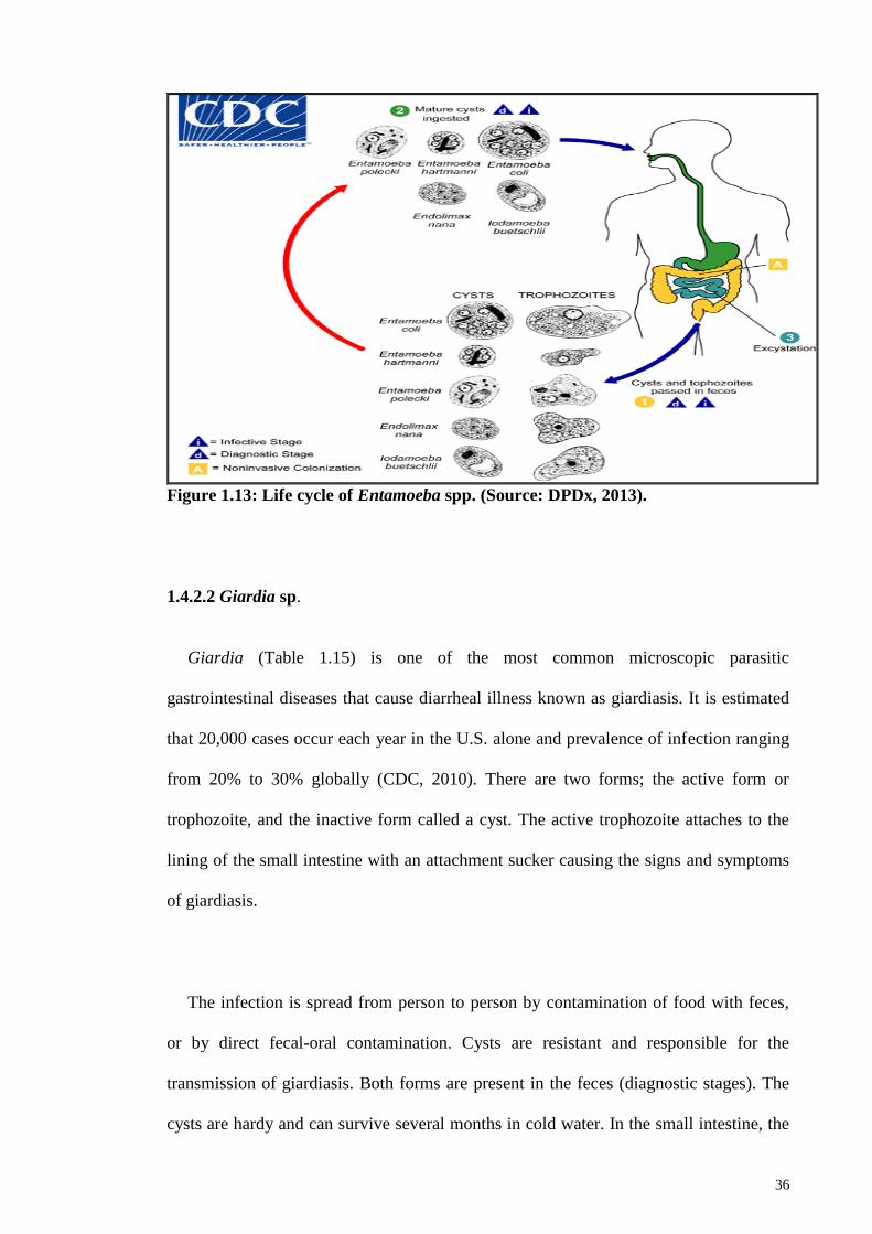
1.4.2.1 Entamoeba spp.
Species in the genus Entamoeba (Table 1.14) colonizes humans, but not all are
associated with disease. Six human intestinal species Entamoeba spp. include
Entamoeba histolytica, E. dispar, E. moshkovskii, E. coli, E. hartmanni and E. polecki.
Amoebiasis is a global health problem caused by the protozoan E. histolytica and is
well recognized as a pathogenic amoeba associated with intestinal and extraintestinal
infections. E histolytica is a pseudopod-forming, non-flagellated protozoal parasite that
causes proteolysis and tissue lysis and can induce host-cell apoptosis. Worldwide,
approximately 50 million cases of invasive E histolytica disease occur each year with
high incidences in developing countries (Stauffer et al., 2006) resulting in as many as
100,000 deaths with only 10%-20% infected individuals become symptomatic
(Valenzuela et al., 2007; Van Hal et al., 2007; Ximenez et al., 2009).
Entamoeba is transmitted via ingestion of the cystic form (infective stage). Cysts and
trophozoites are passed in feces with cysts typically found in formed stool, whereas
trophozoites in diarrheal stool. Infection occurs by ingestion of mature cysts in feces
contaminated food, water, or hands. Excystation occurs in the small intestine releasing
trophozoites that migrate to the large intestine. Trophozoites multiply by binary fission
and produce cysts, and both stages are passed out through the feces. The cysts can
survive days to weeks in the external environment due to the protection conferred by
their walls, and are responsible for transmission. Trophozoites are rapidly destroyed
once outside the body, and if ingested, would not survive exposure to the gastric
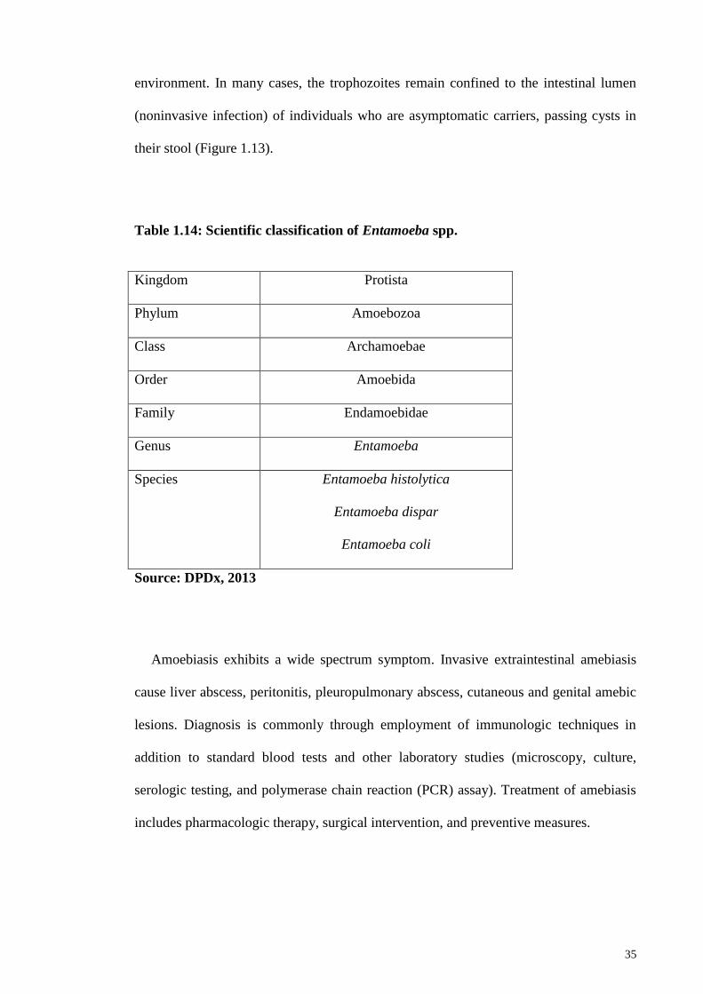
35
environment. In many cases, the trophozoites remain confined to the intestinal lumen
(noninvasive infection) of individuals who are asymptomatic carriers, passing cysts in
their stool (Figure 1.13).
Table 1.14: Scientific classification of Entamoeba spp.
Kingdom Protista
Phylum Amoebozoa
Class Archamoebae
Order Amoebida
Family Endamoebidae
Genus Entamoeba
Species Entamoeba histolytica
Entamoeba dispar
Entamoeba coli
Source: DPDx, 2013
Amoebiasis exhibits a wide spectrum symptom. Invasive extraintestinal amebiasis
cause liver abscess, peritonitis, pleuropulmonary abscess, cutaneous and genital amebic
lesions. Diagnosis is commonly through employment of immunologic techniques in
addition to standard blood tests and other laboratory studies (microscopy, culture,
serologic testing, and polymerase chain reaction (PCR) assay). Treatment of amebiasis
includes pharmacologic therapy, surgical intervention, and preventive measures.
36
Figure 1.13: Life cycle of Entamoeba spp. (Source: DPDx, 2013).
1.4.2.2 Giardia sp.
Giardia (Table 1.15) is one of the most common microscopic parasitic
gastrointestinal diseases that cause diarrheal illness known as giardiasis. It is estimated
that 20,000 cases occur each year in the U.S. alone and prevalence of infection ranging
from 20% to 30% globally (CDC, 2010). There are two forms; the active form or
trophozoite, and the inactive form called a cyst. The active trophozoite attaches to the
lining of the small intestine with an attachment sucker causing the signs and symptoms
of giardiasis.
The infection is spread from person to person by contamination of food with feces,
or by direct fecal-oral contamination. Cysts are resistant and responsible for the
transmission of giardiasis. Both forms are present in the feces (diagnostic stages). The
cysts are hardy and can survive several months in cold water. In the small intestine, the
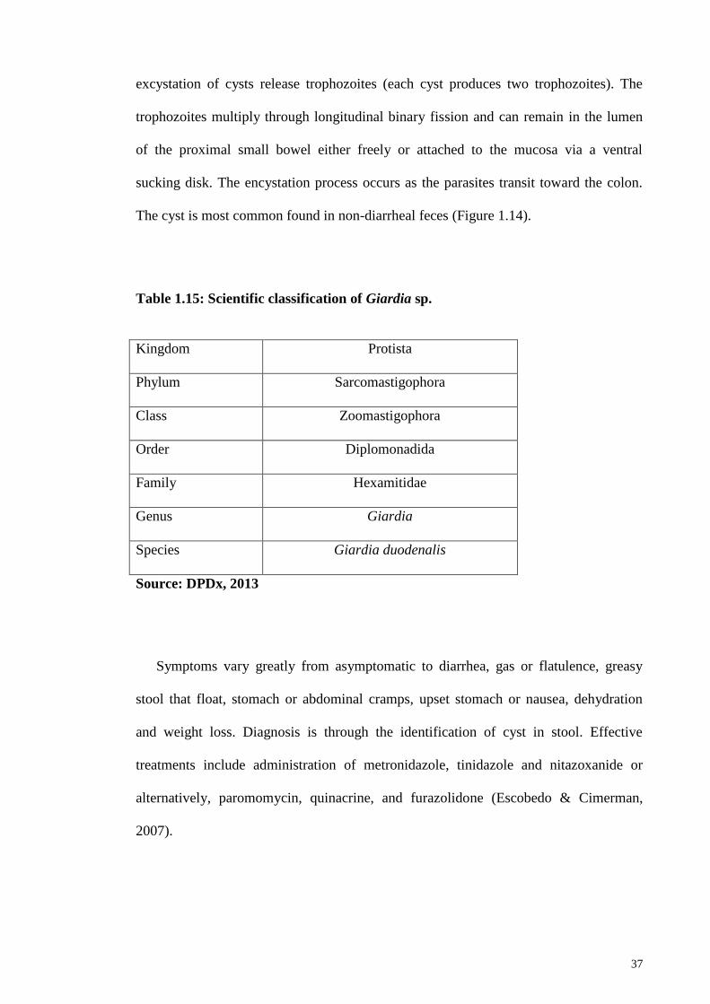
37
excystation of cysts release trophozoites (each cyst produces two trophozoites). The
trophozoites multiply through longitudinal binary fission and can remain in the lumen
of the proximal small bowel either freely or attached to the mucosa via a ventral
sucking disk. The encystation process occurs as the parasites transit toward the colon.
The cyst is most common found in non-diarrheal feces (Figure 1.14).
Table 1.15: Scientific classification of Giardia sp.
Kingdom Protista
Phylum Sarcomastigophora
Class Zoomastigophora
Order Diplomonadida
Family Hexamitidae
Genus Giardia
Species Giardia duodenalis
Source: DPDx, 2013
Symptoms vary greatly from asymptomatic to diarrhea, gas or flatulence, greasy
stool that float, stomach or abdominal cramps, upset stomach or nausea, dehydration
and weight loss. Diagnosis is through the identification of cyst in stool. Effective
treatments include administration of metronidazole, tinidazole and nitazoxanide or
alternatively, paromomycin, quinacrine, and furazolidone (Escobedo & Cimerman,
2007).
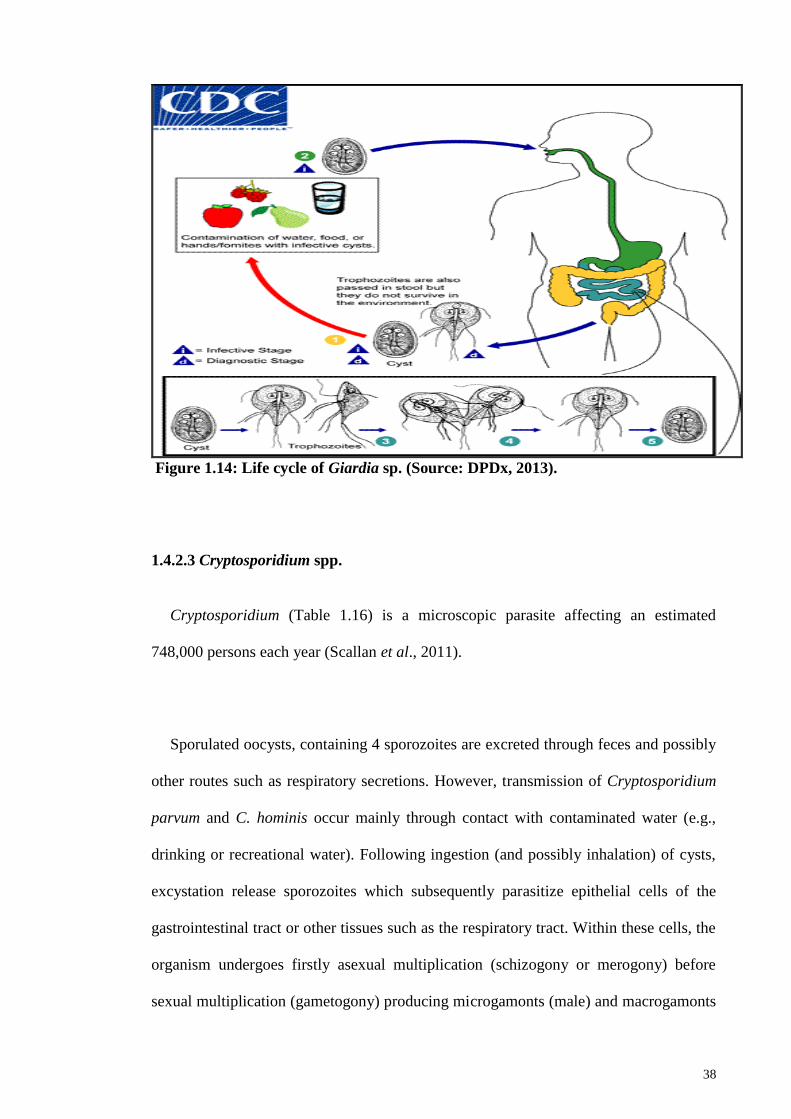
38
Figure 1.14: Life cycle of Giardia sp. (Source: DPDx, 2013).
1.4.2.3 Cryptosporidium spp.
Cryptosporidium (Table 1.16) is a microscopic parasite affecting an estimated
748,000 persons each year (Scallan et al., 2011).
Sporulated oocysts, containing 4 sporozoites are excreted through feces and possibly
other routes such as respiratory secretions. However, transmission of Cryptosporidium
parvum and C. hominis occur mainly through contact with contaminated water (e.g.,
drinking or recreational water). Following ingestion (and possibly inhalation) of cysts,
excystation release sporozoites which subsequently parasitize epithelial cells of the
gastrointestinal tract or other tissues such as the respiratory tract. Within these cells, the
organism undergoes firstly asexual multiplication (schizogony or merogony) before
sexual multiplication (gametogony) producing microgamonts (male) and macrogamonts
39
(female). Upon fertilization, the oocysts sporulate in the infected host into two types; a
thick-walled commonly excreted out from the host, or a thin-walled oocyst primarily
involved in autoinfection. Oocysts are infective upon excretion, thus permitting direct
and immediate fecal-oral transmission (Figure 1.15).
Table 1.16: Scientific classification of Cryptosporidium spp.
Kingdom Protista
Phylum Sarcomastigophora
Class Conoidasida
Order Eucoccidiorida
Family Cryptosporidiidae
Genus Cryptosporidium
Species Cryptosporidium parvum
Cryptosporidium hominis
Source: DPDx, 2013
Symptoms generally appear 2 to 10 days (average 7 days) after infection with watery
diarrhea, stomach cramps or pain, dehydration, nausea, vomiting, fever and weight loss.
Diagnosis of cryptosporidiosis is via examination of stool samples using several
techniques (e.g., acid-fast staining, direct fluorescent antibody [DFA], and/or enzyme
immunoassays for the detection of Cryptosporidium sp. antigens) and molecular
methods. Those with healthy immune systems will recover without treatment. Diarrhea
can be managed by drinking plenty of fluids to prevent dehydration.
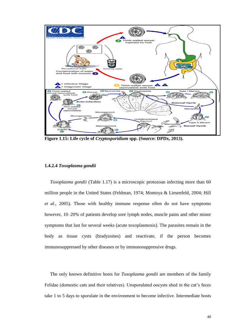
40
Figure 1.15: Life cycle of Cryptosporidium spp. (Source: DPDx, 2013).
1.4.2.4 Toxoplasma gondii
Toxoplasma gondii (Table 1.17) is a microscopic protozoan infecting more than 60
million people in the United States (Feldman, 1974; Montoya & Liesenfeld, 2004; Hill
et al., 2005). Those with healthy immune response often do not have symptoms
however, 10–20% of patients develop sore lymph nodes, muscle pains and other minor
symptoms that last for several weeks (acute toxoplasmosis). The parasites remain in the
body as tissue cysts (bradyzoites) and reactivate, if the person becomes
immunosuppressed by other diseases or by immunosuppressive drugs.
The only known definitive hosts for Toxoplasma gondii are members of the family
Felidae (domestic cats and their relatives). Unsporulated oocysts shed in the cat‘s feces
take 1 to 5 days to sporulate in the environment to become infective. Intermediate hosts
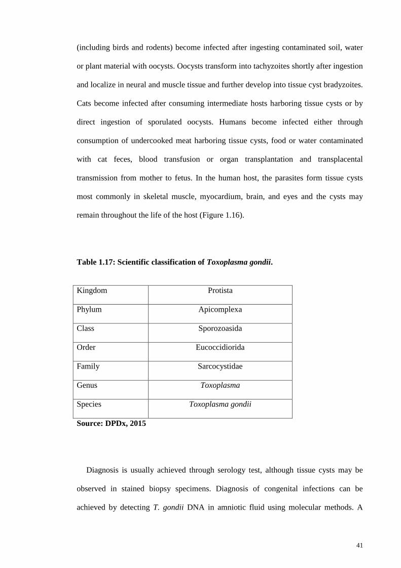
41
(including birds and rodents) become infected after ingesting contaminated soil, water
or plant material with oocysts. Oocysts transform into tachyzoites shortly after ingestion
and localize in neural and muscle tissue and further develop into tissue cyst bradyzoites.
Cats become infected after consuming intermediate hosts harboring tissue cysts or by
direct ingestion of sporulated oocysts. Humans become infected either through
consumption of undercooked meat harboring tissue cysts, food or water contaminated
with cat feces, blood transfusion or organ transplantation and transplacental
transmission from mother to fetus. In the human host, the parasites form tissue cysts
most commonly in skeletal muscle, myocardium, brain, and eyes and the cysts may
remain throughout the life of the host (Figure 1.16).
Table 1.17: Scientific classification of Toxoplasma gondii.
Kingdom Protista
Phylum Apicomplexa
Class Sporozoasida
Order Eucoccidiorida
Family Sarcocystidae
Genus Toxoplasma
Species Toxoplasma gondii
Source: DPDx, 2015
Diagnosis is usually achieved through serology test, although tissue cysts may be
observed in stained biopsy specimens. Diagnosis of congenital infections can be
achieved by detecting T. gondii DNA in amniotic fluid using molecular methods. A
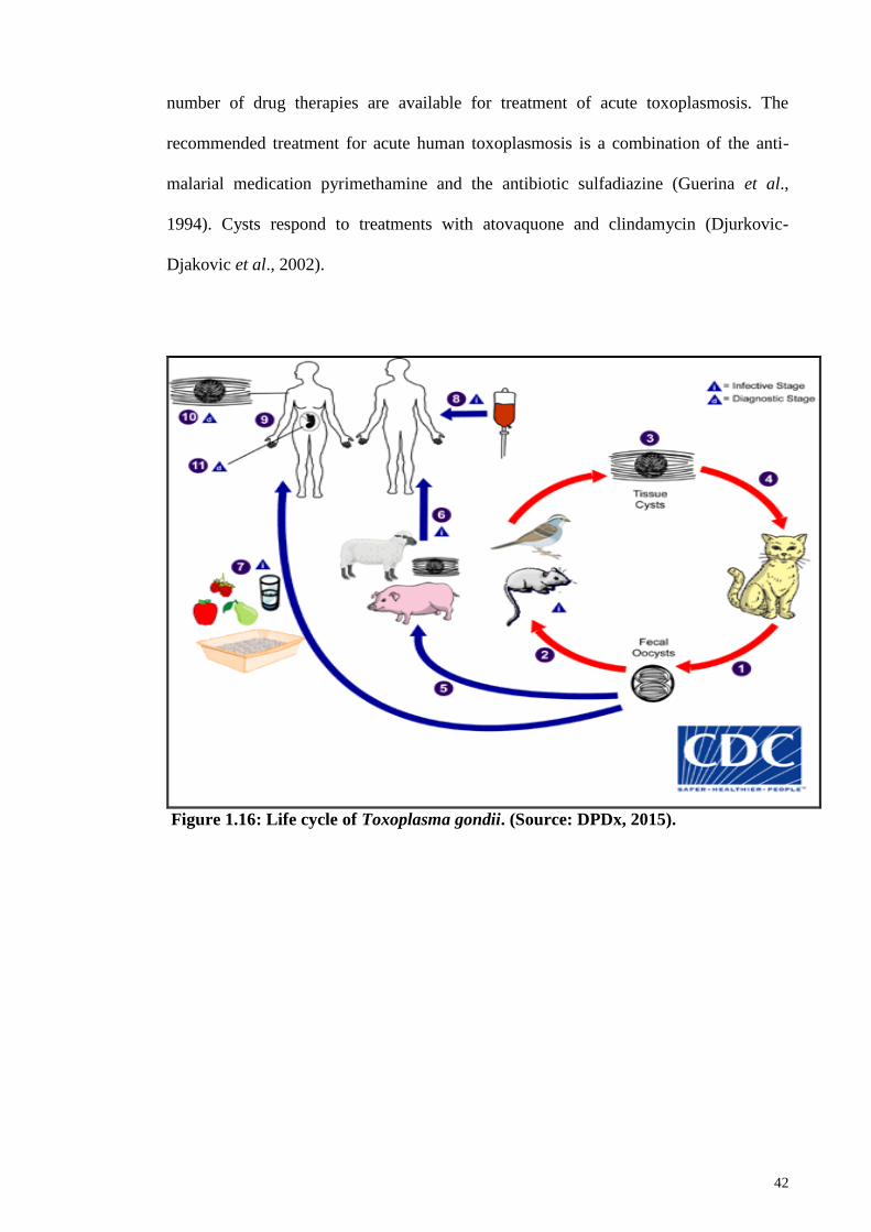
42
number of drug therapies are available for treatment of acute toxoplasmosis. The
recommended treatment for acute human toxoplasmosis is a combination of the anti-
malarial medication pyrimethamine and the antibiotic sulfadiazine (Guerina et al.,
1994). Cysts respond to treatments with atovaquone and clindamycin (Djurkovic-
Djakovic et al., 2002).
Figure 1.16: Life cycle of Toxoplasma gondii. (Source: DPDx, 2015).
43
1.5 Studies on the status of parasitic infections amongst migrant workers
Many health status studies of parasitic infections amongst migrant workers have
been conducted worldwide, particularly in Asia (Thailand (Nuchprayoon et al., 2009;
Ngrenngarmlert et al., 2012), Taiwan (Lo & Lee, 1996; Wang, 1998; 2004; Lu & Sung,
2008; Hsieh et al., 2011), Taipei (Cheng & Shieh, 2000)) and in the Middle East
primarily in the Kingdom of Saudi Arabia (Abha district (Al-Madani & Mahfouz,
1995), Riyadh (Kalantan, 2001), Al-Khobar (Abahussain, 2005), Makkah (Wakid et al.,
2009), Al-Baha (Mohammad & Koshak, 2011) and Medina (Taha et al., 2013)).
Meanwhile in Qatar, Abu-Madi et al. (2008; 2010; 2011) extensively studied the
parasitic infections in migrant workers. In Malaysia, a large study scope was conducted
more than a decade ago (Zaini et al., 2002) that included physical examination and
basic haematological findings (Chia, 2002), microbiological analysis (Ngeow et al.,
2002), health status of woman respondent (Siti Norazah, 2002a), sexual health (Siti
Norazah, 2002b), oral health problem (Ishak, 2002), lifestyle habit and risk behaviors
(Wong, 2002) and psychiatric morbidity (Mohd Hussain, 2002). However, limited
studies were conducted on parasitic infections (Suresh et al., 2002; Kamarulzaman &
Khairul Anuar, 2002; Zurainee et al., 2002; Khairul Anuar et al., 2002; Zurainee, 2002
& Rajah et al, 2002).
1.5.1 Studies in Asia
In Thailand, most studies focused among workers from Myanmar. A cross-sectional
survey showed that the overall prevalence of intestinal parasitic infections was low
(13.6%) primarily with helminth infections (10.3%) being more common than protozoa
(8.5%) particularly with E. histolytica/dispar (3.8%), followed by A. lumbricoides
(3.3%), and T. trichiura (2.3%) (Ngrenngarmlert et al., 2012). Another study focused
44
on Myanmar workers in the food industry reported with more than half screened were
infected (62.3%) with Blastocystis hominis (41.5%) followed by T. trichiura (22.2%),
G. lamblia (14.1%) and A. lumbricoides (1.8%) (Nuchprayoon et al., 2009).
In Taiwan, 20.2% (188/932) immigrants were reported infected with B. hominis (Lu
& Sung, 2008). While another study in Taiwan showed low infection (7.4%, 214/2875)
among foreigners, with hookworm (2.92%) being the highest followed by T. trichiura
(1.18%), and B. hominis (1.14%) (Hsieh et al., 2011). In Taipei, of the 302 Thailand
laborers examined, 196 (64.9%) were infected with Opisthorchis viverrini (43.0%)
followed by hookworm (38.4%) and S. stercoralis (13.9%) (Cheng & Shieh, 2000).
In Saudi Arabia, many studies were conducted throughout the kingdom, Abha
district (Al-Madani & Mahfouz, 1995), Riyadh (Kalantan, 2001), Al-Khobar
(Abahussain, 2005), Makkah (Wakid et al., 2009), Al-Baha (Mohammad & Koshak,
2011), and Medina (Taha et al., 2013). More recently, in Medina (Taha et al., 2013) of
the 2732 stool samples collected and screened, 407 (14.9%) stool samples were positive
with intestinal parasites particularly among farmers, food handlers and shepherds and
those who originated from Pakistan (23.2%). Prevalence of G. lamblia (21.9%)
infections was the highest, followed by E. histolytica/ E. dispar (17.8%), T. trichiura
(16.2%), A, lumbricoides (15.8%), hookworm (13%), H. nana (8.9%), S. sterocoralis
(3.5%), Schistosoma mansoni (2.2%) and E. vermicularis (0.43%) (Taha et al., 2013).
Infections were lower among expatriate workers in cities, such as Al-Khobar (31.4%)
(Abahussain, 2005). In Al-Khobar, Abahussain (2005) reported that A. lumbricoides
infection was common among Indonesians, T. trichiura and E. histolytica were common
among Philippines and hookworms among Sri Lankan workers. Another study by Al-
45
Madani & Mahfouz (1995) reported 46.5% of Asian female domestic workers were
positive with parasitic infection. In Makkah, 31.9% of food handlers working in the
hospitals were positive with parasitic infections (Wakid et al., 2009). Similar to the
report by Taha et al. (2013), in Makkah (Wakid et al., 2009), G. lamblia was the most
prevalent infection encountered in the examined populations.
In Qatar, Abu-Madi et al. reported the occurrence of parasitic infections among
migrant workers in three subsequent years; 33.9% in 2008, 10.2% in 2009 and 21.5% in
2011. In 2008, seven species of intestinal parasitic infections (IPI) amongst food
handlers and housemaids from different geographical regions were recorded with three
nematode species (13.6%) i.e. T. trichiura, hookworms and A. lumbricoides and four
protozoans (24.8%) (E. histolytica/dispar, non-pathogenic Entamoeba, Blastocystis
hominis and G. duodenalis). In 2009, Abu-Madi et al. looked into the trends of IPI
among long-term-residents and settled immigrants after introduction of routine
albendazole treatment as a condition of entry, residence and issuance of a work permit.
Results reported low infection rate (10.2%) with at least one species of intestinal
parasite (2.6% with helminths and 8.0% with protozoan species). Infections were
primarily found in 20-30 year old male subjects from Nepal with hookworms (69%).
While in 2011, Abu-Madi et al. compared IPI between newly arrived and resident
workers in Qatar and results showed that 21.5% of the subjects were infected with at
least one of the species recorded (8 helminthes and 4 protozoan species) with the high
infection being hookworms (8.3%). Most helminth infections declined in subjects that
acquired residency status in Qatar, especially among female subjects, however one male
Nepalese worker continued to harbor helminth infections (notably hookworms) after he
became resident.
46
1.5.2 Migrant health status studies in Malaysia
Health status of foreign workers was conducted more than a decade ago (Zaini et al.,
2002) that included physical examination and basic haematological (Chia, 2002),
parasitic infection (Suresh et al., 2002; Kamarulzaman & Khairul Anuar, 2002;
Zurainee et al., 2002; Khairul Anuar et al., 2002; Zurainee, 2002 & Rajah et al, 2002),
microbiological (Ngeow et al., 2002), health status of women (Siti Norazah, 2002),
male sexual health (Siti Norazah, 2002), oral health problem (Ishak, 2002), lifestyle
habit and risk behaviors (Wong, 2002) and psychiatric morbidity (Mohd Hussain,
2002).
Among the results reported were 36% from 173 stool samples were infected with
Blastocystis hominis (Suresh et al., 2002) with smaller forms (3-5 µm in size) of B.
hominis in Indonesian immigrants compared to other nationalities (10-15 µm) (Rajah et
al., 2002). One case of visceral leishmaniasis (Kala-azar) in a 28 years old Bangladesh
migrant worker was also reported (Kamarulzaman & Khairul Anuar, 2002).
Eight serological tests were performed on 698 foreign workers including amoebiasis,
echinococcosis, filariasis (Brugia malayi and Wuchereria bancrofti), leishmaniasis,
malaria, schistosomiasis and trypanosomiasis (Zurainee, 2002; Zurainee et al., 2002)
with 38.1% positive with at least one parasitic infection. One Bangladeshi was found
positive for Wuchereria bancrofti and an Indonesian positive for malaria (Plasmodium
falciparum) from the 241 blood samples screened (Khairul Anuar et al., 2002).
47
1.6 Justification of the study
To date, the only study to determine the health status among foreign workers in
Malaysia was conducted by Zaini et al. (2002) more than a decade ago. This study
provided useful methodological enquiries however, was not designed for policy
recommendations. Over the last decade, the number of migrant workers has grown
exponentially. Despite compulsory medical screening for workers prior to entering the
Malaysian workforce, screening for parasitic infections is grossly inadequate or lacking.
Therefore, there is an acute need for more accurate and up-to-date information on the
parasitic infections in this particular group of workers and an understanding of the
factors associated with transmission of these infections, especially as they are likely to
impact significantly upon the local community through close contact, lost productivity
and the heighten cost of healthcare. This study therefore is timely in adopting a
scientific approach to address an important public health problem and to provide
conclusions that can inform the design of effective public health policies.
48
1.7 Objectives
The main objective of this study is to analyze the current parasitic infections status
among migrant workers in Malaysia and to evaluate the health dangers posed by the
migrant workers to the local population.
The specific objectives of the present study are as follows:
To analyze the socio-demographic profile of the respondents in the present study
in Malaysia.
To determine the intestinal parasitic infections from stool samples among
migrant workers.
To determine the status of parasitic infections among migrant workers by using
serological tests.
To associate the prevalence of infections in the migrant populations to factors
such as host age, gender, geographical origin and employment sector with a
view to provide a descriptive epidemiological assessment.
To characterize parasite using molecular techniques where appropriate.
49
CHAPTER 2: MIGRANT WORKERS IN MALAYSIA:
SOCIO-DEMOGRAPHY BACKGROUND
2.1 Introduction
Mass migration from less developed to more developed country has created a
shift in the global population. Urbanization and extensive industrialization of
developing nations have resulted in millions of migrants travelling to major urban cities
around the globe to join the expanding workforces. The International Labour
Organization (ILO) estimates that there are approximately 232 million international
migrant workers worldwide. Globalization, demographic shifts, conflicts, income
inequalities and climate change are some of the influences that drive workers and their
families to cross borders in search of better employment and security (International
Labour Organization, 2015). In Malaysia, the robust economic growth of the different
sectors has led to the mushrooming of small to large enterprises requiring high demand
of a low-skilled workforce primarily in sectors such as construction, domestic, food
services, manufacturing and plantation. This has attracted many to flock to the country
both legally and illegally (Ministry of Human Resources, 2015; Bardan, 2014) from
South East Asian (Indonesia, Cambodia, Vietnam, the Philippines and Myanmar) and
South Asian countries (Nepal, India and Bangladesh) (Ministry of Human Resources,
2015; Bardan, 2014) where endemic infections are very much prevalent and most likely
to pose public health problems to the local community (Abu-Madi et al., 2008; 2010;
2011; Taha et al., 2013).
Malaysia is a middle-income country whose economy has transformed into an
emerging multi-sector economy and since the 1970s it has been facilitated largely by
50
imported migrant workers. Malaysia has a higher standard of living compared with
other neighbouring countries in the South East Asian and South Asian region. A total of
74.7% of the population in Malaysia has undergone urbanization with 2.66% annual
rate of change (2010 - 2015) (Central Intelligence Agency, 2016). Access to sanitation
facilities in Malaysia has improved also in both urban and rural areas up to 96.0% of the
population. Meanwhile drinking water sources have improved up to 98.2% of the
population (Central Intelligence Agency, 2016). The percentage of the population in
Malaysia still living below the poverty line is 3.8%, considerably lower than that of
other nationalities recruited in the present study including Myanmar (32.7%),
Bangladesh (31.5%), India (29.8%), Nepal (25.2%), Vietnam (11.3%) and Indonesia
(11.3%). The push factors for migration include poor remuneration and slim
employment opportunities in their home country. Meanwhile the main factors for
choosing Malaysia as a destination country are perceived abundant opportunities, high
wage levels and attractive job offers (Abdul-Aziz, 2001).
In Malaysia, Zaini et al. (2002) conducted a study to determine the health status
of migrant worker more than a decade ago involving clinical subjects from University
Malaya Medical Centre (UMMC) and Peremba Group of Companies (PEREMBA),
which provided useful methodological enquiries. In the last decade, there has been an
influx in number of migrant workers from 1.06 in 2002 to 2.07 million in 2014 to
Malaysia in the seeking for better job opportunities. This has lead to a change in the
demographic background of the working forces in this country. Therefore, the objective
of this chapter is to determine the socio-demographic profile of the migrant respondent
in the different working sectors in Malaysia.
51
2.2 Materials and Methods
2.2.1 Subjects
Migrant workers in Malaysia primarily employed in the low skilled and semi-
skilled of five working sectors only, namely manufacturing, food services sector,
agriculture and plantation, construction and domestic work were selected as the main
study subjects in this study. Workers who voluntarily participated in this study were
recruited from September 2014 to August 2015 from agencies and companies in
Peninsular Malaysia. A minimum sample size using Leedy and Ormrod (2001) was
calculated based on the last study of intestinal parasitic infections in migrant workers
(36%) in Malaysia (Zaini et al., 2002). Using this formula, a minimum of 355 subjects
were required however, a total of 610 migrant workers were successfully recruited.
2.2.2 Questionnaire
Questionnaires were distributed to gather relevant information related to the
study to all respondents. An individual clinical interview with questionnaire was
performed in order to collect information pertaining socio-demographic (nationality,
sex, age, religion, marital status, educational level and employment sector), migration
history (area in country origin, years of residence in Malaysia, mode of entry, working
history), environmental health (current residential area, type of accommodation,
amenities), life-style habits (smoker, consumption of alcohol and illegal drugs), access
to healthcare and episodes of illness (health care utilization, mode of payment, health
history) and occupational health and safety (safety hazard, personal protective
equipment). The interview process was performed through an interpreter in situations
where the respondents had difficulty in understanding Malay or English. All
52
participants were fully informed of the nature of the study to enable maximum co-
operation and completed the consent forms (Appendix B).
2.2.3 Ethical considerations
An ethical clearance was obtained from the ethics committee, University Malaya
Medical Centre (UMMC), Malaysia prior to commencement of the study (Reference
number: MECID NO: 20143-40). All respondents were adults and made aware of the
nature of the study and provided written consent to participate in the study.
2.2.4 Data analysis
Data were statistically analyzed using SPSS software statistic program version
22 to determine values prevalence of the set factors. Figures and tables were constructed
using the Descriptive Statistics of the software. Factors were analyzed with each other
to calculate prevalence and presented in tables and figures.
53
2.3 Results
2.3.1 Socio-demographic profile
A total of 610 migrant workers from five working sectors in Malaysia were
successfully recruited. The highest number of volunteers came from the domestic
(n=148; 24.3%) workers, followed by construction (n=139; 22.8%), food service
(n=128; 21.0%), plantation (n=102; 16.7%) and manufacturing sector (n=93; 15.2%).
Six nationalities were recruited with a majority originating from Indonesia (n=302,
49.5%), followed by Bangladesh (n=117, 19.2%), Nepal (n=100, 16.4%), India (n=64,
10.5%), Myanmar (n=26, 4.3%) and Vietnam (n=1; 0.2%). The majority of the
volunteers were Muslims (74.9%) with more than half married (67.4%). The socio-
demographic description of volunteers (sex, age, educational level, religion and marital
status) is shown in Table 2.1.
Analysis of the employment sectors by educational level showed most workers
had basic primary level education. Workers from the manufacturing and food service
sector had most workers with higher education with 88.2% and 63.3%, respectively.
Meanwhile, the plantation sector were dominated by workers with primary education
(72.5%) and with no formal education (27.5%) (Figure 2.1). Those involved in the
domestic sector were primarily Indonesian (144/148, 97.3%). Indonesian workers also
dominated the construction sector (54.0%) followed by Bangladeshi (34.5%), Burmese
(7.9%) and Indian (3.6%). In manufacturing sector, Nepalese (76/93; 81.7%) were
mostly employed (Figure 2.2).
54
Table 2.1: Demographic profile of migrant workers according to sex, age,
employment sector, nationality, educational level, religion and marital status.
(N=610)
Factors Total samples Percentage (%)
Sex Male 474 77.7
Female 136 22.3
Age <25 168 27.5
25-34 239 39.2
35-44 148 24.3
45-54 40 6.6
>55 15 2.5
Employment Construction 139 22.8
Sector Manufacturing 93 15.2
Plantation 102 16.7
Food Service 128 21.0
Domestic 148 24.3
Nationality Indonesia 302 49.5
Bangladesh 117 19.2
Myanmar
Vietnam
26
1
4.3
0.2
India 64 10.5
Nepal 100 16.4
Educational Primary 297 48.7
Level Secondary 227 37.2
University 12 2.0
No formal schooling 74 12.1
Religion Muslim 457 74.9
Buddhist 22 3.6
Hindu 121 19.8
Christian 10 1.6
Marital status Married 411 67.4
Divorced
Single
8
191
1.3
31.3
55
Figure 2.1: Education profile of migrant workers according to employment
sectors.
Figure 2.2: Worker’s nationality profile in relation to employment sectors
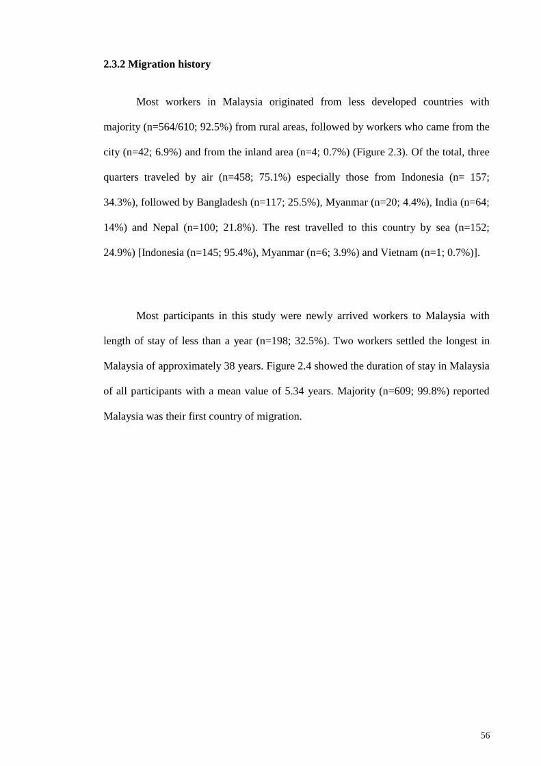
56
2.3.2 Migration history
Most workers in Malaysia originated from less developed countries with
majority (n=564/610; 92.5%) from rural areas, followed by workers who came from the
city (n=42; 6.9%) and from the inland area (n=4; 0.7%) (Figure 2.3). Of the total, three
quarters traveled by air (n=458; 75.1%) especially those from Indonesia (n= 157;
34.3%), followed by Bangladesh (n=117; 25.5%), Myanmar (n=20; 4.4%), India (n=64;
14%) and Nepal (n=100; 21.8%). The rest travelled to this country by sea (n=152;
24.9%) [Indonesia (n=145; 95.4%), Myanmar (n=6; 3.9%) and Vietnam (n=1; 0.7%)].
Most participants in this study were newly arrived workers to Malaysia with
length of stay of less than a year (n=198; 32.5%). Two workers settled the longest in
Malaysia of approximately 38 years. Figure 2.4 showed the duration of stay in Malaysia
of all participants with a mean value of 5.34 years. Majority (n=609; 99.8%) reported
Malaysia was their first country of migration.
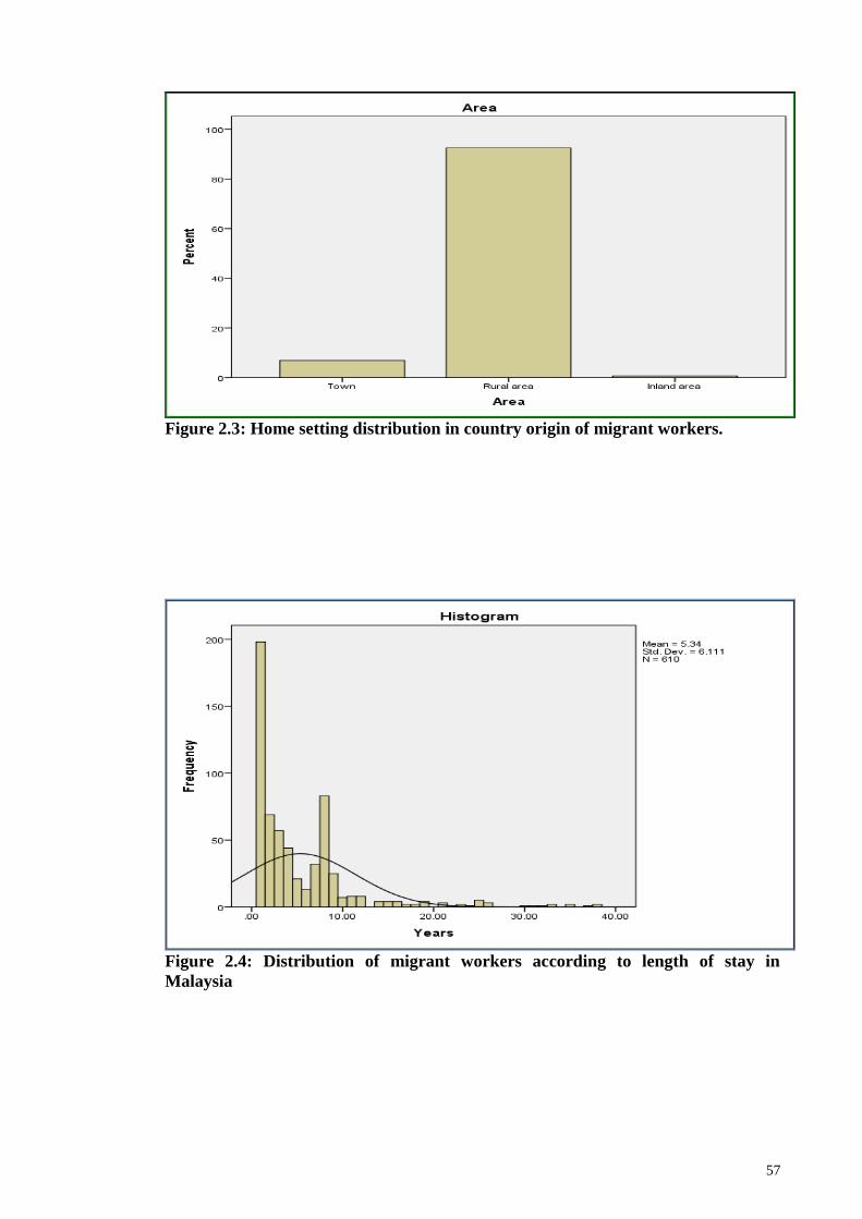
57
Figure 2.3: Home setting distribution in country origin of migrant workers.
Figure 2.4: Distribution of migrant workers according to length of stay in
Malaysia
58
2.3.3 Environmental health
The majority of respondents were provided housing by their employers. The
types of accommodation provided were mostly hostel (n=374; 61.3%), followed by
work-site accommodation (for construction workers) (n=56; 9.2%) and employer‘s
residence (n=3; 0.5%). A total of 173 (28.4%) owned or rented accommodation and
only 4 (0.7%) workers lived in a squatter home (Table 2.2). Majority of the workers
lived together with their fellow workers (n=340; 55.7%) and only 84 workers (13.8%)
lived with family.
Most of the workers were provided better amenities such as piped water and
sanitary toilets in their present accommodation, compared to their home countries. We
were also given a site visit to confirm their claim and were provided with suitable
accommodation, equipped with clean water system, proper sewage toilet and efficient
waste disposal system despite being overcrowded with too many workers to a room.
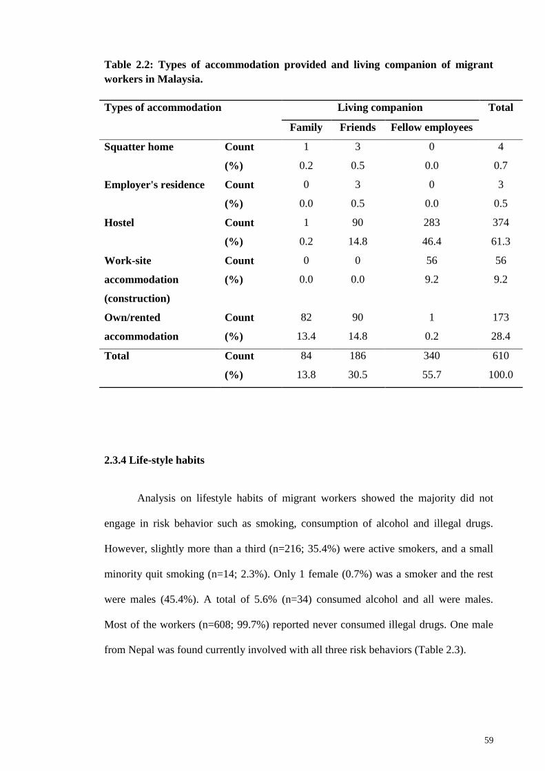
59
Table 2.2: Types of accommodation provided and living companion of migrant
workers in Malaysia.
Types of accommodation Living companion Total
Family Friends Fellow employees
Squatter home Count 1 3 0 4
(%) 0.2 0.5 0.0 0.7
Employer's residence Count 0 3 0 3
(%) 0.0 0.5 0.0 0.5
Hostel Count 1 90 283 374
(%) 0.2 14.8 46.4 61.3
Work-site
accommodation
(construction)
Count 0 0 56 56
(%) 0.0 0.0 9.2 9.2
Own/rented
accommodation
Count 82 90 1 173
(%) 13.4 14.8 0.2 28.4
Total Count 84 186 340 610
(%) 13.8 30.5 55.7 100.0
2.3.4 Life-style habits
Analysis on lifestyle habits of migrant workers showed the majority did not
engage in risk behavior such as smoking, consumption of alcohol and illegal drugs.
However, slightly more than a third (n=216; 35.4%) were active smokers, and a small
minority quit smoking (n=14; 2.3%). Only 1 female (0.7%) was a smoker and the rest
were males (45.4%). A total of 5.6% (n=34) consumed alcohol and all were males.
Most of the workers (n=608; 99.7%) reported never consumed illegal drugs. One male
from Nepal was found currently involved with all three risk behaviors (Table 2.3).
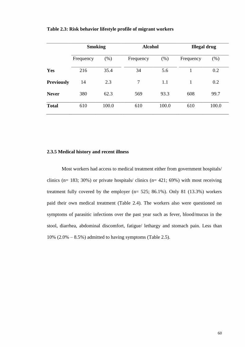
60
Table 2.3: Risk behavior lifestyle profile of migrant workers
Smoking Alcohol Illegal drug
Frequency (%) Frequency (%) Frequency (%)
Yes
216 35.4 34 5.6 1 0.2
Previously
14 2.3 7 1.1 1 0.2
Never
380 62.3 569 93.3 608 99.7
Total
610 100.0 610 100.0 610 100.0
2.3.5 Medical history and recent illness
Most workers had access to medical treatment either from government hospitals/
clinics (n= 183; 30%) or private hospitals/ clinics (n= 421; 69%) with most receiving
treatment fully covered by the employer (n= 525; 86.1%). Only 81 (13.3%) workers
paid their own medical treatment (Table 2.4). The workers also were questioned on
symptoms of parasitic infections over the past year such as fever, blood/mucus in the
stool, diarrhea, abdominal discomfort, fatigue/ lethargy and stomach pain. Less than
10% (2.0% – 8.5%) admitted to having symptoms (Table 2.5).
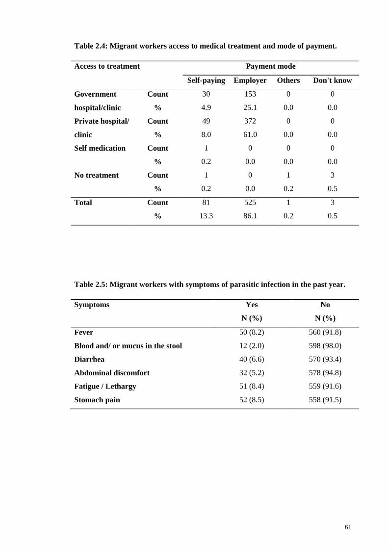
61
Table 2.4: Migrant workers access to medical treatment and mode of payment.
Access to treatment Payment mode
Self-paying Employer Others Don't know
Government
hospital/clinic
Count 30 153 0 0
% 4.9 25.1 0.0 0.0
Private hospital/
clinic
Count 49 372 0 0
% 8.0 61.0 0.0 0.0
Self medication Count 1 0 0 0
% 0.2 0.0 0.0 0.0
No treatment Count 1 0 1 3
% 0.2 0.0 0.2 0.5
Total Count 81 525 1 3
% 13.3 86.1 0.2 0.5
Table 2.5: Migrant workers with symptoms of parasitic infection in the past year.
Symptoms Yes
N (%)
No
N (%)
Fever 50 (8.2) 560 (91.8)
Blood and/ or mucus in the stool 12 (2.0) 598 (98.0)
Diarrhea 40 (6.6) 570 (93.4)
Abdominal discomfort 32 (5.2) 578 (94.8)
Fatigue / Lethargy 51 (8.4) 559 (91.6)
Stomach pain 52 (8.5) 558 (91.5)
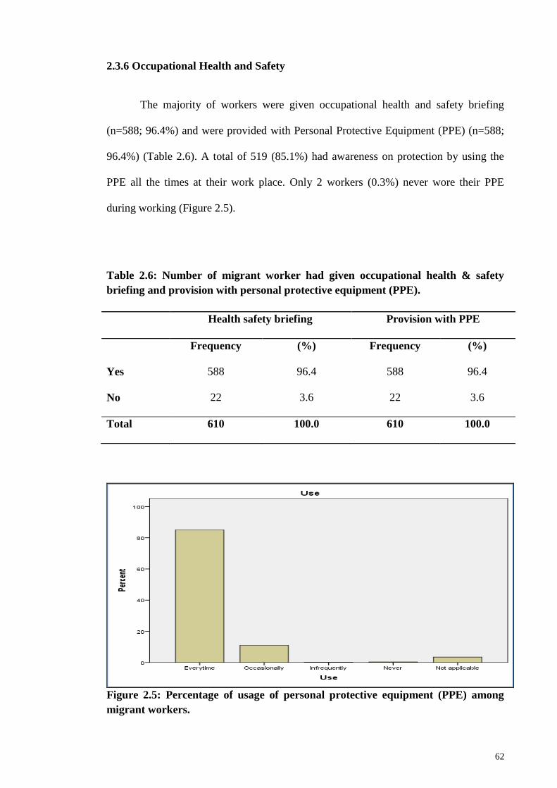
62
2.3.6 Occupational Health and Safety
The majority of workers were given occupational health and safety briefing
(n=588; 96.4%) and were provided with Personal Protective Equipment (PPE) (n=588;
96.4%) (Table 2.6). A total of 519 (85.1%) had awareness on protection by using the
PPE all the times at their work place. Only 2 workers (0.3%) never wore their PPE
during working (Figure 2.5).
Table 2.6: Number of migrant worker had given occupational health & safety
briefing and provision with personal protective equipment (PPE).
Health safety briefing Provision with PPE
Frequency (%) Frequency (%)
Yes 588 96.4 588 96.4
No 22 3.6 22 3.6
Total 610 100.0 610 100.0
Figure 2.5: Percentage of usage of personal protective equipment (PPE) among
migrant workers.
63
2.4 Discussion
The demand for low and semi-skilled workers in plantation/agriculture,
construction, factories and domestic service in Malaysia saw a dramatic rise in the
number of workers from 1.06 million in 2002 to 2.07 million in 2014 (Bardan, 2014)
from countries including Indonesia, Bangladesh, the Philippines, Nepal, Myanmar, and
other Asian countries labor. This study reports the socio-demographic profile of 610
migrant workers successfully recruited from the five main working sectors
(construction, manufacturing, plantation, food service and domestic service) compared
to the previous study by Zaini et al., (2002) with only 250 respondents from three
working sectors (construction, service and domestic). Recruitment were on a voluntary
basis at their work sites compared to Zaini et al., (2002) which focused mainly on
clinical data from University Malaya Medical Centre (UMMC) and PEREMBA group.
Six nationalities (Indonesia, Bangladesh, Myanmar, Vietnam, India and Nepal)
participated compared to only three (Indonesia, The Philippines and Bangladesh)
previously (Zaini et.al., 2002) with most workers particularly from Indonesia and
Bangladesh were Muslims.
Most workers originated from less developed countries of Southeast Asia with a
majority from rural locations (92.5%) in search for better remuneration and security.
Malaysia is a country rich in natural resources such as palm oil, rubber, tin, petroleum,
natural gas and timber. In addition, its robust economic development is unable to meet
the labor demand in the different sectors. Hence, Malaysia opens its borders to allow
workers from neighboring country to engage in employment. According to the Central
Intelligence Agency (2016), only 3.8% of population in Malaysia is living below the
poverty line compared to other nationalities. Myanmar is reported to have the largest
64
proportion of the population living under the poverty line (32.7%), followed by
Bangladesh (31.5%), India (29.8%), Nepal (25.2%), Vietnam (11.3%) and Indonesia
(11.3%). The push factor for migration for most workers were the poor remuneration
and slim employment opportunities in their home country. Among factors Malaysia was
chosen as a popular destination for those seeking employment were due to the perceived
abundant job opportunities, the high wage levels and attractive job offers (Abdul-Aziz,
2001). Most of the workers travel by low-cost air flights (75.1%) to Malaysia although
other options such as sea routes were also opted particularly for workers from Indonesia
and Myanmar. Abdul-Aziz (2001) also reported travel preferences among Bangladeshi
workers to Malaysia were firstly by air (71.6%), followed by land (14.9%) and sea
(13.5%). The option for travel to Malaysia is normally managed by an employment
agency. Generally, almost all of the participants in this study have some form of travel
documents, either the passport, work permit or both and they do not have any trouble to
travel to Malaysia.
The majority of workers were provided with hostel accommodation by their
employers (61.3%), sharing with other fellow employees. Only a small minority
(13.8%) mainly Indonesians, lived on their own with their families. The provision of
accommodation to the migrant workers is part of an agreement in a Memoranda of
Understanding (MOU) between the Malaysian government and the worker‘s
government stipulating the obligation for employers to provide accommodation in
addition to free access to water and electricity as well as transportation to their working
places. This was further confirmed as better amenities were provided to all workers
compared to their own home country where majority came from low socio-economic
locations in their home country which lacked basic utilities such as proper piped water,
electricity and toilet facilities. In general, sanitation facilities in both urban and rural
65
area in this country were available almost to all (96%) of the population compared to
other countries; Vietnam (78%), Myanmar (77.4%), Indonesia (60.8%), Bangladesh
(60.6%), Nepal (45.8%) and India (39.6%) (Central Intelligence Agency, 2016).
More than a third of the workers were active smokers (35.4%). World Health
Organization (2016) also reported similar global prevalence values of active smokers
aged 15 years old with more males (36.1%) engaging in this behavior than females
(6.8%). This values were also similar between nationalities in this region [i.e., Indonesia
(39.7%), Bangladesh (36.8%), Myanmar (34.6%), India (32.8%) and Nepal (22.0%)]
with more active male smokers (32.1%) compared to females (2.6%) (World Health
Organization, 2016). With regards to alcohol consumption, only a small minority
(6.7%) engaged in this activity similar to global values with more males involved
(7.4%) compared to females (1.4%). Alcohol consumption was among the lifestyle
activities evaluated among the workers. This habit may impact the surrounding people
in many ways as it can harm family members, friends and co-workers when the drinkers
are intoxicated. The risks can be determined by volume of alcohol consumed, patterns
of drinking and quality of alcohol consumed (World Health Organization, 2016).
Monitoring and surveillance of alcohol intake can reduce the negative health and social
implications.
In the present study, almost all workers (99.7%) have access to modern
healthcare services in this country. Majority had history of treatment fully supported by
their employer either at private hospitals/clinics (61.0%) or government hospitals/clinics
(25.1%). Zaini et al. (2002) also reported similar results that indicated good
accessibility of the healthcare provisions in this country to migrant workers.
66
Personal protective equipment (PPE) as defined by the Occupational Safety and
Health Administration (OSHA) is the ―specialized clothing or equipment, worn by an
employee for protection against infectious materials‖. Results also indicate that most
workers were briefed on occupational health and safety (96.4%), provided with
Personal Protective Equipment (PPE) (96.4%) and adhered to the wearing of PPE at all
times during work (85.1%). Awareness of occupational safety and health and the used
of PPE plays a crucial role to make the workplace safe and in the prevention of
occupational injuries and diseases (Lugah et al., 2010). In the latest report of the
occupational accidents by the Department of Occupational Safety and Health, Ministry
of Human Resources Malaysia in 2015, the manufacturing (58.5%) sector accounts the
highest incidents followed by agriculture and plantation (13.6%) and construction
(5.8%) sector (Department of Occupational Safety and Health, 2015). The enactment of
the Occupational Safety and Health Act (OSHA), 1994 implemented various programs
by different agencies to increase awareness and knowledge of OSH in the workplace
including providing instructions, procedures, training and supervision to encourage
people to work safely and responsibly.
67
2.5 Conclusion
The majority of migrant worker in this country were provided with suitable
living accommodations, clean water system, proper sewage toilets, and efficient waste
disposal system. Majority of the workers did not engage in risk behavior such as
smoking, consumption of alcohol or illegal drugs. More than 80% of the migrant
workers also were fully covered for medical treatment with the accessibility to private
or government hospitals/clinics. The workers have all been briefed on the occupational
health and safety and were provided with personal protective equipment (PPE) and
adhered to wearing the gear at work at all times.
68
CHAPTER 3: CURRENT IMPLICATIONS OF SOCIO-DEMOGRAPHIC AND
ENVIRONMENTAL CHARACTERISTICS IN THE TRANSMISSION OF
INTESTINAL PARASITIC INFECTIONS (IPIS)
3.1 Introduction
Neglected intestinal parasitic infections (IPIs) such as soil-transmitted helminths
(STH) have been recognized as one of the main causes of illnesses especially among
disadvantaged communities (Ngui et al., 2011a; Sinniah et al., 2014). According to the
World Health Organization (WHO), STH have been identified as one of 17 neglected
tropical diseases, with more than 1.5 billion people or 24% of the world‘s population
infected (World Health Organization, 2015) with roundworm (Ascaris lumbricoides),
whipworm (Trichuris trichiura) and hookworms (Necator americanus and Ancylostoma
duodenale) primarily through soil contaminated by human faeces. These infections can
cause anaemia, vitamin A deficiency, stunted growth, malnutrition, intestinal
obstruction and impaired development (Hotez et al., 2007). It is estimated that currently
up to 800 million people are infected with A. lumbricoides, 600 million people with T.
trichiura and 600 million with hookworms (Norhayati et al., 2003; Hotez, 2009; Ngui et
al., 2011a).
In addition, common human intestinal protozoan infections such as Entamoeba
histolytica/ dispar, Giardia duodenalis and Cryptosporidium spp. (Ngui et al., 2011a;
Norhayati et al., 2003) are also widespread. It is estimated that there are 50 million
cases of invasive E. histolytica disease each year, resulting in as many as 100,000
deaths. In several parts of the world, Entamoeba infection affects 50% of the population
especially in areas of Central and South America, Africa, and Asia (Tengku et al.,
69
2011). Whilst G. duodenalis, a parasite that is frequently associated with cases of
diarrheal disease throughout the world, affects approximately 200 million people
worldwide (Flanagan, 1992; Mineno & Avery, 2003). On the other hand,
Cryptosporidium spp. infection has been reported in every region of the United States
(Scallan et al., 2011) and throughout the world, with approximately 4% of people in
developed countries infected (Davies & Chalmers, 2009). Intestinal protozoan
infections are spread by the faecal-oral route, so infections are widespread particularly
in areas with inadequate sanitation and water treatment (Ngui et al., 2011a; Norhayati et
al., 2003; Bertrand et al., 2004; Yoder & Beach, 2007).
There is continuous migration of populations from rural to urban areas as well as
mass influx of immigrants from neighbouring countries to big cities. This sudden influx
of people has contributed to the mushrooming of numerous mega urban slums where
the environment is conducive for the transmission of intestinal pathogens (Sinniah et
al., 2014). Studies on parasitic infections amongst migrant workers have been
conducted worldwide particularly in Asia, for example in Thailand (Nuchprayoon et al.,
2009; Ngrenngarmlert et al., 2012), Taiwan (Lo & Lee, 1996; Wang, 1998; 2004; Lu &
Sung, 2008; Hsieh et al., 2011), Taipei (Cheng & Shieh, 2000)) and in the Middle East
primarily in the Kingdom of Saudi Arabia (Abha district (Al-Madani & Mahfouz,
1995), Riyadh (Kalantan, 2001), Al-Khobar (Abahussain, 2005), Makkah (Wakid et al.,
2009), Al-Baha (Mohammad & Koshak, 2011) and Medina (Taha et al., 2013). In
Qatar, Abu-Madi et al. (2008; 2010; 2011) have also extensively studied the parasitic
infections in migrant workers. In Malaysia, Suresh et al. (2002) conducted a similar
study more than a decade ago among migrant workers however, the study only involved
clinically ill subjects from University Malaya Medical Centre (UMMC). The findings of
this study provided useful data but the study was not robustly designed to identify
70
priorities for policy recommendations to the health and political authorities. Studies on
intestinal parasitic infections have been conducted also among the Malaysian population
and infections continue to be a public health problem especially among the poverty-
stricken communities. Studies analyzing parasitic infections among various
communities in Malaysia include; the Orang Asli (indigenous group) (Dunn, 1972;
Dissanaike et al., 1977; Al-Mekhlafi et al., 2005a; 2006; Ngui et al., 2011a; Nasr et al.,
2013; Sinniah et al., 2014), plantation and rural communities (Bisseru & Aziz, 1970; Lo
et al., 1979; Al-Mekhlafi et al., 2007; 2008; Sinniah et al., 2011), slum dwellers (Chia
et al., 1978; Sinniah et al., 2014), fishing communities (Heyneman et al., 1967;
Balasingam et al., 1969; Nawalinski & Roundy, 1978; Anuar et al., 1978; Sinniah et al.,
1988) and flat dwellers (Kan, 1983; Che Ghani et al., 1989; Sinniah et al., 2002; 2014).
The current study is timely as in the past decade, the number of migrant workers
has grown exponentially with a percentage increase of 49% between 2002 (1.06
million) and 2014 (2.07 million) (Bardan, 2014). Despite compulsory medical screening
for workers prior to entering the Malaysian workforce, screening for parasitic infections
is grossly inadequate or lacking. Therefore, there is an acute need for more accurate and
up-to-date information on the parasitic infections in this particular group of workers and
an understanding of the factors associated with transmission of these infections,
especially as they are likely to impact significantly upon the local community through
close contact, lost productivity and the heightened cost of healthcare.
71
3.2 Materials and Methods
3.2.1 Study population and sample collection
A total of 388 from 610 volunteers of migrant workers in Malaysia agreed to
provide stool sample. Questionnaires were distributed to the participants to gather
relevant information related to the study. All participants were fully informed of the
nature of the study for their maximum co-operation and completion of consent forms.
Details of the participants, composition of the questionnaire and ethical clearance of this
study are as described in Chapter 2.
After consent was obtained and the questionnaire completed, each individual
was provided with a plastic container marked with a specific identification number and
the name of the participant. The participant was instructed to scoop a thumb size faecal
sample into the container, ensuring that the sample was not contaminated with urine.
All samples were preserved in 2.5% potassium dichromate solution and brought back to
the laboratory at the Institute of Biological Science, Faculty of Science, University of
Malaya.
3.2.2 Analysis of faecal sample
For the formalin ether concentration technique, approximately 1 to 2g of sample
were mixed with 7 ml of formalin and 3 ml ethyl acetate and centrifuged for 5 minutes
at 2500 rpm. After centrifugation, 4 layers were seen, composed of ethyl acetate, debris,
formalin and pellets containing parasites. A drop of pellet was taken and stained with
Lugol‘s iodine on a clean glass slide (Ngui et al., 2011a). The slide was examined under
72
a light microscope at 10x and 40x magnification for helminths and protozoa,
respectively.
For Cryptosporidium sp., modified Ziehl-Neelsen staining technique was
conducted. A smear was made on a glass slide and allowed to dry. Then the smear was
fixed with methanol for about 5 minutes and afterwards flooded with cold strong neat
carbol fuchsin for 5 to 10 minutes. The slide was washed in tap water and differentiated
in 1% acid alcohol until colour ceased to leach. The smear was next rinsed under tap
water, again followed by counter staining with malachite green for 30 seconds. Slides
were blotted dry and examined using 1000x oil immersion objective (Ngui et al.,
2011a). Three slides per sample were examined and further confirmed by supervisors in
the Department of Parasitology, Faculty of Medicine in the University of Malaya.
3.2.3 Statistical analysis
Prevalence data (percentage of subjects infected) are shown with 95%
confidence limits (CL95), as described by Rohlf & Sokal (1995) using bespoke software.
Prevalences were analyzed using maximum likelihood techniques based on log linear
analysis of contingency tables using the software package SPSS (Version 22). Analysis
was conducted in two phases. In the first phase, full factorial models were fitted with
the intrinsic factors sex (2 levels, males and females), age (5 age classes comprising
those <25 years old, 25-34 years old, 35-44 years old, 45-54 years old and those >54
years) and nationality (5 countries, Indonesia, Bangladesh, Myanmar, India and Nepal).
Infection was considered as a binary factor (presence/absence of parasites). These
explanatory factors were fitted initially to all models that were evaluated. For each level
of analysis in turn, beginning with the most complex model, all possible main effects
73
and interactions were investigated and those combinations that did not contribute
significantly to explaining variation in the data were eliminated in a stepwise fashion
beginning with the highest-level interaction (backward selection procedure). A
minimum sufficient model was then obtained, for which the likelihood ratio of χ2 was
not significant, indicating that the model was sufficient in explaining the data. The
importance of each term (i.e. interactions involving infection) in the final model was
assessed by the probability that its exclusion would alter the model significantly and
these values relating to interactions including presence/absence of infection are given in
the text. The remaining terms in the final model that did not include presence/absence
of infections are not given but can be made available by the authors upon request.
In the second phase, models were fitted with four environmental factors
(employment sector [Construction, manufacturing, plantation workers, food services
and domestic services], educational level [no formal education, primary education only,
education to high school level and to university level], accommodation
[hostel/employer provided or own/rented] and years of residency [less than one year or
more than 1 year] and presence/absence of infections). The most significant of the
intrinsic factors detected in the first phase of the analysis was also included and the
model re-run as explained above.
74
3.3 Results
3.3.1 Socio-demographic characteristics
A total of 388 volunteers of migrant workers provided stool specimens. The
socio-demographic profile of this subset comprised 304 males (78.4%) and 84 females.
Among the males, 37.4% were between 25 to 34 years old (n=145), 29.4% were
younger than 25 (n=114) and 23.2% older (n=90 for 35 to 44 years). Most respondents
were from Indonesia (n=167, 43%) followed by Nepal (n=81, 20.9%), Bangladesh
(n=70, 18%), India (n=47, 12.1%) and Myanmar (n=23, 5.9%). The majority were
involved in the domestic sector (n=105, 27.1%), followed closely by the food service
sector (n=104, 26.8%), while, only a small proportion were from among those working
on plantations (n=71, 18.3%), manufacturing (n=61, 15.7%) and construction (n=47,
12.1%) sectors.
3.3.2 Prevalence of intestinal parasitic infections (IPIs)
The stool screening showed a high proportion of the workers positive (n=244,
62.9%) for intestinal parasitic infections (IPIs) with more than 50% infected in each
working sector except for domestic sector (48.6%). Higher infections were recovered
from workers primarily in the manufacturing (77%) and food service sector (74%). A
total of 8 species were recovered consisting of 5 helminthes [Ascaris lumbricoides
(43.3%), hookworm (13.1%), Trichuris trichiura (9.5%), Hymenolepis nana (1.8%) and
Enterobius vermicularis (0.5%)] and 3 protozoans [Entamoeba spp. (11.6%), Giardia
spp. (10.8%) and Cryptosporodium spp. (3.1%)] (Figure 3.1 – 3.8). The workers were
primarily infected with intestinal helminth (68.3%) compared to intestinal protozoa
(25.5%) infections (Table 3.1). Of which, A. lumbricoides (43.3%) was the most
75
common helminth recovered, while Entamoeba spp. infection (11.6%) was the most
predominant intestinal protozoa. A. lumbricoides infection was the most common
according to each working sector. With regards to the protozoa infections, Entamoeba
spp. and Giardia spp. were also found to be common in all working sectors.
Single species (37.9%) infection was most common followed by infections with
two species (19.3%), three species (5.4%) and four parasite species 0.3%). A.
lumbricoides (24.6%) showed the highest prevalence for the single parasite infection.
Among the mixed infections, combination of helminth and protozoa, namely A.
lumbricoides and Entamoeba sp. showed the highest prevalence (4.6%) in the two
parasite species infections, meanwhile in the three parasite species infections,
combination of three soil-transmitted helminthes (STH); A. lumbricoides, T. trichiura
and hookworm were the most common with 1.3% (Table 3.2).
3.3.3 Intrinsic effects on prevalence of intestinal parasitic infections
3.3.3.1 Higher taxa
Stool screening revealed a high proportion of workers positive for intestinal
helminths and protozoan infections (both helminths and protozoa combined = 62.9%
[56.87-68.55]). There was no significant effect of age or sex, but a highly significant
effect of nationality was found (χ2
4=38.1, P<0.001). Prevalence was higher among the
Nepalese and Indians (Table 3.3) compared with Indonesians, Bangladeshi and
Myanmar. Analyses of combined helminth infections yielded a similar outcome, with
again only nationality showing a significant effect on prevalence (χ2
4=47.4, P<0.001).
The highest prevalence was also among the Nepalese and Indians with lower values
76
among the remaining three national groups (Table 3.3). In contrast to the above, none of
the main effects (sex, age or nationality; see Table 3.3 for nationality) were significant
in the case of combined protozoan infections, but there was a weak interaction between
sex, age and infection (χ2
4=10.3, P=0.036). This arose primarily through relatively
small differences in prevalence in age class 3 (males =8.8%, n=57; females =24.2%,
n=33), age class 4 (males =28.6%, n=21; females =0%, n=8), and age class 5 (males
=0.0%, n=4; females = 33.3%, n=6), but sample sizes in some subsets were small.
3.3.3.2 Individual helminth species
A. lumbricoides. This was the most common species with an overall prevalence
of 43.3% [37.45-49.32]. Prevalence was almost twice as high among males (47.7%
[42.39-53.00]) compared with females (27.4% [17.28-39.89]). This was a significant
difference when fitted only with infection (χ2
1=11.5, P=0.001), but when nationality
was taken into account (χ2
4=68.5, P<0.001), the effect of host sex disappeared.
Prevalence did not differ significantly between different age classes.
Hookworms. The overall prevalence of hookworms was 13.1% [9.56-17.78].
There was no difference between prevalence in male and female subjects (males =
13.2% [9.95-17.19] and females 13.1% [6.51-24.04]), but there was a significant effect
of age (χ2
4=18.8, P=0.001). Prevalence was highest in the youngest age class and none
of the ten subjects in the oldest age class was infected (for age classes 1-5, prevalence =
23.7%, 8.3%, 12.2%, 3.4% and 0% respectively). Prevalence did not differ significantly
between the 5 nationality classes.
77
T. trichiura. This was the rarest of the 3 major intestinal nematode species with
a prevalence of 9.5% [6.50-13.72]. There was a marked difference between sexes with
prevalence among males (11.5% [8.48-15.35]) being more than 4 times that among
females (2.4% [0.33-10.10]), a difference that was highly significant (χ2
1=11.5,
P=0.001). Prevalence also varied significantly between age classes (χ2
4=13.2,
P=0.010), with the highest prevalence among the youngest individuals and no infection
recorded among the oldest (for age classes 1-5, prevalence = 17.5%, 8.3%, 3.3%, 6.9%
and 0% respectively). With both age and sex taken into account, prevalence also varied
significantly between the different nationalities (Table 3.4, (χ2
4=13.2, P=0.010)).
Prevalence was highest among those from Myanmar and lowest among subjects from
India.
H. nana. This species was recorded in just 7 subjects (1.8% [0.72-4.31]) and
therefore statistical analysis was not robust. Four subjects were from Nepal, two from
Bangladesh, and one from India (Table 3.4) and no infections were detected among the
Indonesians or subjects from Myanmar. All seven infected subjects were male.
E. vermicularis. Only two cases of E. vermicularis were detected (0.5% [0.14-
2.22], both among male subjects, one from Indonesia and the other from Bangladesh.
78
3.3.3.3 Individual protozoan species
Three species of intestinal protozoans were recorded.
E. histolytica/dispar. This was the most common protozoan infecting 45 subjects
(11.6% [8.19-16.02]. Prevalence did not vary significantly between age classes or
nationalities, but there was a significant difference between the sexes (χ21=5.2,
P=0.022). Prevalence was twice as high among female subjects (19.0% [10.83-31.00]
compared with males (9.5% [6.81-13.16]).
Giardia sp. The overall prevalence of Giardia was 10.8% [7.51-15.18].
Prevalence was not affected by host age or nationality, although a marginal significance
was found with host age classes (Table 3.5, (χ2
4=9.9, P=0.042; for age classes 1-5,
prevalence = 14.0%, 13.8%, 5.6%, 3.4% and 0% respectively).
Cryptosporidium spp. This species was detected in 12 subjects (3.1% [1.55-
5.95), and none of the intrinsic factors significantly affected prevalence.
3.3.4 Extrinsic (environmental) effects on intestinal parasitic infections
3.3.4.1 Higher taxa
With nationality taken into account, the prevalence of all parasitic infections
differed between subjects who had resided in Malaysia for less than a year and those
who have been there for longer (more than one year; Table 3.3 ; χ2
1=10.7, P=0.001).
Prevalence also differed between subjects from different employment sectors (Table
3.3; χ2
4=38.1, P<0.001), with the highest prevalence among workers in manufacturing
79
and the food service sector and the least in those working in domestic employment, but
there were no significant effects of education or accommodation.
Analysis of combined helminth infections by 1-way tests fitting only individual
factors with infection in turn (see table 3.3 for prevalence values for all factors and
levels) showed that there were highly significant effects of employment sector
(χ2
4=33.0, P<0.001; highest among those in manufacturing and least among those
employed in the domestic industry), accommodation type (χ2
1=23.2, P<0.001; higher in
those living in hostels or employer provided residences) and years of residence
(χ2
1=21.7, P<0.001; higher among those with less than 1 year residence) but not of
education level (χ2
3=4.9, P=0.2). Fitting a full factorial model resulted in a more
complex outcome with 4 significant interactions affecting prevalence of combined
helminth infections. The strongest interaction was between education, employment and
infection (χ2
12=25.8, P=0.012). The highest prevalence was among workers in
manufacturing and the food service industry and the least in those working in domestic
employment (Table 3.3). However, there were exceptions among the 4 education
classes. Thus, for those employed in the food service industries, prevalence was highest
if the subjects had no formal education (66.7% [27.14-93.71]), only primary education
(82.6% [61.13-93.83]) or university education (80.0% [34.26-98.97]), but among those
with high school education, although high for those employed in the food service
industry (62.9% [51.26-73.27]), prevalence was higher among those working in
manufacturing (81.8% [72.5-88.66]). In contrast, there were no cases of helminth
infections among those in manufacturing if they had not experienced any formal
education, or just primary education, but not surprisingly, sample sizes were very small
in these latter categories. The other three interactions were between accommodation
type and education (χ2
3=9.3, P=0.025), nationality and employment sector (χ2
16=28.6,
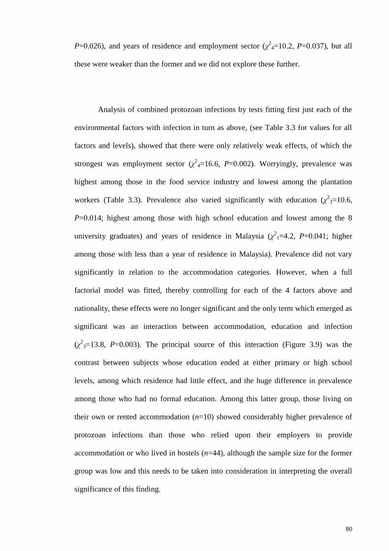
80
P=0.026), and years of residence and employment sector (χ2
4=10.2, P=0.037), but all
these were weaker than the former and we did not explore these further.
Analysis of combined protozoan infections by tests fitting first just each of the
environmental factors with infection in turn as above, (see Table 3.3 for values for all
factors and levels), showed that there were only relatively weak effects, of which the
strongest was employment sector (χ2
4=16.6, P=0.002). Worryingly, prevalence was
highest among those in the food service industry and lowest among the plantation
workers (Table 3.3). Prevalence also varied significantly with education (χ2
3=10.6,
P=0.014; highest among those with high school education and lowest among the 8
university graduates) and years of residence in Malaysia (χ2
1=4.2, P=0.041; higher
among those with less than a year of residence in Malaysia). Prevalence did not vary
significantly in relation to the accommodation categories. However, when a full
factorial model was fitted, thereby controlling for each of the 4 factors above and
nationality, these effects were no longer significant and the only term which emerged as
significant was an interaction between accommodation, education and infection
(χ2
3=13.8, P=0.003). The principal source of this interaction (Figure 3.9) was the
contrast between subjects whose education ended at either primary or high school
levels, among which residence had little effect, and the huge difference in prevalence
among those who had no formal education. Among this latter group, those living on
their own or rented accommodation (n=10) showed considerably higher prevalence of
protozoan infections than those who relied upon their employers to provide
accommodation or who lived in hostels (n=44), although the sample size for the former
group was low and this needs to be taken into consideration in interpreting the overall
significance of this finding.
81
3.3.4.2 Individual helminth species
A. lumbricoides. With nationality taken into account, prevalence varied
significantly in relation to each of the 4 environmental factors examined (Table 3.4;
years of residence, χ2
1=18.5, P<0.001; Educational level, χ2
3=14.9, P=0.002;
accommodation, χ2
1=15.3, P<0.001; Employment sector, χ2
4=54.0, P<0.001). The
highest prevalence was among subjects in manufacturing, those living in hostels,
residents under one year and perhaps surprisingly among those with a university
education, although in the latter case, the sample size was small. There were also
several relatively weak more complex interactions with infection (Accommodation x
Nationality χ2
4=9.5, P=0.049, Years resident x Employment sector χ2
4=10.8, P=0.029,
Educational level x Nationality χ2
12=28.3, P=0.005 and Educational level x
Employment sector χ2
12=24.5, P=0.017) which we did not explore further.
Hookworms. With host age taken into account, none of the environmental
factors affected prevalence of hookworms significantly (Table 3.4).
T. trichiura. With sex taken into account, the only environmental factors that
significantly affected prevalence were employment sector (χ2
4=19.7, P=0.001) and
years of residency (χ2
1=8.1, P=0.004). Prevalence was highest among those employed
in construction and lowest among those in the manufacturing industries (Table 3.4), and
higher among subjects with less than a year‘s residency relative to those with more than
year‘s residency. Accommodation type was marginally significant when fitted on its
own with infection (χ21=4.1, P=0.044) but not when other factors were also part of the
model.
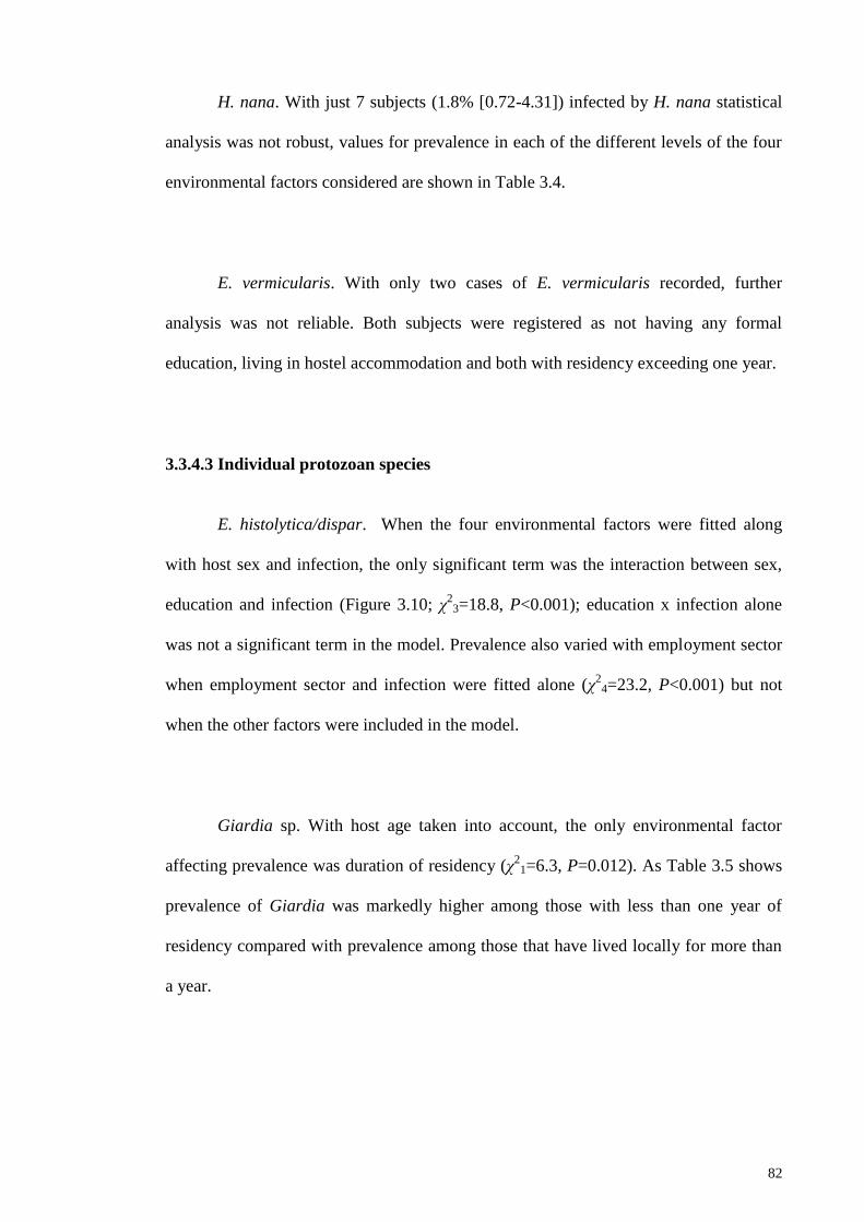
82
H. nana. With just 7 subjects (1.8% [0.72-4.31]) infected by H. nana statistical
analysis was not robust, values for prevalence in each of the different levels of the four
environmental factors considered are shown in Table 3.4.
E. vermicularis. With only two cases of E. vermicularis recorded, further
analysis was not reliable. Both subjects were registered as not having any formal
education, living in hostel accommodation and both with residency exceeding one year.
3.3.4.3 Individual protozoan species
E. histolytica/dispar. When the four environmental factors were fitted along
with host sex and infection, the only significant term was the interaction between sex,
education and infection (Figure 3.10; χ2
3=18.8, P<0.001); education x infection alone
was not a significant term in the model. Prevalence also varied with employment sector
when employment sector and infection were fitted alone (χ2
4=23.2, P<0.001) but not
when the other factors were included in the model.
Giardia sp. With host age taken into account, the only environmental factor
affecting prevalence was duration of residency (χ2
1=6.3, P=0.012). As Table 3.5 shows
prevalence of Giardia was markedly higher among those with less than one year of
residency compared with prevalence among those that have lived locally for more than
a year.
83
Cryptosporidium spp. This species was detected in 12 subjects (3.1% [1.55-
5.95). We fitted a model with nationality, the 4 environmental factors and infection. The
only significant factor to emerge from this analysis was employment sector (Table 3.5;
χ2
4=12.8, P=0.012). Prevalence was clearly highest among food service workers, and
much lower among those in other types of employment, with no infections at all
detected among those in the domestic sector.
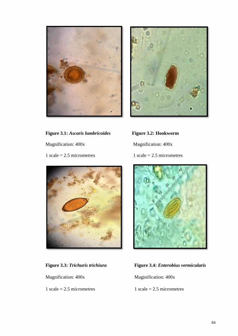
84
Figure 3.1: Ascaris lumbricoides Figure 3.2: Hookworm
Magnification: 400x Magnification: 400x
1 scale = 2.5 micrometres 1 scale = 2.5 micrometres
Figure 3.3: Trichuris trichiura Figure 3.4: Enterobius vermicularis
Magnification: 400x Maginification: 400x
1 scale = 2.5 micrometres 1 scale = 2.5 micrometres
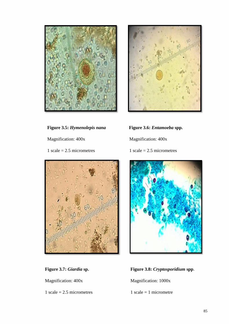
85
Figure 3.5: Hymenolepis nana Figure 3.6: Entamoeba spp.
Magnification: 400x Magnification: 400x
1 scale = 2.5 micrometres 1 scale = 2.5 micrometres
Figure 3.7: Giardia sp. Figure 3.8: Cryptosporidium spp.
Magnification: 400x Magnification: 1000x
1 scale = 2.5 micrometres 1 scale = 1 micrometre
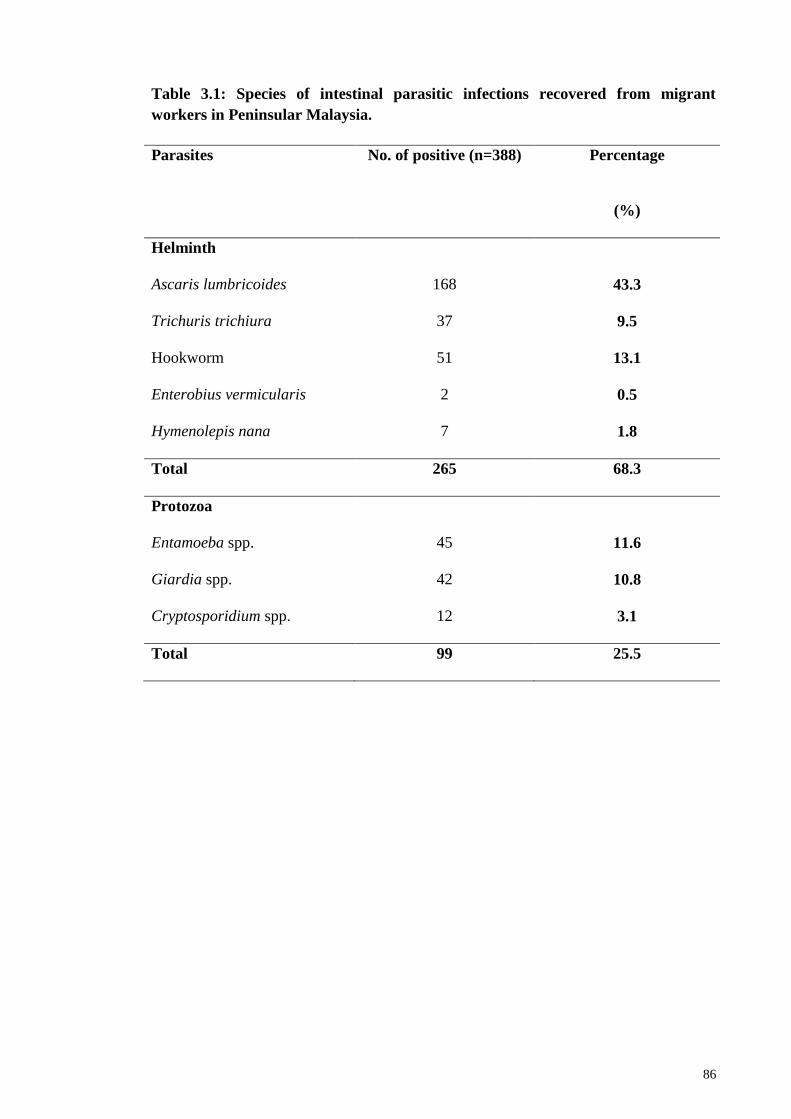
86
Table 3.1: Species of intestinal parasitic infections recovered from migrant
workers in Peninsular Malaysia.
Parasites No. of positive (n=388) Percentage
(%)
Helminth
Ascaris lumbricoides 168 43.3
Trichuris trichiura 37 9.5
Hookworm 51 13.1
Enterobius vermicularis 2 0.5
Hymenolepis nana 7 1.8
Total 265 68.3
Protozoa
Entamoeba spp. 45 11.6
Giardia spp. 42 10.8
Cryptosporidium spp. 12 3.1
Total 99 25.5
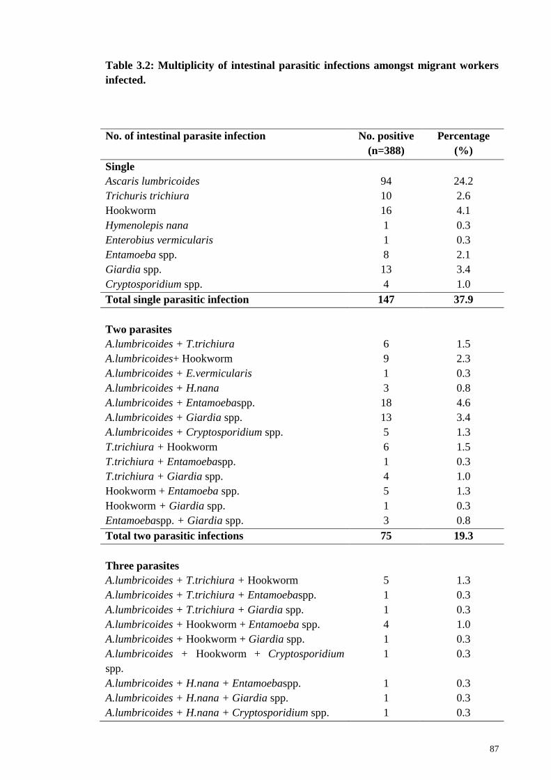
87
Table 3.2: Multiplicity of intestinal parasitic infections amongst migrant workers
infected.
No. of intestinal parasite infection No. positive
(n=388)
Percentage
(%)
Single
Ascaris lumbricoides 94 24.2
Trichuris trichiura 10 2.6
Hookworm 16 4.1
Hymenolepis nana 1 0.3
Enterobius vermicularis 1 0.3
Entamoeba spp. 8 2.1
Giardia spp. 13 3.4
Cryptosporidium spp. 4 1.0
Total single parasitic infection 147 37.9
Two parasites
A.lumbricoides + T.trichiura 6 1.5
A.lumbricoides+ Hookworm 9 2.3
A.lumbricoides + E.vermicularis 1 0.3
A.lumbricoides + H.nana 3 0.8
A.lumbricoides + Entamoebaspp. 18 4.6
A.lumbricoides + Giardia spp. 13 3.4
A.lumbricoides + Cryptosporidium spp. 5 1.3
T.trichiura + Hookworm 6 1.5
T.trichiura + Entamoebaspp. 1 0.3
T.trichiura + Giardia spp. 4 1.0
Hookworm + Entamoeba spp. 5 1.3
Hookworm + Giardia spp. 1 0.3
Entamoebaspp. + Giardia spp. 3 0.8
Total two parasitic infections 75 19.3
Three parasites
A.lumbricoides + T.trichiura + Hookworm 5 1.3
A.lumbricoides + T.trichiura + Entamoebaspp. 1 0.3
A.lumbricoides + T.trichiura + Giardia spp. 1 0.3
A.lumbricoides + Hookworm + Entamoeba spp. 4 1.0
A.lumbricoides + Hookworm + Giardia spp. 1 0.3
A.lumbricoides + Hookworm + Cryptosporidium
spp.
1 0.3
A.lumbricoides + H.nana + Entamoebaspp. 1 0.3
A.lumbricoides + H.nana + Giardia spp. 1 0.3
A.lumbricoides + H.nana + Cryptosporidium spp. 1 0.3
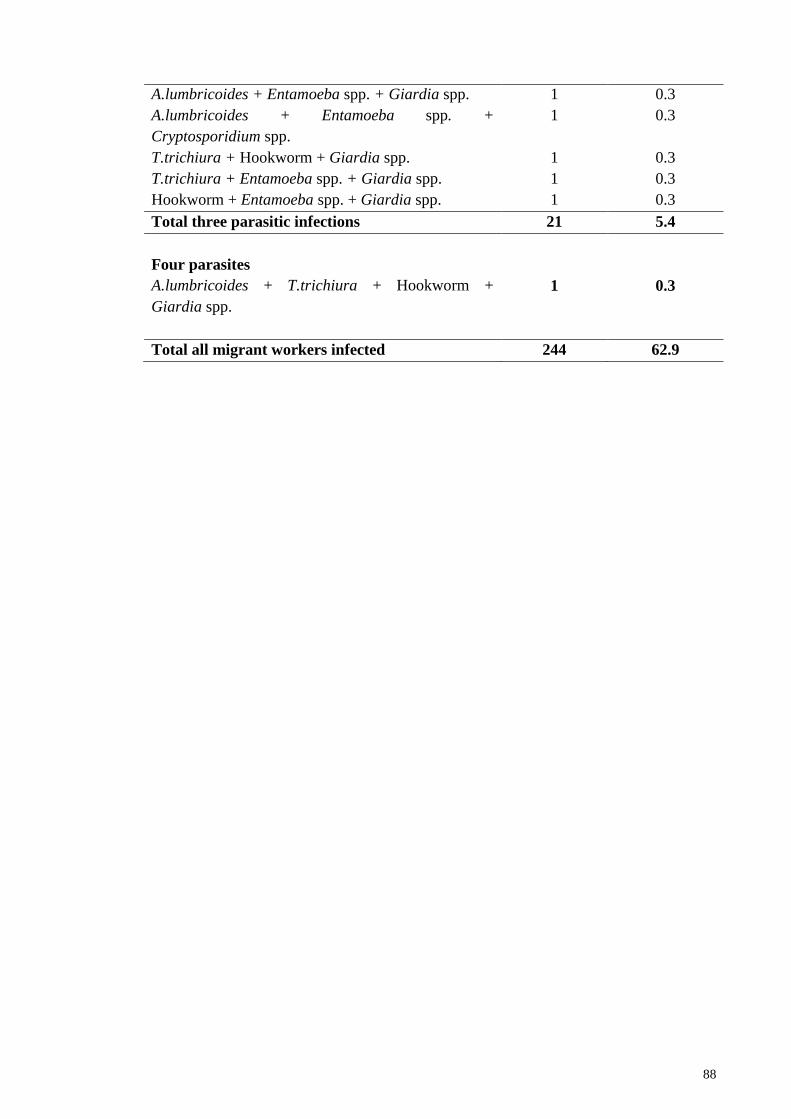
88
A.lumbricoides + Entamoeba spp. + Giardia spp. 1 0.3
A.lumbricoides + Entamoeba spp. +
Cryptosporidium spp.
1 0.3
T.trichiura + Hookworm + Giardia spp. 1 0.3
T.trichiura + Entamoeba spp. + Giardia spp. 1 0.3
Hookworm + Entamoeba spp. + Giardia spp. 1 0.3
Total three parasitic infections 21 5.4
Four parasites
A.lumbricoides + T.trichiura + Hookworm +
Giardia spp.
1 0.3
Total all migrant workers infected 244 62.9
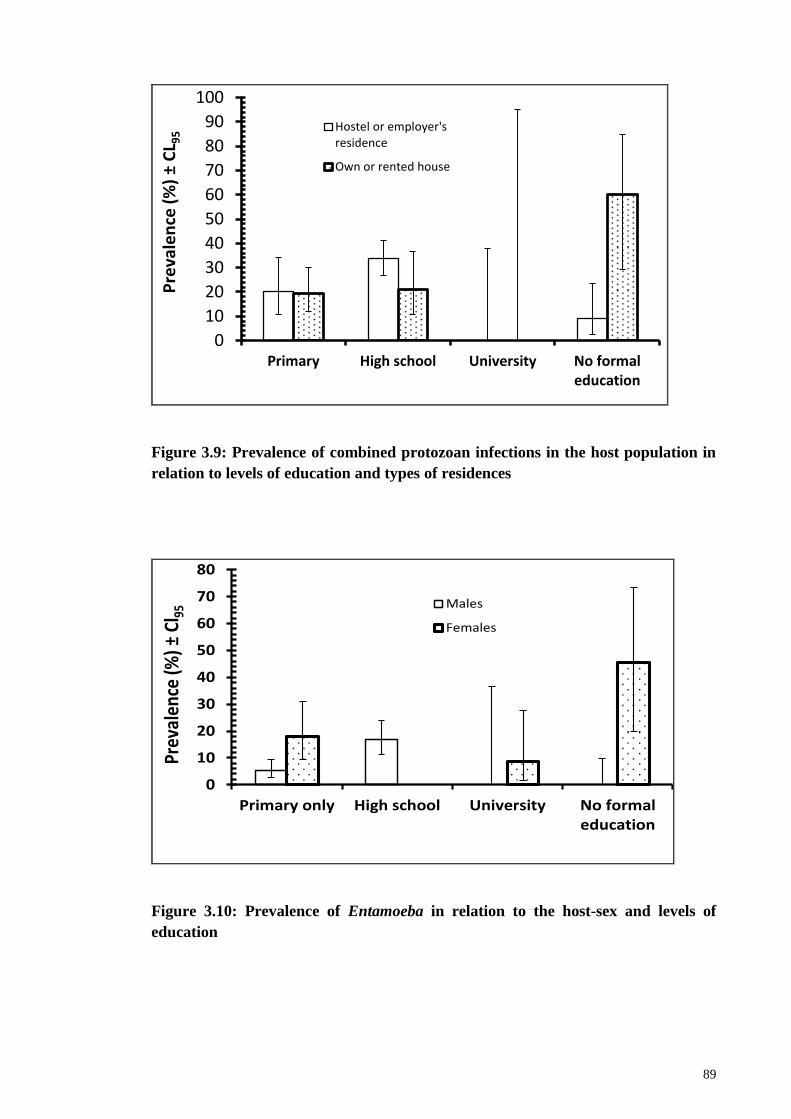
89
0
10
20
30
40
50
60
70
80
90
100
Primary High school University No formaleducation
Hostel or employer'sresidence
Own or rented houseP
reva
len
ce (
%)
±C
L 95
Figure 3.9: Prevalence of combined protozoan infections in the host population in
relation to levels of education and types of residences
0
10
20
30
40
50
60
70
80
Primary only High school University No formaleducation
Males
Females
Prev
alen
ce (%
) ±Cl
95
Figure 3.10: Prevalence of Entamoeba in relation to the host-sex and levels of
education
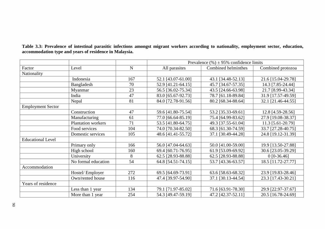
90
Table 3.3: Prevalence of intestinal parasitic infections amongst migrant workers according to nationality, employment sector, education,
accommodation type and years of residence in Malaysia.
Prevalence (%) ± 95% confidence limits
Factor Level N All parasites Combined helminthes Combined protozoa
Nationality
Indonesia 167 52.1 [43.07-61.00] 43.1 [34.48-52.13] 21.6 [15.04-29.78]
Bangladesh 70 52.9 [41.21-64.15] 45.7 [34.67-57.35] 14.3 [7.85-24.44]
Myanmar 23 56.5 [36.02-75.34] 43.5 [24.66-63.98] 21.7 [8.99-43.34]
India 47 83.0 [65.67-92.73] 78.7 [61.18-89.84] 31.9 [17.57-49.59]
Nepal 81 84.0 [72.78-91.56] 80.2 [68.34-88.64] 32.1 [21.46-44.55]
Employment Sector
Construction 47 59.6 [41.80-75.54] 53.2 [35.33-69.61] 12.8 [4.59-28.56]
Manufacturing 61 77.0 [66.64-85.19] 75.4 [64.99-83.62] 27.9 [19.08-38.37]
Plantation workers 71 53.5 [41.80-64.75] 49.3 [37.55-61.04] 11.3 [5.61-20.79]
Food services 104 74.0 [70.34-82.50] 68.3 [61.30-74.59] 33.7 [27.28-40.75]
Domestic services 105 48.6 [41.41-55.72] 37.1 [30.49-44.28] 24.8 [19.12-31.39]
Educational Level
Primary only 166 56.0 [47.04-64.63] 50.0 [41.00-59.00] 19.9 [13.50-27.88]
High school 160 69.4 [60.71-76.95] 61.9 [53.09-69.92] 30.6 [23.05-39.29]
University 8 62.5 [28.93-88.88] 62.5 [28.93-88.88] 0 [0-36.46]
No formal education 54 64.8 [54.51-74.15] 53.7 [43.36-63.57] 18.5 [11.72-27.77]
Accommodation
Hostel/ Employer 272 69.5 [64.69-73.91] 63.6 [58.63-68.32] 23.9 [19.83-28.46]
Own/rented house 116 47.4 [39.97-54.90] 37.1 [30.13-44.54] 23.3 [17.43-30.21]
Years of residence
Less than 1 year 134 79.1 [71.97-85.02] 71.6 [63.91-78.30] 29.9 [22.97-37.67]
More than 1 year 254 54.3 [49.47-59.19] 47.2 [42.37-52.11] 20.5 [16.78-24.69]
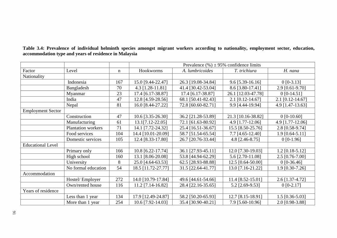
91
Table 3.4: Prevalence of individual helminth species amongst migrant workers according to nationality, employment sector, education,
accommodation type and years of residence in Malaysia
Prevalence (%) ± 95% confidence limits
Factor Level n Hookworms A. lumbricoides T. trichiura H. nana
Nationality
Indonesia 167 15.0 [9.44-22.47] 26.3 [19.08-34.84] 9.6 [5.39-16.16] 0 [0-3.13]
Bangladesh 70 4.3 [1.28-11.81] 41.4 [30.42-53.04] 8.6 [3.80-17.41] 2.9 [0.61-9.70]
Myanmar 23 17.4 [6.17-38.87] 17.4 [6.17-38.87] 26.1 [12.03-47.78] 0 [0-14.51]
India 47 12.8 [4.59-28.56] 68.1 [50.41-82.43] 2.1 [0.12-14.67] 2.1 [0.12-14.67]
Nepal 81 16.0 [8.44-27.22] 72.8 [60.60-82.71] 9.9 [4.44-19.94] 4.9 [1.47-13.63]
Employment Sector
Construction 47 10.6 [3.35-26.30] 36.2 [21.28-53.89] 21.3 [10.16-38.82] 0 [0-10.60]
Manufacturing 61 13.1[7.12-22.05] 72.1 [61.63-80.92] 4.9 [1.77-12.06] 4.9 [1.77-12.06]
Plantation workers 71 14.1 [7.72-24.32] 25.4 [16.51-36.67] 15.5 [8.50-25.76] 2.8 [0.58-9.74]
Food services 104 14.4 [10.01-20.09] 58.7 [51.54-65.54] 7.7 [4.65-12.40] 1.9 [0.64-5.11]
Domestic services 105 12.4 [8.33-17.80] 26.7 [20.76-33.44] 4.8 [2.46-8.75] 0 [0-1.96]
Educational Level
Primary only 166 10.8 [6.22-17.74] 36.1 [27.93-45.11] 12.0 [7.30-19.03] 1.2 [0.18-5.12]
High school 160 13.1 [8.06-20.08] 53.8 [44.94-62.29] 5.6 [2.70-11.08] 2.5 [0.76-7.00]
University 8 25.0 [4.64-63.53] 62.5 [28.93-88.88] 12.5 [0.64-50.00] 0 [0-36.46]
No formal education 54 18.5 [11.72-27.77] 31.5 [22.64-41.77] 13.0 [7.16-21.22] 1.9 [0.30-7.26]
Accommodation
Hostel/ Employer 272 14.0 [10.79-17.84] 49.6 [44.61-54.66] 11.4 [8.52-15.01] 2.6 [1.37-4.72]
Own/rented house 116 11.2 [7.14-16.82] 28.4 [22.16-35.65] 5.2 [2.69-9.53] 0 [0-2.17]
Years of residence
Less than 1 year 134 17.9 [12.49-24.87] 58.2 [50.20-65.93] 12.7 [8.15-18.91] 1.5 [0.36-5.03]
More than 1 year 254 10.6 [7.92-14.03] 35.4 [30.90-40.21] 7.9 [5.60-10.96] 2.0 [0.98-3.88]
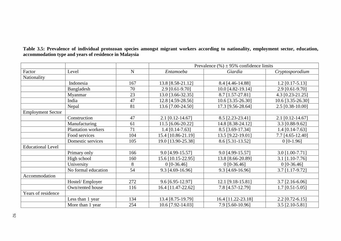
92
Table 3.5: Prevalence of individual protozoan species amongst migrant workers according to nationality, employment sector, education,
accommodation type and years of residence in Malaysia
Prevalence (%) ± 95% confidence limits
Factor Level N Entamoeba Giardia Cryptosporodium
Nationality
Indonesia 167 13.8 [8.58-21.12] 8.4 [4.46-14.88] 1.2 [0.17-5.13]
Bangladesh 70 2.9 [0.61-9.70] 10.0 [4.82-19.14] 2.9 [0.61-9.70]
Myanmar 23 13.0 [3.66-32.35] 8.7 [1.57-27.81] 4.3 [0.23-21.25]
India 47 12.8 [4.59-28.56] 10.6 [3.35-26.30] 10.6 [3.35-26.30]
Nepal 81 13.6 [7.00-24.50] 17.3 [9.56-28.64] 2.5 [0.38-10.00]
Employment Sector
Construction 47 2.1 [0.12-14.67] 8.5 [2.23-23.41] 2.1 [0.12-14.67]
Manufacturing 61 11.5 [6.06-20.22] 14.8 [8.38-24.12] 3.3 [0.88-9.62]
Plantation workers 71 1.4 [0.14-7.63] 8.5 [3.69-17.34] 1.4 [0.14-7.63]
Food services 104 15.4 [10.86-21.19] 13.5 [9.22-19.01] 7.7 [4.65-12.40]
Domestic services 105 19.0 [13.90-25.38] 8.6 [5.31-13.52] 0 [0-1.96]
Educational Level
Primary only 166 9.0 [4.99-15.57] 9.0 [4.99-15.57] 3.0 [1.00-7.71]
High school 160 15.6 [10.15-22.95] 13.8 [8.66-20.89] 3.1 [1.10-7.76]
University 8 0 [0-36.46] 0 [0-36.46] 0 [0-36.46]
No formal education 54 9.3 [4.69-16.96] 9.3 [4.69-16.96] 3.7 [1.17-9.72]
Accommodation
Hostel/ Employer 272 9.6 [6.95-12.97] 12.1 [9.18-15.81] 3.7 [2.16-6.06]
Own/rented house 116 16.4 [11.47-22.62] 7.8 [4.57-12.79] 1.7 [0.51-5.05]
Years of residence
Less than 1 year 134 13.4 [8.75-19.79] 16.4 [11.22-23.18] 2.2 [0.72-6.15]
More than 1 year 254 10.6 [7.92-14.03] 7.9 [5.60-10.96] 3.5 [2.10-5.81]
93
3.4 Discussion
The demand for low and semi-skilled workers in several sectors in Malaysia has
seen a dramatic rise in the number of workers entering the country from 1.06 million in
2002 to 2.07 million in 2014 (Bardan, 2014). The presence of such a substantial foreign
work force originating from countries where parasitic infections are endemic is a major
concern especially as this community is highly dynamic, and the emerging and re-
emerging infectious diseases that they may carry are a great concern. For the present
study we successfully recruited 388 migrant workers from their workplace who
provided stool specimens compared to 173 stool specimens of clinically ill subjects
from the University Malaya Medical Centre (UMMC) in the previous study (Suresh et
al., 2002). Recruiting migrant workers to participate in the present study was very
difficult since most workers and employers in Malaysia refused to participate. The main
reason given by employers was that this procedure was not compulsory by the
FOMEMA (agency involved in the implementation, management and supervision of a
nationwide mandatory health screening programme for all legal migrant workers in
Malaysia), Ministry of Health and Immigration Department of Ministry of Home
Affairs Malaysia upon entry / residing in Malaysia. Other reasons often given by the
workers and their employers included lack of interest in participating, disgust with
faeces and preoccupation with matters related to work and achieving important
deadlines.
Our study identified a two-fold increase of IPIs (62.9%) among workers
compared to a decade ago (36.0%) (Suresh et al., 2002). Studies reporting analyses of
parasitic infections among various communities in Malaysia have been conducted also
among the Orang Asli (44.33%-99.2%) (Dunn, 1972; Dissanaike et al., 1977; Al-
94
Mekhlafi et al., 2005a; 2006; Ngui et al., 2011a; Nasr et al., 2013; Sinniah et al., 2014),
plantation and rural communities (32.3%-70.0%) (Bisseru & Aziz, 1970; Lo et al.,
1979; Al-Mekhlafi et al., 2007; 2008; Sinniah et al., 2011), slum dwellers (20.6%-
90.9%) (Chia et al., 1978; Sinniah et al., 2014), fishing communities (54.2%-98.0%)
(Heyneman et al., 1967; Balasingam et al., 1969; Nawalinski & Roundy, 1978; Anuar
et al., 1978; Sinniah et al., 1988) and flat dwellers (5.1%-57.0%) (Kan, 1983; Che
Ghani et al., 1989; Sinniah et al., 2002; 2014). Our findings based on migrant workers
are in agreement with other studies on poverty- stricken communities in Malaysia
although some studies have reported fluctuations in prevalence values especially among
the slum dwellers (90.9 % in 1978 to 20.6% in 2014) (Chia et al., 1978; Sinniah et al.,
2014), flat dwellers (57% in 1983 to 5.5% in 2014) (Kan, 1983; Sinniah et al., 2014)
and rural communities (90.0% in 1970 to 32.3% in 2014) (Bisseru & Aziz, 1970;
Sinniah et al., 2014). A total of 8 species of parasites were identified (A. lumbricoides,
T. trichiura, hookworm, E. vermicularis, H. nana, Entamoeba sp., Giardia sp. and
Cryptosporidium spp.), compared to only 6 species recorded previously (A.
lumbricoides, T. trichiura, hookworm, H. nana, Giardia sp. and Blastocystis sp.) among
migrant workers (Suresh et al., 2002). This outcome is not surprising as it reflects the
government‘s failure to include mandatory STH screening as part of the requirement for
working in this country.
Soil-transmitted helminth (STH) (68.3%) infections were more prevalent
compared to protozoan infections (25.5%). Of the three common intestinal nematodes,
A. lumbricoides (43.3%) infections were the most frequently identified, followed by
hookworm (13.1%) and T. trichiura (9.5%). In contrast, a study more than a decade ago
highlighted hookworm infections as the most prevalent (Suresh et al., 2002). However,
our result concurs with global findings highlighting A. lumbricoides infections as the
95
most common helminth among the underprivileged communities (World Health
Organization, 2015). A high presence of A. lumbricoides eggs contaminating public
parks in Peninsular Malaysia has also been reported recently (Rahman et al., 2015).
The demographic profiles of respondents comprised predominantly volunteers
from rural areas in their respective countries of origin where IPIs are still very much
prevalent and a major concern among the poor and in deprived communities,
particularly among workers from India and Nepal where prevalence can exceed 80%.
The latest study in the low socio-economic areas of South Chennai documented a
prevalence of 75.7% with IPI (Dhanabal et al., 2014), especially in children from rural
and urban locations among whom prevalence with A. lumbricoides ranged between 60
to 91% (Fernandez et al., 2002). This was the most common helminth infection in this
community (52.8%). Both studies suggest that inadequate sanitation and poor drainage
is likely to have contributed to disease prevalence. Similarly, parasitic infections in
Nepal have also been reported as being linked to rapid, unplanned urbanization, open
defaecation and other unhygienic habits, as well as a lack of health awareness (Singh et
al., 2013; Rabindranath et al., 2006; Uga et al., 2004).
Among the significant explanatory factors associated with the high prevalence
of parasitic infections in this country were two main factors i.e, the number of working
years in Malaysia and anthelmintic treatment. Workers with an employment history of
less than a year or newly arrive workers in Malaysia were those who were most likely to
be infected. In addition, they were also most likely to have no history of taking any
anthelmintic drugs in the last 12 months. This is not surprising as the mandatory
medical screening procedure upon entry to this country excludes examination for IPIs
96
and does not require administration of anthelmintic drugs to newly arrived workers
(FOMEMA, 2015). Therefore our findings call for an improvement in health screening
in future to include screening for parasitic infections and compulsory administration of
anthelmintic drugs to workers upon entering Malaysia for employment. Such
requirement is already implemented in some countries, that depend on an immigrant
workforce, as for example in Qatar where currently prospective workers are required to
undergo health checks at approved health clinics in their country of origin and if
infection with helminths is detected, are routinely given albendazole prior to arrival as a
condition for entry, residence and issuance of a work permit (Abu-Madi et al., 2010;
2011). Moreover those working in the food service industry have to undergo subsequent
annual compulsory examinations by the Medical Commission as a condition of the
continuation of their work permits.
Transmission of intestinal nematode infections within the community is
predominantly dependent on human behaviour, particularly during eating and
defaecation, personal hygiene, and cleanliness. The high prevalence of parasitic
infections among the immigrant community sampled in this study provides an insight
into the conditions under which the subjects live, and reflects the availability of
environmental sanitation as well as the socioeconomic status of this sector of the
population in Malaysia (World Health Organization, 2015).
97
3.5 Conclusion
High intestinal parasitic infections amongst migrant workers of all five working
sectors in Malaysia was reported in this study and highlight the urgent need to refine
the current health polices for workers entering the country for employment to include
mandatory screening for parasitic infections. It is also recommended that workers be
exposed to health education campaigns and programs aimed at increasing in the
community awareness of the importance of personal hygiene, sanitation, cleanliness and
healthy behaviors in controlling parasitic infections.
98
CHAPTER 4: SEROPREVALENCE OF TOXOPLASMA GONDII INFECTIONS
AMONG MIGRANT WORKERS IN MALAYSIA
4.1 Introduction
Toxoplasma gondii is one of the most common protozoan parasites affecting up
to one-third of the world's population (Feldman, 1974; Montoya & Liesenfeld, 2004;
Hill et al., 2005). Human infection may occur via ingestion of food or water
contaminated with oocysts shed in faeces of infected cats, consumption of undercooked
or raw meat of venison, rabbits, raw oysters, clams, or mussels containing tissue cysts
(Jacobs, 1974; Abu Bakar et al., 1992; Kolbekova´ et al., 2007; Ferguson, 2009; Jones
et al., 2009), exposure to contaminated soil through activities such as gardening or
children playing in sandpits (Petersen et al., 2010), blood transfusion or organ
transplantation from infected donors (Siegel et al., 1971; Derouin & Pelloux, 2008) and
vertical transmission from mother to fetus (Husni et al., 1994; Remington & Klein,
2001). Damage to the brain, eyes, or other organs can occur from a severe acute
infection or through reactivation of past infection. Infections acquired during pregnancy
may cause severe damage to the fetus (Remington & Klein, 2001). In
immunocompromised patients, reactivation of latent disease can cause life-threatening
encephalitis (Hanifa et al., 1996; Montoya & Liesenfeld, 2004). The standard method of
diagnosis of this infection is through serological testing, based on the detection of
Toxoplasma-specific immunoglobulin IgG and IgM antibodies in serum and is routinely
practiced in many parts of the world (Alvarado-Esquivel et al., 2006; Binnicker et al.,
2010; Mwambe et al., 2013).
99
Over the years, the number of migrant workers in Malaysia has grown
exponentially. The recruitment of workers to Malaysia from neighboring countries has
raised concerns that diseases endemic to their countries may be inadvertently brought
into the country (Chan et al., 2009a). Despite compulsory medical screening prior to
entering the workforce, parasitic infections screening including for T. gondi is lacking.
In Malaysia, the previous reports on seroprevalence of T. gondii used indirect
hemagglutination (IHA) test, Sabin Feldman dye test, indirect fluorescent antibody test
(Yahaya, 1991) and the enzyme-linked immunosorbent assay (ELISA) (Nissapatorn et
al., 2002).
To date, several studies among migrant workers in Malaysia highlighted the
high presence of T. gondi infection, up to 42% (138/336) with positive IgG while
twenty workers (6%) were positive with IgM particularly among Indonesian plantation
workers and workers in detention camps (Chan et al., 2009a). Prior to that, Chan et al.
in 2008 recorded the highest infection rate among Nepalese workers (46.2%) compared
to other ethnic groups. Similarly, another serological study showed that just over a third
(34.1%, 171/501) of migrant plantation workers and individuals in detention camps
were IgG positive and 5.2% (26/501) were IgM positive (Chan et al., 2009b).
Subsequently, Chan et al. (2009b) also noted that high numbers of local workers (n=89,
44.9%) were IgG positive in plantations as compared to migrants (n=171, 34.1%).
However, workers with raised IgM were lower among locals (n=17, 8.6%), although not
significantly, compared to migrants (n=26, 5.2%). On the other hand, Amal et al. (2008)
noted lower rate of raised specific IgG among workers (n=16, 18.8%) from the Indian
subcontinent from the same plantation and detention camp compared to locals (n=89,
44.9%).
100
There is a need to determine the health status among migrant workers with
regard to T. gondii infection as most workers originate from countries with low
socioeconomic backgrounds and live in deprived environments with poor sanitation and
low hygiene practices (Norhayati et al., 2003). Therefore, this study was undertaken to
determine the seroprevalence of T. gondii in the migrant workers and factors associated
with the infection.
101
4.2 Materials and Methods
4.2.1 Study population and sample collection
A total of 484 from 610 volunteers of migrant workers in Malaysia agreed to
participate in blood sample collection. Questionnaires were distributed to the
participants to gather relevant information related to the study. All participants were
fully informed of the nature of the study for their maximum co-operation and
completion of consent forms. Details of the participants, composition of the
questionnaire and ethical clearance of this study are as described in Chapter 2.
After consent was obtained and questionnaire answered, approximately 5ml of
venous blood were drawn by trained medical assistants and nurses using disposable
syringes and needles into a plain tube (without anticoagulant) from the participants. The
bloods were transported back to the Parasitology Lab, Institute of Biological Science,
Faculty of Science, University of Malaya. Blood samples were spun at 1,500 rpm for 10
minutes and the sera were kept in -20°C until use.
4.2.2 Detection of immunoglobulin G and M antibodies to T. gondii
Toxoplasmosis was screened using enzyme-linked immunosorbent assay
(ELISA) commercial kit for immunoglobulin G (IgG) and M (IgM) (The Trinity
Biotech CaptiaTM
, New York) in accordance to the manufacturer‘s instructions. The
process was performed at the Biohealth Laboratory, Institute of Biological Science,
Faculty of Science, University of Malaya.
102
All reagents were removed from refrigeration and allowed to come to room
temperature before use (21° to 25° C). All samples and controls were vortexed before
use. 50 mL of the 20X Wash Buffer Type I were diluted to 1 L with distilled and/or
deionized H20. The desired numbers of strips were placed into a microwell frame. Four
(4) calibrator were determined (one Negative Control, two Calibrators and one Positive
Control) per run. A reagent blank (RB) was ran on each assay.
For the IgG assays, test sera, Calibrator and Control sera 1:21 (e.g., 10 μL + 200
μL) were diluted in Serum Diluent Plus. 100 μL of diluted patient sera, Calibrator and
Control sera were added to individual wells. 100μL of Serum Diluent Plus was added to
the reagent blank well. Each well was incubated at room temperature (21° to 25° C) for
25 minutes +/- 5 minutes. Liquid was aspirated or shake out from all wells. Using semi-
automated or automated washing equipment, 250-300 μL of diluted Wash Buffer was
added to each well. The wash procedure was repeated two times (for a total of three
washes) for semi-automated equipment or four times (for a total of five washes) for
automated equipment. After the final wash, the plate was blotted dry on paper toweling
to remove all liquid from the wells. 100 μL Conjugate was added to each well,
including the reagent blank well. Each well was incubated 25 minutes +/- 5 minutes at
room temperature (21° to 25° C). Washing procedure was repeated as described above.
100 μL Chromogen/Substrate (TMB) solutions was added to each well, including
reagent blank well, maintaining a constant rate of addition across the plate. Each well
was incubated at room temperature (21° to 25° C) for 10-15 minutes. 100 μL of Stop
Solution (1N H2SO4) was added to stop the reaction following the same order of
Chromogen/Substrate addition, including reagent blank well. The plate was tap gently
along the outsides to mix contents of the wells. The plate may be held up to one (1)
hour after addition of the Stop Solution before reading. The developed color should be
103
read on an ELISA plate reader equipped with a 450 nm filter. The instrument was
blanked on air. The reagent blank must be less than 0.150 Absorbance at 450 nm. If the
reagent blank was > 0.150, the run was repeated. Positive results were defined as >51
IU/ml, indicating latent or pre-existing Toxoplasma infection.
For IgM assays, test sera, Calibrator and Control sera 1:81 (e.g., 10 μL + 800
μL) were diluted in Serum Diluent Plus. 100 μL of diluted patient sera, Calibrator and
Control sera were added to individual wells. 100μL of Serum Diluent Plus was added to
the reagent blank well. Each well was incubated at room temperature (21° to 25° C) for
30 minutes +/- 2 minutes. Liquid was aspirated or shake out from all wells. Using semi-
automated or automated washing equipment, 250-300 μL of diluted Wash Buffer was
added to each well. The wash procedure was repeated two times (for a total of three
washes) for semi-automated equipment or four times (for a total of five washes) for
automated equipment. After the final wash, the plate was blotted dry on paper toweling
to remove all liquid from the wells. 100 μL Conjugate was added to each well,
including the reagent blank well. Each well was incubated 30 minutes +/- 2 minutes at
room temperature (21° to 25° C). Washing procedure was repeated as described above.
100 μL Chromogen/Substrate (TMB) solutions was added to each well, including
reagent blank well, maintaining a constant rate of addition across the plate. Each well
was incubated at room temperature (21° to 25° C) for 15 minutes +/- 2 minutes. 100 μL
of Stop Solution (1N H2SO4) was added to stop the reaction following the same order
of Chromogen/Substrate addition, including reagent blank well. The plate was tap
gently along the outsides to mix contents of the wells. The plate may be held up to one
(1) hour after addition of the Stop Solution before reading. The developed color should
be read on an ELISA plate reader equipped with a 450 nm filter. The instrument was
blanked on air. The reagent blank must be less than 0.150 Absorbance at 450 nm. If the
104
reagent blank was > 0.150, the run was repeated. Positive results for IgM assays were
defined as >51 IU/ml, indicating recently acquired Toxoplasma infection.
In addition, all samples that were both IgG-positive and IgM-positive were
tested using the IgG avidity assay (IgG; NovaLisa, Dietzenbach, Germany) according to
manufacturer‘s instruction. All samples were diluted 1+100 with IgG Sample Diluent.
10μl sample and 1ml IgG Sample Diluent were dispensed into tubes to obtain a 1+100
dilution and thoroughly mixed with a Vortex. Wells were covered with the foil supplied
in the kit and incubated for 1 hour ± 5 min at 37±1°C. Then, the content off the wells
were aspirated and washed three times with 300μl of washing solution. 100μl
Toxoplasma anti-IgG Conjugate was dispensed into all wells except for the blank well,
covered with foil and incubated for 30 min at room temperature (20 to 25°C). Washing
procedure was repeated. 100μl TMB Substrate Solution was dispensed into all wells and
incubated for exactly 15 min at room temperature (20 to 25°C) in the dark. 100μl Stop
Solution was dispensed into all wells in the same order and at the same rate as for the
TMB Substrate Solution. Measure the absorbance of the specimen at 450/620nm within
30 min after addition of the Stop Solution. Toxoplasma antibodies with high avidity
(>40%) indicates past infection, meanwhile Toxoplasma antibodies with low avidity
(≤40) indicates acute or recent infections.
4.2.3 Data analysis
Prevalence data (percentage of subjects infected) are shown with 95%
confidence limits (CL95), as described by Rohlf & Sokal (1995) using bespoke software.
Prevalences were analyzed using maximum likelihood techniques based on log linear
analysis of contingency tables using the software package SPSS (Version 22). Analysis
105
was conducted with the intrinsic factors sex (2 levels, males and females), age (5 age
classes comprising those <25 years old, 25-34 years old, 35-44 years old, 45-54 years
old and those >54 years) and nationality (5 countries, Indonesia, Bangladesh, Myanmar,
India and Nepal). Infection was considered as a binary factor (presence/absence of
parasites). For extrinsic factors including employment sector (5 sectors: construction,
manufacture, plantation, food service and domestic), years of residence in Malaysia (2
categories: less than 1 year and more than 1 year), accommodation (3 types: hostel
provided by the employer, construction site and own/rent house) and education (4
levels: primary school, secondary school, university and no formal schooling).
106
4.3 Results
4.3.1 Sociodemographic characteristics
A total of 484 migrant workers in Malaysia originated from rural areas in
neighboring countries including; Indonesia (n=247, 51.0%), Nepal (n=99, 20.5%),
Bangladesh (n=72, 14.9%), India (n=52, 10.7%) and Myanmar (n=14, 2.9%) were
included in this study. Out of the 485 participants, just slightly over three quarters
(n=375, 77.5%) were males, and the rest (n=109, 22.5%) females. Most were between
the ages of 25 to 34 years old (n=183, 37.8%), followed by less than 25 years (n=142,
29.3%), 35 to 44 years (n=111, 22.9%), 45 to 54 years (n=35, 7.2%) and more than 55
years (n=13, 2.7%). According to the working sectors, the majority of volunteers were
from the food service sector (n=115, 23.8%), followed by domestic (n=106, 21.9%),
plantation (n=102, 21.1%), manufacturing (n=93, 19.2%) and construction (n=68,
14.0%) sectors. Most participants had at least a primary level education (n=228, 47.1%)
followed by high school (n=201, 41.5%) and university level (n=10, 2.1%), and 45
participants (9.3%) did not receive any formal education.
4.3.2 Seroprevalence of Toxoplasma gondii infections
The overall T. gondii seroprevalence among 484 migrant workers was 57.4%
(n= 278; CI = 52.7-61.8%) with 52.9% (n= 256; CI= 48.4-57.2%) being seropositive for
anti-Toxoplasma IgG only, 0.8% (n= 4; CI = 0.2-1.7%) seropositive for anti-
Toxoplasma IgM only and 3.7% (n= 18; CI = 2.1-5.4%) seropositive with both IgG and
IgM antibodies (Table 4.1). All 18 positive samples with both IgG and IgM antibodies
showed high avidity (> 40%), suggesting chronic infection.
107
Infection with T. gondii was found almost similar between males (213/375;
56.8%) and female workers (65/109; 59.6%). Based on age factor, highest prevalence
was found among workers age more than 55 years old (10/13; 76.9%), followed by 45
to 54 years old (26/35; 74.3%), less than 25 years old (84/142; 59.2%), 35 to 44 years
old (61/111; 55.0%) and 25 to 34 years old (97/183; 53.0%). Meanwhile, according to
country of origin, the highest prevalence were among Nepalese (77.8%), followed by
Indonesian (58.3%), Bangladeshi (45.8%), Indian (38.5%) and Myanmaris (28.6%).
The highest seroprevalence according to working sector were from the manufacturing
(76.3%) sector followed by construction (61.8%), domestic (57.5%), food service
(50.4%), and plantation (45.1%). A total of 66.1% of the infected workers have been in
Malaysia for less than a year compared to those has been living in Malaysia for more
than a year (52.3%). Based on the accommodation of the workers, prevalence of
infection was almost similar among the workers living in the construction site (62.3%),
hostel by the employer (56.4%) and own/rent house (57.9%).
4.3.2.1 Intrinsic effects on seroprevalence of IgG and IgM antibodies to T. gondii
infections
The seropositivity of T. gondii was examined in relation to sociodemographic
factors using maximum likelihood techniques based on log linear analysis. The analysis
showed that two factors were found associated with seropositivity of anti-Toxoplasma
IgG including age (Χ2
4 = 9.761, P = 0.045) and nationality (Χ2
4 = 30.046, P = <0.001)
(Table 4.2). Meanwhile, based on seropositivity of anti-Toxoplasma IgM and
seropositivity with both IgG and IgM antibodies, none of the factors were found
significant (Table 4.2).
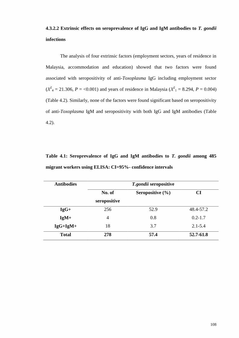
108
4.3.2.2 Extrinsic effects on seroprevalence of IgG and IgM antibodies to T. gondii
infections
The analysis of four extrinsic factors (employment sectors, years of residence in
Malaysia, accommodation and education) showed that two factors were found
associated with seropositivity of anti-Toxoplasma IgG including employment sector
(Χ2
4 = 21.306, P = <0.001) and years of residence in Malaysia (Χ2
1 = 8.294, P = 0.004)
(Table 4.2). Similarly, none of the factors were found significant based on seropositivity
of anti-Toxoplasma IgM and seropositivity with both IgG and IgM antibodies (Table
4.2).
Table 4.1: Seroprevalence of IgG and IgM antibodies to T. gondii among 485
migrant workers using ELISA: CI=95%- confidence intervals
Antibodies T.gondii seropositive
No. of
seropositive
Seropositive (%) CI
IgG+ 256 52.9 48.4-57.2
IgM+ 4 0.8 0.2-1.7
IgG+IgM+ 18 3.7 2.1-5.4
Total 278 57.4 52.7-61.8
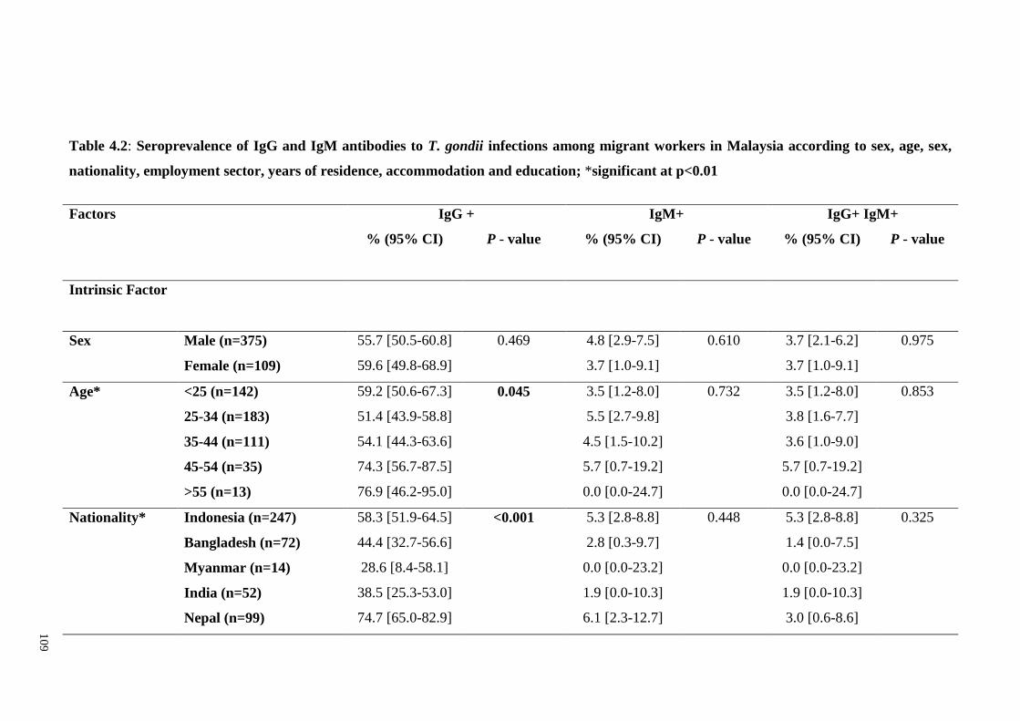
10
9
Table 4.2: Seroprevalence of IgG and IgM antibodies to T. gondii infections among migrant workers in Malaysia according to sex, age, sex,
nationality, employment sector, years of residence, accommodation and education; *significant at p<0.01
Factors
IgG + IgM+ IgG+ IgM+
% (95% CI) P - value % (95% CI) P - value % (95% CI) P - value
Intrinsic Factor
Sex Male (n=375) 55.7 [50.5-60.8] 0.469 4.8 [2.9-7.5] 0.610 3.7 [2.1-6.2] 0.975
Female (n=109) 59.6 [49.8-68.9] 3.7 [1.0-9.1] 3.7 [1.0-9.1]
Age* <25 (n=142) 59.2 [50.6-67.3] 0.045 3.5 [1.2-8.0] 0.732 3.5 [1.2-8.0] 0.853
25-34 (n=183) 51.4 [43.9-58.8] 5.5 [2.7-9.8] 3.8 [1.6-7.7]
35-44 (n=111) 54.1 [44.3-63.6] 4.5 [1.5-10.2] 3.6 [1.0-9.0]
45-54 (n=35) 74.3 [56.7-87.5] 5.7 [0.7-19.2] 5.7 [0.7-19.2]
>55 (n=13) 76.9 [46.2-95.0] 0.0 [0.0-24.7] 0.0 [0.0-24.7]
Nationality* Indonesia (n=247) 58.3 [51.9-64.5] <0.001 5.3 [2.8-8.8] 0.448 5.3 [2.8-8.8] 0.325
Bangladesh (n=72) 44.4 [32.7-56.6] 2.8 [0.3-9.7] 1.4 [0.0-7.5]
Myanmar (n=14) 28.6 [8.4-58.1] 0.0 [0.0-23.2] 0.0 [0.0-23.2]
India (n=52) 38.5 [25.3-53.0] 1.9 [0.0-10.3] 1.9 [0.0-10.3]
Nepal (n=99) 74.7 [65.0-82.9] 6.1 [2.3-12.7] 3.0 [0.6-8.6]
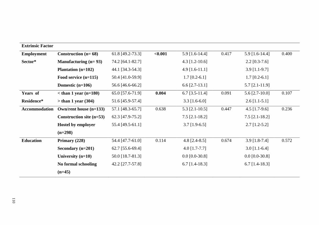
11
0
Extrinsic Factor
Employment Construction (n= 68) 61.8 [49.2-73.3] <0.001 5.9 [1.6-14.4] 0.417 5.9 [1.6-14.4] 0.400
Sector* Manufacturing (n= 93) 74.2 [64.1-82.7] 4.3 [1.2-10.6] 2.2 [0.3-7.6]
Plantation (n=102) 44.1 [34.3-54.3] 4.9 [1.6-11.1] 3.9 [1.1-9.7]
Food service (n=115) 50.4 [41.0-59.9] 1.7 [0.2-6.1] 1.7 [0.2-6.1]
Domestic (n=106) 56.6 [46.6-66.2] 6.6 [2.7-13.1] 5.7 [2.1-11.9]
Years of < than 1 year (n=180) 65.0 [57.6-71.9] 0.004 6.7 [3.5-11.4] 0.091 5.6 [2.7-10.0] 0.107
Residence* > than 1 year (304) 51.6 [45.9-57.4] 3.3 [1.6-6.0] 2.6 [1.1-5.1]
Accommodation Own/rent house (n=133) 57.1 [48.3-65.7] 0.638 5.3 [2.1-10.5] 0.447 4.5 [1.7-9.6] 0.236
Construction site (n=53) 62.3 [47.9-75.2] 7.5 [2.1-18.2] 7.5 [2.1-18.2]
Hostel by employer
(n=298)
55.4 [49.5-61.1] 3.7 [1.9-6.5] 2.7 [1.2-5.2]
Education Primary (228) 54.4 [47.7-61.0] 0.114 4.8 [2.4-8.5] 0.674 3.9 [1.8-7.4] 0.572
Secondary (n=201) 62.7 [55.6-69.4] 4.0 [1.7-7.7] 3.0 [1.1-6.4]
University (n=10) 50.0 [18.7-81.3] 0.0 [0.0-30.8] 0.0 [0.0-30.8]
No formal schooling
(n=45)
42.2 [27.7-57.8] 6.7 [1.4-18.3] 6.7 [1.4-18.3]
111
4.4 Discussion
This study investigated T. gondii infection among migrant workers in Malaysia
using standard commercial kits that detect anti-Toxoplasma IgG and IgM antibodies in
order to determine the seroprevalence and factors that may contribute to the infection.
The results showed that more than half of the workers had latent infection (53.0%), an
indication of previous exposure to T. gondi. Four males (0.8%) were positive for
specific IgM but negative for IgG antibodies, indicating possible acute infection. All
respondents in this study possessed valid working permits and were provided with
proper housing, equipped with clean water, toilets and efficient waste disposal systems.
The workers were also provided with full medical benefits and personal protective
equipment (PPE) whilst at work. Thus the high prevalence of latent T. gondii infection
among these workers probably indicated previous infection acquired from their home
countries where the infections are prevalent (Gandahusada, 1991; Rai et al., 1994;
1999).
This is the first study to determine the seroprevalence of T.gondii infections in
migrant workers from multi-sectors. Previous studies have only reported amongst
workers involved in plantation sector (Chan et al., 2008; 2009a; 2009b; Amal et al.,
2008) and presently, T. gondii seroprevalence was slightly higher (57.4%) compared to
previous reports of 34.1% to 54.4% (Chan et al., 2008; 2009a; 2009b; Amal et al.,
2008). This is not surprising as human infection is widely prevalent, with nearly one-
third of the world population are exposed to this parasite (Dubey & Beattie, 1988;
Montoya & Liesenfeld, 2004; Dubey, 2010). This disease is not exclusive to migrants
only but has been reported in healthy persons (13.9%-30.2%) (Tan & Zaman, 1973;
Thamas et al., 1980; Sinniah et al., 1984), pregnant women (23.0%-31.6%) (Cheah et
112
al., 1975; Tan et al., 1976; Khairul Anuar et al., 1991; Ravichandran et al., 1998), HIV
patients (21.0%-41.2%) (Nissapatorn et al., 2002; 2003a; 2003b; 2003c; 2003d),
newborn babies (2.0%) (Tan & Mak, 1985) and the indigenous communities (10.6%-
37.0%) (Lokman et al., 1994; Ngui et al., 2011b) in Malaysia. In Southeast Asia, T.
gondii seroprevalence varied from < 2% up to 70% (Nissapatorn, 2007). In developed
countries such as the United States and United Kingdom, it was estimated that 10–40%
of people are infected (Bhatia et al., 1974; Dubey, 1994; Sukthana, 2006), whereas
infections in Central and South America and continental Europe ranged from 50 to 80
% (Jones et al., 2007).
In the present study, two internal factors showed significant association with T.
gondii 2
4= 15.99, p=0.003) where
infections were higher in workers above 45 years old (74.3-76.9%) compared with those
below this age group (53.0%-59.2%). Higher infection with age was also in agreement
with previous studies (Tenter et al., 2000; Nissapatorn & Khairul Anuar, 2004; Sobral
et al., 2005; Ngui et al., 2011b). Most infections were acquired early in age and
increased with age (Tan & Zaman 1973; Nissapatorn et al., 2003a). As the host aged,
the probability of other Toxoplasma transmission mechanisms are also increased (Apt et
al., 1973; Amendoeira et al., 1999; Ngui et al., 2011b). Similarly, a study among the
indigenous communities (Orang Asli) showed that the seroprevalence was
comparatively higher in participants above 12 years old compared to those below (Ngui
et al., 2011b). Nevertheless, no significant difference was noted between infection rate
and host-sex. Probable recently acquired infections (positive anti-IgM, negative anti-
IgG) in 4 males (3 Nepalese, 1 Bangladeshi) were possibly due to personal hygiene and
dietary habits.
113
According to nationality, the infections were highest among workers from Nepal
(77.8%), followed by Indonesia (58.3%), Bangladesh (45.8%), India (38.5%) and
Myanmar (28.6%). All nationalities examined for Toxoplasma antibodies were
seropositive which is also in agreement with the previous study in 2008 from Malaysia
(Chan et al2
4= 30.046, p =
<0.001) was a significant factor for seropositivity. Variations in prevalence rates among
migrants from different nationality are most likely due to differences in dietary habits,
behavioral risks, environmental conditions, socioeconomic status and hygiene (Chan et
al., 2008). The high infection among Nepalese could be due to the habitual ingestion of
minced raw meat or insufficiently cooked meat by ethnic groups as reported in the past
with positive rates of 57.9% and 65.3% (Rai et al., 1994; 1999). T. gondi infection is
also one of the most frequently observed food-borne diseases reported in Indonesia.
Gandahusada (1991) reported that Toxoplasma antibodies correlated with the presence
of cats and with eating raw or partly cooked meat. This parasite is widespread, with
seroprevalence rates of 2-63% in humans, 35-73% in cats, 75% in dogs, 11-36% in pigs,
11-61% in goats, and less than 10% in cows (Gandahusada, 1991). In the present study,
100% of workers (n=485) originated from rural areas in their respective countries where
parasitic infections are still very much prevalent and a major concern among the poor
and deprived communities. Significant correlations between consumption of unboiled
water and the T. gondii seropositivity also have been noted in many studies, particularly
among disadvantaged and indigenous communities living in rural and remote areas (de
Moura et al., 2006; Sroka et al., 2006; 2010a; 2010b).
According to the working sector, there were significant differences (Χ2
4 =
21.306, P = <0.001) with infections higher among workers from the manufacturing
(76.3%) compared with other sectors. The nature of one‘s occupation increases the risks
114
acquring T. gondii infection as Rai et al (1999) described high prevalence among those
engaged in agricultural activities. However, the present results showed the lowest
prevalence (45.1%) amongst plantation workers compared to other sectors. The current
results may be biased since most (91.1%) working sectors were dominated by a
particular nationality. Most Nepalese dominated the manufacturing sector (81.7%) and
this corroborated with high infections amongst workers in the manufacturing sector
(76.3%) were Nepalese (77.8%).
Workers with an employment history of less than a year or newly arrive workers
(Χ2
1 = 8.294, P = 0.004) in Malaysia were those who were mostly infected with T.
gondii. The differences in dietary habits, behavioral risks, environmental condition,
socioeconomic status and lack of hygiene were more likely to be the risk factors among
these workers that probably indicated previous infection acquired from their home
countries where the infections are prevalent (Gandahusada, 1991; Rai et al., 1994;
1999).
115
4.5 Conclusion
High seroprevalence of T. gondii latent infection, an indication of previous
exposure to T. gondi was recorded among migrant workers of all five working sectors in
Malaysia with significant association with host-age, nationality, working sector and
period of residence. This indicate that infections were acquired from their home
countries where the infections are prevalent. This calls for public health authorities to
include health education program on transmission of this disease; however not only
among migrant workers but also the general public in an effort to prevent the infection.
116
CHAPTER 5: SEROPREVALENCE OF STRONGYLOIDES STERCORALIS
INFECTIONS AMONG MIGRANT WORKERS IN MALAYSIA
5.1 Introduction
Strongyloides stercoralis is one of four known soil-transmitted helminth
infections categorized as neglected tropical diseases (NTD) which infects an estimated
30-100 million people worldwide (Olsen et al., 2009). Infection is through penetration
of the infective larvae into the intact skin or through auto-infection by the rhabditiform
larvae. Infected individuals may be symptomatic or asymptomatic depending on the
host immune response and number of larvae. Respiratory or gastrointestinal symptoms
may develop include diarrhea, pneumonia, gastrointestinal bleeding and hemorrhagic
pneumonitis (Montes et al., 2010; Ahmad et al., 2013).
Strongyloidiasis is prevalent in socioeconomic deprived communities where
hygiene and sanitation is lacking. In Malaysia, the limited number of studies to detect
the presence of S. stercoralis was previously reported among the rural communities of
the Orang Asli (Rahmah et al., 1997; Ahmad et al., 2013). Rahmah and colleagues
conducted a study among the aborigine children in 6 villages in Post Brooke, Kelantan
using formal-ether concentration technique and detected only 1.2% of the children
infected (Rahmah et al., 1997). While Ahmad et al. (2013) reported no presence of S.
stercoralis larvae from 54 stool samples screened, however, serological examination of
the same individuals revealed a prevalence of 31.5%. Subsequently, a nested PCR
confirmed that only 3 (5.6%) samples were positive (Ahmad et al., 2013). Another
study among patients reporting gastrointestinal symptoms from a hospital in Sarawak
recorded prevalence of 39% using pentaplex-PCR (Basuni et al., 2011).
117
Similar studies were also conducted among refugees and immigrants from
developed countries with infection rates varying substantially up to 75% dependent on
the refugees‘ country of origin (Schar et al., 2013). In Canada, a prevalence of 11.8%
was recorded among Vietnamese and 76.6% among Cambodian refugees (Gyorkos et
al., 1990). Other worldwide studies conducted among refugees and immigrants included
Australia (22.3% - 28.5%) (Martin & Mak, 2006; Rice et al., 2003; Fisher et al., 1993;
de Silva et al., 2002; Caruana et al., 2006; Gibney et al., 2009), Canada (61.3% -
73.5%) (Gyorkos et al., 1989; 1990; 1992), China (15.2% - 19.2%) (Peng et al., 1993;
Cheng & Shieh, 2000; Wang, 1998), France (5.6%) (Lamour et al., 1994), Israel (27.0%
- 35.1%) (Nahmias et al., 1991; Berger et al., 1989), Italy (3.3%) (Gualdieri et al.,
2011), Libya (1.1%) (Al Kilani et al., 2008), Saudi Arabia (5.5% - 9.0%) (Al-Madani &
Mahfouz, 1995; Mohammad & Koshak, 2011), Spain (2.8% - 6.1%) (Diaz et al., 2002;
Martin et al., 2004; Vilalta et al., 1995), Sudan (98.9%) (Marnell et al., 1992), Sweden
(1.0%) (Persson & Rombo, 1994) and United States of America (37.8% - 43.0%) (Lurio
et al., 1991; Buchwald et al., 1995; Ciesielski et al., 1992; Garg et al., 2005; Geltman et
al., 2005; Lifson et al., 2002; Miller et al., 2000; Seybolt et al., 2006; Posey et al.,
2007; Brodine et al., 2009; Hochberg et al., 2011).
Study of this infection among migrant workers was reported among several
female Asian nationalities working as domestic helpers in Saudi Arabia. In this study,
Al-Madani and Mahfouz (1995) recorded low prevalence (0.6%) (0.4% in Filipinos,
0.5% in Indonesians, 1.5% in Sri Lankans, 2.6% in Indians and 3.4% in Thais).
Most workers to Malaysia come from the neighbouring countries where this
infection is endemic (Hall et al., 1994; Hoge et al. in 1995; Lanjewar et al., 1996; Singh
118
et al., 2004; Singh et al., 1993; Kang et al., 1998; Joshi et al., 2002; Bangs et al., 1996;
Widjana & Sutisna, 2000; Hasegawa et al., 1992; Mangali et al., 1993; 1994; Toma et
al. 1999). The last record on parasitic infections among migrant workers in Malaysia
was conducted more than a decade ago however this study excluded screening for S.
stercoralis infections (Suresh et al., 2002). Despite compulsory medical screening for
workers prior to entering the workforce, parasitic infections screening including
detection of S. stercoralis is not carried out. Therefore, this study is necessary to
determine the seroepidemiology of S. stercoralis infection among migrant workers to
this country as it could possibly have public health implications and to identify factors
associated to this infection.
119
5.2 Materials and Methods
5.2.1 Study population and sample collection
A total of 306 from 610 volunteers of migrant workers in Malaysia consented to
blood collection for the detection of Strongyloides stercoralis. Questionnaires were
distributed to the participants to gather relevant information related to the study. All
participants were fully informed of the nature of the study for their maximum co-
operation and completion of consent forms. Details of the participants, composition of
the questionnaire and ethical clearance of this study are described in Chapter 2.
After consent was obtained and questionnaire answered, approximately 5ml of
venous blood were drawn by trained medical assistants and nurses using disposable
syringes and needles into a plain tube (without anticoagulant) from each volunteer. The
blood samples were transported back to the Parasitology Lab, Institute of Biological
Science, Faculty of Science, University of Malaya. Blood samples were spun at 1,500
rpm for 10 minutes and the sera were kept in -20°C until further use.
5.2.2 Detection of immunoglobulin G to Strongyloides stercoralis infection
Strongyloidiasis was screened using enzyme-linked immunosorbent assay
(ELISA) commercial kit for immunoglobulin G (IgG) (Scimedx Corporation, NJ, USA)
in accordance to the manufacturer‘s instructions. The process was performed at the
Biohealth Laboratory, Institute of Biological Science, Faculty of Science, University of
Malaya.
120
All reagents were removed from refrigeration and left at room temperature
before use (15° to 25° C). All samples and controls were vortexed before use. Aliquot
of 25 mL of the 20X wash concentrate was diluted to 500 mL with distilled and/or
deionized H20. The desired numbers of strips were placed into a microwell frame. Three
calibrators were determined; one negative control, one positive control and a reagent
blank (RB), was run on each assay. Patient sera were diluted 1:64 in dilution buffer (5µl
sera and 315 µl dilution buffer). 100µl (or two drops) of the negative control was added
to the well number 1, 100µl of the positive control was added to well number 2 and
100µl of the diluted test samples were added to the remaining wells. Wells were
incubated at room temperature (15 to 25˚C) for 10 minutes, and then washed. Washing
procedure consisted of vigorously filling each well to overflow and decanting contents
for three separate times. The wells were slapped on a clean absorbent towel to remove
excess wash buffer. Two drops (100µl) of enzyme conjugate were added to each well.
The wells were incubated at room temperature for 5 minutes and then washed. Two
drops (100µl) of the chromogen were added to every well. The wells were incubated at
room temperature for 5 minutes. Two drops (100µl) of the stop solution were added to
each well. The wells were mixed by gently tapping the side of the strip holder with
index finger for approximately 15 seconds.
The wells were read at 450/ 620-650nm by the ELISA Reader. Positive results
were defined when absorbance reading greater than or equal to 0.2 OD units that
indicated possible Strongyloides infection. Negative results were defined when
absorbance reading was less than 0.2 OD units indicating no detectable level of
antibodies due to no infection or poor immune response.
121
5.2.3 Statistical analysis
Prevalence data (percentage of subjects infected) are shown with 95%
confidence limits (CL95), as described by Rohlf & Sokal (1995) using bespoke software.
Prevalences were analyzed using maximum likelihood techniques based on log linear
analysis of contingency tables using the software package SPSS (Version 22). Analysis
was conducted with the intrinsic factors sex (2 levels, males and females), age (5 age
classes comprising those <25 years old, 25-34 years old, 35-44 years old, 45-54 years
old and those >54 years) and nationality (5 countries, Indonesia, Bangladesh, Myanmar,
India and Nepal). Infection was considered as a binary factor (presence/absence of
parasites). For extrinsic factors including employment sector (5 sectors: construction,
manufacture, plantation, food service and domestic), years of residence in Malaysia (2
categories: less than 1 year and more than 1 year), accommodation (3 types: hostel
provided by the employer, construction site and own/rent house) and education (4
levels: primary school, secondary school, university and no formal schooling).
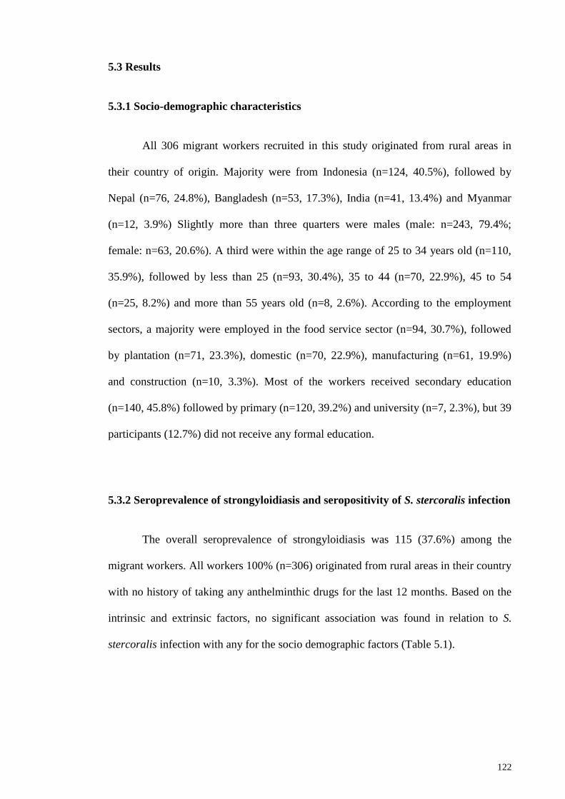
122
5.3 Results
5.3.1 Socio-demographic characteristics
All 306 migrant workers recruited in this study originated from rural areas in
their country of origin. Majority were from Indonesia (n=124, 40.5%), followed by
Nepal (n=76, 24.8%), Bangladesh (n=53, 17.3%), India (n=41, 13.4%) and Myanmar
(n=12, 3.9%) Slightly more than three quarters were males (male: n=243, 79.4%;
female: n=63, 20.6%). A third were within the age range of 25 to 34 years old (n=110,
35.9%), followed by less than 25 (n=93, 30.4%), 35 to 44 (n=70, 22.9%), 45 to 54
(n=25, 8.2%) and more than 55 years old (n=8, 2.6%). According to the employment
sectors, a majority were employed in the food service sector (n=94, 30.7%), followed
by plantation (n=71, 23.3%), domestic (n=70, 22.9%), manufacturing (n=61, 19.9%)
and construction (n=10, 3.3%). Most of the workers received secondary education
(n=140, 45.8%) followed by primary (n=120, 39.2%) and university (n=7, 2.3%), but 39
participants (12.7%) did not receive any formal education.
5.3.2 Seroprevalence of strongyloidiasis and seropositivity of S. stercoralis infection
The overall seroprevalence of strongyloidiasis was 115 (37.6%) among the
migrant workers. All workers 100% (n=306) originated from rural areas in their country
with no history of taking any anthelminthic drugs for the last 12 months. Based on the
intrinsic and extrinsic factors, no significant association was found in relation to S.
stercoralis infection with any for the socio demographic factors (Table 5.1).
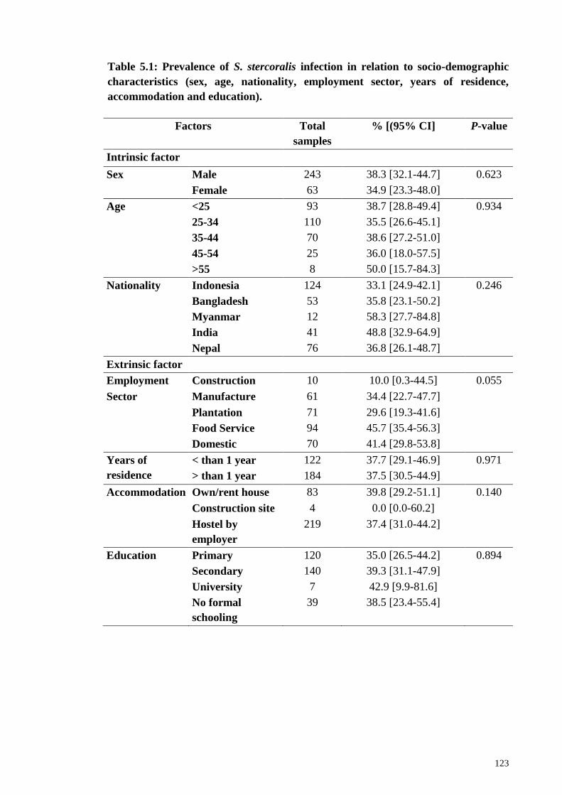
123
Table 5.1: Prevalence of S. stercoralis infection in relation to socio-demographic
characteristics (sex, age, nationality, employment sector, years of residence,
accommodation and education).
Factors Total
samples
% [(95% CI] P-value
Intrinsic factor
Sex Male 243 38.3 [32.1-44.7] 0.623
Female 63 34.9 [23.3-48.0]
Age <25 93 38.7 [28.8-49.4] 0.934
25-34 110 35.5 [26.6-45.1]
35-44 70 38.6 [27.2-51.0]
45-54 25 36.0 [18.0-57.5]
>55 8 50.0 [15.7-84.3]
Nationality Indonesia 124 33.1 [24.9-42.1] 0.246
Bangladesh 53 35.8 [23.1-50.2]
Myanmar 12 58.3 [27.7-84.8]
India 41 48.8 [32.9-64.9]
Nepal 76 36.8 [26.1-48.7]
Extrinsic factor
Employment Construction 10 10.0 [0.3-44.5] 0.055
Sector Manufacture 61 34.4 [22.7-47.7]
Plantation 71 29.6 [19.3-41.6]
Food Service 94 45.7 [35.4-56.3]
Domestic 70 41.4 [29.8-53.8]
Years of
residence
< than 1 year 122 37.7 [29.1-46.9] 0.971
> than 1 year 184 37.5 [30.5-44.9]
Accommodation Own/rent house 83 39.8 [29.2-51.1] 0.140
Construction site 4 0.0 [0.0-60.2]
Hostel by
employer
219 37.4 [31.0-44.2]
Education Primary 120 35.0 [26.5-44.2] 0.894
Secondary 140 39.3 [31.1-47.9]
University 7 42.9 [9.9-81.6]
No formal
schooling
39 38.5 [23.4-55.4]
124
5.4 Discussion
Malaysia has a strong economic presence in the region over since the early
1970‘s and experienced mass migration of workers particularly from rural background
for employment from neighbouring countries where S. stercoralis infections remain
endemic. Presently, screening for strongyloidiasis is not mandatory for employment to
this country. Therefore, this is the first study to report the presence of anti-Strongyloides
antibody in sera among migrant workers using an enzyme-linked immunosorbent assay
(ELISA) commercial kit for immunoglobulin G (IgG). Socio-demographic description
showed in this study provided evidence that all the workers originated mainly from
rural areas in their respective countries where parasitic infections were still very much
prevalent among the poor and deprived communities. Results also show an
overwhelming over a third of the workers screened were seropositive and was in
agreement with global values between 10% and 40% in endemic countries in the
tropical and subtropical region (Schar et al., 2013). In South-East Asia region, reports
have shown that prevalence of S. stercoralis infection among the population varied
[Cambodia (2.6%-44.7%), Thailand (0.1%-57.0%), Indonesia (0.8%-5.4%), China
(0.04%-17.9%), Lao PDR (1.4%-41.0%) and Vietnam (0.1%)] (Schar et al., 2016).
Most studies previously conducted in Malaysia focused on other soil-transmitted
helminth (STH) ignoring S. stercoralis specifically. Other works have used the
conventional microscopy technique for the detection of S. stercoralis larvae in faecal
samples which have low level of sensitivity (Rahmah et al., 1997; Ahmad et al., 2013).
Based on microscopic examination conducted in this study, all of the 306 faecal
samples provided were negative for larvae. Siddiqui and Berk (2001) reported that
microscopy examination performed to detect S. stercoralis larvae using only single
125
stool sample lacks sensitivity and failed up to 70 % of cases particularly if the intestinal
worm load is very low as commonly occurs in most asymptomatic individuals.
Although it was suggested that using multiple faecal samples will improve sensitivity
by ≥50% (Siddiqui & Berk, 2001), obtaining repeat samples was not possible in this
study. Based on the results from the ELISA method, high seropositivity was detected in
the targeted population. However, this result was not unequivocally able to prove
current active infection and is possible the result from a persisting level of antibody
from a past infection (Yori et al., 2006; Ahmad et al., 2013). The ELISA method has
more advantage over the microscopy in which, large-scale screening can be performed
simultaneously. In addition, the assay is also easy to perform as it is based on the
interpretation of the absorbance reading and can be performed without expertise in
morphological identification (Caruana et al., 2006; Yori et al., 2006; Ahmad et al.,
2013).
In the present study, infections in male (38.3%) were slightly higher than female
workers (34.9%) however not significant. A review on S. stercoralis infections in South
East Asia recognized gender as one of the risk factors (Schar et al., 2016). In rural Lao
PDR, Conlan et al. (2012) showed that males were at higher risk of infection with an
odds ratio (OR) of 2.76. Similar findings were recorded in Cambodia with S. stercoralis
infection having an OR of 1.4 in males (p = 0.001) (Koga-Kita, 2004) and higher
prevalence recorded among males than females and in all age groups (OR: 1.7; p <
0.001) in another study (Khieu et al., 2014b). In Thailand, infections were significantly
different in boys (35.2%) than girls (16.8%) (p = 0.003) (Khampitak et al., 2006). While
in Northern Ghana, significantly higher infection was recorded in males (12.7%)
compared to females (10.6%) (Yelifari et al., 2005) as well as in Okinawa, Japan
(males: 14.0%; females: 6.8%) (Arakaki et al., 1992). Gender played a significant role
126
particularly among males due to possibly higher involvement in outdoor activities
compared to females especially in the plantation/agricultural sector (Ahmad et al.,
2013). The frequent contact with contaminated soil or water may predispose them to S.
stercoralis infection, as this parasite was transmitted by penetration of the infective
larvae to the intact skin.
Infections from this study also showed workers involved in the food services
(45.7%) with the highest seropositivity, followed closely by the domestic (41.4%)
sector, manufacturing (34.4%), plantation (29.6%) and finally construction (10.0%).
Results were not able to show any significant association to occupation in this study. In
contrast, other studies in South Eastern provinces of China (Wang et al., 2013) and
Yunnan province in the South of China (Steinmann et al., 2007; 2008) have found high
S. stercoralis infections among those involved in agriculture particularly farmers. In
Europe and in the United States, infections were also predominantly among those in
plantation and mining sectors (Schar et al., 2013) and among miners in Germany
(Arbeitsmedizin, 2009). The present results suggest that infections were acquired from
their home country where infections were more prevalent however, using the current
detection method, we were unable to provide evidence to substantiate this statement.
This result highlights public health implications as transmission is basically due
to poor personal hygiene and sanitation, in addition to improper method of handling
sewage disposal may promote S. stercoralis infections (Lim et al., 2009, Schar et al.,
2013). A community based survey of the urban slum community in Bangladesh showed
high prevalence of infection (29.8%) (Hall et al., 1994) as also recorded in Nepal
(22.8%) (Hoge et al. in 1995). However, India (Lanjewar et al., 1996; Singh et al.,
127
2004; Singh et al., 1993; Kang et al., 1998; Joshi et al., 2002) and Indonesia (Bangs et
al., 1996; Widjana & Sutisna, 2000; Hasegawa et al., 1992; Mangali et al., 1993; 1994;
Toma et al. 1999), recorded lower prevalence of 6.6% and 7.6%, respectively. High
infection rates up to 60% can be expected in the resource-poor countries with ecological
and socioeconomic conditions that favor the spread of S. stercoralis and very low
prevalence in societies where faecal contamination is rare especially in urban and
developed countries, with the exception of slum areas in the big cities (Schar et al.,
2013). A study in rural Cambodia found households with latrine were significantly least
infected and a reduction of 39% of strongyloidiasis cases in those households with
proper latrine used for defecation (Khieu et al., 2014a).
Other factors such as behavioral patterns are known to also play a role in the
transmission with most acquired infection gotten during childhood and sustained
through auto-infection (Schar et al., 2013). This is possible due to the parasites‘
capability to reproduce within a human host (endogenous autoinfection) resulting in
long-lasting infection with several studies reporting individuals with S. stercoralis
infections for more than 75 years (Concha et al., 2005; Genta, 1992; Keiser & Nutman,
2004; Vadlamudi et al., 2006; Prendki et al., 2011; Gill et al., 2004).
The current result provided baseline information of the epidemiology of S.
stercoralis infections among the migrant workers and highlights the importance of
using an appropriate technique in the detection of this infection. Similarly, most studies
on refugees and migrants worldwide also used copro-diagnostic procedures with low
sensitivity (71.8%) compared to copro-diagnostic procedures with moderate sensitivity
(7.7%) and serological diagnostic procedures with high sensitivity (20.5%) (Schär et al.,
128
2013). The frequent choice of diagnosis for S. stercoralis infections were the Koga Agar
culture method and Baermann method, which are considerably time consuming and
labour intensive. These methods involve examining stool samples collected over a few
days and could mitigate the risk of missing low-intensity infections. Therefore, further
analysis needed in order to obtain a clearer picture of the prevalence of this parasite. In
addition to serology, further studies such as molecular characterization is necessary to
confirm current active infections which will be described in chapter 6.
129
5.5 Conclusion
High seroprevalence of S. stercoralis infections among migrant workers was
reported for the first time in Malaysia. This data provided baseline information on
epidemiology of S. stercoralis infections among the workers and highlights the
importance of using an appropriate technique in the detection of this infection. The high
infection among these workers calls for health education on transmission and the
importance of good personal hygiene and sanitation. Despite failing to differentiate
current active or past S. stercoralis infection, results suggest that serological method
such as ELISA as one of the alternative diagnostic tool for detection.
130
CHAPTER 6: MOLECULAR CHARACTERIZATION OF HUMAN
INTESTINAL PARASITE INFECTIONS
6.1 Introduction
The development of molecular biology especially polymerase chain reaction
(PCR) has resulted in a powerful tool to screen, characterize and evaluate genetic
diversity (Karp & Edwards, 1997). This method is widely used in many studies to assist
identification of organisms particularly those that are morphologically similar.
Therefore, the present study was conducted to characterize the genetic makeup of
human intestinal parasites recovered in this study namely; Strongyloides stercoralis,
hookworms, Entamoeba spp., Giardia spp. and Cryptosporidium spp.
Strongyloides stercoralis is commonly found in tropical and subtropical regions
of the world including Europe, America and Southeast Asia (Siddiqui & Berk, 2001).
The actual prevalence of S. stercoralis is often queried due to the lack of standard
diagnostic tools (Olsen et al., 2009; Verweij et al., 2009). Previous chapter showed the
importance of using serological analysis (ELISA) as an alternative diagnostic tool for
detecting S. stercoralis infection however, this method failed to detect current active S.
stercoralis infection. On the other hand, the conventional microscopic examination of
stool specimens is only useful when the larvae load is high, such as acute S. stercoralis
infection. Therefore, this helminth will be tested further using nested PCR targeting the
internal transcribed spacer 1 (ITS1) region of the ribosomal DNA gene to assist in
determining current active infections.
131
Human hookworm infection is commonly caused by two species of hookworm
namely Necator americanus and Ancylostoma duodenale. The current diagnosis of
hookworm infection in human is via the identification of the parasite eggs in the host
faeces which is technically simple and low cost. However, this technique is hampered
by the morphological similarities between the eggs species of N. americanus,
Ancylostoma spp. and other strongylid nematodes including Oesophagostoum spp. and
Trichostrongylus spp. Therefore, a two-step semi-nested PCR was employed for DNA
amplification of internal transcribed spacer 2 and 28S ribosomal RNA region of both N.
americanus and Ancylostoma spp. to determine the hookworm species recovered.
Entamoeba spp. inhabits the human intestine and is commonly comprised of six
species including Entamoeba histolytica, E. dispar, E. moshkovskii, E. coli, E.
hartmanni and E. polecki. Amoebiasis is a global health problem caused by the
protozoan Entamoeba histolytica affecting approximately 40 to 50 million people.
Those infected develop colitis or additional intestinal disease causing annual deaths up
to 40,000-110,000 (Schmunis & Lopez-Antunano, 2005; Garlington et al., 2011).
Entamoeba infections are normally diagnosed through microscopic examination of
faecal samples. However, E. histolytica cysts and trophozoites are morphologically
undistinguishable from E. dispar and E. moshkovskii. Therefore, molecular
characterization was conducted by nested PCR targeting 16S-like ribosomal RNA gene
to genetically characterize E. histolytica, E. dispar and E. moshkovskii recovered from
the human samples obtained.
Giardiasis is a diarrheal disease in a wide range of vertebrate hosts. The
taxonomy of Giardia at species level is complicated to distinguish due to limited
132
morphological differences. The genus currently comprises six species namely, Giardia
duodenalis (commonly found in humans), G. agilis (in amphibians), G. ardeae and G.
psittaci (in birds), G. microti and G. muris (in rodents). Molecular tools have been used
recently to characterize the epidemiology of human giardiasis, G. duodenalis. Two
major groups of G. duodenalis have been recognized infecting humans worldwide;
assemblage A and B (Maryhofer et al., 1995). Therefore, this chapter will describe the
development of nested polymerase chain reaction (PCR) to amplify the triosephosphate
isomerase (TPI) gene to differentiate Giardia parasite at species and genotype levels
among the migrant workers.
Cryptosporidium spp. causes cryptosporodiasis, a frequent cause of diarrheal
disease in humans. There are more than 27 species of Cryptosporidium being
considered valid by investigators infecting human and other animals (Fayer, 2010;
Traversa, 2010). The main causative agents of human cryptosporidiosis and of greater
significance to public health are C. parvum and C. hominis (Fayer et al., 2000).
Identification of Cryptosporidium species is usually difficult by simple traditional
microscopic measurements and the use of recent advancement in molecular
characterization has made it possible to differentiate Cryptosporidium spp. oocyst at
species, genotype and subgenotype level (Xiao et al., 2000). Therefore, present study
will perform PCR-RFLP assay to detect the presence of Cryptosporodium spp. based on
SSU-rRNA sequences.
133
6.2 Materials and Methods
6.2.1 Samples collection
A total of 388 stool specimens were collected from the workers (as described in
Chapter 2 and Chapter 3) meanwhile for S. stercoralis screening, 306 stool specimens
have been collected. All stool samples were subjected for preliminary screening by
microscopy and subsequently subjected to molecular analysis for specific parasite
identification up to species level. These parasites include Strongyloides stercoralis,
hookworm, Entamoeba spp., Giardia spp. and Cryptosporidium spp. (Figure 6.1).
6.2.2 Extraction of genomic DNA
DNA was extracted from microscopically positive faecal samples using
NucleoSpin® Soil (MACHEREY-NAGEL, Düren, Germany) according to the
manufacturer‘s instructions. Approximately, 250-500 mg fresh sample material was
transferred to a NucleoSpin® Bead Tube containing the ceramic beads. Then, a total of
700 µl Buffer SL2 was added to the tube. To adjust lysis conditions, 150 µl Enhancer
SX was added to the tube and the cap was closed. To lyse the sample, the NucleoSpin®
Bead Tubes was attached horizontally to a vortexer and was vortexed at full speed in
room temperature (18-25˚C) for 5 minutes. The tube was then centrifuged for 2 minutes
at 11,000x g to eliminate the foam caused by detergent. A volume of 150µl Buffer SL3
was added to the tube and vortexed for 5 seconds. Then the tube was incubated for 5
minutes at 0-4°C and was centrifuged again at 11,000 x g for 1 minute.
134
To filter the lysate, a Nucleospin® Inhibitor Removal Column (red ring) was
placed in a collection tube (2mL, lid). Up to 700µl of clear supernatant was loaded onto
the filter and the tube was centrifuged for 1 minute at 11,000x g. The Nucleospin®
Inhibitor Removal Column was then discarded and if a pellet was visible in the flow-
through, the clear supernatant was transferred to a new collection tube. A total of 250µl
Buffer SB was added to adjust binding conditions and then, the tube was vortexed for 5
seconds. To bind the DNA, a Nucleospin® Soil Column (green ring) was placed in a
collection tube (2mL). 550µL of samples was loaded onto the column and centrifuged
for 1 minute at 11,000 x g. The flow-through was discarded and the column was placed
back into the collection tube. The remaining sample was loaded onto the column,
centrifuged and flow-through was discarded again. The column was then placed back
into the collection tube.
Washing procedure was followed by adding 500µL Buffer SB to the
Nucleospin® Soil column and centrifuged for 30 seconds at 11,000 x g. The flow-
through was discarded and placed back into the collection tube. Then, 550µL of Buffer
SW1 was added to the Nucleospin® Soil column, followed by centrifuged for 30
seconds, flow-through was discarded and placed back into the collection tube. Washing
procedure was then continued by adding 700µL Buffer SW2 to the Nucleospin® Soil
Column, vortexed for 2 seconds and centrifuged for 30 seconds at 11,000x g. The flow-
through was discarded and the column was placed back again into the collection tube.
The procedure was repeated again by adding 700µL of Buffer SW2, vortexed,
centrifuged, flow-through discarded and column was placed back into the collection
tube. The tube was then centrifuged for 2 minutes at 11,000x g to dry the silica
membrane. To elute the DNA, the Nucleospin® Soil Column was placed into a new
microcentrifuge tube and 40µL of Buffer SE was added to the column. The lid of the

135
tube was let open and incubated for 1 minute at room temperature (18-25°C). The lid
was closed and centrifuged for 30 seconds at 11,000 x g. The extracted DNA was stored
at −20°C until required for PCR amplification.
6.2.3 Nested polymerase chain reaction (nested PCR)
6.2.3.1 Strongyloides stercoralis
A nested PCR targeting the internal transcribed spacer 1 (ITS1) region of the
ribosomal DNA gene was used to amplify S. stercoralis DNA. The assay in this study
used two sets of primers and was performed in two separate reactions. The primary
reaction consisted of a forward primer SS–FO: 5‘- ATC CTT CCA ATC GCT GTT GT
-3‘ and reverse primer SS– RO: 5‘- TTT CGT GAT GGG CTA ATT CC -3‘. The
secondary reaction forward and reverse primers were SS–FI: 5‘- GTA ACA AGG TTT
TCG TAG GTG A –3‘ and SS–RI: 5‘- ATT TAG TTT CTT TTC CTC CGC TT –3‘
respectively (Nilforoushan et al. 2007). The nested PCR was performed in a Maxime
PCR PreMix Kit (i-Taq) (iNtRON Biotechnology, Inc.) in 20μl volume reaction. The
reaction contained i-TaqTM
DNA polymerase (5U/µl) (2.5U), deoxynucleoside
triphosphate (dNTPs) (2.5 mM each), 1X reaction buffer (10x) and gel loading buffer
(1x). DNA template (2µl), primers (100 nM each) and distilled water were added to the
premix. The conditions for the primary PCR were initial denaturation at 94 °C for 5
minutes and 35 cycles of 94 °C for 45 seconds, 58 °C for 1 minute, 72 °C for 1 minute
and a final extension at 72 °C for 5 minutes. Subsequently, 2μl of the primary PCR
products were subjected for a secondary PCR performed at 94 °C for 2 minutes and 30
cycles of 94 °C for 45 seconds, 60°C for 45 seconds, 72 °C for 1 minute and a final
extension at 72 °C for 5 minutes. PCR products were subjected to electrophoresis
136
through 2 % (w/v) agarose and visualized in a UV transilluminator after staining with
RedSafeTM
Nucleic Acid Staining Solution (iNtRON Biotechnology, Inc, Korea).
6.2.3.2 Hookworms
A two-step semi-nested PCR was used for DNA amplification of hookworm
species. For the first amplification, forward primer NC1 (5‘- ACG TCT GGT TCA
GGG TTC TT -3‘) and reverse primer NC2 (5‘- TTA GTT TCT TTT CCT CCG CT -
3‘) were used to amplify approximately 310-basepair and 420-basepair regions of
internal transcribed spacer 2 and 28S ribosomal RNA region of N. americanus and
Ancylostoma spp. The nested PCR was performed in a Maxime PCR PreMix Kit (i-Taq)
(iNtRON Biotechnology, Inc.) in 20μl volume reaction. The reaction contained i-TaqTM
DNA polymerase (5U/µl) (2.5U), deoxynucleoside triphosphate (dNTPs) (2.5 mM
each), 1X reaction buffer (10x) and gel loading buffer (1x). DNA template (2µl),
primers (10 pM each) and distilled water were added to the premix. The conditions for
the primary PCR were initial denaturation at 94°C for 5 minutes, followed by 30 cycles
at 94°C for 30 seconds, 55°C for 30 seconds, and 72°C for 30 seconds, and a final
extension at 72°C for 7 minutes. Samples containing N. americanus and Ancylostoma
spp. genomic DNA (positive control) was included in each PCR. Subsequently, 2μl of
the primary PCR products were subjected for a secondary PCR. Amplification was
conducted by using forward primer NA (5‘- ATG TGC ACG TTA TTC ACT -3‘) for N.
americanus, AD1 (5‘- CGA CTT TAG AAC GTT TCG GC -3‘) for Ancylostoma spp.
and NC2 as a common reverse primer. The secondary amplification reagent
concentrations were similar to those of the first round of PCR except that 2 µl of
primary PCR product was added instead of DNA. The cycling conditions for the second
round of amplification were 94°C for 5 minutes, followed by 35 cycles at 94°C for 1
137
minute, 55°C for 1 minute and 72°C for 1 minute, and a final extension at 72°C for 7
minutes. PCR products were subjected to electrophoresis through 2 % (w/v) agarose
and visualized in a UV transilluminator after staining with RedSafeTM
Nucleic Acid
Staining Solution (iNtRON Biotechnology, Inc, Korea).
6.2.3.3 Entamoeba spp.
Nested PCR targeting 16S-like ribosomal RNA gene was used to genetically
characterize E. histolytica, E. dispar and E. moshkovskii according to Que and Reed
(1991). Primary PCR for the detection of Entamoeba genus used forward primer E1 (5‘-
TAA GAT GCA GAG CGA AA -3‘) and reverse primer E2 (5‘- GTA CAA AGG GCA
GGG ACG TA -3‘). The nested PCR was performed in a Maxime PCR PreMix Kit (i-
Taq) (iNtRON Biotechnology, Inc.) in 20μl volume reaction. The reaction contained i-
TaqTM
DNA polymerase (5U/µl) (2.5U), deoxynucleoside triphosphate (dNTPs) (2.5
mM each), 1X aeaction buffer (10x) and gel loading buffer (1x). DNA template (2µl),
primers (100 pM each) and distilled water were added to the premix. The conditions for
the primary PCR were 96°C for 2 minutes, followed by 35 cycles of 92°C for 1 minute,
58°C for 1 minute, 72°C for 1 minute 30 seconds and a final extension at 72°C for 7
minutes. Subsequently, 2µl of the primary PCR products were subjected to secondary
PCR for Entamoeba species-specific characterization using primer sets EH1 (5‘- AAG
CAT TGT TTC TAG ATC TGA G -3‘) and EH2 (5‘- AAG AGG TCT AAC CGA
AAT TAG -3‘) to detect E. histolytica (439 bp); ED1 (5‘- TCT AAT TTC GAT TAG
AAC TCT -3‘) and ED2 (5‘-TCC CTA CCTATT AGA CAT AGC -3‘) to detect E.
dispar (174 bp); Mos1 (5‘- GAA ACC AAG AGT TTC ACA AC -3‘) and Mos2 (5‘-
CAA TAT AAG GCT TGG ATG AT -3‘) to detect E. moshkovskii (553 bp) (Troll et
al., 1997; Khairnar & Parija, 2007). The cycling conditions for the second round of
138
amplification were 96°C for 2 minutes, followed by 35 cycles at 92°C for 1 minute,
47°C for 1 minute and 72°C for 1 minute, and a final extension at 72°C for 7 minutes.
PCR products were subjected to electrophoresis through 2 % (w/v) agarose and
visualized in a UV transilluminator after staining with RedSafeTM
Nucleic Acid Staining
Solution (iNtRON Biotechnology, Inc, Korea).
6.2.3.4 Giardia spp.
A nested PCR protocol was developed to amplify the triosephosphate isomerase
(TPI) fragment from various Giardia isolates. For the primary PCR, a PCR product of
605 bp was amplified by using primers AL3543 (5′- AAA TIA TGC CTG CTC GTC G
-3′) and AL3546 (5′- CAA ACC TTI TCC GCA AAC C -3′). The nested PCR was
performed in a Maxime PCR PreMix Kit (i-Taq) (iNtRON Biotechnology, Inc.) in 20 μl
volume reaction. The reaction contained i-TaqTM
DNA polymerase (5U/µl) (2.5U),
deoxynucleoside triphosphate (dNTPs) (2.5 mM each), 1X reaction buffer (10x) and gel
loading buffer (1x). DNA template (2µl), primers (100 nM each) and distilled water
were added to the premix. The conditions for the primary PCR were initial denaturation
at 94°C for 5 minutes, followed by 35 cycles at 94°C for 45 seconds, 50°C for 45
seconds, and 72°C for 1 minute, and a final extension at 72°C for 10 minutes. For the
secondary PCR, a fragment of 530 bp was amplified using 2 μl of primary PCR reaction
and primers set AL3544 (5′- CCC TTC ATC GGI GGT AAC TT -3′) and AL3545 (5′-
GTG GCC ACC ACI CCC GTG CC -3′). The conditions for the secondary PCR were
initial denaturation at 94°C for 5 minutes, followed by 35 cycles at 94°C for 45 seconds,
58°C for 45 seconds, and 72°C for 1 minute, and a final extension at 72°C for 10
minutes. PCR products were subjected to electrophoresis through 2 % (w/v) agarose
139
and visualized in a UV transilluminator after staining with RedSafeTM
Nucleic Acid
Staining Solution (iNtRON Biotechnology, Inc, Korea).
6.2.3.5 Cryptosporidium spp.
A nested PCR protocol was used to identify Cryptosporidium spp. using small
subunit rRNA as described by Xiao et al. (1999). The primary reaction consisted of a
forward primer SSU-F2: 5‘- TTC TAG AGC TAA TAC ATG CG -3‘ and reverse
primer, SSU-R2: 5‘- CCC ATT TCC TTC GAA ACA GGA -3‘. The secondary
reaction forward and reverse primers were SSU-F3: 5‘- GGA AGG GTT GTA TTT
ATT AGA TAA AG -3‘ and SSU-R4: 5‘- CTC ATA AGG TGC TGA AGG AGT A -3‘
respectively. The nested PCR was performed in a Maxime PCR PreMix Kit (i-Taq)
(iNtRON Biotechnology, Inc.) in 20 μl volume reaction. The reaction contained i-TaqTM
DNA polymerase (5U/µl) (2.5U), deoxynucleoside triphosphate (dNTPs) (2.5 mM
each), 1X reaction buffer (10x) and gel loading buffer (1x). DNA template (2µl),
primers (200 nM each) and distilled water were added to the premix. The conditions for
both primary and secondary PCR were initial denaturation at 94 °C for 4 minutes and
35 cycles of 94 °C for 45 seconds, 56 °C for 90 seconds, 72 °C for 1 minute and a final
extension at 72 °C for 7 minutes. PCR products were subjected to electrophoresis
through 2 % (w/v) agarose and visualized in a UV transilluminator after staining with
RedSafeTM
Nucleic Acid Staining Solution (iNtRON Biotechnology, Inc, Korea).
6.2.4 Purification of PCR product
The PCR product was purified using MEGAquick-spinTM
Total Fragment DNA
Purification Kit (iNtRON Biotechnology, 2011, Korea) according to the manufacturer‘s
140
protocol. 5 volume of BNL buffer was added to the PCR reaction product, and mixed
well by vortexed. A total of 100µl of BNL buffer was added to the PCR tube directly
for PCR product that was 20µl. For PCR product <200bp, 1.5 volume of isopropanol
was added to the sample and mixed by pipetting several times. One MEGAquick-spinTM
column was placed in a collection tube for each DNA mixture. The DNA mixture was
transferred to the MEGAquick-spinTM
column assembled. To bind DNA, the sample
was applied to the MEGAquick-spinTM
column and centrifuged for 1 minute. The flow-
through was discarded and the MEGAquick-spinTM
column was placed back in the
same 2 ml collection tube. 700µl of washing buffer was added to the column and
centrifuged at 13,000 rpm for 1 minute. The flow-through was discarded and the
MEGAquick-spinTM
column was placed back in the same 2 ml collection tube. The
assembled column was centrifuged for 1 minute at 13,000 rpm to dry the spin
membrane. The MEGAquick-spinTM
column was placed to a clean 1.5 ml
microcentrifuge tube. 30µl of the Elution Buffer was applied directly to the center of the
column without touching the membrane with the pipette tip. The column was incubated
at room temperature for 1 minute and then centrifuged for 1 minute at 13,000 rpm. The
MEGAquick-spinTM
column was discarded and the microcentrifuge tube contained the
eluted DNA was stored at -20°C.
6.2.5 DNA sequencing
6.2.5.1 Strongyloides stercoralis
Sequencing was carried out by First Base Laboratories Sdn. Bhd. For
Strongyloides stercoralis targeting the internal transcribed spacer 1 (ITS1) region of the
ribosomal DNA gene, sequencing was done in both directions using forward primer
141
(SS–FI: 5‘- GTA ACA AGG TTT TCG TAG GTG A –3‘) and reverse primer (SS–RI:
5‘- ATT TAG TTT CTT TTC CTC CGC TT –3‘).
6.2.5.2 Hookworm
To sequence hookworm species, sequencing was conducted by using forward
primer NA (5‘- ATG TGC ACG TTA TTC ACT -3‘) for N. americanus, AD1 (5‘-
CGA CTT TAG AAC GTT TCG GC -3‘) for Ancylostoma spp. and NC2 (5‘- TTA
GTT TCT TTT CCT CCG CT -3‘) as a common reverse primer.
6.2.5.3 Entamoeba spp.
Sequencing of 16S-like ribosomal RNA gene of Entamoeba spp. was conducted
using primer sets EH1 (5‘- AAG CAT TGT TTC TAG ATC TGA G -3‘) and EH2 (5‘-
AAG AGG TCT AAC CGA AAT TAG -3‘) to detect E. histolytica, ED1 (5‘- TCT
AAT TTC GAT TAG AAC TCT -3‘) and ED2 (5‘-TCC CTA CCTATT AGA CAT
AGC -3‘) to detect E. dispar and Mos1 (5‘- GAA ACC AAG AGT TTC ACA AC -3‘)
and Mos2 (5‘- CAA TAT AAG GCT TGG ATG AT -3‘) to detect E. moshkovskii.
6.2.5.4 Giardia spp.
For Giardia spp. targeting the TPI gene, sequencing was done in both directions
using forward primer AL3544 (5′- CCC TTC ATC GGI GGT AAC TT -3′) and reverse
primer AL3545 (5′- GTG GCC ACC ACI CCC GTG CC -3′).
142
6.2.6 Sequencing analysis
Sequences were edited via Applied Biosystems Sequence Scanner software v1.0
Sequence Trace Viewer and Editor (www. en.bio-soft.net/dna/ss). Edited sequences
were aligned and consensus sequences were created for each isolates using the BioEdit
(www.mbio.ncsu.edu) programme. Each consensus sequence was used for the
identification of the parasite genotypes and sequences were searched using basic local
alignment search tool (BLAST) (www.ncbi.nlm.nih.gov/blast) in order to get the 100%
similarity with parasites genotypes sequences deposited in the GenBank.
6.2.7 RFLP (Restriction Fragment Length Polymorphism) for Cryptosporidium
spp.
Restriction assays were conducted in a 20μL volume with 0.5 units of restriction
enzymes and 10μL of PCR product per reaction. Mixes were incubated at 37°C for 8
hours. Digested products were subjected to electrophoresis through 1 % (w/v) agarose
and visualized in a UV transilluminator after staining with RedSafeTM
Nucleic Acid
Staining Solution (iNtRON Biotechnology, Inc, Korea). The endonuclease enzymes
used were SspI and VspI (Vivantis, Malaysia).
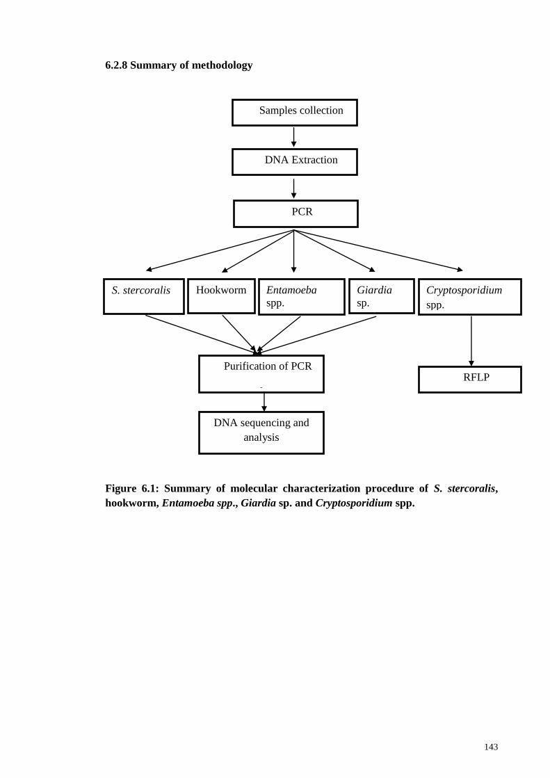
143
6.2.8 Summary of methodology
Figure 6.1: Summary of molecular characterization procedure of S. stercoralis,
hookworm, Entamoeba spp., Giardia sp. and Cryptosporidium spp.
Samples collection
DNA Extraction
Cryptosporidium
spp.
Giardia
sp.
Entamoeba
spp.
Hookworm S. stercoralis
PCR
Purification of PCR
products
DNA sequencing and
analysis
RFLP
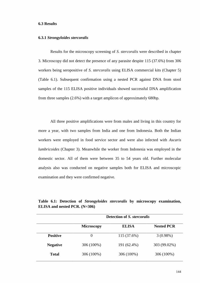
144
6.3 Results
6.3.1 Strongyloides stercoralis
Results for the microscopy screening of S. stercoralis were described in chapter
3. Microscopy did not detect the presence of any parasite despite 115 (37.6%) from 306
workers being seropositive of S. stercoralis using ELISA commercial kits (Chapter 5)
(Table 6.1). Subsequent confirmation using a nested PCR against DNA from stool
samples of the 115 ELISA positive individuals showed successful DNA amplification
from three samples (2.6%) with a target amplicon of approximately 680bp.
All three positive amplifications were from males and living in this country for
more a year, with two samples from India and one from Indonesia. Both the Indian
workers were employed in food service sector and were also infected with Ascaris
lumbricoides (Chapter 3). Meanwhile the worker from Indonesia was employed in the
domestic sector. All of them were between 35 to 54 years old. Further molecular
analysis also was conducted on negative samples both for ELISA and microscopic
examination and they were confirmed negative.
Table 6.1: Detection of Strongyloides stercoralis by microscopy examination,
ELISA and nested PCR. (N=306)
Detection of S. stercoralis
Microscopy ELISA Nested PCR
Positive 0 115 (37.6%) 3 (0.98%)
Negative 306 (100%) 191 (62.4%) 303 (99.02%)
Total 306 (100%) 306 (100%) 306 (100%)
145
6.3.2 Hookworm
From a total of 388 stool samples collected from migrant workers, 51 samples
(13.1%) were found positive by microscopy for hookworm infection (Chapter 3). The
51 positive hookworm samples were subjected to nested PCR analysis to detect Necator
americanus and Ancylostoma spp. The PCR amplicons were successfully obtained from
42 (82.4%) of 51 samples with 81.0% (34 of 42) being N. americanus (approximately
250bp), 16.7% (7 of 42) Ancylostoma spp. (approximately 130bp) and the remaining
one (2.4%) sample has mixed infection of both species (Table 6.2). When screened via
PCR, all 337 samples found to be negative by microscopy showed no amplification.
Sequence comparison using the Basic Local Alignment Search Tool (BLAST)
confirmed that all eight Ancylostoma spp. were Ancylostoma duodenale (Figure 6.2).
Prevalence of N. americanus was markedly higher in male (82.4%, 28 of 34)
compared to female (17.6%, 6 of 34). Workers aged below 25 years old showed highest
infection rate with N. americanus (58.8%, 20 of 34). According to their nationality,
Indonesia (44.1%, 15 of 34) reported the highest prevalence followed by Nepal (26.5%,
9 of 34), Myanmar (11.8%, 4 of 34), Bangladesh (8.8%, 3 of 34) and India (8.8%, 3 of
34). Meanwhile, according to their employment sector, food services (32.3%, 11 of 34)
recorded the highest prevalence followed by domestic (20.6%, 7 of 34), plantation
(20.6%, 7 of 34), construction (14.7%, 5 of 34) and manufacturing (11.8%, 4 of 34)
(Table 6.2).
As for A. duodenale, higher prevalence also was reported in male (71.4%, 5 of
7) and in those workers aged below 25 years old (85.7%, 6 of 7). The species was
reported only from workers in Indonesia (71.4%, 5 of 7) and Nepal (28.6%, 2 of 7) from
146
all working sectors except construction. One sample of mix infections was from
Indonesian female from domestic sector (Table 6.2).
Sequences from all eight A. duodenale samples together with six reference
sequences obtained from the GenBank database and one sequence from Necator
americanus used as an outgroup, were analyzed in MEGA6 software and phylogenetic
tree was constructed (Figure 6.2). All A. duodenale sequences were grouped together
with A. duodenale reference sequence (EU344797.1). All sequences generated in this
study were deposited in GenBank under accession numbers KX650194- KX650201.
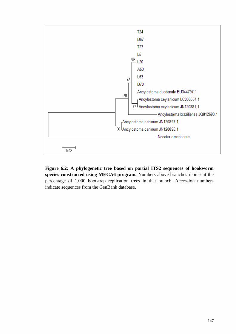
147
Figure 6.2: A phylogenetic tree based on partial ITS2 sequences of hookworm
species constructed using MEGA6 program. Numbers above branches represent the
percentage of 1,000 bootstrap replication trees in that branch. Accession numbers
indicate sequences from the GenBank database.
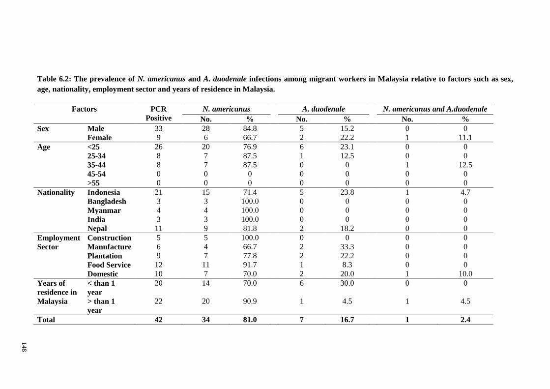
14
8
Table 6.2: The prevalence of N. americanus and A. duodenale infections among migrant workers in Malaysia relative to factors such as sex,
age, nationality, employment sector and years of residence in Malaysia.
Factors PCR
Positive
N. americanus A. duodenale N. americanus and A.duodenale
No. % No. % No. %
Sex Male 33 28 84.8 5 15.2 0 0
Female 9 6 66.7 2 22.2 1 11.1
Age <25 26 20 76.9 6 23.1 0 0
25-34 8 7 87.5 1 12.5 0 0
35-44 8 7 87.5 0 0 1 12.5
45-54 0 0 0 0 0 0 0
>55 0 0 0 0 0 0 0
Nationality Indonesia 21 15 71.4 5 23.8 1 4.7
Bangladesh 3 3 100.0 0 0 0 0
Myanmar 4 4 100.0 0 0 0 0
India 3 3 100.0 0 0 0 0
Nepal 11 9 81.8 2 18.2 0 0
Employment
Sector
Construction 5 5 100.0 0 0 0 0
Manufacture 6 4 66.7 2 33.3 0 0
Plantation 9 7 77.8 2 22.2 0 0
Food Service 12 11 91.7 1 8.3 0 0
Domestic 10 7 70.0 2 20.0 1 10.0
Years of
residence in
Malaysia
< than 1
year
20 14 70.0 6 30.0 0 0
> than 1
year
22 20 90.9 1 4.5 1 4.5
Total 42 34 81.0 7 16.7 1 2.4
149
6.3.3 Entamoeba spp.
A total of 45 (11.6%) from 388 stool samples were found positive with
Entamoeba spp. via microscopy screening. Of the 45 microscopy positive samples, 31
(68.9%) samples were successfully amplified using nested PCR. From the 31 PCR
positive results, E. dispar (20/31; 64.5%) appeared to be the most predominant,
followed by E. histolytica (8/31; 25.8%). Three samples were found having mixed
infections of E. dispar and E. histolytica (3/31; 9.7%) (Table 6.3). No sample was found
positive with E. moshkovskii.
Similar prevalence of infections was found between male and female for both E.
dispar and E. histolytica. For E. dispar, higher infection rates were recorded from
Indonesian (9/31; 29.0%), followed by Indian (5/31; 16.1%), Nepali (3/31; 9.7%),
Myanmarian (2/31; 6.5%) and Bangladeshi (1/31; 3.2%). According to employment
sector, higher prevalence was reported in food service sector (10/31; 32.3%), followed
by domestic (8/31; 25.8%) and manufacturing (2/31; 64.5%) sectors.
As for E. histolytica infections, higher prevalence was recorded in workers from
Indonesia (4/31; 12.9%), followed by workers from Nepal (2/31; 6.5%), India (1/31;
3.2%) and Myanmar (1/31; 3.2%). Meanwhile, according to employment sector, higher
prevalence was found in domestic service (4/31; 12.9%), followed by food services
(2/31; 6.5%) and manufacturing (2/31; 6.5%). Mix infection of both E. dispar and E.
histolytica were reported in workers from Indonesian (2/31; 6.5%) and Nepali (1/31;
3.2%) with each of them working in domestic, food service and manufacturing sectors.
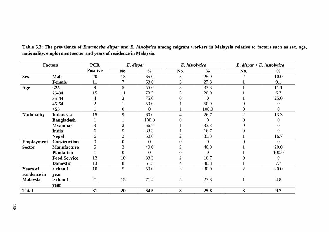
15
0
Table 6.3: The prevalence of Entamoeba dispar and E. histolytica among migrant workers in Malaysia relative to factors such as sex, age,
nationality, employment sector and years of residence in Malaysia.
Factors PCR
Positive
E. dispar E. histolytica E. dispar + E. histolytica
No. % No. % No. %
Sex Male 20 13 65.0 5 25.0 2 10.0
Female 11 7 63.6 3 27.3 1 9.1
Age <25 9 5 55.6 3 33.3 1 11.1
25-34 15 11 73.3 3 20.0 1 6.7
35-44 4 3 75.0 0 0 1 25.0
45-54 2 1 50.0 1 50.0 0 0
>55 1 0 0 1 100.0 0 0
Nationality Indonesia 15 9 60.0 4 26.7 2 13.3
Bangladesh 1 1 100.0 0 0 0 0
Myanmar 3 2 66.7 1 33.3 0 0
India 6 5 83.3 1 16.7 0 0
Nepal 6 3 50.0 2 33.3 1 16.7
Employment
Sector
Construction 0 0 0 0 0 0 0
Manufacture 5 2 40.0 2 40.0 1 20.0
Plantation 1 0 0 0 0 1 100.0
Food Service 12 10 83.3 2 16.7 0 0
Domestic 13 8 61.5 4 30.8 1 7.7
Years of
residence in
Malaysia
< than 1
year
10 5 50.0 3 30.0 2 20.0
> than 1
year
21 15 71.4 5 23.8 1 4.8
Total 31 20 64.5 8 25.8 3 9.7
151
6.3.4 Giardia spp.
From the total of 388 stool specimens from migrant workers, 42 (10.8%)
specimens were found positive with Giardia spp. using microscopy technique. The 42
microscopy-positive specimens were analyzed by nested PCR to amplify the
triosephosphate isomerase (TPI) gene of Giardia duodenalis and PCR amplicons were
successfully obtained from 30 (30/42; 71.4%) samples (approximately 530bp). At the
tpi gene, assemblages A and B were found in 13 (13/30; 43.3%) and 17 (17/30; 56.7%)
samples, respectively. Based on the analysis targeting tpi gene, 13 isolates of the
assemblages A were classified as A2 based on phylogenetic analysis. The neighbor-
joining tree placed six representative sequences (A40, S2, S13, S35, T42 and T66) in
one cluster with AII sequence references with high bootstrap support. Meanwhile, 8
sequences (B50, L101, L44, B1, A11, C31, T78 and A62) representing 17 isolates were
identified as assemblage B (Figure 6.1).
Higher G. duodenalis infection was found in male (26/30; 86.7%) compared to
female (4/30; 13.3%). Based on age factor, workers aged 34 and below (86.7%)
recorded higher infection in both assemblages. Workers with assemblage A were
Nepalese (7/13; 53.8%), Indonesian (4/13; 30.8%) and Indian (2/13; 15.4%) meanwhile
assemblage B was recovered from Indonesian (6/17; 35.3%), Bangladeshi (6/17;
35.3%), Nepalese (4/17; 23.5%) and Indian (1/17; 5.9%). Based on employment sector,
assemblage A was recorded only from workers in manufacturing (5/13; 38.5%), food
service (5/13; 38.5%) and domestic sector (3/13; 23.1%), meanwhile assemblage B was
reported in all 5 working sectors (domestic, 29.4%; food service, 23.5%; plantation,
17.6%; manufacturing, 17.6%; construction, 11.8%).
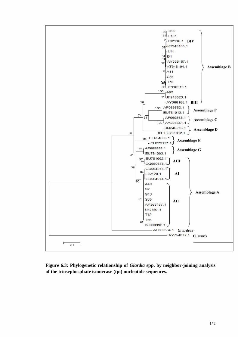
152
Figure 6.3: Phylogenetic relationship of Giardia spp. by neighbor-joining analysis
of the triosephosphate isomerase (tpi) nucleotide sequences.
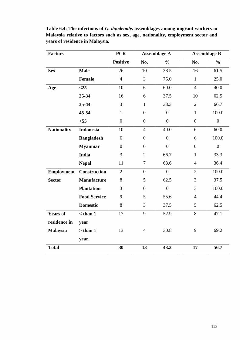
153
Table 6.4: The infections of G. duodenalis assemblages among migrant workers in
Malaysia relative to factors such as sex, age, nationality, employment sector and
years of residence in Malaysia.
Factors PCR
Positive
Assemblage A Assemblage B
No. % No. %
Sex Male 26 10 38.5 16 61.5
Female 4 3 75.0 1 25.0
Age <25 10 6 60.0 4 40.0
25-34 16 6 37.5 10 62.5
35-44 3 1 33.3 2 66.7
45-54 1 0 0 1 100.0
>55 0 0 0 0 0
Nationality Indonesia 10 4 40.0 6 60.0
Bangladesh 6 0 0 6 100.0
Myanmar 0 0 0 0 0
India 3 2 66.7 1 33.3
Nepal 11 7 63.6 4 36.4
Employment
Sector
Construction 2 0 0 2 100.0
Manufacture 8 5 62.5 3 37.5
Plantation 3 0 0 3 100.0
Food Service 9 5 55.6 4 44.4
Domestic 8 3 37.5 5 62.5
Years of
residence in
Malaysia
< than 1
year
17 9 52.9 8 47.1
> than 1
year
13 4 30.8 9 69.2
Total 30 13 43.3 17 56.7
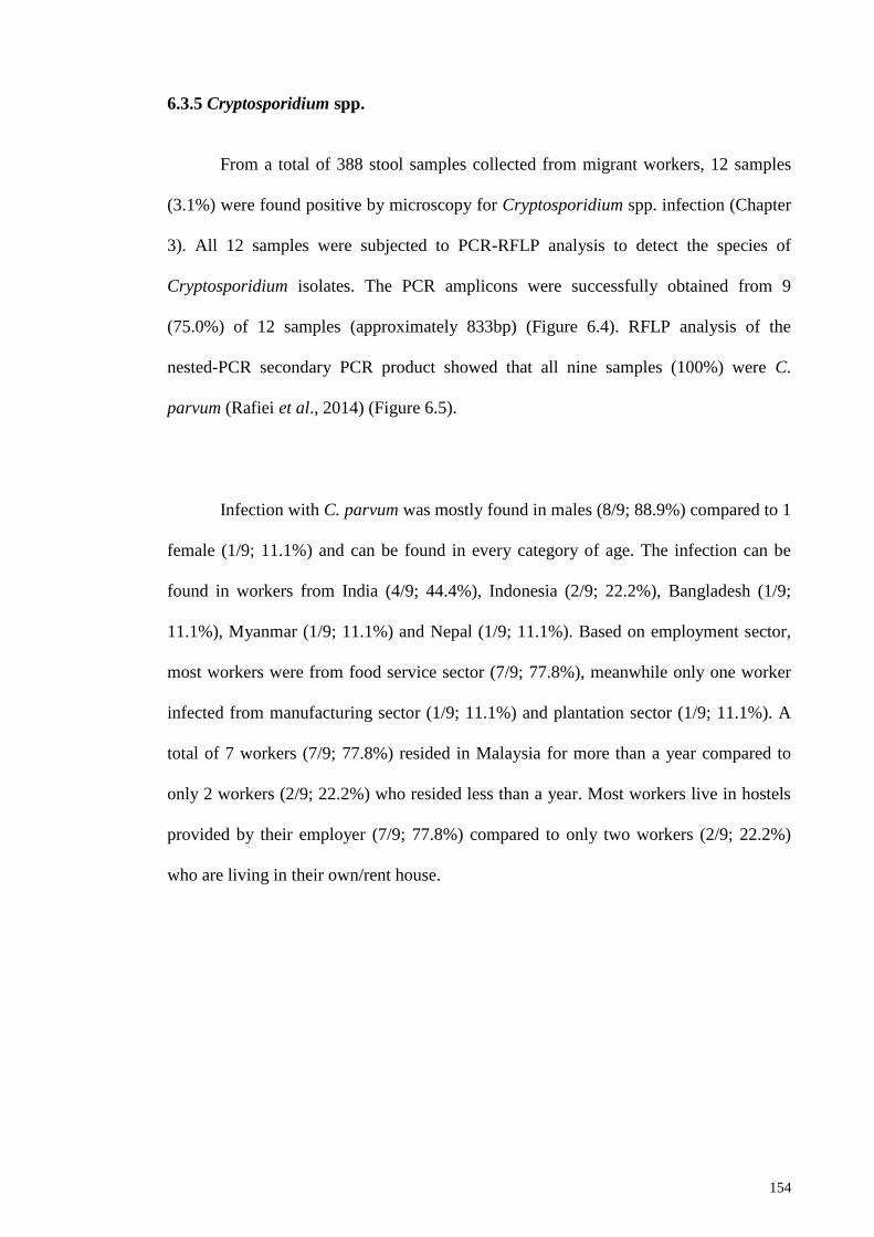
154
6.3.5 Cryptosporidium spp.
From a total of 388 stool samples collected from migrant workers, 12 samples
(3.1%) were found positive by microscopy for Cryptosporidium spp. infection (Chapter
3). All 12 samples were subjected to PCR-RFLP analysis to detect the species of
Cryptosporidium isolates. The PCR amplicons were successfully obtained from 9
(75.0%) of 12 samples (approximately 833bp) (Figure 6.4). RFLP analysis of the
nested-PCR secondary PCR product showed that all nine samples (100%) were C.
parvum (Rafiei et al., 2014) (Figure 6.5).
Infection with C. parvum was mostly found in males (8/9; 88.9%) compared to 1
female (1/9; 11.1%) and can be found in every category of age. The infection can be
found in workers from India (4/9; 44.4%), Indonesia (2/9; 22.2%), Bangladesh (1/9;
11.1%), Myanmar (1/9; 11.1%) and Nepal (1/9; 11.1%). Based on employment sector,
most workers were from food service sector (7/9; 77.8%), meanwhile only one worker
infected from manufacturing sector (1/9; 11.1%) and plantation sector (1/9; 11.1%). A
total of 7 workers (7/9; 77.8%) resided in Malaysia for more than a year compared to
only 2 workers (2/9; 22.2%) who resided less than a year. Most workers live in hostels
provided by their employer (7/9; 77.8%) compared to only two workers (2/9; 22.2%)
who are living in their own/rent house.
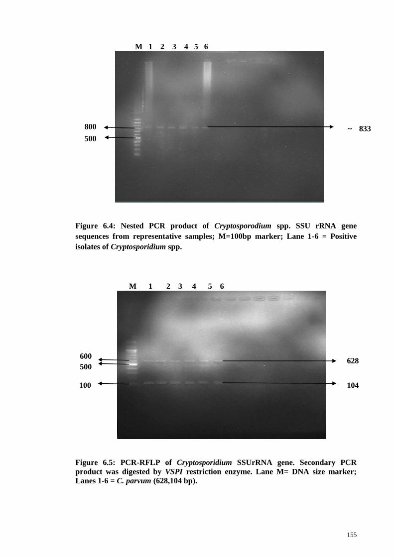
155
M 1 2 3 4 5 6
Figure 6.4: Nested PCR product of Cryptosporodium spp. SSU rRNA gene
sequences from representative samples; M=100bp marker; Lane 1-6 = Positive
isolates of Cryptosporidium spp.
M 1 2 3 4 5 6
Figure 6.5: PCR-RFLP of Cryptosporidium SSUrRNA gene. Secondary PCR
product was digested by VSPI restriction enzyme. Lane M= DNA size marker;
Lanes 1-6 = C. parvum (628,104 bp).
500
bp
800
bp
~ 833
bp
600
bp
100
bp
500
bp 104
bp
628
bp
156
6.4 Discussion
This study confirms the advantages of molecular characterization technique in
differentiating organisms at species levels for the 5 human intestinal parasitic infections.
6.4.1 Strongyloides stercoralis
Nested PCR successfully characterized current active infections of S. stercoralis
in three (2.6%) of 115 serological positive workers. Nested PCR was able to greatly
improve sensitivity compared to faecal examination. Developments in the PCR have
now permitted for reliable results to be obtained (Ahmad et al., 2013).
All three positive samples were males and could be due to the higher
involvement of males with outdoor activities compared to females especially in the
plantation sector (Ahmad et al., 2013). However, in the present study, two males were
employed in the food service sector and one in the domestic sector. The infections could
be due to bad personal hygiene and improper way of handling sewage disposal (Lim et
al., 2009; Ahmad et al., 2013).
In the developed countries, S. stercoralis infections remain an issue among
migrant workers as infections are still endemic in their country of origin. According to
Schar et al. (2013), high infection rates up to 60% can be expected in the resource-poor
countries with ecological and socioeconomic conditions which favour the spread of S.
stercoralis while low prevalence in urban and developed countries where faecal
contamination is rare with the exception of slum areas in the big cities (Schar et al.,
2013).
157
Most studies in Malaysia used the low sensitivity conventional microscopy
technique for the detection of S. stercoralis larvae in faecal samples (Rahmah et al.,
1997; Ahmad et al., 2013). Although this study adopted this technique, it failed to
detect any infection in any of the workers. In addition, most studies focused on STH
only and excluded screening for S. stercoralis and studies that did however, used low
sensitivity methods (Suresh et al., 2002). Similarly, most studies on refugees and
migrants worldwide also used copro-diagnostic procedures with low sensitivity (71.8%)
compared to copro-diagnostic procedures with moderate sensitivity (7.7%) and
serological diagnostic procedures with high sensitivity (20.5%) (Schar et al., 2013). The
frequent choice of diagnosis for S. stercoralis infections were the Koga Agar culture
method and Baermann method, which are considerably time consuming and labour
intensive. These methods involve examining stool samples collected over a few days
and could mitigate the risk of missing low-intensity infections. Furthermore, the
screening should include a minimum of three consecutive days‘ stool samples per
person. Recently, the advancement in molecular studies has enabled high sensitivity
diagnostic method of S. stercoralis. The current use of nested PCR has been successful
applied for the screening of this parasite and should be applied in all future analysis and
studies. In addition to better screening, workers should be exposed to health education
campaigns and programs aimed at increasing community awareness of the importance
of personal hygiene, sanitation, cleanliness and healthy behaviors in controlling S.
stercoralis infections.
6.4.2 Hookworm
Slightly over 10% of the workers screened in this study were positive for
hookworms which was consistent with previous studies in Malaysia conducted among
158
Orang Asli, rural communities, urban squatters and children with infection ranging
between 3.0% to 12.8% (Nor Aini et al., 2007; Hakim et al., 2007; Al-Mekhlafi et al.,
2008; Lim et al., 2009; Ngui et al., 2011a; Sinniah et al., 2014). However, current
diagnostic methods used were unable to identify the species of hookworm from the eggs
and therefore employing molecular methods and sequencing to overcome this limitation
is vital (Gruijter et al., 2005).
The PCR amplicons were successfully obtained with mostly N. americanus
(81%) and the remaining were of Ancylostoma spp and only one sample was with mix
infections. This finding was in agreement with previous studies highlighting N.
americanus as the predominant human hookworm species (Gruijter et al., 2005; Ngui et
al., 2012a). Sequence comparison using the Basic Local Alignment Search Tool
(BLAST) confirmed that all 8 Ancylostoma spp. were Ancylostoma duodenale and is the
first report of Ancylostoma duodenale in Malaysia.
Most Asian studies also reported high N. americanus infections compared to A.
duodenale. In southern part of Thailand, high prevalence of N. americanus (99.9%) was
reported among school children compared to A. duodenale (0.1%) (Anantaphruti et al.,
2002). Another study in rural community in central Thailand recorded 92.0% N.
americanus infection compared to A. duodenale (2.0%) (Jiraanankul et al., 2011).
While a study in northern Vietnam also recorded high infections of N. americanus
(95%) (Verle et al., 2003). Conversely, a study in rural area of Laos found higher
Ancylostoma spp. infection (9.4%) compared to N. americanus (5.9%), however the
species was not elucidated (Sato et al., 2010). In West Bengal, India (Pal et al., 2007)
and Hainan Province, China (Gandhi et al., 2001) also reported that hookworm
159
infections were more predominant to N. americanus with prevalence of 42.8% and
60.0%, respectively.
In the present study, A. duodenale infected workers were mainly from Indonesia
and Nepal. Six workers were newly arrived workers to Malaysia, suggesting infections
originated from their home country where infections were endemic due to poverty, poor
hygiene practices and inadequate sanitation services. Infection in humans with A.
duodenale can cause more severe pathological effects and produce symptoms with
fewer worms compared to N. americanus (Bogitsh & Cheng, 1999). Furthermore, A.
duodenale infection causes approximately five times greater blood loss resulting in a
higher degree of iron deficiency than N. americanus infection (Pawlowski et al., 1991).
6.4.3 Entamoeba spp.
A relative small proportion of the workers were found positive for Entamoeba
spp. with similar values with other studies conducted among rural communities in
Malaysia ranging from 9.4% to 21.0% (Ngui et al., 2011a; 2012; Nor et al., 2003;
Rajeswari et al., 1994; Hakim et al., 2007). However, these findings were based on
microscopy screening which could not differentiate species (Ngui et al., 2012b). E.
histolytica cysts and trophozoites were morphologically similar and difficult to
differentiate between E. dispar and E. moshkovskii. Newer techniques have been
developed for the detection of E. histolytica antigen using an enzyme-linked
immunosorbent assay (ELISA) (Gonin & Louise, 2003; Redondo et al., 2006; Zeehaida
et al., 2008) and PCR to amplify Entamoeba DNA (Fotedar et al., 2007; Katzwinkel-
Wladarsch et al., 1994; Troll et al., 1997; Ngui et al., 2012b).
160
Close to 70% of the positive samples were successfully amplified using nested
PCR which resulted in identity of E. dispar (20/31; 64.5%) as the most predominant
species, followed by E. histolytica (8/31; 25.8%) and mixed infections (3/31; 9.7%).
The finding was in agreement with worldwide distribution of Entamoeba spp. (10%);
pathogenic E. histolytica constitute 10% of the infections and non-pathogenic E. dispar
for the remaining 90%. India also reported similar findings with 49.5% patients being
infected with E. dispar meanwhile only 7.4% were infected with E. histolytica
(Khairnar & Parija, 2007). Other worldwide studies including from Australia reported
70.8% patients infected with E. dispar compared to 4.5% with E. histolytica and 61.8%
with E. moshkovskii (Fotedar et al., 2007). A study in northern Ghana also reported
higher prevalence of E. dispar compared to E. histolytica with 92.3% and 1.3%,
respectively (Verweij et al., 2003). In Brazil, a study in an urban slum area in Fortaleza
reported higher prevalence of E. dispar (90.0%) compared to E. histolytica (10.0%)
(Braga et al., 2001). In Netherland, 91.2% were identified as E. dispar and 6.7% were
E. histolytica (Visser et al.,2006) meanwhile in Canada, 97.1% of the samples were E.
dispar compared to 2.9% for E. histolytica (Gonin & Louise, 2003).
However, result from this study was in contrast with previous studies conducted
in Malaysia. Ngui et al. (2012b) reported higher prevalence of E. histolytica (75.0%)
compared to E. dispar (30.8%) from five rural villages in Peninsular Malaysia and
among aborigine community in Pahang with E. histolytica (13.2%) being more
prevalent compared to E. dispar (5.6%) (Noor Azian et al., 2006). Ngui et al., (2012b)
also reported the first detection of E. moshkovskii in Malaysia. A study in Bangladesh
highlighted that infection with E. moshkovskii was common in children aged 2 to 5
years (Ali et al., 2003). Ngui et al. (2012b) also reported that 5.8% of infected E.
161
moshkovskii were children. However, E. moshkovskii was not present in the present
study.
E. histolytica has the potential to cause dysentery and extraintestinal disease,
meanwhile E. dispar is considered to be a harmless commensal (Petri, 1996; Walsh,
1986). Migrant workers in the present study were provided with suitable
accommodation, clean water system, proper sewage toilets, and efficient waste disposal
system. High prevalence of Entamoeba infection may be due to low standards of
hygiene practice and lack of education about the diseases. The presence of E. histolytica
in the study population must be considered as a public health problem, therefore
parasite control strategies especially mass treatment and health education are rec-
ommended for all migrant workers as well as local population of Malaysia.
6.4.4 Giardia spp.
This study is the first in Malaysia to report on Giardia spp. infections among
migrant workers (10.8%) via microscopy observation. The results were similar to other
studies conducted in Malaysia among Orang Asli communities and children in rural
areas with prevalence ranging between 0.2% - 29.2% (Noor Azian et al., 2007; Lim et
al., 2008, Al-Mekhlafi et al., 2005b; 2010; Anuar et al., 2012). PCR amplicons were
successfully obtained from 30 isolates (30/42; 71.4%) and based on BLAST search of
the GenBank database, all of the nucleotide sequences belonged to G. duodenalis which
is in agreement with worldwide data that G. duodenalis is the only species found in
human (Thompson et al., 2000). G duodenalis also has been found infecting other
mammals including cattle, cats, dogs, horses, sheep and pigs (Thompson et al., 2000;
Xiao, 1994; Olson et al., 1995).
162
Phylogenetic analysis highlighted that G. duodenalis was placed in two distinct
lineage; assemblages A (13/30; 43.3%) and B (17/30; 56.7%). The isolates of
assemblage A in this study belonged to sub-assemblage AII as also reported by other
studies (Hussein et al., 2009; Lebbad et al., 2011; Bonhomme et al., 2011; Huey et al.,
2013). Assemblage B also commonly reported in human infected other mammals
including beavers (Fayer et al., 2006), cattle (Coklin et al., 2007), dogs (Lalle et al.,
2005; Read et al., 2004; Traub et al., 2004), horses (Traub et al., 2005), monkeys
(Itagaki et al., 2005), muskrats (Sulaiman et al., 2003), rabbits (Sulaiman et al., 2003),
and sheep (Castro-Hermida et al., 2007). This strongly suggests the potential zoonotic
transmission between animal and human (Sulaiman et al., 2003). However, the isolates
of assemblage B could not be assigned to any sub-assemblages due to high degree of
nucleotide variation (Bonhomme et al., 2011; Caccio et al., 2008; Lalle et al., 2009;
Lebbad et al., 2011, Levecke et al., 2009; Huey et al., 2013).
The presence of assemblage B (56.7%) was slightly higher compared to
assemblage A (43.3%) in the current study. Global incidence of assemblage A and B
differs from country to country (Feng & Xiao, 2011). In the present study, assemblage
B was reported in Bangladeshi workers (35.3%) only, which was in agreement with a
case control study in Bangladesh (Haque et al., 2005) where the presence of assemblage
B (231/267; 86.5%) was significantly higher compared to assemblage A (20/267;
7.5%). In Nepal, higher prevalence of assemblage B (26/35; 74%) also was found in the
isolates compared with assemblage A (7/35; 20%) (Singh et al., 2009). However,
present study recorded higher prevalence of assemblage A (63.6%) compared to
assemblage B (36.4%) among the Nepalese. A study in India by Caccio et al. (2005)
reported slightly higher prevalence of assemblage B (47.0%) compared to assemblage A
163
(32.0%) as also reported by Paintlia et al., (1998) (assemblage B=58%; assemblage A=
42.0%).
The presence of assemblage B and sub-assemblage AII in the samples of present
study suggest that the mode of transmission of giardiasis among migrant workers in
Malaysia may be human-to-human. However, further investigation should include
multilocus genotyping of parasites from human and animals to understand the
epidemiology, possibility of zoonotic transmission and public health importance of G.
duodenalis among migrant workers in Malaysia.
6.4.5 Cryptosporidium spp.
The present molecular analysis of Cryptosporidium spp. among migrant workers
in Malaysia successfully amplified SSU rRNA gene of Cryptosporidium spp. from 9
(9/388; 2.3%) stool samples. The prevalence reported was low compared to other
previous studies conducted in Malaysia including among the Orang Asli with
prevalence ranging from 5.5% to 20.1% (Kamel et al., 1994a; Lim et al., 1997),
community cases of children below 7 years with 10.6% (Lai, 1992), HIV patients with
prevalence ranging from 3.0% to 23.0% (Kamel et al., 1994b; Lim et al., 2005; Zaidah
et al., 2008; Iqbal et al., 2012; Iqbal et al., 2015) and among the pediatric hospital cases
ranging from 0.9% to 11.4% (Mendez et al., 1988; Mat Ludin et al., 1991; Lai, 1992;
Ng & Shekhar, 1993; Menon et al., 1999; Menon et al., 2001).
Based on the SSU rRNA sequences, more than 20 Cryptosporidium species have
been recognized (Plutzer & Karanis, 2009) and cryptosporidiosis in human was mainly
164
caused by C. parvum and C. hominis (Xiao et al., 1999; Kosek et al., 2001). PCR-RFLP
analysis based on SSU rRNA gene on all 9 isolates of Cryptosporidium sp. inferred C.
parvum. The dominance of C. parvum in this analysis was in agreement with recent
study conducted among HIV/AIDS patients in Malaysia with 84.3% of 32
Cryptosporidium isolates was C. parvum, followed by 6.3% C. hominis, 6.3% C.
meleagridis and 3.1% C. felis (Iqbal et al., 2015). Another study among HIV/AIDS
patients in Malaysia also reported the dominant of C. parvum with 64% from 25
samples, followed by 24.0% C. hominis, 8.0% C. meleagridis and 4% C. felis (Lim et
al., 2011). Zaidah et al., (2008) also reported that C. parvum was the only species found
among 9 HIV patients in Kota Bharu. Previously, the distribution and genotyping of
Cryptosporidium species base on gp60 gene among the 18 isolates of HIV-infected
patients in Malaysia also reported that 72.2% was C. parvum and 27.7% was C. hominis
(Iqbal et al., 2012).
However, results from previous studies failed to show significance between
Cryptosporidium infection and gender. The result in this study reported more male
workers (8/9; 88.9%) were infected with C. parvum compared to female (1/9; 11.1%)
with infections were found in all age levels. However this study was only limited to
adult workers and was not unable to compare infections with ages younger than 21
years old.
Based on nationality factor, most infected workers were from India (4/9;
44.4%), followed by Indonesia (2/9; 22.2%), Bangladesh (1/9; 11.1%), Nepal (1/9;
11.1%) and Myanmar (1/9; 11.1%). Recent study in Andhra Pradesh, India reported
high prevalence of Cryptosporidium infection (25%) from 306 cases, with 35% to 36%

165
adults, 17% children and 20% infants (Manocha et al., 2014). In North India also
reported high prevalence of infection with 73.0% C. hominis and 24% C. parvum
(Sharma et al., 2013). Similarly, Das et al. (2006) also reported C. hominis (87.5%), C.
parvum (10%) and C. felis (2.5%) from children in Kolkata, India. The high infections
in the population were possibly due to the close contact with animals in rural areas and
farms and the highly endemic occurrence of cryptosporidiosis among livestock could be
the sources of zoonotic transmission to human (Kali, 2014). In Indonesia, a community
based study in Surabaya reported 8.2% of C. parvum oocysts were detected in diarrhea
samples and indicated that close contact with cats, rain, flood and crowded living
condition as significant risk factors (Katsumata et al., 1998).
In the present study, most of the infected workers resided in Malaysia for more
than a year. The transmission among the workers could be due to improper personal
hygiene and in the crowded living condition as most of them are living in the hostel
provided by the employer. The outcome of this study is important in order to enable
proper control strategy and hygiene programme by the responsible agency to prevent
Cryptosporidium sp. infections among this migrant population (Lim et al., 2008).
166
6.5 Conclusion
Molecular characterization has been successful applied for the identification up
to species level of all IPIs.
The use of nested PCR targeting the internal transcribed spacer 1 (ITS1) region
of the ribosomal DNA gene was successfully applied in the detection of S. stercoralis
infection in three migrant workers.
Using internal transcribed spacer 2 and 28S ribosomal RNA region of N.
americanus and Ancylostoma spp., PCR amplicons resulted in the detection of both N.
americanus and for the first time in Malaysia, A. duodenale in this targeted group.
While, nested PCR targeting 16S-like ribosomal RNA gene was used to
genetically characterize E. histolytica and E. dispar in which E. dispar was the most
predominant infection in migrant workers. The presence of E. histolytica albeit low in
the study population still plays a role in public health.
Molecular characterization of G. duodenalis isolated from migrant workers in
Malaysia was conducted and successfully amplificated the triosephosphate isomerase
(TPI) gene. Targeting the tpi gene, the presence of assemblage B and sub-assemblage
AII suggest that the mode of transmission of giardiasis amongst these workers were
possibly human-to-human. However, further investigation is required to include
multilocus genotyping of parasites from human and animals to have a better
understanding of the epidemiology of this infection.
167
Based on the SSU rRNA gene, the C. parvum amplicons were successfully
detected in all 9 human isolates. The possible associated risk factor could be due to
crowded living conditions among the workers. Further studies should be conducted to
determine the significance of Cryptosporidium sp. transmission among the migrant
workers in Malaysia.
168
CHAPTER 7: GENERAL DISCUSSION AND CONCLUSION
7.1 General discussion
The health status of marginalized population in Malaysia is well documented
particularly on parasitic infections and toxoplasmosis. A review of one hundred and one
published articles on intestinal parasitic infections in Malaysia (1970 to 2013)
highlighted that intestinal parasitic infections continue to be a public health concern
especially among the poverty-stricken communities including the Orang Asli, plantation
and rural communities, squatter, fishing communities, new villages and the flat dwellers
(Sinniah et al., 2014). Some studies have shown that prevalence of infections fluctuated
among slum dwellers (Chia et al., 1978; Sinniah et al., 2014), flat dwellers (Kan, 1983;
Sinniah et al., 2014) and rural communities (Bisseru & Aziz, 1970; Sinniah et al.,
2014). In addition, human toxoplasmosis also persists among the Malaysian population
particularly in the clinically suspected cases and HIV-AIDS patients (Nissapatorn &
Anuar, 2004) and noted several factors linked to the acquisition of infection. Another
target study group is among migrants with a few studies among the clinical suspected
patients and workers of specific sectors (Table 7.1).
Large numbers of workers originating from more than 12 countries in Asia
(Indonesia, Bangladesh, Thailand, Philippines, Pakistan, Myanmar, Nepal, India,
Cambodia, China, Vietnam, Laos and Sri Lanka) enter Malaysia annually to fill the
low-skilled labor market primarily in sectors such as construction, domestic, food
services, manufacturing and plantation. Therefore, there is great interest to determine
the health status of these workers as they come from countries endemic to many
diseases. Despite the mandatory health screening prior to entering the country for
169
communicable diseases such as AIDS, malaria and tuberculosis, little is known on the
non-communicable disease status. These diseases have public health implications as it
can potentially be transmitted to the general public through bad hygiene and sanitation.
The present study successfully recruited 610 migrant workers from five working sectors
to determine their parasitic health status based on a questionnaire survey, microscopy
and molecular characterisation. However, only 388 stool samples in addition to 484
blood samples were available for screening (Table 7.2). Many refused to donate either
blood or/and stool samples as recruitment was on voluntary basis and the screening was
non-mandatory under FOMEMA (agency involved in the implementation, management
and supervision of a nationwide mandatory health screening programme for all legal
migrant workers in Malaysia), Ministry of Health and Immigration Department of
Ministry of Home Affairs Malaysia upon entry or residing in Malaysia. Other reasons
for not participating include disgusted with faeces handling and/or preoccupied with
matters related to work.
Most workers recruited were from domestic sector (n=148), followed by
construction (n=139), food service (n=128), plantation (n=102) and manufacturing
(n=93) sector. The participants came from varied demographic background (age, level
of education, nationality, religion and marital status, types of accommodation), however
most were housed with clean water, proper sewage toilets and waste disposal system.
Most workers did not engage in any risk behavior, such as smoking, consumption of
alcohol and illegal drugs. More than 80% were fully covered for medical treatment with
accessibility to private or government hospital/clinic. The workers also were briefed on
occupational health and safety background and were provided with personal protective
equipment (PPE) at work.
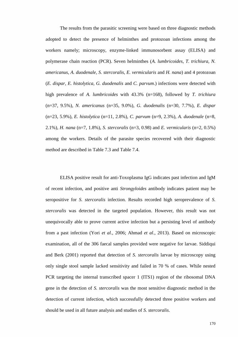
170
The results from the parasitic screening were based on three diagnostic methods
adopted to detect the presence of helminthes and protozoan infections among the
workers namely; microscopy, enzyme-linked immunosorbent assay (ELISA) and
polymerase chain reaction (PCR). Seven helminthes (A. lumbricoides, T. trichiura, N.
americanus, A. duodenale, S. stercoralis, E. vermicularis and H. nana) and 4 protozoan
(E. dispar, E. histolytica, G. duodenalis and C. parvum.) infections were detected with
high prevalence of A. lumbricoides with 43.3% (n=168), followed by T. trichiura
(n=37, 9.5%), N. americanus (n=35, 9.0%), G. duodenalis (n=30, 7.7%), E. dispar
(n=23, 5.9%), E. histolytica (n=11, 2.8%), C. parvum (n=9, 2.3%), A. duodenale (n=8,
2.1%), H. nana (n=7, 1.8%), S. stercoralis (n=3, 0.98) and E. vermicularis (n=2, 0.5%)
among the workers. Details of the parasite species recovered with their diagnostic
method are described in Table 7.3 and Table 7.4.
ELISA positive result for anti-Toxoplasma IgG indicates past infection and IgM
of recent infection, and positive anti Strongyloides antibody indicates patient may be
seropositive for S. stercoralis infection. Results recorded high seroprevalence of S.
stercoralis was detected in the targeted population. However, this result was not
unequivocally able to prove current active infection but a persisting level of antibody
from a past infection (Yori et al., 2006; Ahmad et al., 2013). Based on microscopic
examination, all of the 306 faecal samples provided were negative for larvae. Siddiqui
and Berk (2001) reported that detection of S. stercoralis larvae by microscopy using
only single stool sample lacked sensitivity and failed in 70 % of cases. While nested
PCR targeting the internal transcribed spacer 1 (ITS1) region of the ribosomal DNA
gene in the detection of S. stercoralis was the most sensitive diagnostic method in the
detection of current infection, which successfully detected three positive workers and
should be used in all future analysis and studies of S. stercoralis.
171
Among the significant explanatory factors association with the high prevalence
of parasitic infections in this country were sex, age, nationality, employment sector and
years of residence in Malaysia. A. lumbricoides infection among the workers was
significantly related to sex, nationality, employment sector and years of residence
factor. Similarly, significantly higher infections of A. lumbricoides and T. trichiura
were linked mainly to males compared to females. This could be due to more
involvement of males with more outdoor activities compared to females.
The demographic profiles of respondents comprised predominantly of
volunteers from rural areas in their respective countries where IPIs are still very much
prevalent and a major concern among the poor and deprived communities, particularly
workers from India and Nepal. This finding was in agreement with studies in India
which suggested that inadequate sanitary and poor drainage was likely to have
contributed to disease prevalence (Fernandez et al., 2002; Dhanabal et al., 2014).
Similarly, parasitic infections in Nepal were also reported linked to rapid, unplanned
urbanization, open defaecation and other unhygienic habits, as well as a lack of health
awareness (Uga et al., 2004; Rabindranath et al., 2006; Singh et al., 2013). It was also
shown that workers with employment history of less than a year or newly arrive
workers were most likely to be infected as they were also most likely to have no history
of taking any anthelmintic drugs in the last 12 months. This is not surprising as the
mandatory medical screening procedure upon entry to this country excludes
examination for IPIs and not required to be administrated with any anthelmintic drugs
(FOMEMA, 2015).
172
Serological analysis of T. gondii found four demographic factors associated with
the infections namely; age, nationality, employment sector and years of residence in
Malaysia. Highest prevalence of infection was found amongst workers from Nepal.
Variations in prevalence of infection among workers from different countries were most
likely due to dietary habits, risk behavior, environmental conditions, socioeconomic
status and hygiene (Chan et al., 2008). The high infections among Nepalese could be
due to the habitual ingestion of minced raw meat or cooking meat insufficiently
common among certain ethnic groups (Rai et al., 1994; Rai et al., 1999). In the present
study, all workers (n=484) originated from rural areas in their respective countries
where parasitic infections are still very much prevalent and a major concern among the
poor and deprived communities. Correlations were observed between seropositivity of
toxoplasmosis and consumption of unboiled water. Toxoplasmosis was also reported in
many studies, particularly among disadvantaged and indigenous communities living in
rural and remote areas (de Moura et al., 2006; Sroka et al., 2006; Sroka et al., 2010a)
Despite showing high infections among those in manufacturing, the current results may
be biased as most (91.1%) working sectors were dominated by a particular nationality
i.e., Nepalese.
The high prevalence of parasitic infections among the migrant community in
this study provided an insight into the conditions under which the subjects previously
and currently live, and reflects the availability of environmental sanitation as well as the
socioeconomic status. Transmission of parasitic infections within the community is
predominantly dependent on human behaviour, particularly during food preparation and
intake, defaecation, personal hygiene, and cleanliness. Furthermore, most parasites
found in this study can be easily treated with the appropriate anthelmintic (Table 7.3
and Table 7.4).
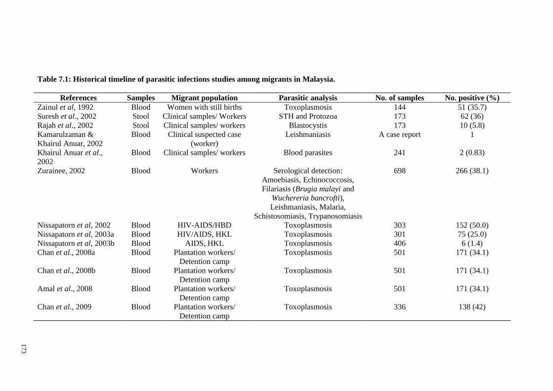
17
3
Table 7.1: Historical timeline of parasitic infections studies among migrants in Malaysia.
References Samples Migrant population Parasitic analysis No. of samples No. positive (%)
Zainul et al, 1992 Blood Women with still births Toxoplasmosis 144 51 (35.7)
Suresh et al., 2002 Stool Clinical samples/ Workers STH and Protozoa 173 62 (36)
Rajah et al., 2002 Stool Clinical samples/ workers Blastocystis 173 10 (5.8)
Kamarulzaman &
Khairul Anuar, 2002
Blood Clinical suspected case
(worker)
Leishmaniasis A case report 1
Khairul Anuar et al.,
2002
Blood Clinical samples/ workers Blood parasites 241 2 (0.83)
Zurainee, 2002 Blood Workers Serological detection:
Amoebiasis, Echinococcosis,
Filariasis (Brugia malayi and
Wuchereria bancrofti),
Leishmaniasis, Malaria,
Schistosomiasis, Trypanosomiasis
698 266 (38.1)
Nissapatorn et al, 2002 Blood HIV-AIDS/HBD Toxoplasmosis 303 152 (50.0)
Nissapatorn et al, 2003a Blood HIV/AIDS, HKL Toxoplasmosis 301 75 (25.0)
Nissapatorn et al, 2003b Blood AIDS, HKL Toxoplasmosis 406 6 (1.4)
Chan et al., 2008a Blood Plantation workers/
Detention camp
Toxoplasmosis 501 171 (34.1)
Chan et al., 2008b Blood Plantation workers/
Detention camp
Toxoplasmosis 501 171 (34.1)
Amal et al., 2008 Blood Plantation workers/
Detention camp
Toxoplasmosis 501 171 (34.1)
Chan et al., 2009 Blood Plantation workers/
Detention camp
Toxoplasmosis 336 138 (42)
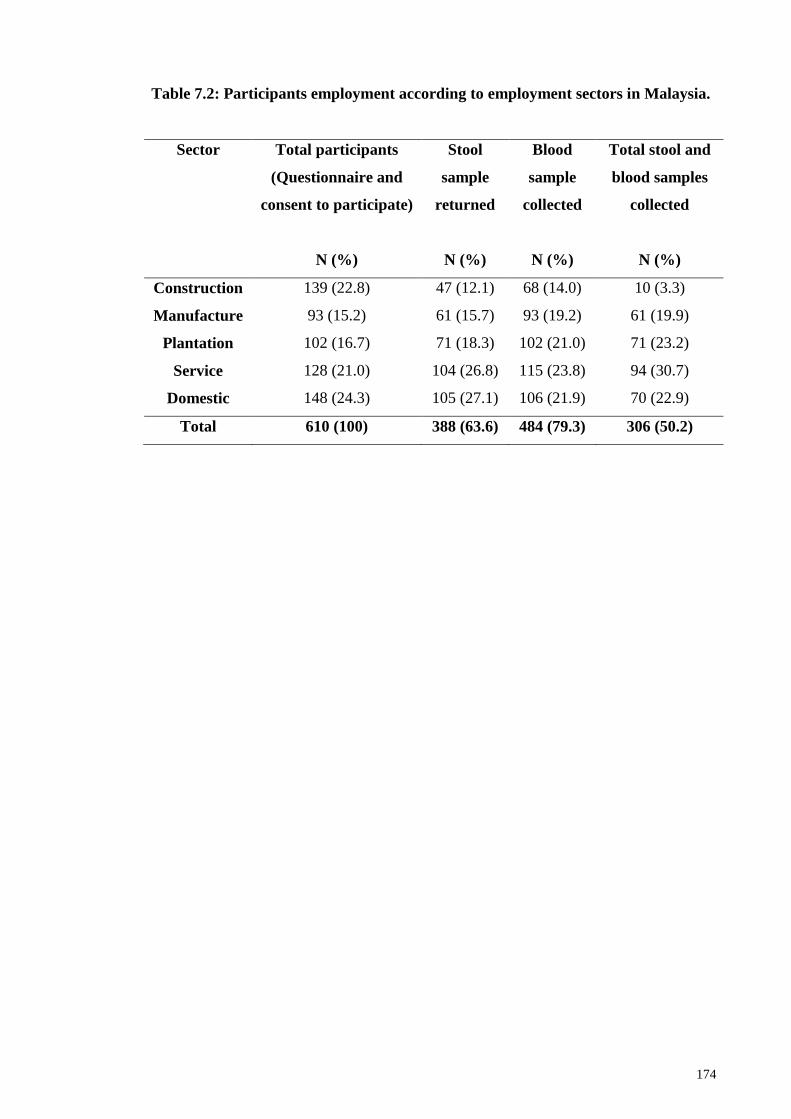
174
Table 7.2: Participants employment according to employment sectors in Malaysia.
Sector Total participants
(Questionnaire and
consent to participate)
N (%)
Stool
sample
returned
N (%)
Blood
sample
collected
N (%)
Total stool and
blood samples
collected
N (%)
Construction 139 (22.8) 47 (12.1) 68 (14.0) 10 (3.3)
Manufacture 93 (15.2) 61 (15.7) 93 (19.2) 61 (19.9)
Plantation 102 (16.7) 71 (18.3) 102 (21.0) 71 (23.2)
Service 128 (21.0) 104 (26.8) 115 (23.8) 94 (30.7)
Domestic 148 (24.3) 105 (27.1) 106 (21.9) 70 (22.9)
Total 610 (100) 388 (63.6) 484 (79.3) 306 (50.2)
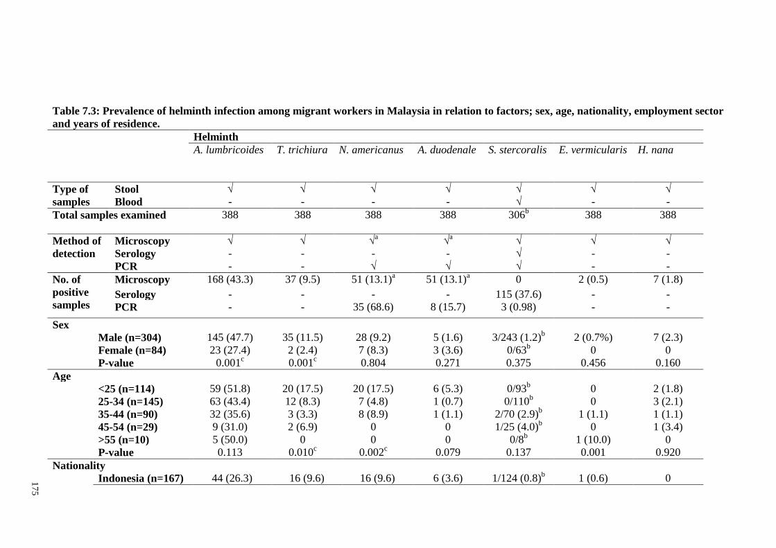
17
5
Table 7.3: Prevalence of helminth infection among migrant workers in Malaysia in relation to factors; sex, age, nationality, employment sector
and years of residence.
Helminth
A. lumbricoides T. trichiura N. americanus A. duodenale S. stercoralis E. vermicularis H. nana
Type of
samples
Stool √ √ √ √ √ √ √
Blood - - - - √ - -
Total samples examined 388 388 388 388 306b
388 388
Method of
detection
Microscopy √ √ √a
√a
√ √ √
Serology - - - - √ - -
PCR - - √
√ √ - -
No. of
positive
samples
Microscopy 168 (43.3) 37 (9.5) 51 (13.1)a
51 (13.1)a
0 2 (0.5) 7 (1.8)
Serology - - - - 115 (37.6) - -
PCR - - 35 (68.6) 8 (15.7) 3 (0.98) - -
Sex
Male (n=304) 145 (47.7) 35 (11.5) 28 (9.2) 5 (1.6) 3/243 (1.2)b
2 (0.7%) 7 (2.3)
Female (n=84) 23 (27.4) 2 (2.4) 7 (8.3) 3 (3.6) 0/63b
0 0
P-value 0.001c
0.001c
0.804 0.271 0.375 0.456 0.160
Age
<25 (n=114) 59 (51.8) 20 (17.5) 20 (17.5) 6 (5.3) 0/93b
0 2 (1.8)
25-34 (n=145) 63 (43.4) 12 (8.3) 7 (4.8) 1 (0.7) 0/110b
0 3 (2.1)
35-44 (n=90) 32 (35.6) 3 (3.3) 8 (8.9) 1 (1.1) 2/70 (2.9)b
1 (1.1) 1 (1.1)
45-54 (n=29) 9 (31.0) 2 (6.9) 0 0 1/25 (4.0)b
0 1 (3.4)
>55 (n=10) 5 (50.0) 0 0 0 0/8b
1 (10.0) 0
P-value 0.113 0.010c
0.002c
0.079 0.137 0.001
0.920
Nationality
Indonesia (n=167) 44 (26.3) 16 (9.6) 16 (9.6) 6 (3.6) 1/124 (0.8)b
1 (0.6) 0
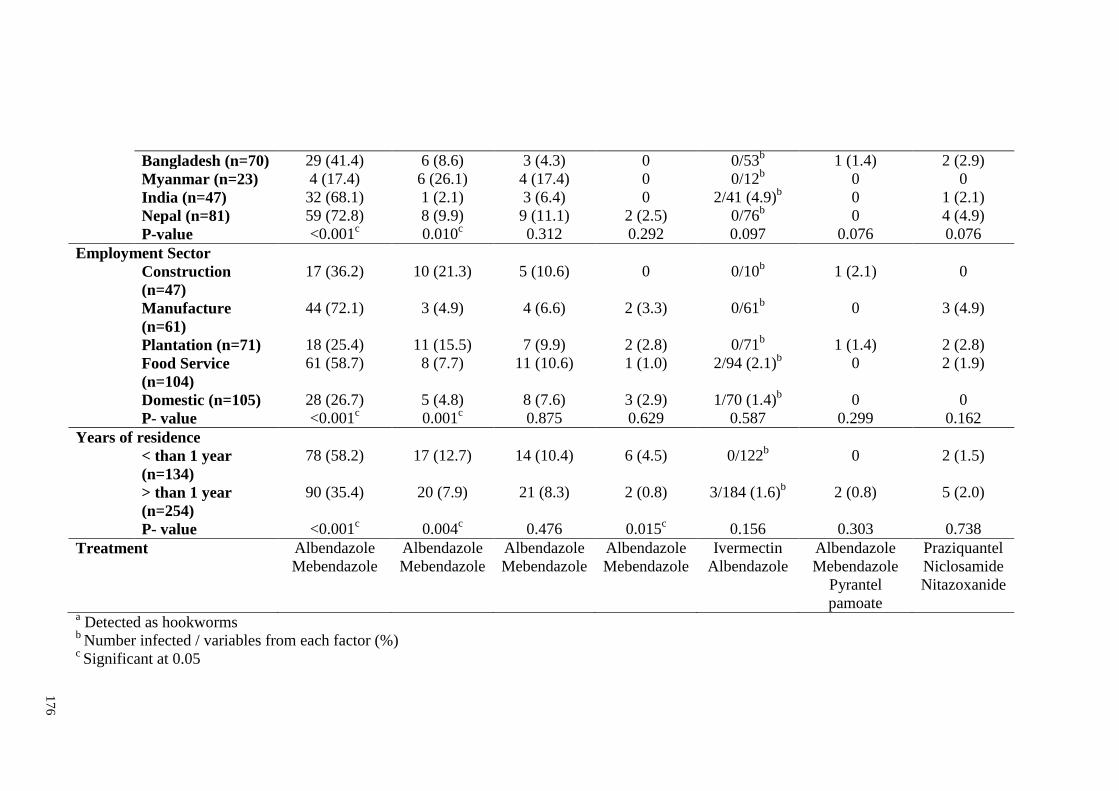
17
6
Bangladesh (n=70) 29 (41.4) 6 (8.6) 3 (4.3) 0 0/53b
1 (1.4) 2 (2.9)
Myanmar (n=23) 4 (17.4) 6 (26.1) 4 (17.4) 0 0/12b
0 0
India (n=47) 32 (68.1) 1 (2.1) 3 (6.4) 0 2/41 (4.9)b
0 1 (2.1)
Nepal (n=81) 59 (72.8) 8 (9.9) 9 (11.1) 2 (2.5) 0/76b
0 4 (4.9)
P-value <0.001c
0.010c
0.312 0.292 0.097 0.076 0.076
Employment Sector
Construction
(n=47)
17 (36.2) 10 (21.3) 5 (10.6) 0 0/10b
1 (2.1) 0
Manufacture
(n=61)
44 (72.1) 3 (4.9) 4 (6.6) 2 (3.3) 0/61b
0 3 (4.9)
Plantation (n=71) 18 (25.4) 11 (15.5) 7 (9.9) 2 (2.8) 0/71b
1 (1.4) 2 (2.8)
Food Service
(n=104)
61 (58.7) 8 (7.7) 11 (10.6) 1 (1.0) 2/94 (2.1)b
0 2 (1.9)
Domestic (n=105) 28 (26.7) 5 (4.8) 8 (7.6) 3 (2.9) 1/70 (1.4)b
0 0
P- value <0.001c
0.001c
0.875 0.629 0.587 0.299 0.162
Years of residence
< than 1 year
(n=134)
78 (58.2) 17 (12.7) 14 (10.4) 6 (4.5) 0/122b
0 2 (1.5)
> than 1 year
(n=254)
90 (35.4) 20 (7.9) 21 (8.3) 2 (0.8) 3/184 (1.6)b
2 (0.8) 5 (2.0)
P- value <0.001c
0.004c
0.476 0.015c
0.156 0.303 0.738
Treatment Albendazole
Mebendazole
Albendazole
Mebendazole
Albendazole
Mebendazole
Albendazole
Mebendazole
Ivermectin
Albendazole
Albendazole
Mebendazole
Pyrantel
pamoate
Praziquantel
Niclosamide
Nitazoxanide
a Detected as hookworms
b Number infected / variables from each factor (%)
c Significant at 0.05
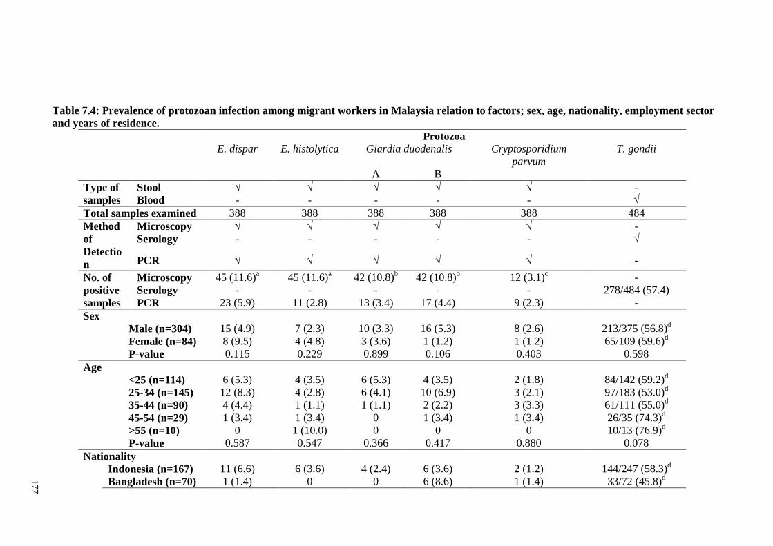
17
7
Table 7.4: Prevalence of protozoan infection among migrant workers in Malaysia relation to factors; sex, age, nationality, employment sector
and years of residence.
Protozoa
E. dispar E. histolytica Giardia duodenalis Cryptosporidium
parvum
T. gondii
A B
Type of
samples
Stool √ √ √ √ √ -
Blood - - - - - √
Total samples examined 388 388 388 388 388 484
Method
of
Detectio
n
Microscopy √ √ √ √ √ -
Serology - - - - - √
PCR √ √ √ √ √ -
No. of
positive
samples
Microscopy 45 (11.6)a
45 (11.6)a
42 (10.8)b
42 (10.8)b
12 (3.1)c
-
Serology - - - - - 278/484 (57.4)
PCR 23 (5.9) 11 (2.8) 13 (3.4) 17 (4.4) 9 (2.3) -
Sex
Male (n=304) 15 (4.9) 7 (2.3) 10 (3.3) 16 (5.3) 8 (2.6) 213/375 (56.8)d
Female (n=84) 8 (9.5) 4 (4.8) 3 (3.6) 1 (1.2) 1 (1.2) 65/109 (59.6)d
P-value 0.115 0.229 0.899 0.106 0.403 0.598
Age
<25 (n=114) 6 (5.3) 4 (3.5) 6 (5.3) 4 (3.5) 2 (1.8) 84/142 (59.2)d
25-34 (n=145) 12 (8.3) 4 (2.8) 6 (4.1) 10 (6.9) 3 (2.1) 97/183 (53.0)d
35-44 (n=90) 4 (4.4) 1 (1.1) 1 (1.1) 2 (2.2) 3 (3.3) 61/111 (55.0)d
45-54 (n=29) 1 (3.4) 1 (3.4) 0 1 (3.4) 1 (3.4) 26/35 (74.3)d
>55 (n=10) 0 1 (10.0) 0 0 0 10/13 (76.9)d
P-value 0.587 0.547 0.366 0.417 0.880 0.078
Nationality
Indonesia (n=167) 11 (6.6) 6 (3.6) 4 (2.4) 6 (3.6) 2 (1.2) 144/247 (58.3)d
Bangladesh (n=70) 1 (1.4) 0 0 6 (8.6) 1 (1.4) 33/72 (45.8)d
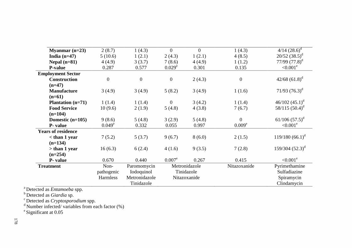
17
8
Myanmar (n=23) 2 (8.7) 1 (4.3) 0 0 1 (4.3) 4/14 (28.6)d
India (n=47) 5 (10.6) 1 (2.1) 2 (4.3) 1 (2.1) 4 (8.5) 20/52 (38.5)d
Nepal (n=81) 4 (4.9) 3 (3.7) 7 (8.6) 4 (4.9) 1 (1.2) 77/99 (77.8)d
P-value 0.287 0.577 0.029e
0.301 0.135 <0.001e
Employment Sector
Construction
(n=47)
0 0 0 2 (4.3) 0 42/68 (61.8)d
Manufacture
(n=61)
3 (4.9) 3 (4.9) 5 (8.2) 3 (4.9) 1 (1.6) 71/93 (76.3)d
Plantation (n=71) 1 (1.4) 1 (1.4) 0 3 (4.2) 1 (1.4) 46/102 (45.1)d
Food Service
(n=104)
10 (9.6) 2 (1.9) 5 (4.8) 4 (3.8) 7 (6.7) 58/115 (50.4)d
Domestic (n=105) 9 (8.6) 5 (4.8) 3 (2.9) 5 (4.8) 0 61/106 (57.5)d
P- value 0.049e
0.332 0.055 0.997 0.009e
<0.001e
Years of residence
< than 1 year
(n=134)
7 (5.2) 5 (3.7) 9 (6.7) 8 (6.0) 2 (1.5) 119/180 (66.1)d
> than 1 year
(n=254)
16 (6.3) 6 (2.4) 4 (1.6) 9 (3.5) 7 (2.8) 159/304 (52.3)d
P- value 0.670 0.440 0.007e
0.267 0.415 <0.001e
Treatment Non-
pathogenic
Harmless
Paromomycin
Iodoquinol
Metronidazole
Tinidazole
Metronidazole
Tinidazole
Nitazoxanide
Nitazoxanide Pyrimethamine
Sulfadiazine
Spiramycin
Clindamycin a Detected as Entamoeba spp.
b Detected as Giardia sp.
c Detected as Cryptosporodium spp.
d Number infected/ variables from each factor (%)
e Significant at 0.05
179
7.2 Conclusion
The parasitic health status of migrant workers in Malaysia was successfully
determined according to the objectives described below;
Migrant workers in Malaysia were provided with suitable living
accommodations and were supplied with complete basic amenities including
clean water system, proper sewage toilets, and efficient waste disposal system.
The workers also were fully covered for medical treatment with the accessibility
to private or government hospitals/clinics and all were provided with personal
protective equipment (PPE) and adhered to wearing the gear at work at all times.
The screening of stool samples using formalin ethyl-acetate concentration
technique and modified ziehl-neelsen staining method recovered a total of 8
species of parasites with four nematode species (Ascaris lumbricoides, Trichuris
trichiura, Enterobius vermicularis and hookworms), one cestode (Hymenolepis
nana) and three protozoan species (Entamoeba histolytica/dispar, Giardia spp.
and Cryptosporidium spp.) infecting the workers with infections significantly
influenced by socio-demographic (nationality), and environmental
characteristics (years of residence in this country, employment sector and
educational level).
180
Slightly more than half of the workers were seropositive for Toxoplasma gondii
(57.4%) with 52.9% being seropositive for anti-Toxoplasma IgG only, 0.8%
seropositive for anti-Toxoplasma IgM only and 3.7% seropositive with both IgG
and IgM antibodies. Samples positive for both IgG and IgM antibodies were
further tested for IgG avidity and all showed high avidity, suggesting chronic
infection. Four significant factors affected seropositivity namely, age,
nationality, employment sector and years of residence in Malaysia.
Over a third of the workers were seropositive (37.6%) to S. stercoralis infection
with no association to any for the factors tested.
Nested PCR targeting the internal transcribed spacer 1 (ITS1) region of the
ribosomal DNA gene of S. stercoralis was highly sensitive in the detection of
current infections and should be applied in all future analysis and studies. This
study successfully detected three workers with current S. stercoralis infections.
PCR methods successfully amplified internal transcribed spacer 2 and 28S
ribosomal RNA region of N. americanus and Ancylostoma spp. The PCR
amplicons successfully obtained N. americanus and A. duodenale. This is the
first time A. duodenale was reported in Malaysia. This finding may have
important implications for public health and for the control of hookworm
diseases in Malaysia, especially A. duodenale which has higher degree of iron
deficiency and anemia compared to N. americanus.
181
Nested PCR targeting 16S-like ribosomal RNA gene successfully recovered E.
dispar as the most dominant infection among workers compared to E.
histolytica. The presence of E. histolytica in the study population was small
however is carries a public health risk, therefore parasite control strategies
especially mass treatment and health education are recommended for all migrant
workers as well as local population of Malaysia.
Amplification of the triosephosphate isomerase (TPI) gene from G. duodenalis
isolates successfully obtained the presence of assemblage B and sub-assemblage
AII. This suggested that the mode of transmission of giardiasis among migrant
workers in Malaysia may be human-to-human. However, further investigation
should be conducted involving multilocus genotyping of parasites from human
and animals to understand the epidemiology of G. duodenalis infection based on
molecular genotyping approach.
Based on the SSU rRNA gene, the C. parvum amplicons were successfully
detected in 9 human isolates. The possible associated risk factor could be due to
crowded living conditions among the workers in the hostel provided by the
employer. Further analysis should be done to determine the significance of
Cryptosporidium sp. transmission among the migrant workers in Malaysia.
182
Limitations of the study
Of the 610 volunteers recruited, only 388 stool samples and 484 blood samples were
available for screening. Many refused to donate either blood or/and stool samples for
reasons including disgusted with faeces handling and/or preoccupied with matters
related to work. Higher participation was expected if this study had the full support
from the Ministry of Health Malaysia and other related agencies such as FOMEMA
including the employer of the migrant workers.
Recommendations
These findings highlight the urgent need especially to refine current health polices
for Malaysia to include STH and other parasites in the mandatory health screening list
of those applying for entry, work permits and residence in Malaysia. Recommendations
also include the implementation of mass drug administration for all newly arrive
workers as stated by World Health Organization (2015). Moreover, this should be
accompanied by health education campaigns and programs aimed at increasing
community awareness of the importance of personal hygiene, sanitation, cleanliness,
healthy behaviors in controlling parasitic infections and the potential in transmission of
diseases.
183
REFERENCES
Abahussain, N. A. (2005). Prevalence of intestinal parasites among expatriate workers
in Al-Khobar, Saudi Arabia. Middle East Journal of Family Medicine, 3(3), 17–
21.
Abdul-Aziz, A. R. (2001). Bangladeshi Migrant workers in Malaysia‘s Construction
Sector. Asia Pacific Population Journal, 16, 3-22.
Abu-Madi, M. A., Behnke, J. M., Ismail, A., Al-Olaqi, N., Al-Zaher, K. & El-Ibrahim,
R. (2011). Comparison of intestinal parasitic infection in newly arrived and
resident workers in Qatar. Parasites & Vectors, 4, 211.
Abu-Madi, M. A., Behnke, J. M. & Doiphode, S. H. (2010). Changing trends in
intestinal parasitic infections among long-term-residents and settled immigrants
in Qatar. Parasites & Vectors, 3, 98.
Abu-Madi, M. A., Behnke, J. M. & Ismail, A. (2008). Patterns of infection with
intestinal parasites in Qatar among food handlers and housemaids from different
geographical regions or origin. Acta Tropica, 106, 213-220.
Afifi, A., Khairul Anuar, A., Ludin, M. & Rahmah, N. (1992). Survey of meat samples
for Toxoplasma gondii in Northern Peninsular Malaysia. Diagnosa, 6(1), 700-
704.
Ahmad, A. F., Hadip, F., Ngui, R., Lim, A. L. & Mahmud, R. (2013). Serological and
molecular detection of Strongyloides stercoralis infection among an Orang Asli
community in Malaysia. Parasitology Research, 112(8), 2811-2816.
Al Kilani, M. K., Dahesh, S. M. & El Taweel, H. A. (2008). Intestinal parasitosis in
Nalout popularity, western Libya. Journal of the Egyptian Society of
Parasitology, 38, 255-264.
184
Al-Madani, A. A. & Mahfouz, A. A. (1995). Prevalence of intestinal parasitic infections
among Asian female house keepers in Abha District, Saudi Arabia. Southeast
Asian Journal of Tropical Medicine and Public Health, 26, 135–137.
Al-Mekhlafi, H. M. S., Azlin, M. & Nor Aini, U. (2006). Prevalence and distribution of
soil-transmitted helminthiases among Orang Asli children living in peripheral
Selangor, Malaysia. Southeast Asian Journal of Tropical Medicine and Public
Health, 37, 40-47.
Al-Mekhlafi, H. M., Atiya, A. S., Lim, Y. A. L., Ariffin, W. A., Abdullah, A. C. &
Surin, J. (2007). An unceasing problem: soil transmitted helminthiases in rural
Malaysian communities. Southeast Asian Journal of Tropical Medicine and
Public Health, 38, 998-1007.
Al-Mekhlafi, H. M., Azlin, M., Nor Aini, U., Shaik, A., Sa‘iah, A., Fatmah, M.S.,
Ismail, M.G., Ahmad Firdaus, M.S., Aisah, M.Y., Rozlida, A.R. & Norhayati,
M. (2005b). Giardiasis as a predictor of childhood malnutrition in Orang Asli
children in Malaysia. Transactions of the Royal Society of Tropical Medicine
and Hygiene, 99, 686–691.
Al-Mekhlafi, H. M., Azlin, M., Nor Aini, U., Shaikh, A., Sa‘iah, A., Fatmah, M. S.,
Ismail, M. G., Firdaus, A. A., Aisah, M. Y., Roslida, A. R. & Norhayati, M.
(2005a). Protein energy malnutrition and soil transmitted helminthiases among
Orang Asli children in Selangor, Malaysia. Asia Pacific Journal of Clinical
Nutrition, 14, 188-194.
Al-Mekhlafi, H. M., Surin, J., Atiya, A. S., Lim, Y. A. L., Ariffin, W. A. & Abdullah,
A. C. (2008). Pattern and predictors of soil transmitted helminth re-infection
among aboriginal school children in rural Peninsular Malaysia. Acta Tropica,
107, 200-204.
Al-Mekhlafi, H.M., Surin, J., Sallam, A.A., Abdullah, A.W. & Mahdy, M.A.K. (2010).
Giardiasis and poor vitamin A status among aboriginal school children in rural
Malaysia. American Journal of Tropical Medicine and Hygiene, 83, 523–527.
Ali, I. K. M., Hossain, M. B., Roy, S., Ayeh-Kumi, P., Petri, J. W. A., Haque, R. &
Clark, C. G. (2003). Entamoeba moshkovskii infections in children, Bangladesh.
Emerging Infectious Diseases, 9, 580–584.
185
Alvarado-Esquivel, C., Sifuentes-Álvarez, A., Narro-Duarte, S. G., Estrada-Martínez,
S., Díaz-García, J. H., Liesenfeld, O., Martínez-García, S. A. & Canales-Molina,
A. (2006). Seroepidemiology of Toxoplasma gondii infection in pregnant
women in a public hospital in northern Mexico. BMC Infectious Diseases, 6,
113.
Amal, R. N., Chan, B. T. E., Noor Hayati, M. I., Kino, H., Anisah, N., Norhayati, M.,
Sulaiman, O., Mohammed Abdullah, M., Fatmah, M. S., Roslida, A. R. &
Ismail, G. (2008). Seroprevalence of toxoplasmosis: comparative study between
migrant workers from the Indian subcontinent and local Malaysian workers. The
New Iraqi Journal of Medicine, 4(1), 9-15.
Amendoeira, M. R. R., Costa, T. & Spalding, S. M. (1999). Toxoplasma gondii Nicolle
& Manceaux, 1909 (Apicomplexa: Sarcocystidae) e atoxoplasmose. Revista de
Sousa Marques, 1, 15 – 35.
Anantaphruti, M. T., Maipanich, W., Muennoo, C., Pubampen, S. & Sanguankiat, S.
(2002). Hookworm infections of schoolchildren in southern Thailand. Southeast
Asian Journal of Tropical Medicine and Public Health, 33, 468–473.
Anuar, K., Ramachandran, C. P. & Paran, T. P. (1978). Parasitic diseases among
fishermen living on Penang Island. Medical Journal of Malaysia, 32, 321-327.
Anuar, T.K., Al-Mekhlafi, H.M., Abdul Ghani, M.K., Osman, E., Mohd Yasin, A.,
Nordin, A., Nor Azreen, S., Md Salleh, F., Ghazali, N., Bernadus, M. & Moktar,
N. (2012). Giardiasis among different tribes of Orang Asli in Malaysia:
highlighting the presence of other family members infected with Giardia
intestinalis as a main risk factor. International Journal of Parasitology, 42, 871–
880.
Apt, W., Thiermann, E., Niedmann, G. & Pasmanik, S. (1973). Toxoplasmosis.
Santiago, Chile: Universidade do Chile.
Arakaki, T., Kohakura, M., Asato, R., Ikeshiro, T., Nakamura, S. & Iwanaga, M.
(1992). Epidemiological aspects of Strongyloides stercoralis infection in
Okinawa, Japan. American Journal of Tropical Medicine and Hygiene, 95, 210–
213.
186
Balasingam, E., Lim, B. L. & Ramachandran, C. P. (1969). A parasitological study of
Pulau Pinang and Pulau Perhentian Kechil, off Terengganu, West Malaysia.
Medical Journal of Malaysia, 23, 300-306.
Bangs, M. J., Purnomo, Andersen, E. M. & Anthony, R. L. (1996). Intestinal parasites
of humans in a highland community of Irian Jaya, Indonesia. Annals of Tropical
Medicine and Parasitology, 90, 49-53.
Bardan S. (2014). Practical guidelines for employers on the recruitment, placement,
employment and repatriation of foreign workers in Malaysia. Malaysian
Employers Federation.
Basuni, M., Muhi, J., Othman, N., Verweij, J. J., Ahmad, M., Miswan, N.,
Rahumatullah, A., Aziz, F. A., Zainudin, N. S. & Noordin, R. (2011). A
pentaplex real-time polymerase chain reaction assay for detection of four species
of soil-transmitted helminths. American Journal of Tropical Medicine and
Hygiene, 84, 338–343.
Berger, S. A., Schwartz, T. & Michaeli, D. (1989). Infectious disease among Ethiopian
immigrants in Israel. Archives of Internal Medicine, 149, 117-119.
Bertrand, I., Gantzer, C., Chesnot, T. & Schwartzbrod, J. (2004). Improved specificity
for Giardia lamblia cyst quantification in wastewater by development of a real-
time PCR method. Journal of Microbiological Methods, 57, 41-53.
Bethony, J., Brooker, S., Albonico, M., Geiger, S. M., Loukas, A. & Diemert, D.
(2006). Soil-transmitted helminth infections: ascariasis, trichuriasis, and
hookworm. The Lancet, 367, 1521–1532.
Bhatia, V. N., Meenakshi, K. & Aggarwal, S. C. (1974). Toxoplasmosis in South India,
a serological study. Indian Journal of Medical Research, 62, 1818-1825.
Binnicker, M. J., Jespersen, D. J. & Harring, J.A. (2010). Multiplex detection of IgM
and IgG class antibodies to Toxoplasma gondii, rubella virus, and
cytomegalovirus using a novel multiplex flow immunoassay. Clinical and
Vaccine Immunology, 17, 1734–1738.
187
Bisseru, B. & Aziz, A. (1970). Intestinal parasites, eosinophilia, hemoglobin and
gamma globulin of Malay, Chinese and Indian school children. Medical Journal
of Malaysia. 25, 29-33.
Bogitsh, B. J. & Cheng, T. C. (1999). Human Parasitology, 2nd edn. (pp. 312–331).
Academic Press, San Diego, CA.
Bonhomme, J., Goff, L. L., Lemee, V., Gargala, G., Ballet, J. J. & Favennec, L. (2011).
Limitations of tpi and bg genes sub-genotyping for characterization of human
Giardia duodenalis isolates. Parasitology International, 60, 327–330.
Braga, L., Gomes, M. L., Silva, M. W., Paiva, C., Sales, A. & Mann, B. J. (2001).
Entamoeba histolytica and Entamoeba dispar infections as detected by
monoclonal antibody in an urban slum in Fortaleza, Northeastern Brazil. Revista
da Sociedade Brasileira de Medicina Tropical, 34, 467–471.
Brodine, S. K., Thomas, A., Huang, R., Harbertson, J., Mehta, S., Leake, J., Nutman, T.,
Moser, K., Wolf, J., Ramanathan, R., Burbelo, P., Nou, J., Wilkins, P. & Reed,
S. L. (2009). Community based parasitic screening and treatment of Sudanese
refugees: application and assessment of Centers for Disease Control guidelines.
American Journal of Tropical Medicine and Hygiene, 80, 425-430.
Buchwald, D., Lam, M. & Hooton, T. M. (1995). Prevalence of intestinal parasites and
association with symptoms in Southeast Asian refugees. Journal of Clinical
Pharmacy and Therapeutics, 20, 271-275.
Caccio, S. M. & Ryan, U. (2008). Molecular epidemiology of giardiasis. Molecular and
Biochemical Parasitology, 160, 75–80.
Caccio, S. M., Thompson, R. C., McLauchlin, J. & Smith, H. V. (2005). Unravelling
Cryptosporidium and Giardia epidemiology. Trends in Parasitology 21, 430–
437.
188
Caruana, S. R., Kelly, H. A., Ngeow, J. Y., Ryan, N. J., Bennett, C. M., Chea, L., Nuon,
S., Bak, N., Skull, S. A. & Biggs, B. A. (2006). Undiagnosed and potentially
lethal parasite infections among immigrants and refugees in Australia. Journal
of Travel Medicine, 13, 233-239.
Castro-Hermida, J. A., Almeida, A., Gonzalez-Warleta, M., Correia da Costa, J. M.,
Rumbo Lorenzo, C. & Mezo, M. (2007). Occurrence of Cryptosporidium
parvum and Giardia duodenalis in healthy adult domestic ruminants.
Parasitology Research, 101, 1443–1448.
CDC (2010). Giardiasis surveillance - United States, 2006-2008. Morbidity and
Mortality Weekly Report, 59(SS06), 15-25.
Central Intelligence Agency (2016). Bangladesh. The World Factbook. Retrieved
March 14, 2015, from https://www.cia.gov/library/publications/the-world-
factbook/geos/bg.html
Central Intelligence Agency (2016). Burma. The World Factbook. Retrieved March 14,
2015, from https://www.cia.gov/library/publications/the-world-
factbook/geos/bm.html
Central Intelligence Agency (2016). India. The World Factbook. Retrieved March 14,
2015, from https://www.cia.gov/library/publications/the-world-
factbook/geos/in.html
Central Intelligence Agency (2016). Indonesia. The World Factbook. Retrieved March
14, 2015, from https://www.cia.gov/library/publications/the-world-
factbook/geos/id.html
Central Intelligence Agency (2016). Malaysia. The World Factbook. Retrieved March
14, 2015, from https://www.cia.gov/library/publications/the-world-
factbook/geos/my.html
Central Intelligence Agency (2016). Nepal. The World Factbook. Retrieved March 14,
2015, from https://www.cia.gov/library/publications/the-world-
factbook/geos/np.html
189
Central Intelligence Agency (2016). Vietnam. The World Factbook. Retrieved March
14, 2015, from https://www.cia.gov/library/publications/the-world-
factbook/geos/vm.html
Chan, B. T. E., Amal, R. N., Noor Hayati, M. I., Hideto, K., Anisah, N. & Norhayati,
M. (2009b). Comparative study of seroprevalence of toxoplasmosis between
local workers and migrant workers in Malaysia. Archives of Medical Science,
5(2), 255-258.
Chan, B. T. E., Amal, R. N., Noor Hayati, M. I., Kino, H., Anisah, N., Norhayati, M.,
Sulaiman, O., Mohammed Abdullah, M., Fatmah, M. S., Roslida, A. R. &
Ismail, G. (2008). Seroprevalence of toxoplasmosis among migrant workers
from different Asian countries working in Malaysia. Southeast Asian Journal of
Tropical Medicine and Public Health, 39 (1), 9-13.
Chan, B. T. E., Amal, R. N. & Noor Hayati, M. I. (2009a). Toxoplasmosis among
Indonesian Migrant Workers in Malaysia. Malaysian Journal of Medicine and
Health Science, 5(1), 31–37.
Che Ghani, M., Noor Hayati, M. I., Ali, O. & Baharam, M. H. (1989). Effect of
rehousing and improved sanitation on the prevalence and intensity of soil
transmitted helminthiases in an urban slum in Kuala Lumpur. Collected papers
on the control of soil-transmitted helminthiases, 4, 51-55.
Cheah, W. C., Fah, C. S. & Fook, C. W. (1975). Pattern of toxoplasma antibodies in
Malaysian pregnant women. Medical Journal of Malaysia, 29, 275-279.
Cheng, H. S. & Shieh, Y. H. (2000). Investigation on subclinical aspects related to
intestinal parasitic infections among Thai laborers in Taipei. Journal of Travel
Medicine, 7, 319-324.
Chia, W. Y., Ishak, F., Goh, L. H., Devaraj, J. M., Jalleh, R. P., Tan, L. P. & Jalil, T. M.
A. (1978). The problem of soil-transmitted helminthes in squatter areas around
Kuala Lumpur. Medical Journal of Malaysia, 32, 33-34.
190
Chia, Y. C. (2002). Physical examination and basic haematological findings in a
selected group of migrant workers in Malaysia. Journal of the University of
Malaya Medical Centre, 7(1), 40-43.
Ciesielski, S. D., Seed, J. R., Ortiz, J. C. & Metts, J. (1992). Intestinal parasites among
North Carolina migrant farmworkers. American Journal of Public Health, 82,
1258-1262.
Coklin, T., Farber, J., Parrington, L. & Dixon, B. (2007). Prevalence and molecular
characterization of Giardia duodenalis and Cryptosporidium spp. in dairy cattle
in Ontario, Canada. Veterinary Parasitology, 150, 297–305.
Concha, R., Harrington, W. Jr. & Rogers, A. I. (2005). Intestinal strongyloidiasis:
recognition, management, and determinants of outcome. Journal of Clinical
Gastroenterology, 39, 203–211.
Conlan, J. V., Khamlome, B., Vongxay, K., Elliot, A., Pallant, L., Sripa, B., Blacksell,
S. D., Fenwick, S. & Thompson, R. C. (2012). Soil-transmitted helminthiasis in
Laos: a community wide cross-sectional study of humans and dogs in a mass
drug administration environment. American Journal of Tropical Medicine and
Hygiene, 86, 624–634.
Cook, G. & Zumla, A. (2003). Manson’s tropical diseases. 21st edn. London: WB
Saunders.
Das, P., Roy, S. S., MitraDhar, K., Dutta, P., Bhattacharya, M. K., Sen, A., Ganguly, S.,
Bhattacharya, S. K., Lal, A. A. & Xiao, L. (2006). Molecular characterization of
Cryptosporidium spp. from children in Kolkata, India. Journal of Clinical
Microbiology, 44, 4246-4249.
Davies, A. P. & Chalmers, R. M. (2009). Cryptosporidiosis. British Medical Journal,
339, b4168.
191
de Gruijter, J. M., van Lieshout, L., Gasser, R. B., Verweij, J. J., Brienen, E. A., Ziem,
J. B., Yelifari, L. & Polderman, A. M. (2005). Polymerase chain reaction-based
differential diagnosis of Ancylostoma duodenale and Necator americanus
infections in human in northern Ghana. Tropical Medicine and International
Health, 10, 574–580.
de Moura, L., Bahia-Oliveira, L. M., Wada, M. Y., Jones, J. L., Tuboi, S. H., Carmo, E.
H., Ramalho, W. M., Camargo, N. J., Trevisan, R., Graça, R. M., da Silva, A. J.,
Moura, I., Dubey, J. P. & Garrett, D. O. (2006). Waterborne toxoplasmosis,
Brazil, from field to gene. Emerging Infectious Diseases, 12, 326-329.
de Silva, S., Saykao, P., Kelly, H., MacIntyre, C. R., Ryan, N., Leydon, J. & Biggs, B.
A. (2002). Chronic Strongyloides stercoralis infection in Laotian immigrants
and refugees 7-20 years after resettlement in Australia. Epidemiology &
Infection, 128, 439-444.
Department of Occupational Safety and Health Malaysia (2015). Occupational
Accidents Statistics by Sector until December 2015. Ministry of Human
Resources Malaysia. Retrieved April 16, 2015, from
http://www.dosh.gov.my/index.php/en/archive-statistics/2015/1713-
occupational-accidents-statistics-by-sector-until-december-2015
Derouin, F. & Pelloux, H. (2008). Prevention of toxoplasmosis in transplant patients.
Clinical Microbiology and Infection, 14, 1089–1101.
Dhanabal, J., Selvadoss, P. P. & Muthuswamy, K. (2014). Comparative Study of the
Prevalence of Intestinal Parasites in Low Socioeconomic Areas from South
Chennai, India. Journal of Parasitology Research. doi: 10.1155/2014/630968.
Diaz, J., Igual, R., Alonso, M. C. & Moreno, M. J. (2002). Intestinal parasitological
study in immigrants in the region of Safor (ComunidadValeciana), Spain.
Medicina Clinica (Barcelona), 119, 36.
Dissanaike, A. S., Kan, S. P. & Thomas, V. (1977). Studies on parasitic infections in
Orang Asli (Aborigines) in Peninsular Malaysia. Medical Journal of Malaysia,
32, 48-55.
192
Djurković-Djaković, O., Milenković, V., Nikolić, A., Bobić, B. & Grujić, J. (2002).
Efficacy of atovaquone combined with clindamycin against murine infection
with a cystogenic (Me49) strain of Toxoplasma gondii. Journal of Antimicrobial
Chemotherapy, 50(6), 981-987.
DPDx (2013). Amebiasis. Centers for Disease Control and Prevention. Retrieved May
17, 2015, from http://www.cdc.gov/dpdx/amebiasis/
DPDx (2013). Ascaris lumbricoides. Centers for Disease Control and Prevention.
Retrieved May 17, 2015, from http://www.cdc.gov/dpdx/ascariasis/index.html
DPDx (2013). Cryptosporidiosis. Centers for Disease Control and Prevention. Retrieved
May 17, 2015, from http://www.cdc.gov/dpdx/cryptosporidiosis/index.html
DPDx (2013). Enterobiasis. Centers for Disease Control and Prevention. Retrieved May
17, 2015, from http://www.cdc.gov/dpdx/enterobiasis/
DPDx (2013). Giardiasis. Centers for Disease Control and Prevention. Retrieved May
17, 2015, from http://www.cdc.gov/dpdx/giardiasis/
DPDx (2013). Hookworm. Centers for Disease Control and Prevention. Retrieved May
17, 2015, from http://www.cdc.gov/dpdx/hookworm/index.html
DPDx (2013). Hymenolepiasis. Centers for Disease Control and Prevention. Retrieved
May 17, 2015, from http://www.cdc.gov/dpdx/hymenolepiasis/
DPDx (2013). Trichuriasis. Centers for Disease Control and Prevention. Retrieved May
17, 2015, from http://www.cdc.gov/dpdx/trichuriasis/index.html
DPDx (2015). Strongyloidiasis. Centers for Disease Control and Prevention. Retrieved
May 17, 2015, from https://www.cdc.gov/dpdx/strongyloidiasis/index.html
193
DPDx (2015). Toxoplasmosis. Centers for Disease Control and Prevention. Retrieved
May 17, 2015, from http://www.cdc.gov/dpdx/toxoplasmosis/
Dubey, J. P. & Beattie, C. P. (1988). Toxoplasmosis of animals and man. (pp. 220).
Boca Raton, Fla.: CRC Press.
Dubey, J. P. (2010). Toxoplasmosis of animals and humans. (pp. 313). Boca Raton,
Fla.: CRC Press.
Dubey, J. P. (1994). Toxoplasmosis. Journal of the American Veterinary Medical
Association, 205, 1593-1598.
Dunn, F. L. (1972). Intestinal parasites in Malaysia Aborigines (Orang Asli). Bulletin of
the World Health Organization, 46, 99-113.
Escobedo, A. A. & Cimerman, S. (2007). Giardiasis: a pharmacotherapy review. Expert
Opinion on Pharmacotherapy, 8(12), 1885-1902.
Fayer, R. (2010). Taxonomy and species delimitation in Cryptosporidium. Experimental
Parasitology, 124, 90–97.
Fayer, R., Morgan, U. & Upton, S. J. (2000). Epidemiology of Cryptosporidium:
transmission, detection and identification. International Journal of Parasitology,
30, 1305-1322.
Fayer, R., Santin, M., Trout, J. M., Destefano, S., Koenen, K. & Kaur, T. (2006).
Prevalence of Microsporidia, Cryptosporidium spp., and Giardia spp. in beavers
(Castor canadensis) in Massachusetts. Journal of Zoo and Wildlife Medicine, 37,
492–497.
Feldman, H. A. (1974). Toxoplasmosis: An Overview. Bulletin of the New York
Academy of Medicine, 50, 110-127.
194
Feng, Y. Y. & Xiao, L. H. (2011). Zoonotic potential and molecular epidemiology of
Giardia Species and giardiasis. Clinical Microbiology Reviews, 24, 110.
Ferguson, D. J. (2009). Identification of faecal transmission of Toxoplasma gondii:
Small science, large characters. International Journal for Parasitology, 39, 871–
875.
Fernandez, M. C., Verghese, S., Bhuvaneswari, R., Elizabeth, S. J., Mathew, T., Anitha,
A. & Chitra, A. K. (2002). A comparative study of the intestinal parasites
prevalent among children living in rural and urban settings in and around
Chennai. Journal of Communicable Diseases, 34(1), 35–39.
Fisher, D., McCarry, F. & Currie, B. (1993). Strongyloidiasis in the Northern Territory.
Under-recognised and under-treated? Medical Journal of Australia, 159, 88-90.
Flanagan, P. A. (1992). Giardia - diagnosis, clinical course and epidemiology. A
review. Epidemiology & Infection, 109, 1-22.
FOMEMA (2015). Fomema Prevention and Care. Medical Screening Process.
Retrieved April 22, 2015, from http://www.fomema.com.my/index.php/medical-
screening-process
Fotedar, R., Stark, D., Beebe, N., Marriot, D., Ellis, J. & Harkness, J. (2007). PCR
detection of Entamoeba histolytica, Entamoeba dispar and Entamoeba
moshkovskii in stool samples from Sydney, Australia. Journal of Clinical
Microbiology, 45, 1035–1037.
Gandahusada, S. (1991). Study on the prevalence of toxoplasmosis in Indonesia: a
review. Southeast Asian Journal of Tropical Medicine and Public Health, 22,
93-98.
195
Gandhi, N. S., Jizhang, C., Khoshnood, K., Fuying, X., Shanwen, L., Yaoruo, L., Bin,
Z., Haechou, X., Chongjin, T., Yan, W., Wensen, W., Dungxing, H., Chong, C.,
Shuhua, X., Hawdon, J. M. & Hotez, P. J. (2001) Epidemiology of Necator
Americanus hookworm infections in Xiulongkan Village, Hainan Province
China: high prevalence and intensity among middle aged and elderly
residents. Journal of Parasitology, 87(4), 739-743.
Garg, P. K., Perry, S., Dorn, M., Hardcastle, L. & Parsonnet, J. (2005). Risk of
intestinal helminth and protozoan infection in a refugee population. American
Journal of Tropical Medicine and Hygiene, 73, 386-391.
Garlington, W., High, K. & Schmader, K. E. H.N.S . (2011). Evaluation of infection in
the older adult.
Geltman, P. L., Cochran, J. & Hedgecock, C. (2003). Intestinal parasites among African
refugees resettled in Massachusetts and the impact of an overseas pre-departure
treatment program. American Journal of Tropical Medicine and Hygiene, 69,
657-662.
Genta, R. M. (1992). Dysregulation of strongyloidiasis: a new hypothesis. Clinical
Microbiology Reviews, 5, 345–355.
Gibney, K. B., Mihrshahi, S., Torresi, J., Marshall, C., Leder, K. & Biggs, B. A. (2009).
The profile of health problems in African immigrants attending an infectious
disease unit in Melbourne, Australia. American Journal of Tropical Medicine
and Hygiene, 80, 805-811.
Gill, G. V., Welch, E., Bailey, J. W., Bell, D. R. & Beeching, N. J. (2004). Chronic
Strongyloides stercoralis infection in former British Far East prisoners of war.
Quarterly Journal of Medicine, 97, 789–795.
Global Atlas of Helminth Infection (2016). Distribution of global STH survey data.
London School of Hygiene and Tropical Medicine. Retrieved May 2, 2015, from
http://www.thiswormyworld.org/maps/by-worm/soil-transmitted-helminths
196
Godue, C. B. & Gyorkos, T. W. (1990). Intestinal parasites in refugee claimants: a case
study for selective screening? Canada Journal of Public Health, 81, 191-195.
Gonin, P. & Louise, T. (2003). Detection and differentiation of Entamoeba histolytica
and Entamoeba dispar isolates in clinical samples by PCR and enzyme linked
immunosorbent assay. Journal of Clinical Microbiology, 41, 237–241.
Gualdieri, L., Rinaldi, L., Petrullo, L., Morgoglione, M. E., Maurelli, M. P., Musella,
V., Piemonte, M., Caravano, L., Coppola, M. G. & Cringoli, G.(2011). Intestinal
parasites in immigrants in the city of Naples (southern Italy). Acta Tropica, 117,
196-201.
Guerina, N. G., Hsu, H. W., Meissner, H. C., Maguire, J. H., Lynfield, R., Stechenberg,
B., Abroms, I., Pasternack, M. S., Hoff, R. & Eaton, R. B. (1994). Neonatal
serologic screening and early treatment for congenital Toxoplasma gondii
infection. The New England Regional Toxoplasma Working Group. The New
England Journal of Medicine, 330, 1858–1863.
Gyorkos, T. W., Frappier-Davignon, L., MacLean, J. D. & Viens, P. (1989). Effect of
screening and treatment on imported intestinal parasite infections: results from a
randomized, controlled trial. American Journal of Epidemiology, 129, 753-761.
Gyorkos, T. W., Genta, R. M., Viens, P. & MacLean, J. D. (1990). Seroepidemiology of
Strongyloides infection in the Southeast Asian refugee population in Canada.
American Journal of Epidemiology, 132, 257-264.
Gyorkos, T. W., MacLean, J. D., Viens, P., Chheang, C. & Kokoskin-Nelson, E. (1992).
Intestinal parasite infection in the Kampuchean refugee population 6 years after
resettlement in Canada. Journal of Infectious Disease, 166, 413-417.
Hakim, S. L., Gan, C. C., Malkit, K., Azian, M. N., Chong, C. K., Shaari, N.,
Zainuddin, W., Chin, C. N., Sara, Y. & Lye, M. S. (2007). Parasitic infections
among Orang Asli (aborigine) in the Cameron Highlands, Malaysia. Southeast
Asian Journal of Tropical Medicine and Public Health, 38, 415–419.
197
Hall, A., Conway, D. J., Anwar, K. S. & Rahman, M. L. (1994). Strongyloides
stercoralis in an urban slum community in Bangladesh: factors independently
associated with infection. Transactions of the Royal Society of Tropical
Medicine and Hygiene, 88, 527-530.
Hanifa, Khairul Anuar, A. & Rahmah, N. (1996). Cerebral toxoplasmosis in AIDS: A
case report and literature review. Tropical Biomedicine, 13(1), 29-33.
Haque, R., Roy, S., Kabir, M., Stroup, S., Mondal, D. & Houpt, E. (2005). Giardia
assemblage A infection and diarrhea in Bangladesh. Journal of Infectious
Diseases, 192, 2171– 2173.
Hasegawa, H., Miyagi, I., Toma, T., Kamimura, K. & Nainggolan, I. J. (1992).
Intestinal parasitic infections in Likupang, North Sulawesi, Indonesia. Southeast
Asian Journal of Tropical Medicine and Public Health, 23, 219-227.
Heyneman, D., Ramachandran, C. P., Balasingam, E. & Umathevy, T. (1967). A
combined parasitology survey 111. Preliminary observation on intestinal
parasitism in the island population. Medical Journal of Malaysia, 2, 265-268.
Hill, D. E., Chirukandoth, S. & Dubey, J. P. (2005). Biology and epidemiology of
Toxoplasma gondii in man and animals. Animal Health Research Reviews, 6,
41–61.
Hochberg, N. S., Moro, R. N., Sheth, A. N., Montgomery, S. P., Steurer, F., McAuliffe,
I. T., Wang, Y. F., Armstrong, W., Rivera, H. N., Lennox, J. L. & Franco-
Paredes, C. (2011). High prevalence of persistent parasitic infections in foreign-
born, HIV-infected persons in the United States. PLOS Neglected Tropical
Diseases, 5(4), e1034.
Hoge, C. W., Echeverria, P., Rajah, R., Jacobs, J., Malthouse, S., Chapman, E.,
Jimenez, L. M. & Shlim, D. R. (1995). Prevalence of Cyclospora species and
other enteric pathogens among children less than 5 years of age in Nepal.
Journal of Clinical Microbiology, 33, 3058-3060.
198
Hotez, P., Molyneux, D., Fenwick, A., Kumaresan, J., Ehrlich Sachs, S., Sachs, J. D. &
Savioli, L. (2007). Control of neglected tropical diseases. The New England
Journal of Medicine, 1018– 1027.
Hotez, P. J. (2009). One world health: neglected tropical diseases in a flat world. PLOS
Neglected Tropical Diseases, 3, 405.
Huey, C. S., Mahdy, M. A. K., Al-Mekhlafi, H. M., Nasr, N. A., Lim, Y. A. L.,
Mahmud, R. & Surin, J. (2013). Multilocus genotyping of Giardia duodenalis in
Malaysia. Infection, Genetics and Evolution, 17, 269–276.
Husni, O. M., Bowman, D. D., Khairul Anuar, A. & Rahmah, N. (1994). Evaluation of
strategies to reduce the risk of congenital Toxoplasmosis. Journal of Eukaryotic
Microbiology, 41(5), 155.
Hussein, A. I. A., Yamaguchi, T., Nakamoto, K., Iseki, M. & Tokoro, M. (2009).
Multiple subgenotype infections of Giardia intestinalis detected in Palestinian
clinical cases using a subcloning approach. Parasitology International, 58, 258–
262.
International Labour Organization (2015). Labour Migration. Retrieved March 24, 2015
from http://www.ilo.org/global/topics/labour-migration/lang--en/index.htm
Iqbal, A., Lim, Y.A.L., Surin, J. & Sim, B.L.H. (2012). High diversity of
Cryptosporidium subgenotypes identified in Malaysian HIV/AIDS individuals
targeting gp60 gene. PLOS One, 7. doi.org/e31139.doi:10.1371/journal.
pone.0031139.
Iqbal, A., Sim, B. L. H., Dixon, B. R., Surin, J. & Lim, Y. A. L. (2015). Molecular
epidemiology of Cryptosporidium in HIV/AIDS patients in Malaysia. Tropical
Biomedicine, 32(2), 310–322.
Ishak, A. R. (2002). Oral health problems among foreign workers. Journal of the
University of Malaya Medical Centre, 7(1), 46-51.
199
Itagaki, T., Kinoshita, S., Aoki, M., Itoh, N., Saeki, H., Sato, N., Uetsuki, J.,
Izumiyama, S., Yagita, K. & Endo, T. (2005). Genotyping of Giardia
intestinalis from domestic and wild animals in Japan using glutamate
dehydrogenase gene sequencing. Veterinary Parasitology, 133, 283–287.
Jacobs, L. J. (1974). Toxoplasma gondii: Parasitology and Transmission. Bulletin of the
New York Academy of Medicine, 50, 128-145.
Jiraanankul, V., Aphijirawat, W., Mungthin, M., Khositnithikul, R., Rangsin, R., Traub,
R. J., Piyaraj, P., Naaglor, T., Taamari, P. & Leelayoova, S. (2011). Incidence
and risk factors of hookworm infection in a rural community of central
Thailand. American Journal of Tropical Medicine and Hygiene, 84, 594–598.
Jones, J. L., Dargelas, V., Roberts, J., Press, C., Remington, J. S. & Montoya, J. G.
(2009). Risk factors for Toxoplasma gondii infection in the United States.
Clinical Infectious Diseases, 49, 878–884.
Jones, J. L., Kruszon-Moran, D. & Wilson, M. (2007). Toxoplasma gondii prevalence,
United States [letter]. Emerging Infectious Diseases, 13, 656–657.
Joshi, M., Chowdhary, A. S., Dalal, P. J. & Maniar, J. K. (2002). Parasitic diarrhoea in
patients with AIDS. The National Medical Journal of India, 15, 72-74.
Kalantan, K. A., Al-Faris, E. A. & Al-Taweel, A. A. (2001). Pattern of intestinal
parasitic infection among food handlers in Riyadh, Saudi Arabia. Journal of
Family and Community Medicine, 8(3), 1–12.
Kali, A. (2014). Cryptosporidiosis in India. International Journal of Pharma and Bio
Sciences, 5(4), 466-472.
Kamarulzaman, A. & Khairul Anuar, A. (2002). A case report of visceral leishmaniasis
in a foreign worker. Journal of the University of Malaya Medical Centre, 7(1),
11.
200
Kamel, A. G. M., Maning, N., Arulmainathan, S., Murad, S., Nasuruddin, A. & Lai, K.
P. F. (1994b). Cryptosporidiosis among HIV positive intravenous drug users in
Malaysia. Southeast Asian Journal of Tropical Medicine and Public Health,
25(4), 650–653.
Kamel, A. G., Kasim, M. S. & Lai, K. P. F. (1994a). Parasitic infections among Orang
Asli community in Pangsun, Hulu Langat (pp. 46-47). Institute for Medical
Research Annual Report. Institute for Medical Research, Kuala Lumpur.
Kan, S. P. (1983). Soil-transmitted helminthiasis in Selangor, Malaysia. Collected
papers on the control of soil transmitted helminthiases, 2, 72-83.
Kang, G., Mathew, M. S., Rajan, D. P., Daniel, J. D., Mathan, M. M., Mathan, V. I. &
Muliyil, J. P. (1998). Prevalence of intestinal parasites in rural Southern Indians.
Tropical Medicine & International Health, 3, 70-75.
Koroma, J. B., Peterson, J., Gbakima, A. A., Nylander, F. E., Sahr, F., Soares
Magalhães, R. J. (2010) Geographical Distribution of Intestinal Schistosomiasis
and Soil-Transmitted Helminthiasis and Preventive Chemotherapy Strategies in
Sierra Leone. PLoS Neglected Tropical Diseases, 4(11), e891.
doi:10.1371/journal.pntd.0000891
Karp, A. & Edwards, K. J. (1997). DNA markers: a global overview. DNA markers:
protocols, applications and overviews. Wiley-Liss Inc, NY. pp 1-14.
Katsumata, T., Hosea, D., Wasito, E. B., Kohno, S., Hara, K., Soeparto, P. & Ranuh, I.
G. (1998). Cryptosporidiosis in Indonesia: A hospital-based study and a
community-based survey. American Journal of Tropical Medicine and Hygiene,
59(4), 628-632.
Katzwinkel-Wladarsch, S., Loscher, T. & Reider, H. (1994). Direct amplification and
differentiation of pathogenic and non-pathogenic Entamoeba histolytica DNA
from stool specimen. American Society of Tropical Medicine and Hygiene, 51,
115–118.
201
Keiser, P. B. & Nutman, T. B. (2004). Strongyloides stercoralis in the
immunocompromised population. Clinical Microbiology Reviews, 17, 208–217.
Khairnar, K. & Parija, S. C. (2007). A novel nested multiplex polymerase chain reaction
(PCR) assay for differential detection of Entameoba histolytica, E. moshkovskii
and E. dispar DNA in stool samples. BMC Microbiology, 7, 47.
Khairul Anuar, A., Afifi, A. B., Dighe, V. C. & Rosiani, A. M. M. (1991). Toxoplasma
antibody in pregnant women in northern Peninsular Malaysia. Diagnosis, 5, 18-
23.
Khairul Anuar, A., Rohela, M., Zurainee, M. N., Abdul Aziz, A. & Sivanandan, S.
(2002). Foreign workers study: blood parasites. Journal of the University of
Malaya Medical Centre, 7(1), 13.
Khampitak, T., Knowles, J., Yongvanit, P., Sithithaworn, P., Tangrassameeprasert, R.,
Boonsiri, P. & Satarug, S. (2006). Thiamine deficiency and parasitic infection in
rural Thai children. Southeast Asian Journal of Tropical Medicine and Public
Health, 37, 441–445.
Khieu, V., Schär, F., Forrer, A., Hattendorf, J., Marti, H., Duong, S., Vounatsou, P.,
Muth, S. & Odermatt, P. (2014a). High prevalence and spatial distribution of
Strongyloides stercoralis in rural Cambodia. PLOS Neglected Tropical
Diseases, 8, e2854.
Khieu, V., Schär, F., Marti, H., Bless, P.J., Char, M.C., Muth, S. & Odermatt, P.
(2014b). Prevalence and risk factors of Strongyloides stercoralis in Takeo
Province, Cambodia. Parasites and Vectors, 7, 221.
Koga-Kita, K. (2004). Intestinal parasitic infections and socioeconomic status in Prek
Russey Commune, Cambodia. Nihon Koshu Eisei Zasshi, 51, 986–992.
Kolbekova´, P., Kourbatova, E., Novotna´, M., Kodym, P. & Flegr, J. (2007). New and
old risk-factors for Toxoplasma gondii infection: prospective cross-sectional
study among military personnel in the Czech Republic. Clinical Microbiology
Infection, 13, 1012–1017.
202
Kosek, M., Alcantara, C., Lima, A. A. M. & Guerrant, R. L. (2001). Cryptosporidiosis:
an update. The Lancet Infectious Diseases, 1, 262-269.
Lai, K. P. F. (1992). Intestinal protozoan infections in Malaysia. Southeast Asian
Journal of Tropical Medicine and Public Health, 23(4), 578–586.
Lalle, M., Bruschi, F., Castagna, B., Campa, M., Pozio, E. & Caccio, S. M. (2009).
High genetic polymorphism among Giardia duodenalis isolates from Sahrawi
children. Transactions of the Royal Society of Tropical Medicine and Hygiene,
103, 834–838.
Lalle, M., Pozio, E., Capelli, G., Bruschi, F., Crotti, D. & Caccio`, S. M. (2005).
Genetic heterogeneity at the ˇ-giardin locus among human and animal isolates
of Giardia duodenalis and identification of potentially zoonotic sub-genotypes.
International Journal of Parasitology, 35, 207–213.
Lamour, P., Bouree, P., Hennequin, C., Lombrail, P., Squinazi, F., Roussel, C. &
Brodin, M. (1994). [Blind treatment or treatment oriented to intestinal
parasitoses in a Parisian health center for refugees]. Sante, 4, 21-26.
Lanjewar, D. N., Rodrigues, C., Saple, D. G., Hira, S. K. & DuPont, H. L. (1996).
Cryptosporidium, isospora and strongyloides in AIDS. The National Medical
Journal of India, 9, 17-19.
Lebbad, M., Petersson, I., Karlsson, L., Botero-Kleiven, S., Andersson, J. O.,
Svenungsson, B. & Svard, S. G. (2011). Multilocus genotyping of human
giardia isolates suggests limited zoonotic transmission and association between
assemblage B and flatulence in children. PLOS Neglected Tropical Diseases,
5(8), e1262.
Leedy, P. D. & Ormrod, J. E. (2001). Practical research: planning and design. Merrill
Prentice Hall, New Jersey.
203
Levecke, B., Geldhof, P., Claerebout, E., Dorny, P., Vercammen, F., Caccio, S.M.,
Vercruysse, J. & Geurden, T. (2009). Molecular characterisation of Giardia
duodenalis in captive non-human primates reveals mixed assemblage A and B
infections and novel polymorphisms. International Journal of Parasitology, 39,
1595–1601.
Lifson, A. R., Thai, D., O'Fallon, A., Mills, W. A. & Hang, K. (2002). Prevalence of
tuberculosis, hepatitis B virus, and intestinal parasitic infections among refugees
to Minnesota. Public Health Reports, 117, 69-77.
Lim, Y. A. L., Ahmad, R. A. & Osman, A. (1997). Prevalence of Giardia and
Cryptosporidium infections in a Temuan (aborigine) village in Malaysia.
Transactions of the Royal Society of Tropical Medicine and Hygiene, 91, 505–
506.
Lim, Y. A. L., Ahmad, R. A. & Smith, H. V. (2008). Current status and future trends in
Cryptosporidium and Giardia epidemiology in Malaysia. Journal of Water and
Health, 239-254. doi: 10.2166/wh.2008.023
Lim, Y. A. L., Rohela, M., Sim, B. L. H., Jamaiah, I. & Nurbayah, M. (2005).
Prevalence of cryptosporidiosis in HIV infected patients in Kajang Hospital,
Selangor. Southeast Asian Journal of Tropical Medicine and Public Health,
36(Supplement 4), 30–33.
Lim, Y. A., Romano, N., Colin, N., Chow, S. C. & Smith, H. V. (2009). Intestinal
parasitic infections amongst Orang Asli (indigenous) in Malaysia: has
socioeconomic development alleviated the problem? Tropical Biomedicine, 26,
110–122.
Lim, Y.A.L., Asma, I., Surin, J., Sim, B.L.H., Jex, A.R., Nolan, M.J., Smith, H.V. &
Gasser, R.B. (2011). First genetic classification of Cryptosporidium and Giardia
from HIV patients in Malaysia. Infection, Genetics and Evolution, 11, 968-974.
Lo, C. T. & Lee, K. M. (1996). Intestinal parasites among the Southeast Asian laborers
in Taiwan during 1993-1994. Chung Hua I Hsueh Tsa Chih (Taipei), 57, 401-
404.
204
Lo, E. K., Varghese, J., Ghouse, A. & Noor, M. (1979). Helminthiases in Peninsular
Malaysia-prevalence and density of infestation of hookworm, Ascaris and
Trichuris in rural school children. Medical Journal of Malaysia, 34, 95-99.
Lokman, H. S., Radzan, T. & Nazma, M. (1994). Distribution of anti- Toxoplasma
gondii antibodies among Orang Asli (Aborigines) in peninsular Malaysia.
Southeast Asian Journal of Tropical Medicine and Public Health, 25, 485-489.
Lugah, V., Ganesh, B., Darus, A., Retneswari, M., Rosnawati, M. R. & Sujatha, D.
(2010). Training of occupational safety and health: knowledge among
healthcare professionals in Malaysia. Singapore Medical Journal, 51(7), 586-
592.
Lurio, J., Verson, H. & Karp, S. (1991). Intestinal parasites in Cambodians: comparison
of diagnostic methods used in screening refugees with implications for treatment
of populations with high rates of infestation. The Journal of the American Board
of Family Practice, 4, 71-78.
Mangali, A., Sasabone, P., Syafruddin, Abadi, K. & Hasegawa, H. (1994). Prevalence
of intestinal helminthic infections in Kao District, north Halmahera, Indonesia.
Southeast Asian Journal of Tropical Medicine and Public Health, 25, 737-744.
Mangali, A., Sasabone, P., Syafruddin, Abadi, K., Hasegawa, H., Toma, T., Kamimura,
K. & Miyagi, I. (1993). Intestinal parasitic infections in Campalagian district,
south Sulawesi, Indonesia. Southeast Asian Journal of Tropical Medicine and
Public Health, 24, 313-320.
Manocha, H., Dua, S., Chander, Y. & Tailang, M. (2014). Cryptosporidiosis, whether it
is more prevalent in Southern India. Tropical Parasitology, 4(2), 125-127.
Marnell, F., Guillet, A. & Holland, C. (1992). A survey of the intestinal helminths of
refugees in Juba, Sudan. Annals of Tropical Medicine and Parasitology, 86,
387-393.
205
Martin, J. A. & Mak, D. B. (2006). Changing faces: A review of infectious disease
screening of refugees by the Migrant Health Unit, Western Australia in 2003
and 2004. Medical Journal of Australia, 185, 607-610.
Martin Sanchez, A. M., Hernandez Garcia, A., Gonzalez Fernandez, M., Afonso
Rodriguez, O., Hernandez Cabrera, M. & Perez Arellano, J. L. (2004).
Intestinal parasitosis in the asymptomatic Subsaharian immigrant population.
Gran Canaria 2000. Revista Clinica Espanola, 204, 14-17.
Maryhofer, G., Andrews, R. H., Ey, P. L. & Chilton, N. B. (1995). Division of Giardia
isolates from humans into two genetically distinct assemblages by
electrophoretic analysis of enzymes coded at 27 loci and comparison with
Giardia muris. Parasitology, 111, 11–17.
Mat Ludin, C. M., Afifi, S. A. B., Hasenan, N., Maimunah, A. & Khairul Anuar, A.
(1991). Cryptosporidiosis among children with acute gastroenteritis in the
pediatric ward in the General Hospital, Penang. Southeast Asian Journal of
Tropical Medicine and Public Health, 22(2), 200-202.
Mendez, N. L., Mohd. Hamdan, M. T. & Ow-Yang, C. K. (1988). The prevalence of
Cryptosporidium as a causative agent in diarrhoea among young children (pp.
46-47). Institute for Medical Research Annual Report. Institute for Medical
Research, Kuala Lumpur.
Meng-Hsuan, H., Ya-Yun, L., Yu-Kuei, H., Jeng-Fu, Y., Yu-Chun, H., Wu-Cheng, C.,
Chia-Yen, D., Ming-Lung, Y. & Chi-Kung, H. (2011). Intestinal parasitic
infections in foreigners detected by stool examination in Taiwan. The Open
Infectious Diseases Journal, 5, 135-141.
Menon, B. S., Abdullah, M. S., Mahamud, F. & Singh, B. (1999). Intestinal parasites in
Malaysian children with cancer. Journal of Tropical Pediatrics, 45, 241–242.
Menon, B. S., Abdullah, S., Mahamud, F., Morgan, U. M., Malik, A. S., Choo, K. E. &
Singh, B. (2001). Low prevalence of Cryptosporidium parvum in hospitalized
children in Kota Bharu, Malaysia. Southeast Asian Journal of Tropical Medicine
and Public Health, 32, 319–322.
206
Miller, J. M., Boyd, H. A., Ostrowski, S. R., Cookson, S. T., Parise, M. E., Gonzaga, P.
S., Addiss, D. G., Wilson, M., Nguyen-Dinh, P., Wahlguist, P., Weld, L. H.,
Wainwright, R. B., Gushulak, B. D. & Cetron, M. S. (2000). Malaria, intestinal
parasites, and schistosomiasis among Barawan Somali refugees resettling to the
United States: a strategy to reduce morbidity and decrease the risk of imported
infections. American Journal of Tropical Medicine and Hygiene, 62, 115-121.
Mineno, T. & Avery, M. A. (2003). Giardiasis: recent progress in chemotherapy and
drug development. Current Pharmaceutical Design, 9, 841–855.
Ministry of Health Malaysia (2012). Foreign Workers Medical Examination (pp. 94-
96). Annual Report Ministry of Health Malaysia.
Ministry of Human Resources (2015). Labour Department Peninsular Malaysia.
Statistics PLKS by citizens and sector. In: Statistics of Employment and Labour.
Malaysia (pp. 24-25). Ministry of Home Affairs.
Mohammad, K. A. & Koshak, E. A. (2011). A prospective study on parasites among
expatriate workers in Al-Baha from 2009-2011, Saudi Arabia. Journal of the
Egyptian Society of Parasitology, 41, 423-432.
Mohd Hussain, H. (2002). Psychiatric morbidity of migrant workers in Malaysia – for
IRPA study on health problem of foreign worker. Journal of the University of
Malaya Medical Centre, 7(1), 62-66.
Montes, M., Sawhney, C. & Barros, N. (2010). Strongyloides stercoralis: there but not
seen. Current Opinion in Infectious Diseases, 23(5), 500–504.
Montoya, J. G. & Liesenfeld, O. (2004). Toxoplasmosis. Lancet, 363, 1965–1976.
Mwambe, B., Mshana, S. E., Kidenya, B. R., Massinde, A. N., Mazigo, H. D., Michael,
D., Majinge, C. & Gross, U. (2013). Sero-prevalence and factors associated with
Toxoplasma gondii infection among pregnant women attending antenatal care in
Mwanza, Tanzania. Parasites & Vectors, 6, 222.
207
Nahmias, J., Greenberg, Z., Djerrasi, L. & Giladi, L. (1991). Mass treatment of
intestinal parasites among Ethiopian immigrants. Israel Journal of Medical
Science, 27, 278-283.
Nasr, N. A., Al-Mekhlafi, M., Ahmed, A., Roslan, M. A. & Bulgiba, A. (2013).
Towards an effective control programme of soil transmitted helminth infections
among Orang Asli in rural Malaysia. Part 1: Prevalence and associated key
factors. Parasites & Vectors, 6, 28.
Nawalinski, T. & Roundy, L. (1978). Intestinal parasitism in a Kampung on Pulau
Pangkor, West Malaysia. Southeast Asian Journal of Tropical Medicine and
Public Health, 3, 440-451.
Ng, K. P. & Shekhar, K. C. (1993). The prevalence of cryptosporidiosis in children and
adults at University Hospital, Kuala Lumpur. Medical Journal of Malaysia, 48,
293–296.
Ngeow, Y. F., Ng, K. P., Savithiri, D. P. & Lam, S. K. (2002). Health problem of
foreign workers-microbiological investigations. Journal of the University of
Malaya Medical Centre, 7(1), 67-69.
Ngrenngarmlert, W., Kritsiriwuthinan, K. & Nilmanee, N. (2012). Prevalence of
intestinal parasitic infections among Myanmar workers in Bangkok and Samut
Sakhon. Asia Journal of Public Health, 3(2), 53-58.
Ngui, R., Angal, L., Fakhrurrazi, S. A., Lim, Y. A. L., Lau, Y. L., Ibrahim, J. &
Mahmud, R. (2012b). Differentiating Entamoeba histolytica, Entamoeba dispar
and Entamoeba moshkovskii using nested polymerase chain reaction (PCR) in
rural communities in Malaysia. Parasites & Vectors, 5, 187.
Ngui, R., Ching, L. S., Kai, T. T., Roslan, M. A. & Lim, Y. A. L. (2012a). Molecular
identification of human hookworm infections in economically disadvantaged
communities in Peninsular Malaysia. American Journal of Tropical Medicine
and Hygiene, 86(5), 837–842.
208
Ngui, R., Ishak, S., Chuen, C. S., Mahmud, R. & Lim, Y. A. L. (2011a). Prevalence and
Risk Factors of Intestinal Parasitism in Rural and Remote West Malaysia. PLOS
Neglected Tropical Diseases, 5(3), e974.
Ngui, R., Lim, Y. A. L., Amir, N. F. H., Nissapatorn, V. & Mahmud, R. (2011b).
Seroprevalence and Sources of Toxoplasmosis among Orang Asli (Indigenous)
Communities in Peninsular Malaysia. American Journal of Tropical Medicine
and Hygiene, 85(4), 660–666.
Nilforoushan, M. R., Mirhendi, H., Rezaie, S., Rezaian, M., Meamar, A. R. & Kia, E. B.
(2007). A DNA-based identification of Strongyloides stercoralis isolates from
Iran. Iranian Journal of Public Health, 36(3),16–20.
Nissapatorn, V., Kamarulzaman, A., Init, I., Chan, L. L., Fong, M. Y. & Khairul Anuar,
A. (2003d). The sensitivity of PCR for detection of Toxoplasma gondii DNA
from HIV infected patients. Journal of University Malaya Medical Centre, (In
Press).
Nissapatorn, V., Kamarulzaman, A., Init, I., Tan, L. H., Rohela, M., Norliza, A., Chan,
L. L., Latt, H. M., Anuar, A. K. & Quek, K. F. (2002). Seroepidemiology of
toxoplasmosis among HIV infected patients and healthy blood donors. Medical
Journal of Malaysia, 57, 304-310.
Nissapatorn, V., Lee, C., Quek, K. F. & Khairul Anuar, A. (2003c). AIDS-related
opportunistic infections in Hospital Kuala Lumpur. Japanese Journal of
Infectious Diseases, 55, 187- 192.
Nissapatorn, V., Lee, C. K. C., Cho, S. M., Rohela, M., Khairul Anuar, A., Quek, K. F.
& Latt, H. M. (2003a). Toxoplasmosis in HIV/AIDS patients in Malaysia.
Southeast Asian Journal of Tropical Medicine and Public Health, 34 (Suppl 2),
80-85.
Nissapatorn, V., Lee, C. K. C. & Khairul Anuar, A. (2003b). Seroprevalence of
toxoplasmosis among AIDS patients in Hospital Kuala Lumpur, 2001.
Singapore Medical Journal, 44, 194-196.
209
Nissapatorn, V. & Abdullah, K. A. (2004). Review on human toxoplasmosis in
Malaysia: the past, present and prospective future. Southeast Asian Journal of
Tropical Medicine and Public Health, 35(1), 24-30.
Nissapatorn, V. (2007). Toxoplasmosis: A Silent Threat in Southeast Asia. Research
Journal of Parasitology, 2(1), 1-12.
Noor Azian, M. Y., San, Y. M., Gan, C. C., Yusri, M. Y., Nurulsyamzawaty, Y.,
Zuhaizam, A. H., Maslawaty, M. N., Norparina, I. & Vythilingam, I. (2006).
Prevalence of intestinal protozoa in an aborigine community in Pahang,
Malaysia. Tropical Biomedicine, 24, 55–62.
Nor Aini, U., Al-Mekhlafi, H. M., Azlin, M., Shaik, A., Sa‘iah, A., Fatmah, M. S.,
Ismail, M. G., Firdaus, M. S., Aisah, M. Y., Rozlida, A. R. & Norhayati, M.
(2007). Serum iron status in Orang Asli children living in endemic areas of soil-
transmitted helminth. Asia Pacific Journal of Clinical Nutrition, 16, 724–730.
Nor, A., Ashley, S. & Albert, J. (2003). Parasitic infection in human communities living
on the fringes of the Crocker Range Park Sabah, Malaysia. In ASEAN Review of
Biodiversity and Environmental Conservation (ARBEC). Retrieved September
16, 2015, from: http://www.arbec.com.my/pdf/art11janmar03.pdf
Norhayati, M., Fatmah, M. S., Yusof, S. & Edariah, A. B. (2003). Intestinal parasitic
infections in man: a review. Medical Journal of Malaysia, 58, 296–305.
Nuchprayoon, S., Sandprasery, V., Kaewzaithim, S. & Saksirisampant, W. (2009).
Screening for intestinal parasitic infections among Myanmar migrant workers in
the Thai food industry: a high risk transmission. Journal of Immigrant and
Minority Health, 11, 115–121.
Olsen, A., van Lieshout, L., Marti, H., Polderman, T., Polman, K., Steinmann, P.,
Stothard, R., Thybo, S., Verweij, J. J. & Magnussen, P. (2009).
Strongyloidiasis–the most neglected of the neglected tropical diseases?
Transactions of the Royal Society of Tropical Medicine and Hygiene, 103, 967–
972.
210
Olson, M. E., McAllister, T. A., Deselliers, L., Morck, D. W., Cheng, K. J., Buret, A. G.
& Ceri, H. (1995). Effects of giardiasis on production in a domestic ruminant
(lamb) model. American Journal of Veterinary Research, 56, 1470–1474.
Paintlia, A. S., Descoteaux, S., Spencer, B., Chakraborti, A., Ganguly, N. K., Mahajan,
R. C. & Samuelson, J. (1998). Giardia lamblia groups A and B among young
adults in India. Clinical Infectious Diseases, 26, 190–191.
Pal, D., Chattopadhyay, U. K. & Sengupta, G. (2007). A study on the prevalence of
hookworm infection in four districts of West Bengal and its linkage with
anemia. Indian Journal of Pathology and Microbiology, 50(2), 449-452.
Pawlowski, Z. S., Scahd, G. A. & Stott, G. J. (1991). Approaches to prevention and
control. Hookworm infection and anaemia. World Health Organization: Geneva.
Peng, H. W., Chao, H. L. & Fan, P. C. (1993). Imported Opisthorchis viverrini and
parasite infections from Thai labourers in Taiwan. Journal of Helminthology,
67, 102-106.
Persson, A. & Rombo, L. (1994). Intestinal parasites in refugees and asylum seekers
entering the Stockholm area, 1987-88: evaluation of routine stool screening.
Scandivanian Journal of Infectious Diseases, 26, 199-207.
Petersen, E., Vesco, G., Villari, S. & Buffolano, W. (2010). What do we know about
risk factors for infection in humans with Toxoplasma gondii and how can we
prevent infections? Zoonoses of Public Health, 57, 8–17.
Petri, W. A. Jr. (1996). Amebiasis and the Entamoeba histolytica Gal/GalNAc lectin:
from lab bench to bedside. Journal of Investigative Medicine, 44, 24–35.
Plutzer, J. & Karanis, P. (2009). Genetic polymorphism in Cryptosporidium species: an
update. Veterinary Parasitology, 165(3-4), 187–199.
211
Posey, D. L., Blackburn, B. G., Weinberg, M., Flagg, E. W., Ortega, L., Wilson, M.,
Secor, W. E., Sanders-Lewis, K., Won, K. & Maguire, J. H. (2007). High
prevalence and presumptive treatment of schistosomiasis and strongyloidiasis
among African refugees. Clinical Infectious Diseases, 45, 1310-1315.
Prendki, V., Fenaux, P., Durand, R., Thellier, M. & Bouchaud, O. (2011).
Strongyloidiasis in man 75 years after initial exposure. Emerging Infectious
Diseases, 17, 931–932.
Que, X, & Reed, S. L. (1991). Nucleotide sequence of a small subunit ribosomal RNA
(16S like rRNA) gene from Entamoeba histolytica: differentiation of pathogenic
from non-pathogenic isolates. Nucleic Acids Research, 19, 5438.
Rabindranath, D., Kumar, P. S. & Biswas, R. (2006). Prevalence of intestinal parasites
& its association with sociodemographic, environmental & behavioral factors in
children in Pokhara valley, Nepal. African Journal of Clinical and Experimental
Microbiology, 7(2), 106-115.
Rafiei, A., Rashno, Z., Samarbafzadeh, A. & Khademvatan, S. (2014). Molecular
characterization of Cryptosporidium spp. isolated from immunocompromised
patients and children. Jundishapur Journal of Microbiology, 7(4), e9183.
Rahmah, N., Ariff, R. H., Abdullah, B., Shariman, M. S., Nazli, M. Z. & Rizal, M. Z.
(1997). Parasitic infections among aborigine children at PostBrooke, Kelantan,
Malaysia. Medical Journal of Malaysia, 52(4), 412–415.
Rahman, R., Mohd Zain, S. N. & Lewis, J. W. (2015). The role of stray cats and dogs
in contaminating soil with geohelminth eggs in playgrounds from Peninsular
Malaysia. Journal of Helminthology, 89(6), 740-747.
Rai, S. K., Matsumura, T., Ono, K., Abe, A., Hirai, K., Rai, G., Sumi, K., Kubota, K.,
Uga, S. & Shrestha, H. G. (1999). High Toxoplasma seroprevalence associated
with meat eating habits of locals in Nepal. Asia-Pacific Journal of Public
Health, 11, 89-93.
212
Rai, S. K., Shibata, H., Sumi, K., Kubota, K., Hirai, K., Matsuoka, A., Kubo, T.,
Tamura, T., Basnet, S. R., Shrestha, H. G. & Mahajan, R. C. (1994).
Seroepidemiological study of Toxoplasmosis in two different geographical areas
in Nepal. Southeast Asian J Trop Med Public Health, 25, 479-484.
Rajah, S., Suresh, K., Vennila, G. D., Khairul Anuar, A. & Saminathan, R. (2002).
Small forms of Blastocystis hominis. Journal of the University of Malaya
Medical Centre, 7(1), 77-79.
Rajeswari, B., Sinniah, B. & Hussein, H. (1994). Socioeconomic factor associate with
intestinal parasites among children living in Gombak, Malaysia. Asia Pacific
Journal of Public Health, 7, 21–25.
Ravichandran, J., Rahmah, N., Kamaruzzaman & Khairul Anuar, A. (1998).
Toxoplasma gondii antibodies among Malaysian pregnant women: A hospital-
based study. Biomedical Research, 1, 25-28.
Read, C. M., Monis, P. T. & Thompson, R. C. (2004). Discrimination of all genotypes
of Giardia duodenalis at the glutamate dehydrogenase locus using PCR-RFLP.
Infection, Genetics and Evolution, 4, 125–130.
Redondo, R. B., Mendez, L. G. M. & Baer, G. (2006). Entamoeba histolytica and
Entamoeba dispar: differentiation by enzyme-linked immunosorbent assay
(ELISA) and its clinical correlation in pediatric patients. Parasitologia
Latinoamericana, 6, 37–42.
Remington, J. S. & Klein, J. O. (2001). Infectious diseases of the fetus and newborn
infant, 5th
Edition. Philadelphia: WB Saunders.
Rice, J. E., Skull, S. A., Pearce, C., Mulholland, N., Davie, G. & Carapetis, J. R. (2003).
Screening for intestinal parasites in recently arrived children from East Africa.
Journal of Paediatrics and Child Health, 39, 456-459.
Rohlf, F. J. & Sokal, R. R. (1995). Statistical Tables 3rd
edition. San Francisco, USA:
W.H. Freeman and Company.
213
Saksirisampant, W., Wiwanitkit, V., Akrabovorn, P. & Nuchprayoon, S. (2002).
Parasitic infections in Thai workers that pursue overseas employment: the need
for a screening program. Southeast Asian Journal of Tropical Medicine and
Public Health, 33(Suppl 3), 110–112.
Sato, M., Sanguankiat, S., Yoonuan, T., Pongvongsa, T., Keomoungkhoun, M.,
Phimmayoi, I., Boupa, B., Moji, K. & Waikagul, J. (2010). Copro-molecular
identification of infections with hookworm eggs in rural Lao PDR. Transactions
of the Royal Society of Tropical Medicine and Hygiene, 104, 617–622.
Scallan, E., Hoekstra, R. M., Angulo, F. J., Tauxe, R. V., Widdowson, M. A., Roy, S.
L., Jones, J. L. & Griffin, P. M. (2011). Foodborne illness acquired in the United
States--major pathogens. Emerging Infectious Diseases, 17(1), 7-15.
Schär, F., Giardina, F., Khieu, V., Muth, S., Vounatsou, P., Marti, H. & Odermatt, P.
(2016). Occurrence of and risk factors for Strongyloides stercoralis infection in
South-East Asia. Acta Tropica, 159, 227-238.
Schär, F., Trostdorf, U., Giardina, F., Khieu, V., Muth, S., Marti, H., Vounatsou, P. &
Odermatt, P. (2013). Strongyloides stercoralis: Global Distribution and Risk
Factors. PLOS Neglected Tropical Diseases, 7(7), e2288.
Schmunis, G. A. & Lopez-Antunano, F. J. (2005). Toply and Wilson Microbiology and
Microbial infections, Parasitology (pp. 24). In Cox, F. E. G., Wakelin, D.,
Gillespie, S. H. & Despommier, D. D. p. London: Edward Arnold.
Seybolt, L. M., Christiansen, D. & Barnett, E. D. (2006). Diagnostic evaluation of
newly arrived asymptomatic refugees with eosinophilia. Clinical Infectious
Diseases, 42, 363-367.
Sharma, P., Sharma, A., Sehgal, R., Malla, N. & Khurana, S. (2013). Genetic diversity
of Cryptosporidium isolates from patients in North India. International Journal
of Infectious Diseases, 17, 16.
Siddiqui, A. A. & Berk, S. L. (2001). Diagnosis of Strongyloides stercoralis infection.
Clinical Infectious Diseases, 33(7), 1040–1047.
214
Siegel, S. E., Lunde, M. N., Gelderman, A. H., Halterman, R. H., Brown, J. A., Levie,
A. S. & Graw Jr., G. R. (1971). Transmission of toxoplasmosis by leukocyte
transfusion. Blood, 37, 388–394.
Singh, A., Janaki, L., Petri, W. A. Jr. & Houpt, E. R. (2009). Giardia
intestinalis assemblages A and B infections in Nepal. American Journal of
Tropical Medicine and Hygiene, 81, 538–539.
Singh, G. K., Parajuli, K. P., Shrestha, M., Pandey, S. & Yadav, S. C. (2013). The
prevalence of intestinal parasitic infestation in tertiary care hospital-A
retrospective study. Journal of Nobel Medical College, 2(1), 3.
Singh, H. L., Singh, N. B. & Singh, Y. I. (2004). Helminthic infestation of the primary
school-going children in Manipur. Journal of Communicable Diseases, 36, 111-
116.
Singh, S., Samantaray, J. C., Singh, N., Das, G. B. & Verma, I. C. (1993). Trichuris
vulpis infection in an Indian tribal population. Journal of Parasitology, 79, 457-
458.
Sinniah, B., Hassan, A. K. R., Sabaridah, I., Soe, M. M., Ibrahim, Z. & Ali, O. (2014).
Prevalence of intestinal parasitic infections among communities living in
different habitats and its comparison with one hundred and one studies
conducted over the past 42 years (1970 to 2013) in Malaysia. Tropical
Biomedicine, 31(2), 190–206.
Sinniah, B., Ramphal, L. & Rajeswari, B. (1988). Parasitic infections among school
children of Pulau Ketam. Journal of Malaysian Society and Health, 6(1), 30-33.
Sinniah, B., Thomas, V. & Yap, P. L. (1984). Toxoplasmosis in West Malaysian
population. Tropical Biomedicine, 1: 81.
Sinniah, N., Rajeswari, B., Sinniah, B. & Harun, M. (2002). Impact of urbanization on
the epidemiology of intestinal parasitic infections. Journal of Malaysian Society
and Health, 20, 59-64.
215
Siti Norazah, Z. (2002a). Selected data on health status of migrant women respondents.
Journal of the University of Malaya Medical Centre, 7(1), 15-23.
Siti Norazah, Z. (2002b). Sexual health of male migrant workers. Journal of the
University of Malaya Medical Centre, 7(1), 52-58.
Sobral, C. A., Amendoeira, M. R., Teva, A., Patel, B. N. & Klein, C. H. (2005).
Seroprevalence of infection with Toxoplasma gondii in indigenous Brazilian
populations. American Journal of Tropical Medicine and Hygiene, 72, 37-41 .
Sroka, J., Wójcik-Fatla, A. & Dutkiewicz, J. (2006). Occurrence of Toxoplasma gondii
in water from wells located on farms. Annals of Agriculture and Environmental
Medicine, 13, 169 – 175.
Sroka, J., Wojcik-Fatla, A., Szymanska, J., Dutkiewicz, J., Zajac, V. & Zwolinski, J.
(2010a). The occurrence of Toxoplasma gondii infection in people and animals
from rural environment of Lublin region—estimate of potential role of water as
a source of infection. Annals of Agriculture and Environmental Medicine, 17,
125 – 132.
Sroka, S., Bartelheimer, N., Winter, A., Heukelbach, J., Ariza, L., Ribeiro, H., Oliveira,
F. A., Queiroz, A. J., Alencar, C. Jr. & Liesenfeld, O. (2010b). Prevalence and
risk factors of toxoplasmosis among pregnant women in Fortaleza, northeastern
Brazil. American Journal of Tropical Medicine and Hygiene, 83, 528 – 533.
Stauffer, W., Abd-Alla, M. & Ravdin, J. I. (2006). Prevalence and incidence of
Entamoeba histolytica infection in South Africa and Egypt. Archives of Medical
Research, 37(2), 266-269.
Stauffer, W. M. Kamat, D. & Walker, P. F. (2002). Screening of international
immigrants, refugees and adoptees. Primary Care Clinical Office Practice, 29,
879-905.
216
Steinmann, P., Zhou, X.N., Du, Z.W., Jiang, J.Y., Wang, L.B., Wang, X.Z., Li, L.H.,
Marti, H. & Utzinger, J. (2007). Occurrence of Strongyloides stercoralis in
Yunnan Province, China, and comparison of diagnostic methods. PLOS
Neglected Tropical Diseases, 1, e75.
Steinmann, P., Zhou, X.N., Du, Z.W., Jiang, J.Y., Xiao, S.H., Wu, Z.X., Zhou, H. &
Utzinger, J. (2008). Tribendimidine and albendazole for treating soil-transmitted
helminthes Strongyloides stercoralis and Taenia spp.: open-label randomized
trial. PLOS Neglected Tropical Diseases, 2, e322.
Sukthana, Y. (2006). Toxoplasmosis: beyond animals to humans. Trends Parasitology,
22, 137-142.
Sulaiman, I. M., Fayer, R., Bern, C., Gilman, R. H., Trout, J. M., Schantz, P. M., Das,
P., Lal, A. A. & Xiao, L. (2003). Triosephosphate isomerase gene
characterization and potential zoonotic transmission of Giardia duodenalis.
Emerging Infectious Diseases, 9, 1444–1452.
Suresh K, Rajah S, Khairul Anuar A, Anuar Zaini MZ, Saminathan R, Ramakrishnan S.
(2002). Faecal pathogens in foreign workers. Journal of the University of
Malaya Medical Centre, 7(1), 11.
Taha, H. A., Soliman, M. I. & Banjar, S. A. N. (2013). Intestinal parasitic infections
among expatriate workers in Al-Madina Al-Munawarah, Kingdom of Saudi
Arabia. Tropical Biomedicine, 30(1), 78–88.
Tan, D. S. K. & Mak, J. W. (1985). The role of toxoplasmosis in congenital disease in
Malaysia. Southeast Asian Journal of Tropical Medicine and Public Health, 16,
88-92.
Tan, D. S. K. & Zaman, V. (1973). Toxoplasma antibody survey in West Malaysia.
Medical Journal of Malaysia, 17, 188-191.
Tan, D. S. K., Cheah, W., Sukumaran, K. D. & Stern, H. (1976). The ―TORCHES‖
(congenital toxoplasmosis) programme 1 in women of child-bearing age.
Singapore Medical Journal, 17, 207-210.
217
Tengku, S. A. & Norhayati, M. (2011). Public health and clinical importance of
amoebiasis in Malaysia: a review. Tropical Biomedicine, 28(2), 194-222.
Tenter, A. M., Heckeroth, A. R. & Weiss, L. M. (2000). Toxoplasma gondii: from
animals to humans. International Journal of Parasitology, 30, 1217–1258.
Thomas, V., Sinniah, B. & Yap, P. L. (1980). Prevalence of antibodies including IgM to
Toxoplasma gondii in Malaysia. Southeast Asian Journal of Tropical Medicine
and Public Health, 11, 119-25.
Thompson, R.C., Hopkins, R.M. & Homan, W.L. (2000). Nomenclature and genetic
groupings of Giardia infecting mammals. Parasitology Today, 16, 210–213.
Toma, A., Miyagi, I., Kamimura, K., Tokuyama, Y., Hasegawa, H., Selomo, M.,
Dahlan, D., Majid, I., Hasanuddi, I., Ngatimin, R., Mogi, M. & Kuwabara, N.
(1999). Questionnaire survey and prevalence of intestinal helminthic infections
in Barru, Sulawesi, Indonesia. Southeast Asian Jourrnal of Tropical Medicine
and Public Health, 30, 68-77.
Traub, R. J., Monis, P., Robertson, I., Irwin, P., Mencke, N. & Thompson, R. C. A.
(2004). Epidemiological and molecular evidence support the zoonotic
transmission of Giardia among humans and dogs living in the same community.
Parasitology, 128, 53–62.
Traub, R., Wade, S. & Read, C., Thompson, A. & Mohammed, H. (2005). Molecular
characterization of potentially zoonotic isolates of Giardia duodenalis in horses.
Veterinary Parasitology, 130, 317–321.
Traversa, D. (2010). Evidence for a new species of Cryptosporidium infecting tortoises:
Cryptosporidium ducismarci. Parasites & Vectors, 3, 21.
Troll, H., Marti, H. & Weiss, N. (1997). Simple differential detection of Entamoeba
histolytica and Entamoeba dispar in fresh stool specimens by sodium acetate-
acetic acid-formalin concentration and PCR. Journal of Clinical Microbiology,
35, 1701–1705.
218
Uga, S., Rai, S. K., Kimura, K., Ganesh, R., Kimura, D., Wakasugi, M., Miyake, Y.,
Ishiyama, S. & Rajbhandari, T. P. (2004). Parasites detected from diarroheal
stool samples collected in Kathmandu, Nepal. Southeast Asian Journal of
Tropical Medicine and Public Health, 35, 19-23.
Vadlamudi, R. S., Chi, D. S. & Krishnaswamy, G. (2006). Intestinal strongyloidiasis
and hyperinfection syndrome. Clinical and Molecular Allergy, 4, 8.
Valenzuela, O., Morán, P., Gómez, A., Cordova, K., Corrales, N., Cardoza, J., Gomez,
N., Cano, M. & Ximenez, C. (2007). Epidemiology of amoebic liver abscess in
Mexico: the case of Sonora. Annals of Tropical Medicine and Parasitology,
101(6), 533-538.
Van Hal, S. J., Stark, D. J., Fotedar, R., Marriott, D., Ellis, J. T. & Harkness, J. L.
(2007). Amoebiasis: current status in Australia. Medical Journal of Australia,
186(8), 412-416.
Verle, P., Kongs, A., De, N. V., Thieu, N. Q., Depraetere, K., Kim, H. T. & Dorny, P.
(2003). Prevalence of intestinal parasitic infections in northern Vietnam.
Tropical Medicine & International Health, 8(10), 961-964.
Verweij, J. J., Oostvogel, F., Brienen, E. A., Nang-Beifubah, A., Ziem, J. & Polderman,
A. M. (2003). Short communication: Prevalence of Entamoeba histolytica and
Entamoeba dispar in northern Ghana. Tropical Medicine and International
Health, 8(12), 1153-1156.
Vilalta, E., Gascon, J., Valls, M. E. & Corachan, M. (1995). Ancylostomiasis and
strongyloidiasis: clinico-epidemiologic comparative study of travelers coming
from endemic areas. Medicina Clinica (Barcelona), 105, 292-294.
Visser, L. G., Verweij, J. J., Van Esbroeck, M., Edeling, W. M., Clerinx, J. &
Polderman, A. M. (2006). Diagnostic methods for differentiation of Entamoeba
histolytica and Entamoeba dispar in carriers: performance and clinical
implications in a non-endemic setting. International Journal of Medical
Microbiology, 296, 397–403.
219
Wakid, M. H., Azhar, E. I. & Zafar, T. A. (2009). Intestinal parasitic infection among
food handlers in the Holy City of Makkah during Hajj season 1428 Hegira
(2007G). Journal of King Abdul Aziz University Medical Science, 16(1), 39–52.
Walsh, J. A. (1986). Problems in recognition and diagnosis of amebiasis: estimation of
the global magnitude of morbidity and mortality. Reviews of Infectious Diseases,
8(2), 228-238.
Wang, L. C. (1998). Parasitic infections among Southeast Asian labourers in Taiwan: a
long-term study. Epidemiology & Infection, 120, 81-86.
Wang, L. C. (2004). Changing patterns in intestinal parasitic infections among
Southeast Asian laborers in Taiwan. Parasitology Research, 92, 18–21.
Wang, C., Xu, J., Zhou, X., Li, J., Yan, G., James, A.A. & Chen, X. (2013).
Strongyloidiasis: an emerging infectious disease in China. Amerian Journal of
Tropical Medicine and Hygiene, 88, 420–425.
Widjana, D. P. & Sutisna, P. (2000). Prevalence of soil-transmitted helminth infections
in the rural population of Bali, Indonesia. Southeast Asian Journal of Tropical
Medicine and Public Health, 31, 454-459.
Wong, Y. L. (2002). Health profile of foreign workers – Lifestyle habits/ risk
behaviours. Journal of the University of Malaya Medical Centre, 7(1), 59-61.
World Atlas (2016). South East Asia Map. Retrieved June 23, 2015, from
http://www.worldatlas.com/webimage/countrys/asia/seasiatm.htm
World Health Organization (WHO) (2015). Soil-transmitted helminth infections.
Retrieved June 23, 2015, from
http://www.who.int/mediacentre/factsheets/fs366/en/
World Health Organization (WHO) (2016). Alcohol. Retrieved June 23, 2015, from
http://www.who.int/gho/alcohol/en/
220
World Health Organization (WHO) (2016). Tobacco control. Retrieved June, 23, 2015,
from http://www.who.int/gho/tobacco/en/
Xiao, L. (1994). Giardia infection in farm animals. Parasitology Today, 10, 436–438.
Xiao, L., Alderisio, K., Limor, J., Royer, M. & Lal, A. A. (2000). Identification of
species and sources of Cryptosporidium oocysts in storm waters with a small-
subunit rRNA-based diagnostic and genotyping tool. Applied and
Environmental Microbiology, 66, 5492–5498.
Xiao, L., Escalante, L., Yang, C., Sulaiman, I., Escalante, A., Montali, R. J., Fayer, R.
& Lal, A. A. (1999). Phylogenetic analysis of Cryptosporidium parasites based
on the small subunit rRNA gene locus. Applied and Environmental
Microbiology, 65 (4), 1578-1583.
Ximénez, C., Morán, P., Rojas, L., Valadez, A. & Gómez, A. (2009). Reassessment of
the epidemiology of amebiasis: state of the art. Infection, Genetics and
Evolution, 9(6), 1023-1032.
Yaeger, R. G. (1996). Protozoa: Structure, Classification, Growth, and Development.
4th
Edition. Medical Microbiology.
Yahaya, N. (1991). Review of toxoplasmosis in Malaysia. Southeast Asian Journal of
Tropical Medicine and Public Health, 22 (suppl), 102-106.
Yelifari, L., Bloch, P., Magnussen, P., van Lieshout, L., Dery, G., Anemana, S.,
Agongo, E. & Polderman, A. M. (2005). Distribution of human
Oesophagostomum bifurcum, hookworm and Strongyloides stercoralis
infections in northern Ghana. Transactions of the Royal Societyof Tropical
Medicine and Hygiene, 99, 32–38.
Yoder, J. & Beach, M. (2007). Giardiasis Surveillance - United States, 2003-2005.
Surveillance Summary, 56(SS07), 11-18.
221
Yori, P. P., Kosek, M., Gilman, R. H., Cordova, J., Bern, C., Chavez, C. B., Olortegui,
M. P., Montalvan, C., Sanchez, G. M., Worthen, B., Worthen, J., Leung, F. &
Ore, C. V. (2006). Seroepidemiology of strongyloidiasis in the Peruvian
Amazon. American Journal of Tropical Medicine and Hygiene, 74(1), 97–102.
Zaidah, A.R., Chan, Y.Y., Siti, A.H., Abdullah, S., Nurhaslindawati, A.R., Salleh, M.,
Zeehaida, M., Lalitha, P., Mustafa, M. & Ravichandran, M. (2008). Detection of
Cryptosporidium parvum in HIV infected patients in Malaysia using molecular
approach. Southeast Asian Journal of Tropical Medicine and Public Health, 39,
511-516.
Zaini, A., KhairulAnuar, A. & Nooriah, M. S. (2002). Health Problems of Foreign
Workers. Eds. Journal of the University of Malaya Medical Centre, 7(1).
Zeehaida, M., Wan Nor Amilah, W. A. W., Amry, A. R., Hassan, S., Sarimah, A. &
Rahmah, N. (2008). A study on the usefulness of Techlab Entamoeba histolytica
II antigen detection ELISA in the diagnosis of amoebic liver abscess (ALA) at
Hospital Universiti Sains Malaysia(HUSM), Kelantan, Malaysia. Tropical
Biomedicine, 25, 209–216.
Zurainee, M. N. (2002). Parasitic infection among foreign workers: serological findings.
Journal of the University of Malaya Medical Centre, 7(1), 70-76.
Zurainee, M. N., Khairul Anuar, A., Khatijah, O., Sri Suriati, A. & Noraishah, S.
(2002). Parasitic infection in foreign workers: serological findings. Journal of
the University of Malaya Medical Centre, 7(1), 12.
222
LIST OF PUBLICATIONS AND PAPERS PRESENTED
PUBLICATIONS
1. Sahimin, N., Yvonne A.L. Lim, Ariffin, F., Behnke, J.M., Lewis, J.W. & Mohd
Zain, S.N. (2016). Migrant Workers in Malaysia: Current Implications of
Sociodemographic and Environmental Characteristics in the Transmission of
Intestinal Parasitic Infections. PLoS Neglected Tropical Diseases, 10(11):
e0005110. doi:10.1371/journal.pntd.0005110. (ISI-Cited Publication)
2. Sahimin, N., Yvonne A.L. Lim, Ariffin, F., Behnke, J. M., Basáñez, M. G.,
Walker, M., Lewis, J.W., Nordin, R., Abdullah, K. A. & Mohd Zain, S.N.
(2017). Socio-demographic determinants of Toxoplasma gondii seroprevalence
in migrant workers of Peninsular Malaysia. Parasites & Vectors. Accepted with
minor revision.
3. Sahimin, N., Yvonne A.L. Lim, Douadi, B., Mohd Khalid, M. K. N., Wilson, J.
J. & Mohd Zain, S.N. (2017). Hookworm infections among migrant workers in
Malaysia: molecular identification of Necator americanus and Ancylostoma
duodenale. Submitted to Acta Tropica.
223
PRESENTATIONS
1. Sahimin, N., Mohd Zain, S. N. & Yvonne A.L. Lim. (2015). Preliminary study
of intestinal parasitic infections amongst migrant workers in Malaysia. 51st
Annual Scientific Conference of The Malaysian Society of Parasitology and
Tropical Medicine.
2. Sahimin, N., Mohd Zain, S. N. & Yvonne A.L. Lim. (2015). Intestinal parasitic
infections amongst migrant workers in Malaysia. 2nd
International Conference
on Tropical Medicine & Infectious Diseases.
3. Sahimin, N., Mohd Zain, S. N., Yvonne A.L. Lim. Behnke, J.M., Lewis, J.W. &
Ariffin, F. (2015). Intestinal parasitic infections amongst migrant workers in
Malaysia. BSP Spring Meeting 2015. British Society for Parasitology.
4. Sahimin, N., Mohd Zain, S. N. & Yvonne A.L. Lim. (2016).The relationship
between seroprevalence of toxoplasmosis with sociodemographic characteristics
and period of residence of migrant workers in Malaysia. BSP Spring Meeting
2016. British Society for Parasitology.
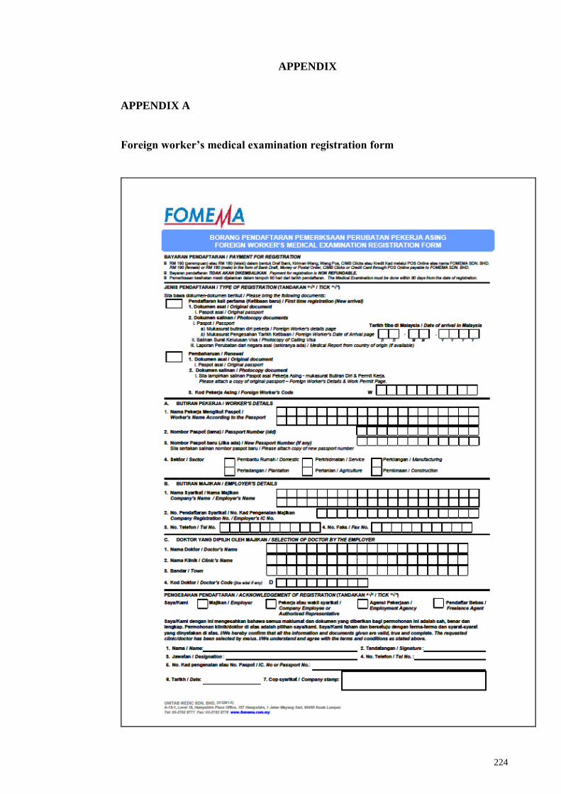
224
APPENDIX
APPENDIX A
Foreign worker’s medical examination registration form
225
APPENDIX B
Consent Form
CONSENT BY PATIENT FOR CLINICAL RESEARCH
I, ………………………………………………….Identity Card No………………………
(Name of Patient)
of ………………………………………………………………………………………………
(Address)
hereby agree to take part in the clinical research (clinical study/questionnaire study/drug trial)
specified below:
Title of Study: Parasitic infections amongst migrant workers in Malaysia
the nature and purpose of which has been explained to me by
Dr. Farnaza Ariffin…………………………….
(Name & Designation of Doctor)
and interpreted by ………………………..……………………………….…..…………
(Name & Designation of Interpreter)
to the best of his/her ability in …………………….…………… language/dialect.
I have been told about the nature of the clinical research in terms of methodology, possible
adverse effects and complications (as in the Patient Information Sheet). After knowing and
understanding all the possible advantages and disadvantages of this clinical research, I
voluntarily consent of my own free will to participate in the clinical research specified above.
I understand that I can withdraw from this clinical research at any time without assigning any
reason whatsoever and in such a situation shall not be denied the benefits of usual treatment
by the attending doctors.
Date: ……………...…… Signature or Thumbprint …………….……………………………
(Patient)
IN THE PRESENCE OF
Name ………………………………………..….……
Identity Card No. ………………………….……… Signature (Witness for Signature of
Patient)
Designation ……………………………….………
I confirm that I have explained to the patient the nature and purpose of the above-mentioned
clinical research.
Date ……………………………Signature ……………………………………………………
(Attending Doctor)
R.N.
Name
Sex
Age
Unit
BK-MIS-1117-E01
226
KEIZINAN OLEH PESAKIT UNTUK PENYELIDIKAN KLINIKAL
Saya,……………………… No. Kad Pengenalan …………………..……
(Nama Pesakit)
beralamat………………………………………………………………………………
(Alamat)
dengan ini bersetuju menyertai dalam penyelidikan klinikal (pengajian
klinikal/pengajian soal-selidik/percubaan ubat-ubatan) disebut berikut:
TajukPenyelidikan: Parasitic infections amongst migrant workers in Malaysia yang
mana sifat dan tujuannya telah diterangkan kepada saya oleh
Dr Farnaza Ariffin.…………………
(Nama & Jawatan Doktor)
mengikut terjemahan ………………….……………………………………………
(Nama & Jawatan Penterjemah)
yang telah menterjemahkan kepada saya dengan sepenuh kemampuan dan kebolehannya
di dalam Bahasa / loghat………………………………………………………
Saya telah diberitahu bahawa dasar penyelidikan klinikal dalam keadaan methodologi,
risiko dan komplikasi (mengikut Kertas Maklumat Pesakit). Selepas mengetahui dan
memahami semua kemungkinan kebaikan dan keburukan penyelidikan klinikal ini, saya
merelakan/mengizinkan sendiri menyertai penyelidikan klinikal tersebut di atas.
Saya faham bahawa saya boleh menarik diri dari penyelidikan klinikal ini pada bila-bila
masa tanpa memberi sebarang alasan dalam situasi ini dan tidak akan dikecualikan dari
kemudahan rawatan dari doktor yang merawat.
Tarikh: …………………….Tandatangan/Cap Jari ………………………
(Pesakit)
DI HADAPAN
Nama ………………………………
No. K/P…………………………….. Tandatangan …………………………………
(Saksi untuk Tandatangan Pesakit)
Jawatan …………..…………………
Saya sahkan bahawa saya telah menerangkan kepada pesakit sifat dan tujuan
penyelidikan klinikal tersebut di atas.
Tarikh: …….…………………. Tandatangan …………………………
(Doktor yang merawat)
No. Pend.
Nama
Jantina
Umur
Unit
BK-MIS-1117-E01
227
CONSENT BY RESPONSIBLE RELATIVE FOR CLINICAL RESEARCH
I, …………………………Identity Card No…………...……………………………
(Name)
of…………………………………………………………………………………
(Address)
hereby agree that my relative ……………I.C. No………..………………………
(Name)
participate in the clinical research (clinical study/questionnaire study/drug trial) specified
below:-
Title of Study: Parasitic infections amongst migrant workers in Malaysia
the nature and purpose of which has been explained to me by Dr. Farnaza Ariffin .
(Name & Designation of Doctor)
and interpreted by ………………………..……………………………….…..…………
(Name & Designation of Interpreter)
to the best of his/her ability in …………………….…………… language/dialect.
I have been informed of the nature of this clinical research in terms of procedure,
possible adverse effects and complications (as in the Patient Information Sheet). I
understand the possible advantages and disadvantages of participating in this research. I
voluntarily give my consent for my relative to participate in this research specified
above.
I understand that I can withdraw my relative from this clinical research at any time
without assigning any reason whatsoever and in such situation, my relative shall not be
denied the benefits of usual treatment by the attending doctors. Should my relative
regains his/her ability to consent, he/she will have the right to remain in this research or
may choose to withdraw.
Relationship Signature or
Date: ……………………. To Patient …………… Thumbprint ……………
IN THE PRESENCE OF
Name …………………………………………….…..
Identity Card No. …………………………………. Signature…………………………
(Witness)
Designation …………………………………………
I confirm that I have explained to the patient‘s relative the nature and purpose of the
above-mentioned clinical research.
Date ……………………………. Signature ……………………………
(Attending Doctor)
R.N.
Name
Sex
Age
Unit
BK-MIS-1117-E01
228
KEIZINAN OLEH WARIS YANG BERTANGGUNGJAWAB UNTUK
PENYELIDIKAN KLINIKAL
Saya,……………………………Kad Pengenalan …………..….………………………
(Nama Waris yang bertanggungjawab)
beralamat………………………………………………………………………………
(Alamat)
dengan ini bersetuju supaya saudara
saya…………………………………………..menyertai
(Nama Pesakit)
dalam penyelidikan klinikal (pengajian klinikal/pengajian soal-selidik/percubaan ubat-
ubatan) disebut berikut:
TajukPenyelidikan: Parasitic infections amongst migrant workers in Malaysia
yang mana sifat dan tujuannya telah diterangkan kepada saya oleh Dr Farnaza Ariffin
(Nama & Jawatan Doktor)
Mengikut terjemahan …………………………..………………… (Nama & Jawatan
Penterjemah)
yang telah menterjemahkan kepada saya dengan sepenuh kemampuan dan
kebolehannya di dalam Bahasa / loghat…………………………………………
Saya telah diberitahu bahawa dasar penyelidikan klinikal dalam keadaan metodologi,
risiko dan komplikasi (mengikut Kertas Maklumat Pesakit). Saya mengetahui dan
memahami semua kemungkinan kebaikan dan keburukan penyelidikan klinikal ini. Saya
merelakan/mengizinkan saudara saya menyertai penyelidikan klinikal tersebut di atas.
Saya faham bahawa saya boleh menarik balik penyertaan saudara saya dalam
penyelidikan klinikal ini pada bila-bila masa tanpa memberi sebarang alasan dalam
situasi ini dan tidak akan dikecualikan dari kemudahan rawatan dari doktor yang
merawat. Sekiranya saudara saya kembali berupaya untuk memberi keizinan, beliau
mempunyai hak untuk terus menyertai kajian ini atau memilih untuk menarik diri.
Tarikh: ………… Pertalian Tandatangan/Cap Jari Waris
dengan Pesakit …..…yang bertanggungjawab ……………
DI HADAPAN
Nama ……………………………………….….
No. K/P……………………………………Tandatangan ……………………………
(Saksi untuk Tandatangan
Jawatan………..……………………………… Waris yang Bertanggungjawab
Saya sahkan bahawa saya telah menerangkan kepada waris yang bertanggungjawab sifat
dan tujuan penyelidikan klinikal tersebut di atas.
Tarikh: …….……………………… Tandatangan …………………………………
(Doktor yang merawat)
No. Pend.
Nama
Jantina
Umur
Unit
BK-MIS-1117-E01
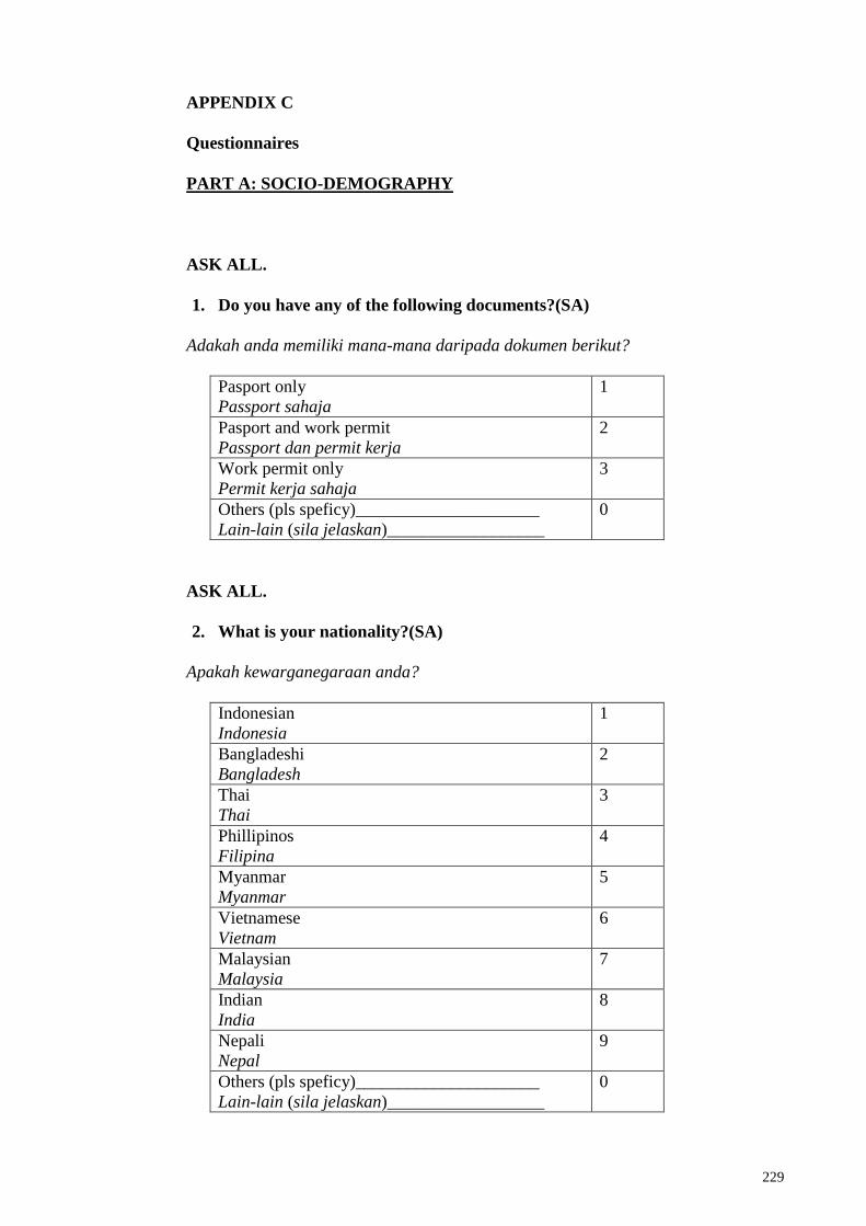
229
APPENDIX C
Questionnaires
PART A: SOCIO-DEMOGRAPHY
ASK ALL.
1. Do you have any of the following documents?(SA)
Adakah anda memiliki mana-mana daripada dokumen berikut?
Pasport only
Passport sahaja
1
Pasport and work permit
Passport dan permit kerja
2
Work permit only
Permit kerja sahaja
3
Others (pls speficy)_____________________
Lain-lain (sila jelaskan)__________________
0
ASK ALL.
2. What is your nationality?(SA)
Apakah kewarganegaraan anda?
Indonesian
Indonesia
1
Bangladeshi
Bangladesh
2
Thai
Thai
3
Phillipinos
Filipina
4
Myanmar
Myanmar
5
Vietnamese
Vietnam
6
Malaysian
Malaysia
7
Indian
India
8
Nepali
Nepal
9
Others (pls speficy)_____________________
Lain-lain (sila jelaskan)__________________
0
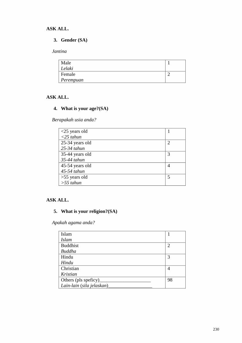
230
ASK ALL.
3. Gender (SA)
Jantina
Male
Lelaki
1
Female
Perempuan
2
ASK ALL.
4. What is your age?(SA)
Berapakah usia anda?
<25 years old
<25 tahun
1
25-34 years old
25-34 tahun
2
35-44 years old
35-44 tahun
3
45-54 years old
45-54 tahun
4
>55 years old
>55 tahun
5
ASK ALL.
5. What is your religion?(SA)
Apakah agama anda?
Islam
Islam
1
Buddhist
Buddha
2
Hindu
Hindu
3
Christian
Kristian
4
Others (pls speficy)_____________________
Lain-lain (sila jelaskan)__________________
98
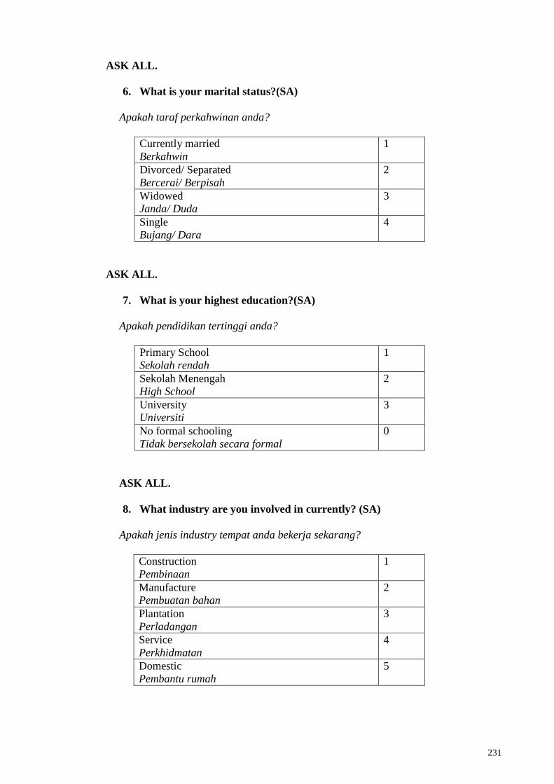
231
ASK ALL.
6. What is your marital status?(SA)
Apakah taraf perkahwinan anda?
Currently married
Berkahwin
1
Divorced/ Separated
Bercerai/ Berpisah
2
Widowed
Janda/ Duda
3
Single
Bujang/ Dara
4
ASK ALL.
7. What is your highest education?(SA)
Apakah pendidikan tertinggi anda?
Primary School
Sekolah rendah
1
Sekolah Menengah
High School
2
University
Universiti
3
No formal schooling
Tidak bersekolah secara formal
0
ASK ALL.
8. What industry are you involved in currently? (SA)
Apakah jenis industry tempat anda bekerja sekarang?
Construction
Pembinaan
1
Manufacture
Pembuatan bahan
2
Plantation
Perladangan
3
Service
Perkhidmatan
4
Domestic
Pembantu rumah
5
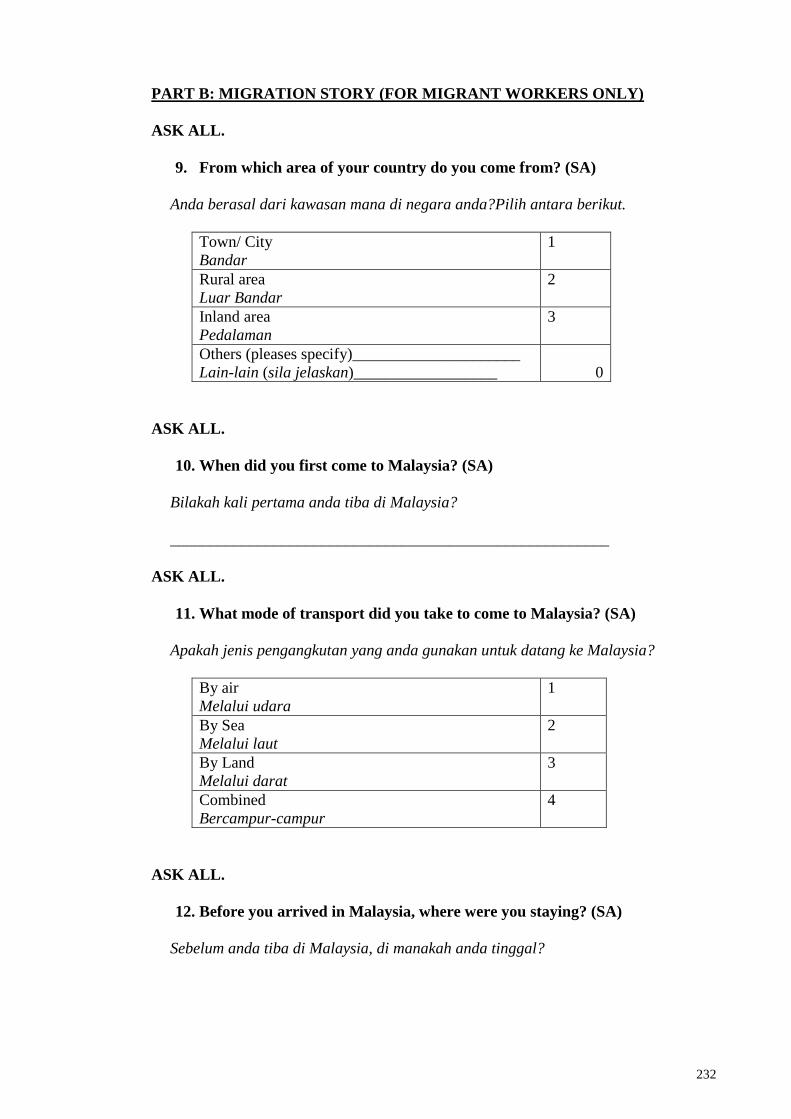
232
PART B: MIGRATION STORY (FOR MIGRANT WORKERS ONLY)
ASK ALL.
9. From which area of your country do you come from? (SA)
Anda berasal dari kawasan mana di negara anda?Pilih antara berikut.
Town/ City
Bandar
1
Rural area
Luar Bandar
2
Inland area
Pedalaman
3
Others (pleases specify)_____________________
Lain-lain (sila jelaskan)__________________
0
ASK ALL.
10. When did you first come to Malaysia? (SA)
Bilakah kali pertama anda tiba di Malaysia?
_______________________________________________________
ASK ALL.
11. What mode of transport did you take to come to Malaysia? (SA)
Apakah jenis pengangkutan yang anda gunakan untuk datang ke Malaysia?
By air
Melalui udara
1
By Sea
Melalui laut
2
By Land
Melalui darat
3
Combined
Bercampur-campur
4
ASK ALL.
12. Before you arrived in Malaysia, where were you staying? (SA)
Sebelum anda tiba di Malaysia, di manakah anda tinggal?
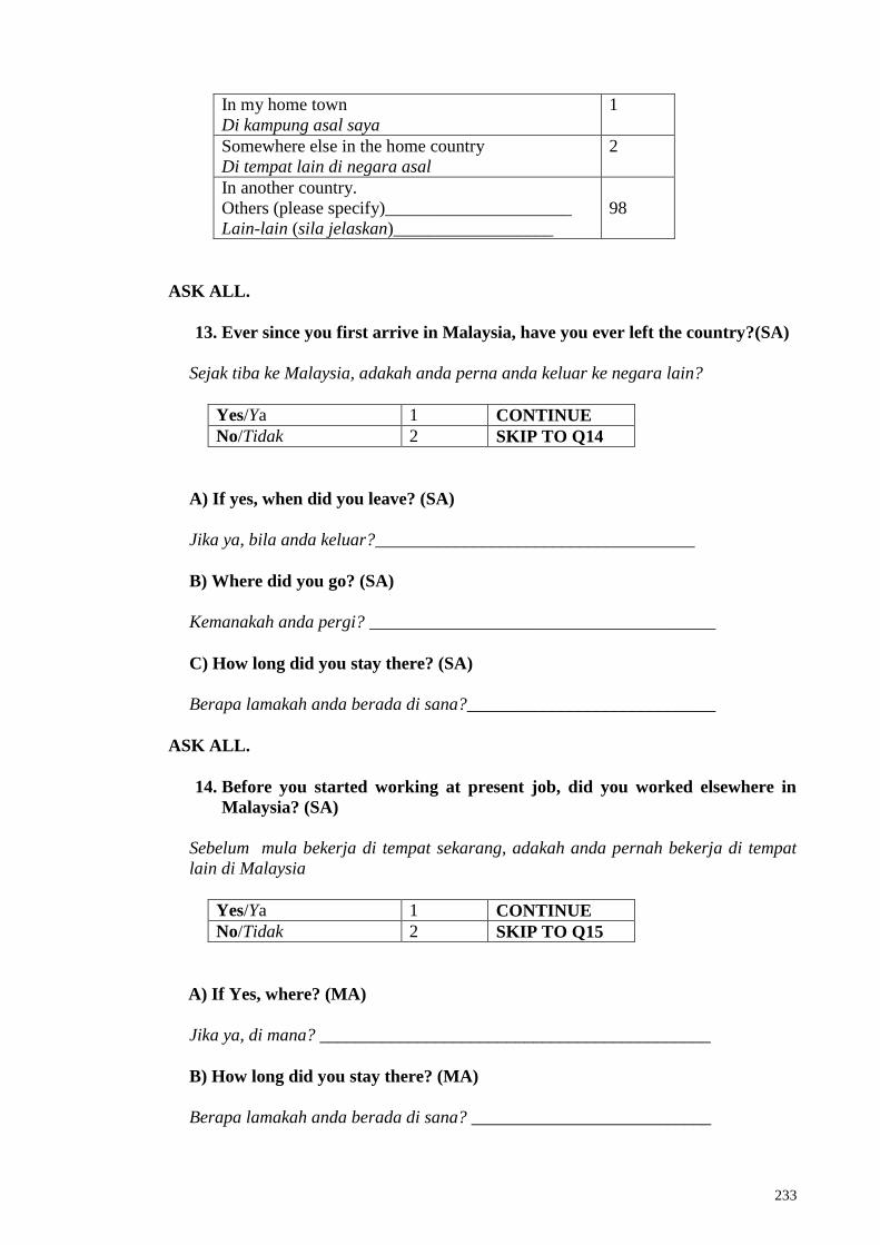
233
In my home town
Di kampung asal saya
1
Somewhere else in the home country
Di tempat lain di negara asal
2
In another country.
Others (please specify)_____________________
Lain-lain (sila jelaskan)__________________
98
ASK ALL.
13. Ever since you first arrive in Malaysia, have you ever left the country?(SA)
Sejak tiba ke Malaysia, adakah anda perna anda keluar ke negara lain?
Yes/Ya 1 CONTINUE
No/Tidak 2 SKIP TO Q14
A) If yes, when did you leave? (SA)
Jika ya, bila anda keluar?____________________________________
B) Where did you go? (SA)
Kemanakah anda pergi? _______________________________________
C) How long did you stay there? (SA)
Berapa lamakah anda berada di sana?____________________________
ASK ALL.
14. Before you started working at present job, did you worked elsewhere in
Malaysia? (SA)
Sebelum mula bekerja di tempat sekarang, adakah anda pernah bekerja di tempat
lain di Malaysia
Yes/Ya 1 CONTINUE
No/Tidak 2 SKIP TO Q15
A) If Yes, where? (MA)
Jika ya, di mana? ____________________________________________
B) How long did you stay there? (MA)
Berapa lamakah anda berada di sana? ___________________________
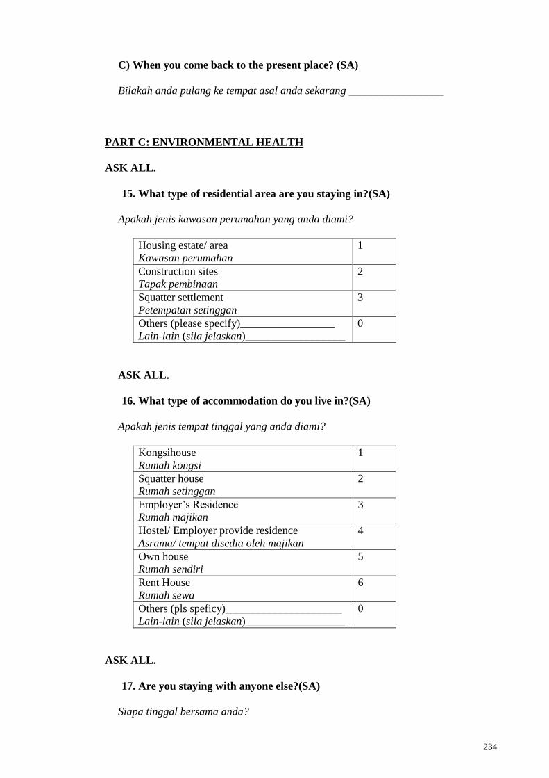
234
C) When you come back to the present place? (SA)
Bilakah anda pulang ke tempat asal anda sekarang _________________
PART C: ENVIRONMENTAL HEALTH
ASK ALL.
15. What type of residential area are you staying in?(SA)
Apakah jenis kawasan perumahan yang anda diami?
Housing estate/ area
Kawasan perumahan
1
Construction sites
Tapak pembinaan
2
Squatter settlement
Petempatan setinggan
3
Others (please specify)_________________
Lain-lain (sila jelaskan)__________________
0
ASK ALL.
16. What type of accommodation do you live in?(SA)
Apakah jenis tempat tinggal yang anda diami?
Kongsihouse
Rumah kongsi
1
Squatter house
Rumah setinggan
2
Employer‘s Residence
Rumah majikan
3
Hostel/ Employer provide residence
Asrama/ tempat disedia oleh majikan
4
Own house
Rumah sendiri
5
Rent House
Rumah sewa
6
Others (pls speficy)_____________________
Lain-lain (sila jelaskan)__________________
0
ASK ALL.
17. Are you staying with anyone else?(SA)
Siapa tinggal bersama anda?
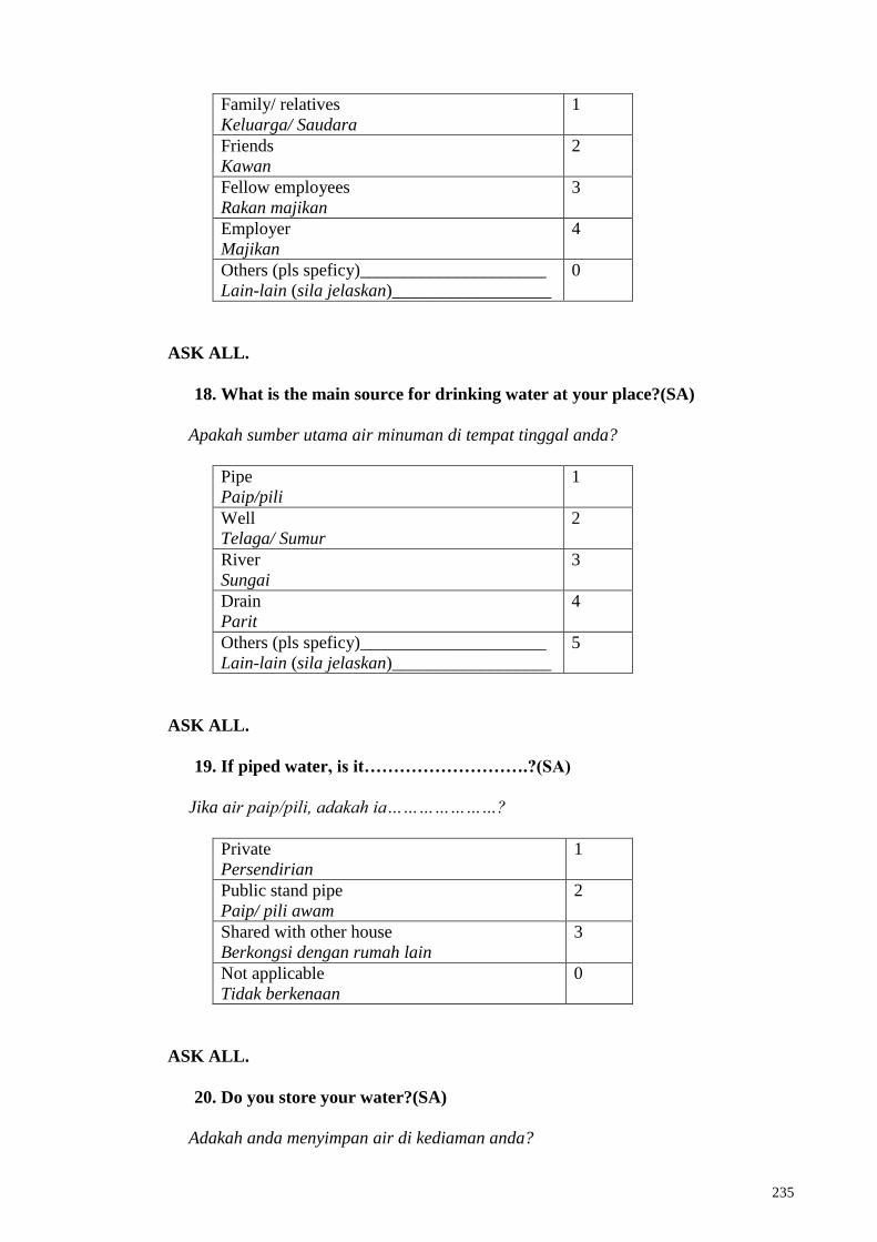
235
Family/ relatives
Keluarga/ Saudara
1
Friends
Kawan
2
Fellow employees
Rakan majikan
3
Employer
Majikan
4
Others (pls speficy)_____________________
Lain-lain (sila jelaskan)__________________
0
ASK ALL.
18. What is the main source for drinking water at your place?(SA)
Apakah sumber utama air minuman di tempat tinggal anda?
Pipe
Paip/pili
1
Well
Telaga/ Sumur
2
River
Sungai
3
Drain
Parit
4
Others (pls speficy)_____________________
Lain-lain (sila jelaskan)__________________
5
ASK ALL.
19. If piped water, is it……………………….?(SA)
Jika air paip/pili, adakah ia…………………?
Private
Persendirian
1
Public stand pipe
Paip/ pili awam
2
Shared with other house
Berkongsi dengan rumah lain
3
Not applicable
Tidak berkenaan
0
ASK ALL.
20. Do you store your water?(SA)
Adakah anda menyimpan air di kediaman anda?
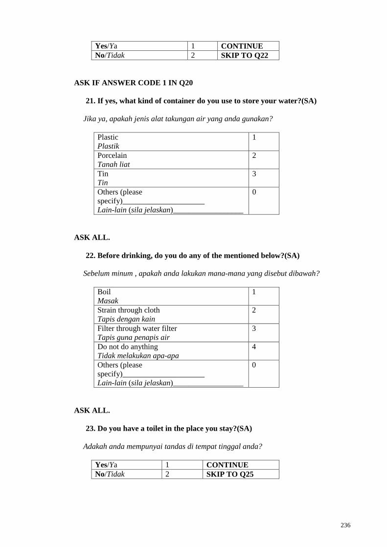
236
Yes/Ya 1 CONTINUE
No/Tidak 2 SKIP TO Q22
ASK IF ANSWER CODE 1 IN Q20
21. If yes, what kind of container do you use to store your water?(SA)
Jika ya, apakah jenis alat takungan air yang anda gunakan?
Plastic
Plastik
1
Porcelain
Tanah liat
2
Tin
Tin
3
Others (please
specify)_____________________
Lain-lain (sila jelaskan)__________________
0
ASK ALL.
22. Before drinking, do you do any of the mentioned below?(SA)
Sebelum minum , apakah anda lakukan mana-mana yang disebut dibawah?
Boil
Masak
1
Strain through cloth
Tapis dengan kain
2
Filter through water filter
Tapis guna penapis air
3
Do not do anything
Tidak melakukan apa-apa
4
Others (please
specify)_____________________
Lain-lain (sila jelaskan)__________________
0
ASK ALL.
23. Do you have a toilet in the place you stay?(SA)
Adakah anda mempunyai tandas di tempat tinggal anda?
Yes/Ya 1 CONTINUE
No/Tidak 2 SKIP TO Q25
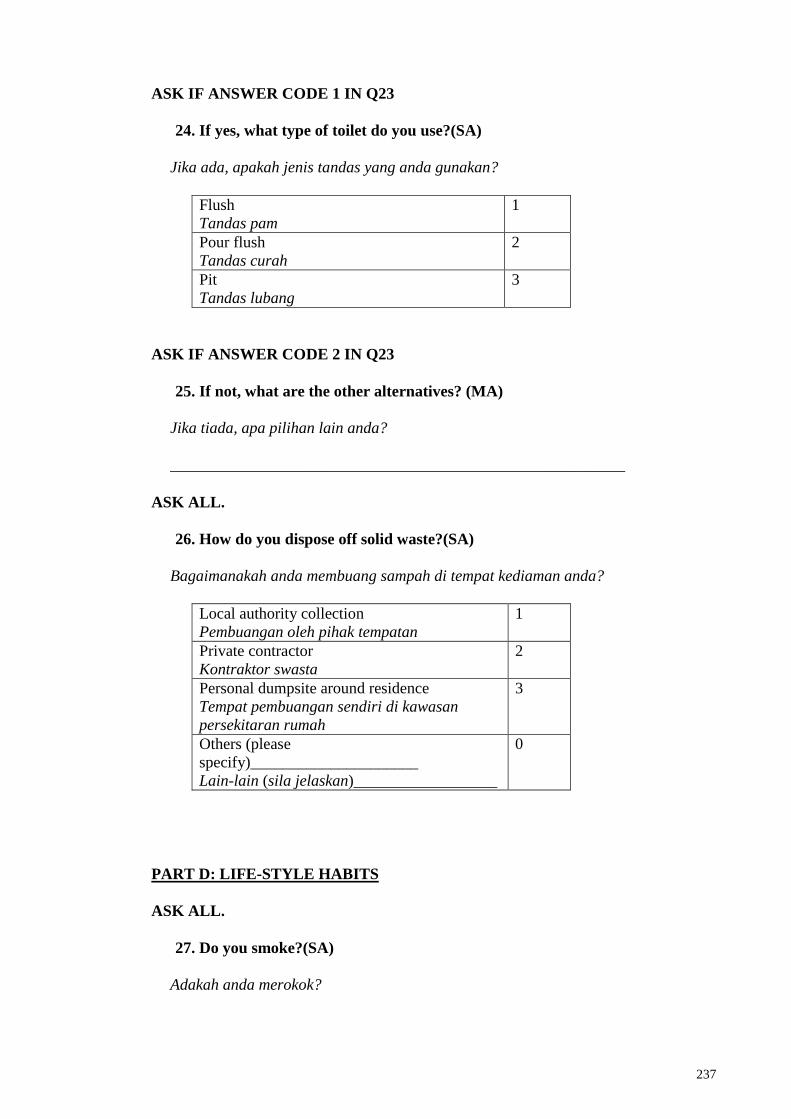
237
ASK IF ANSWER CODE 1 IN Q23
24. If yes, what type of toilet do you use?(SA)
Jika ada, apakah jenis tandas yang anda gunakan?
Flush
Tandas pam
1
Pour flush
Tandas curah
2
Pit
Tandas lubang
3
ASK IF ANSWER CODE 2 IN Q23
25. If not, what are the other alternatives? (MA)
Jika tiada, apa pilihan lain anda?
_________________________________________________________
ASK ALL.
26. How do you dispose off solid waste?(SA)
Bagaimanakah anda membuang sampah di tempat kediaman anda?
Local authority collection
Pembuangan oleh pihak tempatan
1
Private contractor
Kontraktor swasta
2
Personal dumpsite around residence
Tempat pembuangan sendiri di kawasan
persekitaran rumah
3
Others (please
specify)_____________________
Lain-lain (sila jelaskan)__________________
0
PART D: LIFE-STYLE HABITS
ASK ALL.
27. Do you smoke?(SA)
Adakah anda merokok?
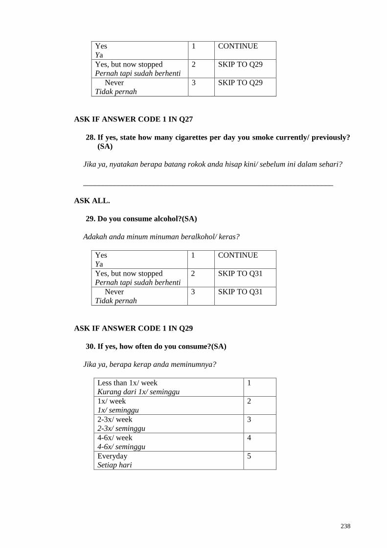
238
Yes
Ya
1 CONTINUE
Yes, but now stopped
Pernah tapi sudah berhenti
2 SKIP TO Q29
Never
Tidak pernah
3 SKIP TO Q29
ASK IF ANSWER CODE 1 IN Q27
28. If yes, state how many cigarettes per day you smoke currently/ previously?
(SA)
Jika ya, nyatakan berapa batang rokok anda hisap kini/ sebelum ini dalam sehari?
________________________________________________________________
ASK ALL.
29. Do you consume alcohol?(SA)
Adakah anda minum minuman beralkohol/ keras?
Yes
Ya
1 CONTINUE
Yes, but now stopped
Pernah tapi sudah berhenti
2 SKIP TO Q31
Never
Tidak pernah
3 SKIP TO Q31
ASK IF ANSWER CODE 1 IN Q29
30. If yes, how often do you consume?(SA)
Jika ya, berapa kerap anda meminumnya?
Less than 1x/ week
Kurang dari 1x/ seminggu
1
1x/ week
1x/ seminggu
2
2-3x/ week
2-3x/ seminggu
3
4-6x/ week
4-6x/ seminggu
4
Everyday
Setiap hari
5
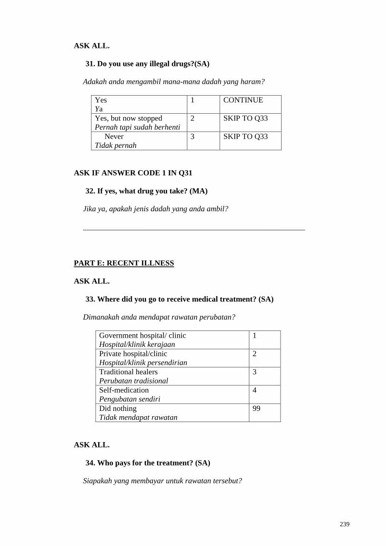
239
ASK ALL.
31. Do you use any illegal drugs?(SA)
Adakah anda mengambil mana-mana dadah yang haram?
Yes
Ya
1 CONTINUE
Yes, but now stopped
Pernah tapi sudah berhenti
2 SKIP TO Q33
Never
Tidak pernah
3 SKIP TO Q33
ASK IF ANSWER CODE 1 IN Q31
32. If yes, what drug you take? (MA)
Jika ya, apakah jenis dadah yang anda ambil?
_________________________________________________________
PART E: RECENT ILLNESS
ASK ALL.
33. Where did you go to receive medical treatment? (SA)
Dimanakah anda mendapat rawatan perubatan?
Government hospital/ clinic
Hospital/klinik kerajaan
1
Private hospital/clinic
Hospital/klinik persendirian
2
Traditional healers
Perubatan tradisional
3
Self-medication
Pengubatan sendiri
4
Did nothing
Tidak mendapat rawatan
99
ASK ALL.
34. Who pays for the treatment? (SA)
Siapakah yang membayar untuk rawatan tersebut?
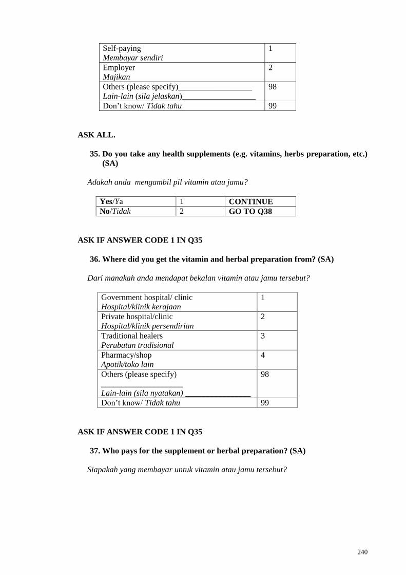
240
Self-paying
Membayar sendiri
1
Employer
Majikan
2
Others (please specify)__________________
Lain-lain (sila jelaskan)__________________
98
Don‘t know/ Tidak tahu 99
ASK ALL.
35. Do you take any health supplements (e.g. vitamins, herbs preparation, etc.)
(SA)
Adakah anda mengambil pil vitamin atau jamu?
Yes/Ya 1 CONTINUE
No/Tidak 2 GO TO Q38
ASK IF ANSWER CODE 1 IN Q35
36. Where did you get the vitamin and herbal preparation from? (SA)
Dari manakah anda mendapat bekalan vitamin atau jamu tersebut?
Government hospital/ clinic
Hospital/klinik kerajaan
1
Private hospital/clinic
Hospital/klinik persendirian
2
Traditional healers
Perubatan tradisional
3
Pharmacy/shop
Apotik/toko lain
4
Others (please specify)
____________________
Lain-lain (sila nyatakan) ________________
98
Don‘t know/ Tidak tahu 99
ASK IF ANSWER CODE 1 IN Q35
37. Who pays for the supplement or herbal preparation? (SA)
Siapakah yang membayar untuk vitamin atau jamu tersebut?
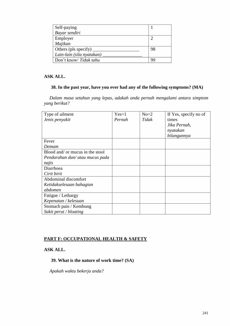
241
Self-paying
Bayar sendiri
1
Employer
Majikan
2
Others (pls specify) ____________________
Lain-lain (sila nyatakan) _________________
98
Don‘t know/ Tidak tahu 99
ASK ALL.
38. In the past year, have you ever had any of the following symptoms? (MA)
Dalam masa setahun yang lepas, adakah anda pernah mengalami antara simptom
yang berikut?
Type of ailment
Jenis penyakit
Yes=1
Pernah
No=2
Tidak
If Yes, specify no of
times
Jika Pernah,
nyatakan
bilangannya
Fever
Demam
Blood and/ or mucus in the stool
Pendarahan dan/ atau mucus pada
najis
Diarrhoea
Cirit birit
Abdominal discomfort
Ketidakselesaan bahagian
abdomen
Fatigue / Lethargy
Kepenatan / kelesuan
Stomach pain / Kembung
Sakit perut / bloating
PART F: OCCUPATIONAL HEALTH & SAFETY
ASK ALL.
39. What is the nature of work time? (SA)
Apakah waktu bekerja anda?
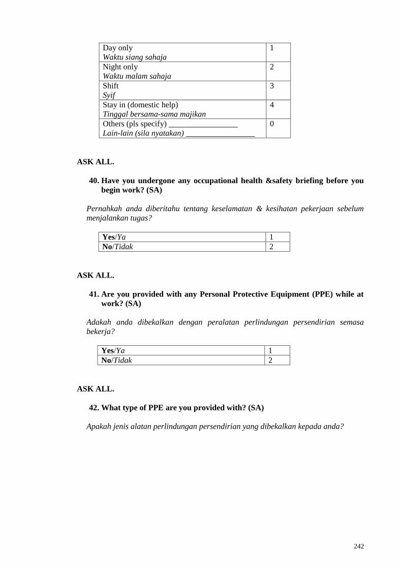
242
Day only
Waktu siang sahaja
1
Night only
Waktu malam sahaja
2
Shift
Syif
3
Stay in (domestic help)
Tinggal bersama-sama majikan
4
Others (pls specify) _________________
Lain-lain (sila nyatakan) _________________
0
ASK ALL.
40. Have you undergone any occupational health &safety briefing before you
begin work? (SA)
Pernahkah anda diberitahu tentang keselamatan & kesihatan pekerjaan sebelum
menjalankan tugas?
Yes/Ya 1
No/Tidak 2
ASK ALL.
41. Are you provided with any Personal Protective Equipment (PPE) while at
work? (SA)
Adakah anda dibekalkan dengan peralatan perlindungan persendirian semasa
bekerja?
Yes/Ya 1
No/Tidak 2
ASK ALL.
42. What type of PPE are you provided with? (SA)
Apakah jenis alatan perlindungan persendirian yang dibekalkan kepada anda?
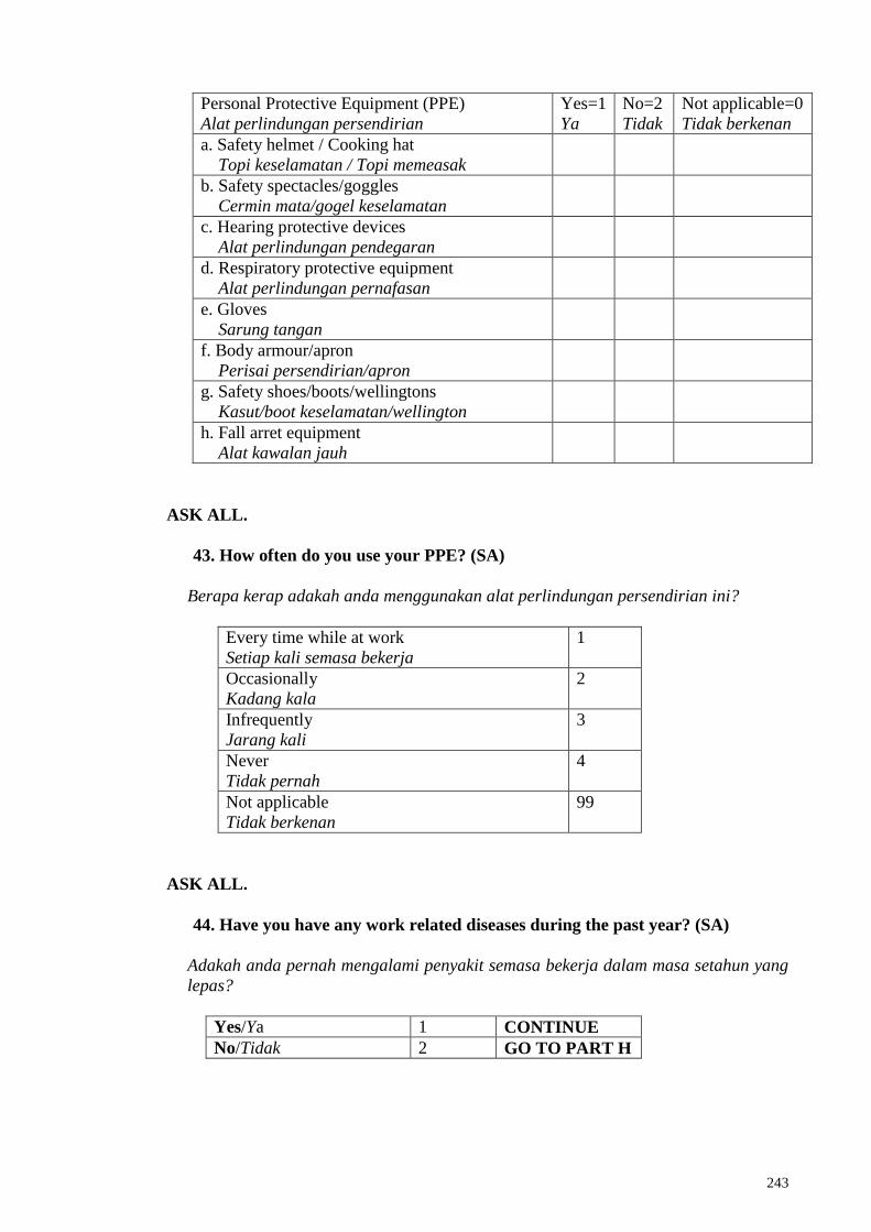
243
Personal Protective Equipment (PPE)
Alat perlindungan persendirian
Yes=1
Ya
No=2
Tidak
Not applicable=0
Tidak berkenan
a. Safety helmet / Cooking hat
Topi keselamatan / Topi memeasak
b. Safety spectacles/goggles
Cermin mata/gogel keselamatan
c. Hearing protective devices
Alat perlindungan pendegaran
d. Respiratory protective equipment
Alat perlindungan pernafasan
e. Gloves
Sarung tangan
f. Body armour/apron
Perisai persendirian/apron
g. Safety shoes/boots/wellingtons
Kasut/boot keselamatan/wellington
h. Fall arret equipment
Alat kawalan jauh
ASK ALL.
43. How often do you use your PPE? (SA)
Berapa kerap adakah anda menggunakan alat perlindungan persendirian ini?
Every time while at work
Setiap kali semasa bekerja
1
Occasionally
Kadang kala
2
Infrequently
Jarang kali
3
Never
Tidak pernah
4
Not applicable
Tidak berkenan
99
ASK ALL.
44. Have you have any work related diseases during the past year? (SA)
Adakah anda pernah mengalami penyakit semasa bekerja dalam masa setahun yang
lepas?
Yes/Ya 1 CONTINUE
No/Tidak 2 GO TO PART H
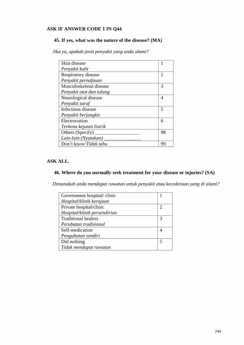
244
ASK IF ANSWER CODE 1 IN Q44
45. If yes, what was the nature of the disease? (MA)
Jika ya, apakah jenis penyakit yang anda alami?
Skin disease
Penyakit kulit
1
Respiratory disease
Penyakit pernafasan
2
Musculoskeletal disease
Penyakit otot dan tulang
3
Neurological disease
Penyakit saraf
4
Infectious disease
Penyakit berjangkit
5
Electrocution
Terkena kejutan listrik
6
Others (Specify) __________________
Lain-lain (Nyatakan) _______________
98
Don‘t know/Tidak tahu 99
ASK ALL.
46. Where do you normally seek treatment for your disease or injuries? (SA)
Dimanakah anda mendapat rawatan untuk penyakit atau kecederaan yang di alami?
Government hospital/ clinic
Hospital/klinik kerajaan
1
Private hospital/clinic
Hospital/klinik persendirian
2
Traditional healers
Perubatan tradisional
3
Self-medication
Pengubatan sendiri
4
Did nothing
Tidak mendapat rawatan
5
245
APPENDIX D
Published Paper I:
Sahimin, N., Yvonne A.L. Lim, Ariffin, F., Behnke, J.M., Lewis, J.W. & Mohd
Zain, S.N. (2016). Migrant Workers in Malaysia: Current Implications of
Sociodemographic and Environmental Characteristics in the Transmission of
Intestinal Parasitic Infections. PLoS Neglected Tropical Diseases, 10(11):
e0005110. doi:10.1371/journal.pntd.0005110. (ISI-Cited Publication)