first report of fusarium species at nesting sites of...
TRANSCRIPT

* To whom correspondence should be addressed.
Malays. Appl. Biol. (2017) 46(3): 195–205
FIRST REPORT OF Fusarium SPECIES AT NESTING SITESOF ENDANGERED SEA TURTLES IN TERENGGANU
AND MELAKA, MALAYSIA
SITI NORDAHLIAWATE MOHAMED SIDIQUE1*, NURUL FARIZAH AZUDDIN2
and JUANITA JOSEPH3
1Laboratory for Pest Disease and Microbial Biotechnology (LAPDiM),School of Food Science and Technology, Universiti Malaysia Terengganu,
21030 Kuala Nerus, Malaysia2School of Biological Sciences, Universiti Sains Malaysia, 11800 Penang, Malaysia
3Institute of Oceanography and Environment, Universiti Malaysia Terengganu,21030 Kuala Nerus, Malaysia
*E-mail: [email protected]
Accepted 13 September 2017, Published online 4 October 2017
ABSTRACT
In recent years, turtle eggs incubated in situ or in protected hatcheries in Malaysia have been reported to show symptoms andsigns of fungal colonisation. However, there are no studies addressing this problem and potential relationship with frequenthatching failures. We identified Fusarium species from nesting sites of green turtle (Chelonia mydas) and hawksbill turtle(Eretmochelys imbricata) situated in Terengganu and Melaka, as well as the environments surrounding those sites. The eggswere incubated on the nesting beach (in situ) or relocated to the beach hatchery and styrofoam boxes (ex situ) in PeninsularMalaysia. Samples were collected from infected eggs, sand, plant roots and debris around the egg chambers. One-hundredand six strains of Fusarium spp. were isolated. They were identified morphologically as member of the Fusarium solanispecies complex (FSSC, 101 strains), F. oxysporum (four strains) and F. proliferatum (one strain). We conducted phylogeneticanalysis based on nucleotide sequences of translation elongation factor 1-alpha gene (TEF-1α). The strains of the FSSCwere further separated into three lineages, F. falciforme, F. lichenicola and F. keratoplasticum. This is the first report onFusarium species isolated from symptomatic green and hawksbill turtle eggs in Peninsular Malaysia. Fusarium colonisationin sea turtle nests poses a serious risk to the survival of endangered sea turtles in Malaysia. It is, therefore, important toexamine the nature of such colonisation and their relationship to hatching failures of the turtles in Malaysia or elsewhere inthe region to mitigate pathogenic fungi impact.
Key words: Fusarium solani species complex, green turtle, hawksbill turtle, hatching failure and Malaysia
INTRODUCTION
Sea turtles are marine reptiles widely distributed intropical, subtropical and temperate seas throughoutthe world. Out of the seven species of sea turtles,four species nest in Malaysia: the green turtle(Chelonia mydas), hawksbill (Eretmochelysimbricata), leatherback (Dermochelys coriacea) andolive ridley (Lepidochelys olivacea) (Chan, 2006).The green turtle is the most abundant sea turtlespecies in Malaysia, nesting in Sabah, Sarawak,Terengganu, Pahang, Perak and Johor. RedangIsland is one of the largest nesting sites for green
turtles in Peninsular Malaysia, with 1000–2500nests per year. The hawksbill turtles nest in Sabah,Melaka, Johor and Terengganu (Chan, 2006). Bothspecies are listed in Appendix I of the Conventionon International Trade in Endangered Species ofWild Fauna and Flora (Seminoff, 2004). TheInternational Union for Conservation of Nature(IUCN, 2004) has listed the green turtle asendangered and the hawksbill turtle as criticallyendangered. This study focused on the green andhawksbill turtles in Peninsular Malaysia.
Most conservation efforts try to restore thedepleted populations by protecting nests andimproving hatching success. Like many otherconservation programmes in the world, they cannot

196 Fusarium SPECIES AT NESTING SITES OF ENDANGERED SEA TURTLES
protect all sea turtle eggs because of poaching, aswell as predation by monitor lizards, ants, ghostcrabs, and maggots (Morita et al., 2008; Garcia etal., 2003). Fungal disease is another threat tosurvive especially due to the increasing number ofpathogenic fungi that threatens wildlife anddomesticated species worldwide (Fisher et al., 2012;Sarmiento-Ramírez et al., 2014). Examples of somefungal diseases that pose substantial threats toanimal such as chytridiomycosis (Batrachochytriumdendrobatidis) that contributed to nearly half of allamphibian species being in decline worldwide(Fisher et al., 2009; Hof et al., 2011) and white-nosesyndrome (Geomyces destructans) found in sixspecies of North American bats that has killedmillions of bats (Frick et al., 2010; Foley et al.,2011). There has been several reports of fungi thathave infected sea turtle eggs and is responsible forthe sea turtles’ decline in the Atlantic, Indian andPacific Oceans from 2005-2012 (Sarmiento-Ramírezet al., 2014).
Researchers in Costa Rica (Mo et al., 1990), theUnited States (Eckert and Eckert, 1990), Australia(Phillott et al., 2004), Turkey (Güçlü et al., 2010)and Spain (Sarmiento-Ramírez et al., 2010) havedemonstrated the association of fungi withunhatched turtle eggs. The loggerhead turtle(Caretta caretta) is susceptible to the ascomycetefungus (Fusarium solani) (Sarmiento-Ramírez et al.,2010). The genus Fusarium has been widelyrecognized as plant pathogens or soil inhabitantsand plays an important role in animal pathology andmycotoxicology (Siti Nordahliawate et al., 2012;Geiser et al., 2013). It has been demonstrated thatapproximately 70 species of Fusarium have beeninvolved in infections in humans and other animalsthat were grouped into about ten phylogeneticspecies complexes (O’Donnell et al., 2010; Guarro,2013). Sarmiento-Ramírez et al. (2014) found F.falciforme and F. keratoplasticum members ofFusarium solani species complex (FSSC) in sixspecies of sea turtles; Chelonia mydas (RaineIsland, Australia; Isla de la Plata at MachalillaNational Park, Ecuador and Ascension Island),Caretta caretta (Boa Vista Island, Cape Verde),Eretmochelys imbricata and Lepidochelys olivacea(La Playita, Machalilla National Park), Dermochelyscoriacea (La Playona, Colombia and Pacuare NatureReserve, Costa Rica) and Natator depressus (CrabIsland, Australia). Nevertheless, Fusarium solanispecies complex (FSSC) is the most common groupof fusaria associated with human infectious diseasesand to date, 60% responsible for fusariosis (Short etal., 2013). Fusariosis is the second most commoncause of mould infections in human afteraspergillosis (Guarro, 2013).
However, the Fusarium isolates weremorphologically indistinguishable and cultures
requires extensive molecular techniques especiallymonophyletic “species complex” such as Fusariumsolani species complex (FSSC) (O’Donnell, 2000;Short et al., 2013). Thus, to identify phylogeneticspecies to date, based on a partial sequence fromtranslation elongation factor-1a (TEF1), and DNA-directed RNA polymerase II largest (RPB1) andsecond largest subunit (RPB2) has proven to be themost reliable (O’Donnell et al., 2015).
Very few studies of microbial contaminants ofsea turtle eggs have been conducted in Malaysia.An unknown species of fungus on leatherback turtleeggs at Rantau Abang, Terengganu was reported in1989 (Chan and Solomon, 1989). In 2010, Daraup(2010) identified morphologically of Fusariumspecies isolated from green turtle eggs incubated atChagar Hutang, Redang Island and Terengganu.However, both studies were rather limited and didnot identify the fungal species. Our study recordedfor the presence of Fusarium species and identifiedFusarium isolates from the shells of unhatched eggs,sands surrounding the egg chamber and debris andplant roots around the turtle nests on natural nestingbeaches and in hatcheries of Peninsular Malaysia.We believe that this study supplies useful data onthe fungi isolated from sea turtle eggs and nests. Ourresults might help the efforts to increase the hatchingsuccess and replenish the depleted populations ofsea turtles in Malaysia.
MATERIALS AND METHODS
Ethics statementCollection of sea turtle eggshells was done
under permissions Sea Turtle Research Unit(SEATRU) and Department of Fisheries Terengganuand Melaka. None of the experiments involvedsacrificing animals and, therefore, we did not requirea specific approval from any institutional animalresearch ethics committee.
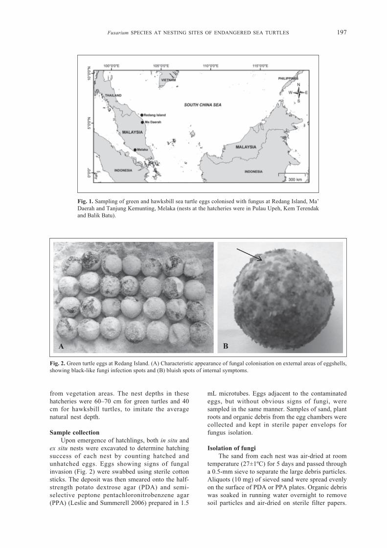
Sampling sitesThree sites in Peninsular Malaysia, using
different methods of turtle nest incubation (in situand ex situ), were selected for sampling (Fig. 1).Samples from in situ nests were obtained fromRedang Island, Terengganu, in 2010. Samples ofgreen turtle eggs were also obtained from ex situnests at the Ma’Daerah hatchery, Terengganu.Samples of hawksbill turtle eggs were obtainedfrom Tanjung Kemunting, Melaka, in 2011. Eggsincubated in Tanjung Kemunting were collectedfrom nearby nesting beaches, namely from PulauUpeh, Kem Terendak and Balik Batu. The nestingsites in Redang Island were near vegetation areas(0–10.76 m) with an average nest depth of 70 cm.Ex situ nesting hatcheries were located far (500 m)
Fusarium SPECIES AT NESTING SITES OF ENDANGERED SEA TURTLES 197
Fig. 1. Sampling of green and hawksbill sea turtle eggs colonised with fungus at Redang Island, Ma’Daerah and Tanjung Kemunting, Melaka (nests at the hatcheries were in Pulau Upeh, Kem Terendakand Balik Batu).
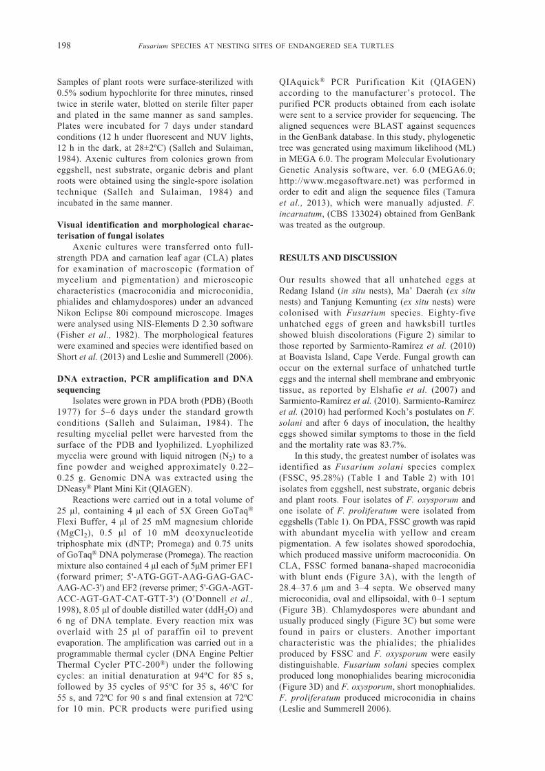
Fig. 2. Green turtle eggs at Redang Island. (A) Characteristic appearance of fungal colonisation on external areas of eggshells,showing black-like fungi infection spots and (B) bluish spots of internal symptoms.
from vegetation areas. The nest depths in thesehatcheries were 60–70 cm for green turtles and 40cm for hawksbill turtles, to imitate the averagenatural nest depth.
Sample collectionUpon emergence of hatchlings, both in situ and
ex situ nests were excavated to determine hatchingsuccess of each nest by counting hatched andunhatched eggs. Eggs showing signs of fungalinvasion (Fig. 2) were swabbed using sterile cottonsticks. The deposit was then smeared onto the half-strength potato dextrose agar (PDA) and semi-selective peptone pentachloronitrobenzene agar(PPA) (Leslie and Summerell 2006) prepared in 1.5
mL microtubes. Eggs adjacent to the contaminatedeggs, but without obvious signs of fungi, weresampled in the same manner. Samples of sand, plantroots and organic debris from the egg chambers werecollected and kept in sterile paper envelops forfungus isolation.
Isolation of fungiThe sand from each nest was air-dried at room
temperature (27±1ºC) for 5 days and passed througha 0.5-mm sieve to separate the large debris particles.Aliquots (10 mg) of sieved sand were spread evenlyon the surface of PDA or PPA plates. Organic debriswas soaked in running water overnight to removesoil particles and air-dried on sterile filter papers.
198 Fusarium SPECIES AT NESTING SITES OF ENDANGERED SEA TURTLES
Samples of plant roots were surface-sterilized with0.5% sodium hypochlorite for three minutes, rinsedtwice in sterile water, blotted on sterile filter paperand plated in the same manner as sand samples.Plates were incubated for 7 days under standardconditions (12 h under fluorescent and NUV lights,12 h in the dark, at 28±2ºC) (Salleh and Sulaiman,1984). Axenic cultures from colonies grown fromeggshell, nest substrate, organic debris and plantroots were obtained using the single-spore isolationtechnique (Salleh and Sulaiman, 1984) andincubated in the same manner.
Visual identification and morphological charac-terisation of fungal isolates
Axenic cultures were transferred onto full-strength PDA and carnation leaf agar (CLA) platesfor examination of macroscopic (formation ofmycelium and pigmentation) and microscopiccharacteristics (macroconidia and microconidia,phialides and chlamydospores) under an advancedNikon Eclipse 80i compound microscope. Imageswere analysed using NIS-Elements D 2.30 software(Fisher et al., 1982). The morphological featureswere examined and species were identified based onShort et al. (2013) and Leslie and Summerell (2006).
DNA extraction, PCR amplification and DNAsequencing
Isolates were grown in PDA broth (PDB) (Booth1977) for 5–6 days under the standard growthconditions (Salleh and Sulaiman, 1984). Theresulting mycelial pellet were harvested from thesurface of the PDB and lyophilized. Lyophilizedmycelia were ground with liquid nitrogen (N2) to afine powder and weighed approximately 0.22–0.25 g. Genomic DNA was extracted using theDNeasy® Plant Mini Kit (QIAGEN).
Reactions were carried out in a total volume of25 μl, containing 4 μl each of 5X Green GoTaq®
Flexi Buffer, 4 μl of 25 mM magnesium chloride(MgCl2), 0.5 μl of 10 mM deoxynucleotidetriphosphate mix (dNTP; Promega) and 0.75 unitsof GoTaq® DNA polymerase (Promega). The reactionmixture also contained 4 μl each of 5μM primer EF1(forward primer; 5'-ATG-GGT-AAG-GAG-GAC-AAG-AC-3') and EF2 (reverse primer; 5'-GGA-AGT-ACC-AGT-GAT-CAT-GTT-3') (O’Donnell et al.,1998), 8.05 μl of double distilled water (ddH2O) and6 ng of DNA template. Every reaction mix wasoverlaid with 25 μl of paraffin oil to preventevaporation. The amplification was carried out in aprogrammable thermal cycler (DNA Engine PeltierThermal Cycler PTC-200®) under the followingcycles: an initial denaturation at 94ºC for 85 s,followed by 35 cycles of 95ºC for 35 s, 46ºC for55 s, and 72ºC for 90 s and final extension at 72ºCfor 10 min. PCR products were purified using
QIAquick® PCR Purification Kit (QIAGEN)according to the manufacturer’s protocol. Thepurified PCR products obtained from each isolatewere sent to a service provider for sequencing. Thealigned sequences were BLAST against sequencesin the GenBank database. In this study, phylogenetictree was generated using maximum likelihood (ML)in MEGA 6.0. The program Molecular EvolutionaryGenetic Analysis software, ver. 6.0 (MEGA6.0;http://www.megasoftware.net) was performed inorder to edit and align the sequence files (Tamuraet al., 2013), which were manually adjusted. F.incarnatum, (CBS 133024) obtained from GenBankwas treated as the outgroup.
RESULTS AND DISCUSSION
Our results showed that all unhatched eggs atRedang Island (in situ nests), Ma’ Daerah (ex situnests) and Tanjung Kemunting (ex situ nests) werecolonised with Fusarium species. Eighty-fiveunhatched eggs of green and hawksbill turtlesshowed bluish discolorations (Figure 2) similar tothose reported by Sarmiento-Ramírez et al. (2010)at Boavista Island, Cape Verde. Fungal growth canoccur on the external surface of unhatched turtleeggs and the internal shell membrane and embryonictissue, as reported by Elshafie et al. (2007) andSarmiento-Ramírez et al. (2010). Sarmiento-Ramírezet al. (2010) had performed Koch’s postulates on F.solani and after 6 days of inoculation, the healthyeggs showed similar symptoms to those in the fieldand the mortality rate was 83.7%.
In this study, the greatest number of isolates wasidentified as Fusarium solani species complex(FSSC, 95.28%) (Table 1 and Table 2) with 101isolates from eggshell, nest substrate, organic debrisand plant roots. Four isolates of F. oxysporum andone isolate of F. proliferatum were isolated fromeggshells (Table 1). On PDA, FSSC growth was rapidwith abundant mycelia with yellow and creampigmentation. A few isolates showed sporodochia,which produced massive uniform macroconidia. OnCLA, FSSC formed banana-shaped macroconidiawith blunt ends (Figure 3A), with the length of28.4–37.6 μm and 3–4 septa. We observed manymicroconidia, oval and ellipsoidal, with 0–1 septum(Figure 3B). Chlamydospores were abundant andusually produced singly (Figure 3C) but some werefound in pairs or clusters. Another importantcharacteristic was the phialides; the phialidesproduced by FSSC and F. oxysporum were easilydistinguishable. Fusarium solani species complexproduced long monophialides bearing microconidia(Figure 3D) and F. oxysporum, short monophialides.F. proliferatum produced microconidia in chains(Leslie and Summerell 2006).
Fusarium SPECIES AT NESTING SITES OF ENDANGERED SEA TURTLES 199
Table 1. Frequency of Fusarium species isolated from turtle eggs shell, debris, nest sands and plant roots in the year2010 and 2011 based on the visual and morphological identification
Natural and relocated nests area (%) Total isolates and source
Fusarium species aRedang aMa’Daerah, bTanjung Percentage Eggs Nest Debris RootIsland, Terengganu Kemunting, shell sands
Terengganu Melaka
F. oxysporum 0.94 2.83 0 3.77 4 0 0 0F. proliferatum 0.94 0 0 0.94 1 0 0 0F. solani 39.62 6.60 49.06 95.28 80 13 2 6
Total number of isolates 44 10 52 106 isolates 85 13 2 6
aGreen turtlebHawksbill turtle
Table 2. Identification of Fusarium species (106 isolates) based on morphological data and TEF-1α sequences obtainedfrom various sources near in situ and ex situ nesting sites of green turtles and hawksbill turtles
Morphological TEF-1α sequencesGenBank
Isolates identification (% similarity)
Sources Origin Accessionnumber
T9839PY F. cf. solani F. falciforme (99%) Root aRedang Island, Terengganu KF035992T9840PY F. cf. solani F. falciforme (99%) Nest sands aRedang Island, Terengganu KF035981T9841PY F. cf. solani F. falciforme (99%) Debris aRedang Island, Terengganu KF035990T9842PY F. cf. solani F. falciforme (99%) Eggs shell bMa’ Daerah, Terengganu KF035995T9843PY F. cf. solani F. falciforme (99%) Debris aRedang Island, Terengganu KF020509T9845PY F. oxysporum F. oxysporum (99%) Eggs shell bMa’ Daerah, Terengganu KF429207T9846PY F. oxysporum F. oxysporum (99%) Eggs shell bMa’ Daerah, Terengganu KF429206T9847PY F. cf. solani F. falciforme (99%) Nest sands aRedang Island, Terengganu KF035973T9848PY F. cf. solani F. falciforme (99%) Eggs shell aRedang Island, Terengganu KF020508T9850PY F. cf. solani F. falciforme (99%) Nest sands aRedang Island, Terengganu KF035972T9851PY F. cf. solani F. falciforme (99%) Eggs shell bMa’ Daerah, Terengganu KF035984T9852PY F. cf. solani F. falciforme (99%) Eggs shell bMa’ Daerah, Terengganu KF035994T9853PY F. cf. solani F. falciforme (99%) Nest sands aRedang Island, Terengganu KF020507T9854PY F. cf. solani F. cf. solani (99%) Nest sands aRedang Island, Terengganu KF429211T9855PY F. cf. solani F. falciforme (99%) Eggs shell aRedang Island, Terengganu KF020497T9856PY F. oxysporum F. oxysporum (99%) Eggs shell bMa’ Daerah, Terengganu KF429208T9857PY F. oxysporum F. oxysporum (99%) Eggs shell bMa’ Daerah, Terengganu KF429209T9859PY F. cf. solani F. falciforme (99%) Eggs shell bMa’ Daerah, Terengganu KF035985T9860PY F. cf. solani F. falciforme (99%) Eggs shell bMa’ Daerah, Terengganu KF035988T9861PY F. cf. solani F. falciforme (99%) Root aRedang Island, Terengganu KF035991T9862PY F. cf. solani F. falciforme (96%) Nest sands aRedang Island, Terengganu KF429197T9863PY F. cf. solani F. falciforme (99%) Eggs shell aRedang Island, Terengganu KF020506T9864PY F. cf. solani F. falciforme (99%) Eggs shell aRedang Island, Terengganu KF035978T9865PY F. cf. solani F. falciforme (99%) Root aRedang Island, Terengganu KF020505T9866PY F. cf. solani F. falciforme (99%) Root aRedang Island, Terengganu KF020498T9867PY F. cf. solani F. falciforme (99%) Root aRedang Island, Terengganu KF020504T9868PY F. cf. solani F. falciforme (99%) Nest sands aRedang Island, Terengganu KF020502T9869PY F. cf. solani F. falciforme (99%) Nest sands aRedang Island, Terengganu KF035993T9870PY F. proliferatum F. proliferatum (99%) Eggs shell aRedang Island, Terengganu KF429210T9871PY F. cf. solani F. falciforme (99%) Nest sands aRedang Island, Terengganu KF035980T9872PY F. cf. solani F. falciforme (100%) Eggs shell aRedang Island, Terengganu KF020503T9873PY F. cf. solani F. falciforme (99%) Eggs shell aRedang Island, Terengganu KF035970T9874PY F. cf. solani F. falciforme (99%) Eggs shell aRedang Island, Terengganu KF035975T9876PY F. cf. solani F. falciforme (99%) Eggs shell aRedang Island, Terengganu KF460430T9877PY F. cf. solani F. cf. solani (99%) Eggs shell aRedang Island, Terengganu KF437474T9878PY F. cf. solani F. falciforme (99%) Eggs shell aRedang Island, Terengganu KF035969T9879PY F. cf. solani F. falciforme (100%) Eggs shell bMa’ Daerah, Terengganu KF020501T9880PY F. cf. solani F. falciforme (99%) Root aRedang Island, Terengganu KF035975T9881PY F. cf. solani F. keratoplasticum (99%) Eggs shell aRedang Island, Terengganu KF429222M9882PY F. cf. solani F. cf. solani (99%) Eggs shell cPulau Upeh, Melaka KF429213T9883PY F. cf. solani F. falciforme (99%) Eggs shell aRedang Island, Terengganu KF035977T9890PY F. cf. solani F. keratoplasticum (99%) Eggs shell aRedang Island, Terengganu KF429223T9891PY F. cf. solani F. keratoplasticum (99%) Eggs shell aRedang Island, Terengganu KF429220T9892PY F. cf. solani F. keratoplasticum (99%) Eggs shell aRedang Island, Terengganu KF429218

200 Fusarium SPECIES AT NESTING SITES OF ENDANGERED SEA TURTLES
Table 2 continued...
Morphological TEF-1α sequencesGenBank
Isolates identification (% similarity)
Sources Origin Accessionnumber
T9893PY F. cf. solani F. falciforme (99%) Eggs shell aRedang Island, Terengganu KF035971T9894PY F. cf. solani F. falciforme (99%) Eggs shell aRedang Island, Terengganu KF035967T9895PY F. cf. solani F. falciforme (99%) Eggs shell aRedang Island, Terengganu KF020500T9896PY F. cf. solani F. cf. solani (99%) Eggs shell aRedang Island, Terengganu KF429216T9897PY F. cf. solani F. cf. solani (99%) Eggs shell aRedang Island, Terengganu KF429214T9898PY F. cf. solani F. cf. solani (99%) Eggs shell aRedang Island, Terengganu KF429217T9899PY F. cf. solani F. falciforme (99%) Eggs shell aRedang Island, Terengganu KF035986T9900PY F. cf. solani F. falciforme (99%) Eggs shell aRedang Island, Terengganu KF035983T9907PY F. cf. solani F. falciforme (99%) Eggs shell aRedang Island, Terengganu KF035987T9908PY F. cf. solani F. falciforme (99%) Eggs shell aRedang Island, Terengganu KF035996T9909PY F. cf. solani F. falciforme (99%) Eggs shell aRedang Island, Terengganu KF035982M9910PY F. cf. solani F. keratoplasticum (99%) Eggs shell cPulau Upeh, Melaka KF429219M9912PY F. cf. solani F. cf. solani (99%) Eggs shell cPulau Upeh, Melaka KF437479M9913PY F. cf. solani F. cf. solani (99%) Eggs shell cPulau Upeh, Melaka KF429212M9914PY F. cf. solani F. keratoplasticum (99%) Eggs shell cPulau Upeh, Melaka KF429228M9916PY F. cf. solani F. keratoplasticum (99%) Eggs shell cPulau Upeh, Melaka KF429229M9917PY F. cf. solani F. falciforme (100%) Eggs shell cPulau Upeh, Melaka KF020496M9919PY F. cf. solani F. cf. solani (99%) Eggs shell cPulau Upeh, Melaka KF429215M9920PY F. cf. solani F. keratoplasticum (99%) Eggs shell cPulau Upeh, Melaka KF429227M9921PY F. cf. solani F. keratoplasticum (99%) Eggs shell cPulau Upeh, Melaka KF429230M9922PY F. cf. solani F. falciforme (99%) Eggs shell cPulau Upeh, Melaka KF035997M9923PY F. cf. solani F. falciforme (99%) Eggs shell cPulau Upeh, Melaka KF020499M9924PY F. cf. solani F. cf. solani (99%) Eggs shell cKem Terendak, Melaka KF437473M9925PY F. cf. solani F. falciforme (99%) Eggs shell cKem Terendak, Melaka KF035999M9926PY F. cf. solani F. cf. solani (99%) Eggs shell cKem Terendak, Melaka KF437472M9927PY F. cf. solani F. cf. solani (99%) Eggs shell cKem Terendak, Melaka KF437471M9928PY F. cf. solani F. cf. solani (99%) Eggs shell cKem Terendak, Melaka KF437470M9929PY F. cf. solani F. cf. solani (99%) Eggs shell cKem Terendak, Melaka KF437469M9931PY F. cf. solani F. falciforme (99%) Eggs shell cKem Terendak, Melaka KF036000M9932PY F. cf. solani F. cf. solani (99%) Eggs shell cBalik Batu, Melaka KF437477M9933PY F. cf. solani F. cf. solani (99%) Eggs shell cBalik Batu, Melaka KF437475M9934PY F. cf. solani F. cf. solani (99%) Eggs shell cBalik Batu, Melaka KF437478M9935PY F. cf. solani F. falciforme (99%) Eggs shell cBalik Batu, Melaka KF020510M9936PY F. cf. solani F. falciforme (99%) Eggs shell cBalik Batu, Melaka KF035998M9937PY F. cf. solani F. cf. solani (99%) Eggs shell cBalik Batu, Melaka KF437468M9938PY F. cf. solani F. cf. solani (99%) Eggs shell cBalik Batu, Melaka KF437467M9939PY F. cf. solani F. cf. solani (99%) Eggs shell cBalik Batu, Melaka KF437466M9940PY F. cf. solani F. cf. solani (99%) Eggs shell cBalik Batu, Melaka KF437464M9941PY F. cf. solani F. falciforme (99%) Eggs shell cBalik Batu, Melaka KF020512M9942PY F. cf. solani F. falciforme (99%) Eggs shell cPulau Upeh, Melaka KF035974M9943PY F. cf. solani F. lichenicola (99%) Eggs shell cPulau Upeh, Melaka KF429198M9944PY F. cf. solani F. lichenicola (99%) Eggs shell cPulau Upeh, Melaka KF429202M9945PY F. cf. solani F. lichenicola (99%) Eggs shell cPulau Upeh, Melaka KF429200M9946PY F. cf. solani F. lichenicola (99%) Eggs shell cPulau Upeh, Melaka KF429204M9947PY F. cf. solani F. lichenicola (99%) Eggs shell cPulau Upeh, Melaka KF429205M9949PY F. cf. solani F. lichenicola (99%) Eggs shell cPulau Upeh, Melaka KF429201M9951PY F. cf. solani F. falciforme (99%) Eggs shell cPulau Upeh, Melaka KF020513M9952PY F. cf. solani F. keratoplasticum (99%) Eggs shell cPulau Upeh, Melaka KF429231M9953PY F. cf. solani F. lichenicola (99%) Eggs shell cPulau Upeh, Melaka KF429203M9954PY F. cf. solani F. keratoplasticum (99%) Eggs shell cPulau Upeh, Melaka KF035989M9956PY F. cf. solani F. keratoplasticum (99%) Eggs shell cPulau Upeh, Melaka KF429232M9959PY F. cf. solani F. keratoplasticum (99%) Eggs shell cPulau Upeh, Melaka KF429233M9960PY F. cf. solani F. keratoplasticum (99%) Eggs shell cPulau Upeh, Melaka KF429225M9961PY F. cf. solani F. keratoplasticum (99%) Eggs shell cPulau Upeh, Melaka KF429226M9962PY F. cf. solani F. lichenicola (99%) Eggs shell cPulau Upeh, Melaka KF429197M9965PY F. cf. solani F. cf. solani (99%) Nest sands cKem Terendak, Melaka KF437476M9966PY F. cf. solani F. cf. solani (99%) Nest sands cKem Terendak, Melaka KF437465M9967PY F. cf. solani F. falciforme (99%) Nest sands cPulau Upeh, Melaka KF035968M9968PY F. cf. solani F. keratoplasticum (99%) Nest sands cPulau Upeh, Melaka KF429234M9969PY F. cf. solani F. keratoplasticum (99%) Eggs shell cPulau Upeh, Melaka KF429221M9970PY F. cf. solani F. falciforme (99%) Eggs shell cBalik Batu, Melaka KF020511M9971PY F. cf. solani F. lichenicola (99%) Eggs shell cPulau Upeh, Melaka KF429199
a = samples from in situ nests; b = samples from ex situ (relocated) nests; c = ex situ samples obtained from Tanjung Kemunting, Melaka(nests at the hatcheries were in Pulau Upeh, Kem Terendak and Balik Batu).

Fusarium SPECIES AT NESTING SITES OF ENDANGERED SEA TURTLES 201
Fig. 3. Morphological characteristics of F. solani. A) Blunt end of macroconidia (arrow) with 3 septa (bar: 10 μm),B) microconidia with 1 septum, of oval and reniform shape (bar: 10 μm), C) single chlamydospores (arrow) (bar: 10μm) and D) long monophialides (arrow) with microconidia (circle) (bar: 50 μm).
Fusarium solani, defined on the basis ofmorphological characteristics, is actually a diversecomplex of approximately 60 phylogeneticallydistinct species (Short et al., 2013). These speciesare morphologically similar and are incorrectlyclassified under the name of F. solani.
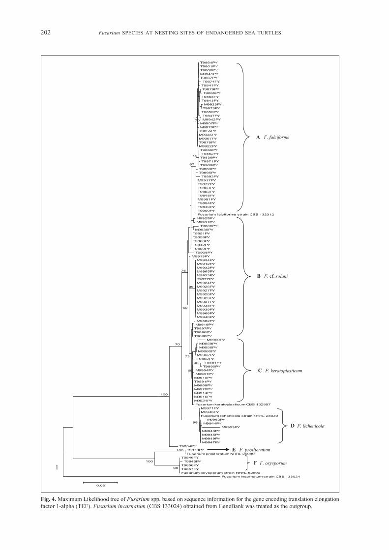
However, our molecular phylogenetic analysisof FSSC demonstrated that Main Clade I consistedof FSSC isolates, divided into four sub-clades(Figure 4): A, B, C and D. Sub-clade A consisted ofF. falciforme isolates, mainly from Terengganu (40isolates) and from Melaka (12 isolates). The isolatesin sub-clade A from Terengganu came from RedangIsland and Ma’ Daerah sites for green turtles. Isolatesfrom Melaka were from a hawksbill turtle hatcherysite. Sub-clade B consisted of 23 isolates of F. cf.solani, mainly from Melaka (18 isolates), and fivefrom Terengganu (Redang Island). Sub-clade Cconsisted of 17 isolates of F. keratoplasticumfrom Terengganu (4 isolates) and from TanjungKemunting, Melaka (13 isolates). Sub-clade Dconsisted of nine isolates of F. lichenicola fromMelaka (eggshells collected from nests in PulauUpeh). Fusarium falciforme and F. keratoplasticumhave been isolated from six species (Cheloniamydas, Caretta caretta, Eretmochelys imbricata,Lepidochelys olivacea, Dermochelys coriacea and
Natator depressus) of sea turtle eggs in nestingbeaches of the Atlantic, Indian and Pacific Oceans,and the Caribbean Sea (Sarmiento-Ramírez et al.,2014). These fungi are considered a threat to theendangered sea turtles (Sarmiento-Ramírez et al.,2014). Main Clade II consisted of two sub-cladeswith isolates only from Terengganu (Figure 4).These were sub-clades E and F: F. proliferatum (oneisolate from eggshells collected in Redang Island,Terengganu) and F. oxysporum (four isolates fromeggshells collected in Ma’ Daerah hatchery,Terengganu), respectively. Only F. lichenicola(8.5%) and F. oxysporum (3.8%) were isolated fromthe eggshells obtained from Tanjung Kemunting(Melaka) and Ma’Daerah hatchery, respectively.
There are three major clades of the FSSC andall the veterinary sources (including sea turtle)are nested within clade 3 (Zhang et al., 2006), themost evolutionarily diverse and species-rich clade(MP=100%; ML=83%) (O’Donnell et al., 2008). Astrong association of FSSC species with marineanimals has been reported.
The FSSC members adapt well to the marineenvironment, causing infections in marine animals(Sarmiento-Ramírez et al., 2014; Zhang et al., 2006).Various environmental stressors (frequent tidalinundation, unsuitable substrates, beach erosion and
202 Fusarium SPECIES AT NESTING SITES OF ENDANGERED SEA TURTLES
Fig. 4. Maximum Likelihood tree of Fusarium spp. based on sequence information for the gene encoding translation elongationfactor 1-alpha (TEF). Fusarium incarnatum (CBS 133024) obtained from GeneBank was treated as the outgroup.
1
T9864PY T9861PY T9880PY M9941PY T9867PY T9874PY T9841PY T9879PY T9865PY
T9868PY T9843PY M9923PY T9873PY
T9850PY T9847PY M9942PY
M9907PY M9970PY T9855PY M9935PY M9967PY T9878PY M9922PY T9869PY T9852PY T9839PY T9871PY T9909PY
T9883PY T9895PY
T9893PY M9917PY T9872PY T9863PY T9853PY T9848PY M9951PY T9894PY T9840PY T9900PY Fusarium falciforme strain CBS 132312 M9925PY M9931PY
T9866PY M9936PY
T9851PY T9859PY T9860PY T9842PY T9899PY
T9908PY M9913PY
M9934PY M9912PY M9932PY M9965PY M9933PY T9877PY M9924PY M9926PY M9927PY M9928PY M9929PY M9937PY M9938PY M9939PY M9966PY M9940PY M9882PY M9919PY T9897PY T9896PY T9898PY
M9960PY M9959PY M9956PY M9968PY M9952PY T9892PY
T9881PY T9890PY
M9954PY M9961PY M9910PY T9891PY M9969PY M9920PY M9914PY M9916PY M9921PY Fusarium keratoplasticum CBS 132897
M9971PY M9946PY Fusarium lichenicola strain NRRL 28030
M9962PY M9944PY
M9953PY M9943PY M9945PY M9949PY M9947PY
T9854PY T9870PY Fusarium proliferatum NRRL 25089
T9846PY T9845PY
T9856PY T9857PY Fusarium oxysporum strain NRRL 52690
Fusarium incarnatum strain CBS 133024
98
100
100
99
100
98
70
99
65
73
69
75
70
74
67
0.05
A F. falciforme
B F. cf. solani
C F. keratoplasticum
D F. lichenicola
E F. proliferatum
F F. oxysporum

Fusarium SPECIES AT NESTING SITES OF ENDANGERED SEA TURTLES 203
changes in sand composition) might increase theopportunities for adaptation of these pathogenicFusarium species and increase their ability tocolonise the sea turtle eggs. Additionally,endangered species whose populations are reducedin terms of genetic diversity could be easily beingdiseased when environmentally stressed orcombination of other threats (Aguirre & Tabor,2008). A single pathogenic species may become thefactor of a host toward extinction (Heard et al.,2013). A recent study has reported that F. falciformeand F. keratoplasticum were always found in deadturtle eggs and can kill up to 90% of the embryos;the optimal temperature for pathogen germinationis similar to the thermal environment of turtle nests,29.7°C (Sarmiento-Ramírez et al., 2014). In RedangIsland, the temperature of turtle nests range between25.6°C to 33.6°C were recorded and we also noticedthat most nests near to vegetation (<3m) had morenumber of infected eggs (Siti Nordahliawate et al.,2017; unpublished data). Recently, apart fromFusarium species we had isolated other fungi fromsoil and debris where a few numbers had beenidentified as Aspergillus species (Siti Nordahliawateet al., 2017; unpublished data).
Although the purpose of eggs relocation is toreduce the threat to the turtle eggs, it might alsoaffect the sea turtle population because ofdifferences in environmental conditions, especiallythe temperature of egg incubation (Chan, 2006). Ithas been reported that the hatching success inrelocated nests (ex situ) is lower than in undisturbednatural nests (in situ) (Wyneken et al., 1988). It ispossible to spread the fungus to a new nest locationunwillingly, particularly if proper handlingprocedures and sterile conditions are not ensured.Another factor that needs some consideration is thereuse of substrate in the hatcheries over severalseasons, which might cause accumulation of thefungal inocula. Recently, we found that there is norelation between fungal infection and hatchlingsuccess even though turtle nests contained eggs withsymptoms of fungal infection (Siti Nordahliawateet al., 2017; unpublished data).
Members of the FSSC can be a serious plant andhuman pathogens (Short et al., 2013; Zhang et al.,2006). Thus, anyone working with Fusariumisolated from turtle eggs should be aware of the risk.Infection can be prevented by wearing masks,gloves or maintaining sterile conditions.
CONCLUSION
This study confirms the presence of several speciesof Fusarium on infected green and hawksbill turtleeggs and nests. All infected egg samples (incubatedin situ and ex situ) showed similar symptoms at all
sampling sites in Terengganu and Melaka. The fungiwere also isolated from sands, debris and roots foundin the nests. Members of FSSC constituted thegreatest proportion of the isolated fungi. The turtleeggs that were damaged by predators such asmonitor lizards are suitable substrate to coloniseby Fusarium and could progressively move to theneighboring eggs. Furthermore, Fusarium hasnecrotroph phase to its lifecycle, the prominenceof Fusarium species on damaged eggs is likelybecasuse it is an opportunist pathogen. We alsoknow that the Fusarium falciforme and F.keratoplasticum implicated in sea turtle eggfusariosis have been isolated from symptomless eggsin nests with high hatching success, suggestingFSSC that present in Terengganu and Melaka nestswith little to no impact on egg mortality. The FSSChas been described as an emerging animal pathogenbut has not been previously reported from sea turtleeggs of any nesting sites in Malaysia. This is thefirst report of the occurrence of Fusarium species onthe green turtle and hawksbill turtle eggshells inPeninsular Malaysia and confirms F. falciforme, F.keratoplasticum, F. lichenicola and F. cf. solani asmembers of the FSSC on the molecular level. Inorder to better understand, further study is neededto investigate the fungi fully on the turtle nestingbeaches and in the hatcheries in Malaysia. We needto assess the diversity and population structure ofFusarium species associated with sand, ocean andsea turtle nests as well as environmental factors thatfavour this opportunistic pathogens. We hope thatour findings will aid in the development ofconservation programmes and increase theawareness of potential pathogens threateningendangered sea turtle species.
ACKNOWLEDGEMENTS
Sampling at Chagar Hutang, Redang Island,Terengganu was sponsored by the SEATRU-INOSTurtle Fund, Universiti Malaysia Terengganu.Sampling in Melaka and laboratory analysis wassponsored by the Fundamental Research GrantScheme (FRGS), Ministry of Education Malaysia,Grant No. 203/PBIOLOGI/6711311USM, School ofBiological Sciences, Universiti Sains Malaysia. Wethank the Department of Fisheries Terengganu andMelaka for giving us the permission to conductsamplings at their turtle hatcheries. The authorswould like to thank Ms Faezah Noor Basir, MrLionel Harith Sebastian Daraup and Mr MohdKamarudin Mohd Maidin, for help with ourfieldwork. We thank the staff of the Laboratory forPest Disease & Microbial Biotechnology (LAPDiM)for their technical assistance. Prof. BaharuddinSalleh, former Professor of plant pathology at the
204 Fusarium SPECIES AT NESTING SITES OF ENDANGERED SEA TURTLES
School of Biological Sciences, Universiti SainsMalaysia, is acknowledged for sharing hisknowledge and insight.
REFERENCES
Aguirre, A.A. & Tabor, G.M. (2008). Global FactorsDriving Emerging Infectious Diseases Impacton Wildlife Populations. Animal Biodiversityand Emerging Diseases: Prediction andPrevention, 1149: 1-3.
Booth, C. (1977). Fusarium, Laboratory guide to theidentification of the major species. Kew, SurreyUK: Commonwealth Mycological Institute.ISBN 0-85198-383-9.
Chan, E.H. & Solomon, S.E. (1989). The structureand function of the eggshell of the leatherbackturtle (Dermochelys coriacea) from Malaysia,with notes on attached fungal forms. AnimalTechnology, 40: 2.
Chan, E.H. (2006). Marine turtles in Malaysia: Onthe verge of extinction?. Aquatic EcosystemHealth & Management, 9: 175-184.
Daraup, L.H.S. (2010). Hatching success in in situand relocated green turtle (Chelonia mydas)nests incubated at Chagar Hutang, RedangIsland, Terengganu. Dissertation, UniversitiMalaysia Terengganu.
Eckert, K.L. & Eckert, S.A. (1990). Embryo mortalityand hatch success in situ and translocatedleatherback sea turtle Dermochelys coriaceaeggs. Biological Conservation, 53: 37-46.
Elshafie, A., Al-Bahry, S.N., Alkindi, A.Y., Ba-Omar,T. & Mahmoud, I. (2007). Mycoflora andaflatoxins in soil, eggshells, and failed eggsof Chelonia mydas at Ras Al-Jinz, Oman.Chelonian Conservation and Biology, 6: 267-270.
Fisher, N.L., Burgess, L.W., Toussoun, T.A. &Nelson, P.E. (1982). Carnation leaves as asubstrate and for preserving cultures ofFusarium species. Phytopathology, 72: 151-153.
Fisher, M.C., Garner, T.W.J. & Walker, S.F. (2009).Global emergence of Batrachochytriumdendrobatidisand amphibian chytridiomycosisin space, time, and host. Annu Rev Microbiol,63: 291-310.
Fisher, M.C., Henk, D.A., Briggs, C.J., Brownstein,J.S., Madoff, L.C., McCraw, S.L. & Gurr, S.J.(2012). Emerging fungal threats to animal, plantand ecosystem health. Nature, 484: 186-194.
Foley, J., Clifford, D., Castle, K., Cryan, P. & Ostfeld,R.S. (2011). Investigating and managing therapid emergence of white-nose syndrome, anovel, fatal, infectious disease of hibernatingbats. Conservation Biology, 25: 223-231.
Frick, W.F. (2010). An emerging disease causesregional population collapse of a commonNorth American bat species. Science, 329: 679-682.
Garcia, A., Ceballos, G. & Adaya, R. (2003).Intensive beach management as an improvedsea turtle conservation strategy in Mexico. BiolConserv, 111: 253-261.
Geiser, D.M., Aoki, T., Bacon, C.W., Baker, S.E.,Bhattacharyya, M.K., Brandt, M.E. et al. (2013).One Fungus, One Name: defining the genusFusarium in a scientifically robust way thatpreserves longstanding use. Phytopathology,103(5): 400-408.
Guarro, J. (2013). Fusariosis, a complex infectioncaused by a high diversity of fungal speciesrefractory to treatment. Eur J Clin MicrobiolInfect Dis, 32: 1491-1500.
Heard, M.J., Smith, K.F., Ripp, K.J., Berger, M.,Chen, J., Dittmeier, J., Goter, M., McGarvey, S.T.& Ryan, E. (2013). The threat of diseaseincreases as species move toward extinction.Conserv Biol., 27(6): 1378-1388.
Hof, C., Araujo, M.B., Jetz, W. & Rahbek, C. (2011).Additive threats from pathogens, climate andland-use change for global amphibian diversity.Nature, 480: 516-519.
IUCN (2004). IUCN Red List of Threatened Species.In: Baillie JEM, Hilton-Taylor C, Stuart SN(eds) A Global Specie Assessment. Gland,Switzerland and Cambridge, UK: IUCN.
Leslie, J.F. & Summerell, B. (2006). The Fusariumlaboratory manual. Iowa: Blackwell Publishing.388.
Mandel, Q.A. (2006). Biodiversity of the genusFusarium in saline soil habitats. Journal ofBasic Microbiology, 46(6): 480-94.
Mo, C.L., Salas, I. & Caballero, M. (1990). Are fungiand bacteria responsible for olive ridley’s egglost? In: Richardson T, Richardson J, DonnellyM (ed) Tenth Annual Workshop on Sea TurtleBiology and Conservation: NOAA TechnicalMemorandum NMFS-SEFC-278. pp. 249-252.
Morita, M., Ahmad, A.H. & Chan, E.H. (2008).Implication of predation incidences by antspecies on green turtle nests in Chagar Hutang,Readang Island. Proc. 3rd Int. Symp. SEASTARand Asian Bio-logging Science.
O’Donnell, K., Kistler, H.C., Cigelnike, E. & Ploetz,R.C. (1998). Multiple evolutionary origins ofthe fungus causing Panama disease of banana:Concordant evidence from nuclear and mito-chondrial gene genealogies. Proc Natl Acad SciUSA 95: 2044-2049.
O’Donnell, K. (2000). Molecular phylogeny of theNectria haematococca- Fusarium solani speciescomplex. Mycologia, 92: 919-938.
Fusarium SPECIES AT NESTING SITES OF ENDANGERED SEA TURTLES 205
O’Donnell, K., Sutton, D.A., Fothergill, A.,McCarthy, D., Rinaldi, M.G., Brandt, M.E.,Zhang, N. & Geiser, D.M. (2008). Molecularphylogenetic diversity, multilocus haplotypenomenclature, and in vitro antifungal resistancewithin the Fusarium solani species complex.Journal of Clinical Microbiology, 46: 2477–2490.
O’Donnell, K., Sutton, D.A., Rinaldi, M.G., Sarver,B.A., Balajee, S.A., Schroers, H.J., Summerbell,R.C., Robert, V.A., Crous, P.W., Zhang, N., Aoki,T., Jung, K., Park, J., Lee, Y.H., Kang, S., Park,B. & Geiser, D.M. (2010). Internet-accessibleDNA sequence database for identifying fusariafrom human and animal infections. J ClinMicrobiol, 48(10): 3708-3718.
O’Donnell, K., Ward, T.J., Robert, V.A.R.G.,Crous, P.W., Geiser, D.M. & Kang, S. (2015).DNA sequence-based identification ofFusarium: Current status and future directions.Phytoparasitica, 43(5): 583-595.
Güçlü, O., Býyýk, H. & Sahiner, A. (2010).Mycoflora identified from loggerhead turtle(Caretta caretta) egg shells and nest sand atFethiye beach, Turkey. African Journal ofMicrobiology Research, 45: 408-413.
Phillott, A.D., Parmenter, C.J. & Limpus, C.J. (2004).Occurrence of mycobiota in eastern Australiansea turtle nests. Memoirs of the QueenslandMuseum, 49: 701-703.
Salleh, B. & Sulaiman, B. (1984). Fusaria associatedwith naturally diseased plants in Penang.Journal of Plant Protection in the Tropics, 1:47-53.
Sarmiento-Ramírez, J.M., Abella, E., Martín, M.P.,Tellería, M.T., López-Jurado, L.F., Marco, A.& Diéguez-Uribeondo, J. (2010). Fusariumsolani is responsible for mass mortalities innests of loggerhead sea turtle, Caretta caretta,in Boavista, Cape Verde. FEMS MicrobiologyLetters, 312: 192-200.
Sarmiento-Ramírez, J.M., Abella, E., Phillott, A.D.,Sim, J., van West, P., Martin, M.P., Marco, A.& Diéguez-Uribeondo, J. (2014). GlobalDistribution of Two Fungal PathogensThreatening Endangered Sea Turtles. PLoSONE, 9: e85853.
Seminoff, J.A. (2004). Chelonia mydas. In: IUCN2012. IUCN Red List of Threatened Species.Version 2012.2. Retrieved from www.iucnredlist.org.
Short, D.P.G., O’Donnell, K., Thrane, U., Nielsen,K.F., Zhang, N., Juba, J.H. & Geiser, D.M.(2013). Phylogenetic relationships amongmembers of the Fusarium solani speciescomplex in human infections and thedescriptions of F. keratoplasticum sp. nov. andF. petroliphilum stat. nov. Fungal Genetics andBiology, 53: 59-70.
Siti Nordahliawate, M.S., Nur Ain Izzati, M.Z.,Nur Azlin, A. & Salleh, B. (2012). Diversity ofFusarium species isolated from soil cultivatedwith cucurbits within East Coast, PeninsularMalaysia. Pertanika. Journal of TropicalAgricultural and Sciences, 35(2): 381-386.
Summerbell, R.C. & Schroers, H.J. (2002). Analysisof phylogenetic relationship of Cylindrocarponlichenicola and Acremonium falciforme to theFusarium solani species complex and a reviewof similarities in the spectrum of opportunisticinfections caused by these fungi. Journal ofClinical Microbiology, 40: 2866-2875.
Tamura, K., Stecher, G., Peterson, D., Filipski, A.& Kumar, S. (2013). MEGA6: MolecularEvolutionary Genetics Analysis version 6.0.Mol Biol Evol, 30: 2725-29.
Wyneken, J., Burke, T.K., Salmon, M. & Pedersen,D.K. (1988). Egg failure in natural and relocatedsea turtle nests. Journal of Herpetology, 22:88-96.
Zhang, N., O’Donnell, K., Sutton, D.A., Nalim, F.A.,Summerbell, R.C., Padhye, A.A. & Geiser, D.M.(2006). Members of the Fusarium solani speciescomplex that cause infections in both humansand plants are common in the environment.Journal of Clinical Microbiology, 44: 2186-2190.
