egjub1 and egerf113 transcription factors as potential
TRANSCRIPT

RESEARCH ARTICLE Open Access
EgJUB1 and EgERF113 transcription factorsas potential master regulators of defenseresponse in Elaeis guineensis against thehemibiotrophic Ganoderma boninenseNurshafika Mohd Sakeh1, Siti Nor Akmar Abdullah1,2*, Mohammad Nazri Abdul Bahari1,Azzreena Mohamad Azzeme3, Noor Azmi Shaharuddin3 and Abu Seman Idris4
Abstract
Background: Hemibiotrophic pathogen such as the fungal pathogen Ganoderma boninense that is destructive tooil palm, manipulates host defense mechanism by strategically switching from biotrophic to necrotrophic phase.Our previous study revealed two distinguishable expression profiles of oil palm genes that formed the basis indeducing biotrophic phase at early interaction which switched to necrotrophic phase at a later stage of infection.
Results: The present report is a continuing study from our previous published transcriptomic profiling of oil palmseedlings against G. boninense. We focused on identifying differentially expressed genes (DEGs) encodingtranscription factors (TFs) from the same RNA-seq data; resulting in 106 upregulated and 108 downregulated TFsbeing identified. The DEGs are involved in four established defense-related pathways responsible for cell wallmodification, reactive oxygen species (ROS)-mediated signaling, programmed cell death (PCD) and plant innateimmunity. We discovered upregulation of JUNGBRUNNEN 1 (EgJUB1) during the fungal biotrophic phase whileEthylene Responsive Factor 113 (EgERF113) demonstrated prominent upregulation when the palm switches todefense against necrotrophic phase. EgJUB1 was shown to have a binding activity to a 19 bp palindromic SNBE1element, WNNYBTNNNNNNNAMGNHW found in the promoter region of co-expressing EgHSFC-2b. Further in silicoanalysis of promoter regions revealed co-expression of EgJUB1 with TFs containing SNBE1 element with singlenucleotide change at either the 5th or 18th position. Meanwhile, EgERF113 binds to both GCC and DRE/CRTelements promoting plasticity in upregulating the downstream defense-related genes. Both TFs were proven to benuclear-localized based on subcellular localization experiment using onion epidermal cells.
(Continued on next page)
© The Author(s). 2021 Open Access This article is licensed under a Creative Commons Attribution 4.0 International License,which permits use, sharing, adaptation, distribution and reproduction in any medium or format, as long as you giveappropriate credit to the original author(s) and the source, provide a link to the Creative Commons licence, and indicate ifchanges were made. The images or other third party material in this article are included in the article's Creative Commonslicence, unless indicated otherwise in a credit line to the material. If material is not included in the article's Creative Commonslicence and your intended use is not permitted by statutory regulation or exceeds the permitted use, you will need to obtainpermission directly from the copyright holder. To view a copy of this licence, visit http://creativecommons.org/licenses/by/4.0/.The Creative Commons Public Domain Dedication waiver (http://creativecommons.org/publicdomain/zero/1.0/) applies to thedata made available in this article, unless otherwise stated in a credit line to the data.
* Correspondence: [email protected] of Plantation Studies, Universiti Putra Malaysia (UPM), 43400Serdang, Selangor, Malaysia2Department of Agriculture Technology, Faculty of Agriculture, UniversitiPutra Malaysia (UPM), 43400 Serdang, Selangor, MalaysiaFull list of author information is available at the end of the article
Sakeh et al. BMC Plant Biology (2021) 21:59 https://doi.org/10.1186/s12870-020-02812-7
(Continued from previous page)
Conclusion: Our findings demonstrated unprecedented transcriptional reprogramming of specific TFs potentially toenable regulation of a specific set of genes during different infection phases of this hemibiotrophic fungalpathogen. The results propose the intricacy of oil palm defense response in orchestrating EgJUB1 during biotrophicand EgERF113 during the subsequent transition to the necrotrophic phase. Binding of EgJUB1 to SNBE motifinstead of NACBS while EgERF113 to GCC-box and DRE/CRT motifs is unconventional and not normally associatedwith pathogen infection. Identification of these phase-specific oil palm TFs is important in designing strategies totackle or attenuate the progress of infection.
Keywords: JUNGBRUNNEN 1, ERF113, SNBE motif, GCC-box, DRE/CRT, Hemibiotrophic
BackgroundGanoderma boninense is a pathogenic species whichcauses basal stem rot (BSR) disease in oil palm. Infectedpalms remain symptomless even though they are alreadyphysiologically impaired [1]. Half of the basal stemwould have been destroyed by the fungus, compromisingintake of water and nutrients, before the first symptomis observed [2, 3]. The fungus, G. boninense is recog-nized as a hemibiotroph, which established an inter-mediate lifestyle of biotroph before switching tonecrotroph [4, 5]. The biotrophic phase involvescolonization of host plant tissues and extraction of nutri-ents for survival by the pathogen while keeping the hostcells intact [3]. Biotrophs thrive in the intracellular re-gion between mesophyll cells through formation of hau-storia that play important role in delivery of pathogeniceffector proteins [6, 7]. The invasion requires minimalrelease of cell wall degrading enzymes (CWDEs) to allowloosening of the plant cell wall [8, 9].Pathogen-associated molecular patterns (PAMPs) are
elicitors such as bacterial flagellin and fungal chitin de-rived from the phytopathogens that are recognized byplant cell surface pattern recognition receptors (PRRs)leading to PAMP-triggered immunity (PTI) [10–12].The susceptibility of plant host to microbial colonizationrelies on effectors secreted by the pathogens to deceivePAMPs recognition, thus suppressing PTI signaling [12,13]. Breaching of the PTI remarks activation of asecond-line of plant defense response termed aseffector-triggered immunity (ETI). ETI comprises resist-ance (R) proteins to counteract a successful invasion ofpathogens by recognizing virulence effectors (Avr) inhost cells [14]. ETI, accompanied by increased signalingof phytohormones including lipid-based jasmonic acid(JA) and gaseous ethylene (ET) stimulate the productionof ROS [15]. The ROS induces a hypersensitive response(HR) at sites of infection to limit the invasion of patho-gens [16].Unfortunately, the accumulation of ROS and induced
programmed cell death (PCD) created a favorable envir-onment for necrotroph to intensify infection strategy[17]. It was postulated that the increasing pressure by
plant defense responses results in switching of thepathogen infection mode from biotrophy to necrotrophybut the time taken differs between specific host-pathogen interaction [18]. Hemibiotrophs may requireextended periods of biotrophic phase to establishcolonization and once sufficed, transition to necro-trophic phase is rapid [19]. Necrotrophs secrete phyto-toxic compounds and excessive CDWEs to induce hostcell death [20]. The dynamic intermediate lifestyle ofhemibiotroph enables manipulation of the host plantdefense mechanisms which ultimately result in the plantsuccumbing to the infection. Salicylic acid (SA) signalingwhich often functions antagonistically to JA-ET signalinghas been widely studied to determine the fine-tuningagainst biotrophic or necrotrophic infection [21–23].However, there is poor molecular information availableexplaining the defense regulation by transcription factors(TFs) during transitions of biotrophic to necrotrophicstate. Our previous transcriptomic profiling via high-throughput RNA-seq analysis has pointed out thecounter-act defense mechanisms executed by plants dur-ing the transition of biotrophic to necrotrophic phase[5]. In the attempt of identifying key defense pathwaysthat are transcriptional regulated, the next generation se-quencing (NGS) data set was mined for TFs responsiblefor triggering the downstream responses.TFs are the ‘master switches’, which regulate the ex-
pression of a large set of genes initiated by unique sig-naling networks in response to stresses [24]. Modulationof defense response gene expression may vary dependingon the intensity and intricacy of multiple stresses [25].There are six major TF families involved in plant defenseresponse including MYB, bHLH, AP2/ERF, NAC, bZIPand WRKY [26]. Regulation by TFs is crucial to mediatethe transcriptional reprogramming which includes in-duced expression of defense-related genes responsible inthe production of antifungal proteins or the antimicro-bial secondary metabolites known as phytoalexins [27].Intriguingly, plants also display cross-tolerance phenom-ena in which a single type of stress may trigger a multi-tude of tolerant levels to different stresses [28]. Forinstance, heat stress transcription factors (HSFs) play an
Sakeh et al. BMC Plant Biology (2021) 21:59 Page 2 of 20
important role as a master regulator of defense responseunder multiple stresses [29, 30]. Over-expressed HSFsconfer resistance against dehydration, bacterial andoomycetes infections and improve yield under water-limited conditions [31].TFs bind to cis-acting elements located in promoters
of either other mediator TFs or downstream target geneswhich results in up- or down-regulation of their expres-sion [32–34]. NAC TFs are recognized as master regula-tors for secondary cell wall biosynthesis mediated byMYB TF through the formation of NAC-MYB-CESAsignaling cascade [35]. In addition, bHLH-MYB associ-ation was involved in the biosynthesis of flavonoids sec-ondary metabolites under phytohormones signaling,wounding and fungal interaction [36, 37]. Interaction tospecific DNA sequences (binding motifs) is dependenton the DNA binding domain (DBD) of TFs [38]. Bindingpreferences of TF during biotic or abiotic stress such asERFs have been suggested to correlate with the compos-ition of amino acid sequences in DBD [38, 39]. Thepresence of multiple cis-acting elements in the promoterregion contributes to overlapping roles in developmentand/or defense against multiple stresses of the expressedproteins [26].In addition to biotic or abiotic stresses [40], in this
study, we reported that the regulation of TFs isdependent on modes of pathogen infection (biotrophicand necrotrophic). Only EgUNE10 TF and a few TF fam-ilies have been reported to regulate defense against laterstages of G. boninense infection [4, 41, 42]. However,there is no comprehensive report on transcriptomic pro-filing of defense-related TFs during different infectionphases of hemibiotroph in plants. Thus, this study is thefirst attempt to recognize specific TFs as ‘key’ bio-markers involved in transcriptional switching from bio-trophic to necrotrophic infection phase based on earlyoil palm-G. boninense interaction. Their potential tar-geted defense response pathways that distinguished thetwo phases are discussed based on the identification ofspecific motifs interacting with the newly discoveredTFs. The findings might allow a more effective diseasemanagement strategy to attenuate the progress of G.boninense infection of oil palm and prevent the spreadof the disease.
ResultsElaeis guineensis defense-related transcription factors andbiomarkers of biotrophy-necrotrophy switchIn order to identify the TF families involved in the earlydefense of oil palm against G. boninense, transcriptomicsanalysis of Ganoderma-treated root tissues at 3, 7 and11 d.p.i was carried out. We identified 106 of upregu-lated and 108 of downregulated TFs upon early inter-action with G. boninense (Fig. 1a and b). The pairwise
comparison was constructed between control againsttime course treatments using stringent cut-off values oflog2 fold change (FC) ≥ |1.0| and P-value < 0.01. Wehave previously reported the transition of biotrophic tonecrotrophic defense mechanism, based on qPCRpreliminary screening using defense-related molecularbiomarkers EgPR1 (biotrophic) and EgMYC2 (necro-trophic) [5]. Based on the report, the expression profileof defense-related TFs was identified as biotrophic-regulated at 3 days post-infection (d.p.i) whilenecrotrophic-regulated at 11 d.p.i, with intermediate at 7d.p.i. We found that the expression patterns of the genesshowed either decreasing or increasing over time. Thehighest TF families upregulated during early interactionwith G. boninense were mainly bHLH > MYB > AP2/ERF, followed by bZIP > MADS > TCP > OFP > NAC >GATA > HSF > NFY > E2F > WRKY> EIN/EIL. On theother hand, the highest downregulated families of TFwere found to be mainly AP2/ERF > bHLH > MYBfollowed by MADS > CAMTA > NAC > TCP > GATA >bZIP > HSF > NFY > E2F > WRKY > OFP. The familiesof oil palm’s TFs involved during defense responseagainst G. boninense were found to be the same but in-volving different members in both upregulated anddownregulated DEGs. Distinctively, EIN/EIL andCAMTA TF family were only found in upregulated anddownregulated DEGs, respectively. AtCAMTA3 andAtCAMTA4 were reported to negatively regulate plantdefense response under SA-mediated signaling pathwayagainst obligate biotroph [43, 44].To further understand plant response against G. boni-
nense interaction, four common pathways regulatingdefense mechanisms were identified from the RNA-seqdata (Fig. 1c). Even though not all genes have been dis-covered in the pathways, important genes associatedwith the different pathways have been identified amongthe DEGs as presented. Reported genes involved in cellwall modification, ROS-mediated signaling, PCD andplant innate immunity were all differentially expressed,indicating active regulation of defense response in E. gui-neensis against hemibiotroph G. boninense. The expres-sion patterns of selected biotic stress-related genes wereimportant to distinguish the defense mechanisms exe-cuted by the plants during two different phases of bio-trophic and necrotrophic infection.Six genes of Cellulose synthase (CESA) and Cellulose
synthase-like D (CSL) were reported in cell wall modifi-cation including EgCESA2, EgCESA4, EgCESA5,EgCESA9, EgCSLD2 and EgCSLD5 demonstrated similarexpression patterns with high upregulation at 3 d.p.i thatsuccessively decreased across time. Three out of sixgenes involved in ROS-mediated signaling includingSuperoxide dismutase (EgSOD), Glutathione S-transferase (EgGSTF12) and Respiratory burst oxidase
Sakeh et al. BMC Plant Biology (2021) 21:59 Page 3 of 20

homolog (EgRBOHA) were increasingly expressed from 3d.p.i to 11 d.p.i, whilst EgGSTU17, EgGST3 and EgR-BOHB demonstrated decreasing expression patterns.Two genes reported in PCD, Metacaspase 9 (EgMC9)and Bifunctional nuclease (EgBFN) showed antagonisticexpression patterns of increasing and decreasingregulation, respectively. Five genes involved in plant in-nate immunity of PTI/ETI signaling were found to bedifferentially expressed. Three of the genes includingNONEXPRESSOR OF PATHOGENESIS-RELATED
GENE 1 (EgNPR1), Calmodulin-like 3 (EgCML3) andCalcium-dependent protein kinase 7 (EgCDPK7) wereupregulated the highest at 3 d.p.i and 7 d.p.i before sub-sequently declined. The other genes which wereEgCML7 and EgCDPK28 were successively upregulatedwith the highest expression at 11 d.p.i. The reliability ofRNA-seq data has been validated in our previous report[5] using representative upregulated and downregulatedDEGs with different levels of fold change compared tountreated seedlings (control).
Fig. 1 Time course of differentially expressed genes (DEGs) during early interaction with Ganoderma boninense. a Heat map clustering ofupregulated transcription factors. Colored blocks indicate ascending expression level from green (0) to red (6). b Heat map clustering ofdownregulated transcription factors. Colored blocks indicate descending expression level from green (0) to red (− 8). c Heat map clustering ofupregulated DEGs commonly expressed during biotic interaction. Colored blocks indicate ascending expression level from white (0) to blue (4).For RNA-seq data analysis, two biological replicates which each consisted of pooled RNA provided equally from six constituent seedlings wereused. Each heatmap data was constructed using an average of pooled biological replicates. Pairwise comparison of RNA-seq data betweencontrol (untreated) and Ganoderma-treated was evaluated according to cut-off values of log2 fold change (FC) ≥ |1.0| and P-value < 0.01
Sakeh et al. BMC Plant Biology (2021) 21:59 Page 4 of 20
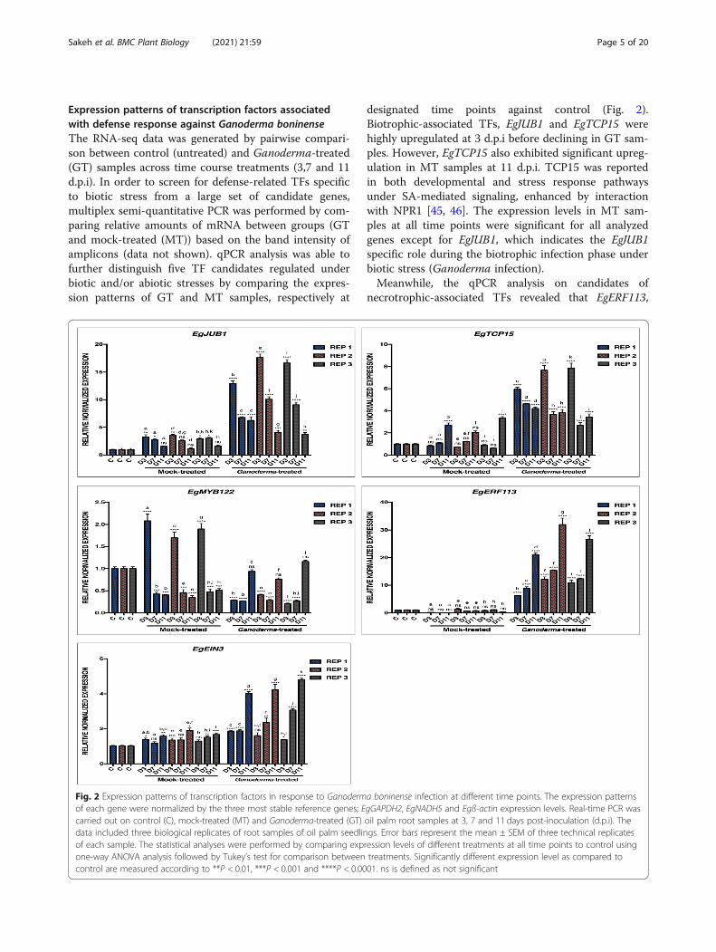
Expression patterns of transcription factors associatedwith defense response against Ganoderma boninenseThe RNA-seq data was generated by pairwise compari-son between control (untreated) and Ganoderma-treated(GT) samples across time course treatments (3,7 and 11d.p.i). In order to screen for defense-related TFs specificto biotic stress from a large set of candidate genes,multiplex semi-quantitative PCR was performed by com-paring relative amounts of mRNA between groups (GTand mock-treated (MT)) based on the band intensity ofamplicons (data not shown). qPCR analysis was able tofurther distinguish five TF candidates regulated underbiotic and/or abiotic stresses by comparing the expres-sion patterns of GT and MT samples, respectively at
designated time points against control (Fig. 2).Biotrophic-associated TFs, EgJUB1 and EgTCP15 werehighly upregulated at 3 d.p.i before declining in GT sam-ples. However, EgTCP15 also exhibited significant upreg-ulation in MT samples at 11 d.p.i. TCP15 was reportedin both developmental and stress response pathwaysunder SA-mediated signaling, enhanced by interactionwith NPR1 [45, 46]. The expression levels in MT sam-ples at all time points were significant for all analyzedgenes except for EgJUB1, which indicates the EgJUB1specific role during the biotrophic infection phase underbiotic stress (Ganoderma infection).Meanwhile, the qPCR analysis on candidates of
necrotrophic-associated TFs revealed that EgERF113,
Fig. 2 Expression patterns of transcription factors in response to Ganoderma boninense infection at different time points. The expression patternsof each gene were normalized by the three most stable reference genes; EgGAPDH2, EgNADH5 and Egß-actin expression levels. Real-time PCR wascarried out on control (C), mock-treated (MT) and Ganoderma-treated (GT) oil palm root samples at 3, 7 and 11 days post-inoculation (d.p.i). Thedata included three biological replicates of root samples of oil palm seedlings. Error bars represent the mean ± SEM of three technical replicatesof each sample. The statistical analyses were performed by comparing expression levels of different treatments at all time points to control usingone-way ANOVA analysis followed by Tukey’s test for comparison between treatments. Significantly different expression level as compared tocontrol are measured according to **P < 0.01, ***P < 0.001 and ****P < 0.0001. ns is defined as not significant
Sakeh et al. BMC Plant Biology (2021) 21:59 Page 5 of 20

EgEIN3 and EgMYC2 were highly upregulated at 11 d.p.ion GT samples. Both EIN3 and MYC2 are JA-dependentwhich were upregulated and downregulated in the RNA-seq data, respectively. The expression of these genes isknown to be mutually exclusive whereby MYC2 relieson co-actions of JA-abscisic acid (ABA) while EIN3 reg-ulates plant defense response through JA-ET signaling[47, 48]. EgERF113 which demonstrated non-significantexpression on MT samples (abiotic stress) at all timepoints were selected for further characterization as anovel potential candidate of necrotrophic-specific TF.
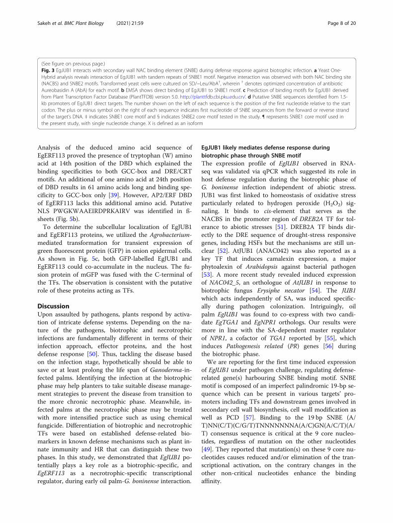
EgJUB1 binds to novel SNBE motif during biotrophicinfectionCharacterization of EgJUB1 TF against G. boninense in-fection was carried out via Y1H assay and EMSA. Threepotential binding motifs; one NAC binding site (NACBS)and two secondary wall NAC binding elements (SNBEs)with respective mutants were tested on EgJUB1 (Fig. 3).NACBS is the established binding motif for JUB1 TFduring abiotic stress while SNBEs are the novel targetmotifs tested in the present study. The SNBE consensussequence was identified as WNNYBTNNNNNN-NAMGNHW, whilst NACBS was RRYGCCGT. Y1Hassay demonstrated positive interaction with SNBE1motif but not to NACBS and SNBE2. The results indi-cate regulation of the alternative pathway by JUB1 in en-hancing resistance during biotic stress. The positivebinding with one of the SNBE1 motif indicates that thebinding affinity is dependent on the core motifs of theSNBE consensus sequence, whereby four nucleotideschange on the core motif of SNBE1 resulted in no inter-action with SNBE2 (Fig. 3a). Colony PCR was carriedout on five positive clones and sequencing was per-formed using T7 promoter primer of pGADT7-Rec vec-tor to confirm genuine positive interaction. p53-AbAiyeast reporter vector was used as a positive control inthe Y1H assay. We further confirmed the protein-DNAinteraction via EMSA by testing the binding of nuclearprotein extracted from a positive clone of Y1H assay onbiotinylated DNA probes. A shifted band was observedupon testing with a biotinylated DNA probe. Unlabeledprobe (molar excess 200-fold) was able to compete ef-fectively for binding with biotinylated target DNA probe.The inability of the mutated fragment to compete withthe biotinylated target DNA probe revealed specificbinding of EgJUB1 to SNBE1 (Fig. 3b). SNBE consensussequence is the predicted DNA binding motif of JUB1(Fig. 3c), based on Plant Transcription Factor Database(PlantTFDB), but thus far there is no reported evidenceto support this.Here, we listed SNBE motifs explored in the 1.5 kb
promoter regions of TFs co-expressed with EgJUB1 dur-ing the biotrophic phase (Fig. 3d). From the RNA-seq
data, EgJUB1 showed significant upregulation at 3 and 7d.p.i but not at 11 d.p.i, based on statistical analysis ofcut-off values log2 fold change |1.0| and P-value < 0.01.Hence, TFs with a similar pattern of expression profileusing the same statistical analysis are categorized as co-expressing with EgJUB1. The core motif of the SNBE1sequence was identified in the promoter region ofEgHSFC-2b, suggesting direct regulation of EgJUB1 withthe EgHSFC-2b. We highlighted the promoter regions ofa few TFs co-expressing with EgJUB1 with a single nu-cleotide change in the SNBE1 core motif tested in thepresent study at the 5th or 18th position, includingEgHSFB-4b, EgGAMYB-X2, EgERF003, EgKAN1-like-X3,EgILI-5-like, EgERF086-like, EgPIF3-X1 and -X2. It isplausible that changes of single nucleotide on SNBE1core motif, still maintain the ability of EgJUB1 to acti-vate these TFs. Changes of two and more nucleotides inthe core motif of SNBE1 are expected to result in a sig-nificant decline in binding affinity [49], which merit ana-lysis in more detail.
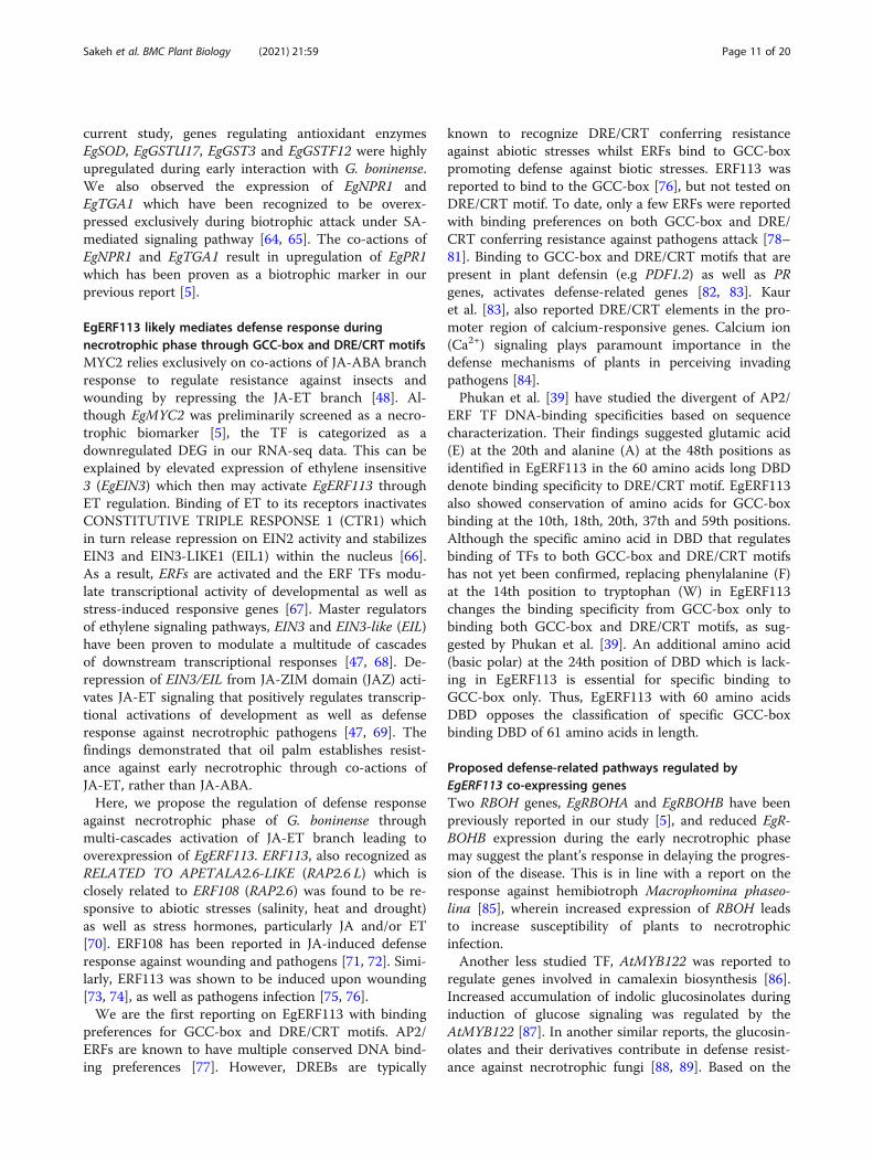
EgERF113 binds to both GCC and DRE/CRT motifs duringnecrotrophic infectionEgERF113 TF was tested via yeast one-hybrid (Y1H)assay and electrophoretic mobility shift assay (EMSA)(Fig. 4) on two AP2/ERF DNA binding preferenceswhich were GCC-box also known as Ethylene-ResponseElement (ERE) and Dehydration Response Element/C-Repeat (DRE/CRT). The GCC-box and DRE/CRT motifsin the study share a 6-bp core sequence of GCCGMC.Our findings revealed recognition of EgERF113 by bothmotifs in the Y1H assay. The binding affinity ofEgERF113 with both binding motifs was supported bythe EMSA results. Unlabelled probes of GCC-box andDRE/CRT (molar excess 200-fold) were able to competewith respective biotinylated target DNA probes. Noshifted band was observed on mutated fragments whichproved binding specificity of EgERF113 with both GCC-box and DRE/CRT motifs. The findings suggest thatEgERF113 can regulate stress-related genes harboringGCC-box and/or DRE/CRT in their promoter region.
EgJUB1 and EgERF113 are localized in the nucleusAnalysis of the deduced amino acid sequence revealedthe NAC binding domain of EgJUB1 with five highlyconserved subdomains A to E at the N-terminal region.The C-terminal region showed a highly conserved do-main between different species which suggested its pos-sible role as a transcriptional activator, repressor, or inbinding to other proteins. Putative nuclear localizationsignals (NLS), KKSLVYYLGSAGKGTKT was identifiedin subdomain D (Fig. 5a).The DBD of EgERF113 TF consists of three-stranded
ß-sheets and one α-helix running almost in parallel.
Sakeh et al. BMC Plant Biology (2021) 21:59 Page 6 of 20
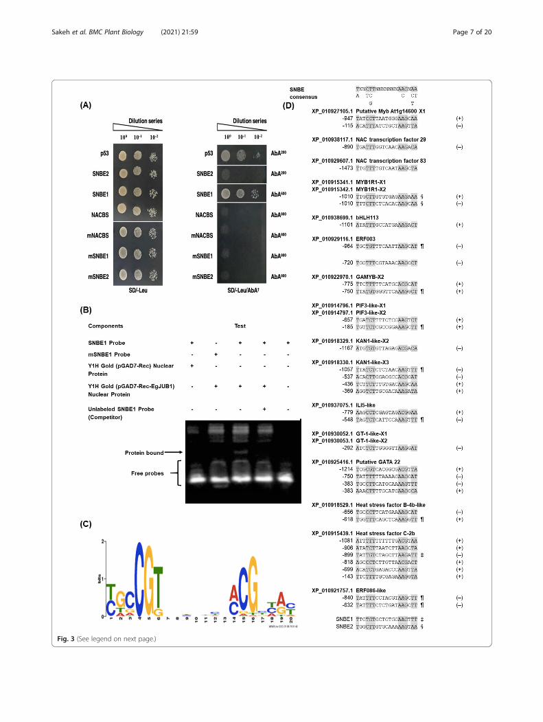
Fig. 3 (See legend on next page.)
Sakeh et al. BMC Plant Biology (2021) 21:59 Page 7 of 20
Analysis of the deduced amino acid sequence ofEgERF113 proved the presence of tryptophan (W) aminoacid at 14th position of the DBD which explained thebinding specificities to both GCC-box and DRE/CRTmotifs. An additional of one amino acid at 24th positionof DBD results in 61 amino acids long and binding spe-cificity to GCC-box only [39]. However, AP2/ERF DBDof EgERF113 lacks this additional amino acid. PutativeNLS PWGKWAAEIRDPRKAIRV was identified in ß-sheets (Fig. 5b).To determine the subcellular localization of EgJUB1
and EgERF113 proteins, we utilized the Agrobacterium-mediated transformation for transient expression ofgreen fluorescent protein (GFP) in onion epidermal cells.As shown in Fig. 5c, both GFP-labelled EgJUB1 andEgERF113 could co-accumulate in the nucleus. The fu-sion protein of mGFP was fused with the C-terminal ofthe TFs. The observation is consistent with the putativerole of these proteins acting as TFs.
DiscussionUpon assaulted by pathogens, plants respond by activa-tion of intricate defense systems. Depending on the na-ture of the pathogens, biotrophic and necrotrophicinfections are fundamentally different in terms of theirinfection approach, effector proteins, and the hostdefense response [50]. Thus, tackling the disease basedon the infection stage, hypothetically should be able tosave or at least prolong the life span of Ganoderma-in-fected palms. Identifying the infection at the biotrophicphase may help planters to take suitable disease manage-ment strategies to prevent the disease from transition tothe more chronic necrotrophic phase. Meanwhile, in-fected palms at the necrotrophic phase may be treatedwith more intensified practice such as using chemicalfungicide. Differentiation of biotrophic and necrotrophicTFs were based on established defense-related bio-markers in known defense mechanisms such as plant in-nate immunity and HR that can distinguish these twophases. In this study, we demonstrated that EgJUB1 po-tentially plays a key role as a biotrophic-specific, andEgERF113 as a necrotrophic-specific transcriptionalregulator, during early oil palm-G. boninense interaction.
EgJUB1 likely mediates defense response duringbiotrophic phase through SNBE motifThe expression profile of EgJUB1 observed in RNA-seq was validated via qPCR which suggested its role inhost defense regulation during the biotrophic phase ofG. boninense infection independent of abiotic stress.JUB1 was first linked to homeostasis of oxidative stressparticularly related to hydrogen peroxide (H2O2) sig-naling. It binds to cis-element that serves as theNACBS in the promoter region of DREB2A TF for tol-erance to abiotic stresses [51]. DREB2A TF binds dir-ectly to the DRE sequence of drought-stress responsivegenes, including HSFs but the mechanisms are still un-clear [52]. AtJUB1 (ANAC042) was also reported as akey TF that induces camalexin expression, a majorphytoalexin of Arabidopsis against bacterial pathogen[53]. A more recent study revealed induced expressionof NAC042_5, an orthologue of AtJUB1 in response tobiotrophic fungus Erysiphe necator [54]. The JUB1which acts independently of SA, was induced specific-ally during pathogen colonization. Intriguingly, oilpalm EgJUB1 was found to co-express with two candi-date EgTGA1 and EgNPR1 orthologs. Our results weremore in line with the SA-dependent master regulatorof NPR1, a cofactor of TGA1 reported by [55], whichinduces Pathogenesis related (PR) genes [56] duringthe biotrophic phase.We are reporting for the first time induced expression
of EgJUB1 under pathogen challenge, regulating defense-related gene(s) harbouring SNBE binding motif. SNBEmotif is composed of an imperfect palindromic 19-bp se-quence which can be present in various targets’ pro-moters including TFs and downstream genes involved insecondary cell wall biosynthesis, cell wall modification aswell as PCD [57]. Binding to the 19 bp SNBE (A/T)NN(C/T)(C/G/T)TNNNNNNNA(A/C)GN(A/C/T)(A/T) consensus sequence is critical at the 9 core nucleo-tides, regardless of mutation on the other nucleotides[49]. They reported that mutation(s) on these 9 core nu-cleotides causes reduced and/or elimination of the tran-scriptional activation, on the contrary changes in theother non-critical nucleotides enhance the bindingaffinity.
(See figure on previous page.)Fig. 3 EgJUB1 interacts with secondary wall NAC binding element (SNBE) during defense response against biotrophic infection. a Yeast One-Hybrid analysis reveals interaction of EgJUB1 with tandem repeats of SNBE1 motif. Negative interaction was observed with both NAC binding site(NACBS) and SNBE2 motifs. Transformed yeast cells were cultured on SD/−Leu/AbA†, wherein † denotes optimized concentration of antibioticAureobasidin A (AbA) for each motif. b EMSA shows direct binding of EgJUB1 to SNBE1 motif. c Prediction of binding motifs for EgJUB1 derivedfrom Plant Transcription Factor Database (PlantTFDB) version 5.0. http://planttfdb.cbi.pku.edu.cn/. d Putative SNBE sequences identified from 1.5-kb promoters of EgJUB1 direct targets. The number shown on the left of each sequence is the position of the first nucleotide relative to the startcodon. The plus or minus symbol on the right of each sequence indicates first nucleotide of SNBE sequences from the forward or reverse strandof the target’s DNA. ‡ indicates SNBE1 core motif and § indicates SNBE2 core motif tested in the study. ¶ represents SNBE1 core motif used inthe present study, with single nucleotide change. X is defined as an isoform
Sakeh et al. BMC Plant Biology (2021) 21:59 Page 8 of 20

It was discovered from our study that EgJUB1 directlyregulates EgHSFC-2b to promote resistance against thebiotrophic phase through the SNBE1 motif. The SNBE1motif tested in this study consists of the nucleotides Gand T at the 5th and 18th position of the core motif, re-spectively. Based on the report by Zhong et al. [49], thebinding affinity can still be maintained if a single nucleo-tide in the core motif of the SNBE consensus sequenceis changed, however, changes of two and more nucleo-tides may reduce the binding affinity significantly. Thus,it is most likely that EgJUB1 is still able to bind to thepromoters of the listed oil palm TFs (Fig. 3d), EgHSFB-4b, EgGAMYB-X2, EgERF003, EgKAN1-like-X3, EgILI-5-like, EgERF086-like, EgPIF3-X1 and -X2, which harboursingle nucleotide change at either the 5th or the 18thposition of the SNBE1 core motif. Among these TFs,heat shock factors EgHSFC-2b and EgHSFB-4b wereidentified. Interestingly it is well known that HSFB is in-volved in transcriptional reprogramming during stressresponse [29]. Thus, defense mechanisms of oil palmagainst G. boninense may be channelled through theHSF pathways.Our findings are in line with a recent study which ob-
served high up-regulation of HSF and heat shock pro-teins (HSPs) against biotrophic fungus [58, 59]. HSF was
also reported during bacterial infection which directlyregulated Enhanced Disease Susceptibility 1 (EDS1) andPR4 under SA-mediated signaling [60].
Proposed defense-related pathways regulated by EgJUB1co-expressing genesHere, we report that high expression of EgCESA4,EgCESA9 and EgCSLD2 correlates with the expression ofEgGAMYB-X2 TF during the biotrophic phase (3 and 7d.p.i) before a subsequent decline in expression. GAMYBTF interacts with GAMYB binding motif to activatedownstream genes [61]. The GAMYB motif was foundin the promoter region of CESA responsible for second-ary cell wall cellulose biosynthesis [35, 62]. Consistently,CESA4, CESA7 and CESA9 were reported as regulatorsof secondary cell wall cellulose synthesis [63]. Besides,local cell wall reinforcement by CSLD2 has been provenunder the biotroph challenge of powdery mildew fungus(Douchkov et al., 2016). Thus, it is strongly postulatedthat EgJUB1 binding to SNBE1 motifs in the promoterregions of EgGAMYB-X2 activates oil palm defense re-sponse through regulation of secondary cell wallbiosynthesis.Increased production of ROS accompanied with PCD
[7] provides evidence on the occurrence of HR. In the
Fig. 4 EgERF113 interacts with both GCC-box and DRE/CRT elements during defense response against necrotrophic infection. a Yeast One-Hybridanalysis reveals positive interactions with both GCC-box and DRE/CRT motifs. Transformed yeast cells were cultured on SD/−Leu/AbA†, wherein †
denotes optimized concentration of antibiotic Aureobasidin A (AbA) for each motif. EMSA shows specific binding of EgERF113 to (b) GCC-boxand (c) DRE/CRT motifs
Sakeh et al. BMC Plant Biology (2021) 21:59 Page 9 of 20
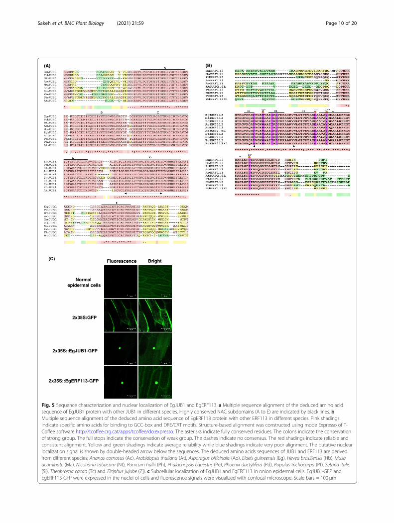
Fig. 5 Sequence characterization and nuclear localization of EgJUB1 and EgERF113. a Multiple sequence alignment of the deduced amino acidsequence of EgJUB1 protein with other JUB1 in different species. Highly conserved NAC subdomains (A to E) are indicated by black lines. bMultiple sequence alignment of the deduced amino acid sequence of EgERF113 protein with other ERF113 in different species. Pink shadingsindicate specific amino acids for binding to GCC-box and DRE/CRT motifs. Structure-based alignment was constructed using mode Expresso of T-Coffee software http://tcoffee.crg.cat/apps/tcoffee/do:expresso. The asterisks indicate fully conserved residues. The colons indicate the conservationof strong group. The full stops indicate the conservation of weak group. The dashes indicate no consensus. The red shadings indicate reliable andconsistent alignment. Yellow and green shadings indicate average reliability while blue shadings indicate very poor alignment. The putative nuclearlocalization signal is shown by double-headed arrow below the sequences. The deduced amino acids sequences of JUB1 and ERF113 are derivedfrom different species; Ananas comosus (Ac), Arabidopsis thaliana (At), Asparagus officinalis (Ao), Elaeis guineensis (Eg), Hevea brasiliensis (Hb), Musaacuminate (Ma), Nicotiana tabacum (Nt), Panicum hallii (Ph), Phalaenopsis equestris (Pe), Phoenix dactylifera (Pd), Populus trichocarpa (Pt), Setaria italic(Si), Theobroma cacao (Tc) and Ziziphus jujube (Zj). c Subcellular localization of EgJUB1 and EgERF113 in onion epidermal cells. EgJUB1-GFP andEgERF113-GFP were expressed in the nuclei of cells and fluorescence signals were visualized with confocal microscope. Scale bars = 100 μm
Sakeh et al. BMC Plant Biology (2021) 21:59 Page 10 of 20
current study, genes regulating antioxidant enzymesEgSOD, EgGSTU17, EgGST3 and EgGSTF12 were highlyupregulated during early interaction with G. boninense.We also observed the expression of EgNPR1 andEgTGA1 which have been recognized to be overex-pressed exclusively during biotrophic attack under SA-mediated signaling pathway [64, 65]. The co-actions ofEgNPR1 and EgTGA1 result in upregulation of EgPR1which has been proven as a biotrophic marker in ourprevious report [5].
EgERF113 likely mediates defense response duringnecrotrophic phase through GCC-box and DRE/CRT motifsMYC2 relies exclusively on co-actions of JA-ABA branchresponse to regulate resistance against insects andwounding by repressing the JA-ET branch [48]. Al-though EgMYC2 was preliminarily screened as a necro-trophic biomarker [5], the TF is categorized as adownregulated DEG in our RNA-seq data. This can beexplained by elevated expression of ethylene insensitive3 (EgEIN3) which then may activate EgERF113 throughET regulation. Binding of ET to its receptors inactivatesCONSTITUTIVE TRIPLE RESPONSE 1 (CTR1) whichin turn release repression on EIN2 activity and stabilizesEIN3 and EIN3-LIKE1 (EIL1) within the nucleus [66].As a result, ERFs are activated and the ERF TFs modu-late transcriptional activity of developmental as well asstress-induced responsive genes [67]. Master regulatorsof ethylene signaling pathways, EIN3 and EIN3-like (EIL)have been proven to modulate a multitude of cascadesof downstream transcriptional responses [47, 68]. De-repression of EIN3/EIL from JA-ZIM domain (JAZ) acti-vates JA-ET signaling that positively regulates transcrip-tional activations of development as well as defenseresponse against necrotrophic pathogens [47, 69]. Thefindings demonstrated that oil palm establishes resist-ance against early necrotrophic through co-actions ofJA-ET, rather than JA-ABA.Here, we propose the regulation of defense response
against necrotrophic phase of G. boninense throughmulti-cascades activation of JA-ET branch leading tooverexpression of EgERF113. ERF113, also recognized asRELATED TO APETALA2.6-LIKE (RAP2.6 L) which isclosely related to ERF108 (RAP2.6) was found to be re-sponsive to abiotic stresses (salinity, heat and drought)as well as stress hormones, particularly JA and/or ET[70]. ERF108 has been reported in JA-induced defenseresponse against wounding and pathogens [71, 72]. Simi-larly, ERF113 was shown to be induced upon wounding[73, 74], as well as pathogens infection [75, 76].We are the first reporting on EgERF113 with binding
preferences for GCC-box and DRE/CRT motifs. AP2/ERFs are known to have multiple conserved DNA bind-ing preferences [77]. However, DREBs are typically
known to recognize DRE/CRT conferring resistanceagainst abiotic stresses whilst ERFs bind to GCC-boxpromoting defense against biotic stresses. ERF113 wasreported to bind to the GCC-box [76], but not tested onDRE/CRT motif. To date, only a few ERFs were reportedwith binding preferences on both GCC-box and DRE/CRT conferring resistance against pathogens attack [78–81]. Binding to GCC-box and DRE/CRT motifs that arepresent in plant defensin (e.g PDF1.2) as well as PRgenes, activates defense-related genes [82, 83]. Kauret al. [83], also reported DRE/CRT elements in the pro-moter region of calcium-responsive genes. Calcium ion(Ca2+) signaling plays paramount importance in thedefense mechanisms of plants in perceiving invadingpathogens [84].Phukan et al. [39] have studied the divergent of AP2/
ERF TF DNA-binding specificities based on sequencecharacterization. Their findings suggested glutamic acid(E) at the 20th and alanine (A) at the 48th positions asidentified in EgERF113 in the 60 amino acids long DBDdenote binding specificity to DRE/CRT motif. EgERF113also showed conservation of amino acids for GCC-boxbinding at the 10th, 18th, 20th, 37th and 59th positions.Although the specific amino acid in DBD that regulatesbinding of TFs to both GCC-box and DRE/CRT motifshas not yet been confirmed, replacing phenylalanine (F)at the 14th position to tryptophan (W) in EgERF113changes the binding specificity from GCC-box only tobinding both GCC-box and DRE/CRT motifs, as sug-gested by Phukan et al. [39]. An additional amino acid(basic polar) at the 24th position of DBD which is lack-ing in EgERF113 is essential for specific binding toGCC-box only. Thus, EgERF113 with 60 amino acidsDBD opposes the classification of specific GCC-boxbinding DBD of 61 amino acids in length.
Proposed defense-related pathways regulated byEgERF113 co-expressing genesTwo RBOH genes, EgRBOHA and EgRBOHB have beenpreviously reported in our study [5], and reduced EgR-BOHB expression during the early necrotrophic phasemay suggest the plant’s response in delaying the progres-sion of the disease. This is in line with a report on theresponse against hemibiotroph Macrophomina phaseo-lina [85], wherein increased expression of RBOH leadsto increase susceptibility of plants to necrotrophicinfection.Another less studied TF, AtMYB122 was reported to
regulate genes involved in camalexin biosynthesis [86].Increased accumulation of indolic glucosinolates duringinduction of glucose signaling was regulated by theAtMYB122 [87]. In another similar reports, the glucosin-olates and their derivatives contribute in defense resist-ance against necrotrophic fungi [88, 89]. Based on the
Sakeh et al. BMC Plant Biology (2021) 21:59 Page 11 of 20
expression patterns of EgMYB122, the TF channeled itsregulation from responding to abiotic stress at 3 d.p.i.into defensing against Ganoderma attack and it is ex-pected that the gene regulation will be more extensive ata later stage of the necrotrophic phase.Rapid and transient increase of cytosolic Ca2+ par-
ticularly during pathogens interaction results in acti-vation of both PTI and ETI signaling [84]. CMLs andCDPKs are known as Ca2+ sensor proteins which areresponsible for perceiving the transduction duringplant innate immunity [90, 91]. Based on our RNA-seq data analysis, both EgCML7 and EgCDPK28 wereupregulated during the necrotrophic phase. Consistentwith the analysis, EgERF113 is suggested to orches-trate defense mechanisms through the regulation ofPR and calcium-responsive genes.
Other transcription factors differentially regulated underthe biotrophic or necrotrophic phaseThe Calmodulin-binding transcription activator(CAMTA) gene was first discovered in Nicotiana taba-cum, regulating senescence and cell death [92], and wasrecently comprehensively studied by Kakar et al. [93].We discovered members of the novel CAMTA TF fam-ily, namely EgCAMTA3 and EgCAMTA4 which weredownregulated at 3 d.p.i. The suppression of both geneswas later reduced across time, which might be the resultof infection phase transition from biotrophic to earlynecrotrophic phase. CAMTA was proven to regulate re-sponses under Ca2+ signaling during both abiotic and bi-otic stresses [94, 95].In general, most downregulated DEGs of TFs showed
de-repression across time. It is reasonable to postulatethat changes of expression patterns might be the resultsof plant immunity interplay against biotrophic andnecrotrophic infection phases. For instance, downregula-tion of EgMYB108 was highest at 7 d.p.i before reducingat 11 d.p.i. Coherently, MYB108 was proven to positivelyregulate the defense mechanism against hemibiotrophVerticillium dahliae in the presence of calmodulin andCa2, antagonistic to the regulation of CAMTA3 [96, 97].In contrast, EgERF9 was found to be downregulatedexclusively during the necrotrophic phase at 11 d.p.i.The result provides agreement with other studieswhich reported repression activity of AtERF9 in en-hanced resistance against necrotrophic Botrytiscinerea [82]. Likewise, expression patterns of TFs inupregulated DEGs were higher at early interactionagainst G. boninense before decreasing over time. Theplant-specific EgTCP15 demonstrated upregulationduring the biotrophic phase before declining butshowing an opposite expression pattern under abioticstress.
ConclusionsTogether with the results presented above, we were ableto recognize TF genes that were regulated duringswitching of the fungal mode of infection. With this firstanalysis of oil palm RNA-seq data encoding TFs, sixcommon major families of TFs were identified to be re-sponsible for promoting oil palm defense responseagainst G. boninense attack which include MYB, bHLH,AP2/ERF, NAC, bZIP and WRKY. Reported genes in-volved in cell wall modification, ROS-mediated signaling,PCD and plant innate immunity were all differentiallyexpressed; indicating active regulation of oil palmdefense response against the hemibiotrophic G. boni-nense. The biotrophic and necrotrophic infection phasesof G. boninense were further supported through gene ex-pression of biotrophy-specific, EgJUB1 and necrotrophy-specific, EgERF113. Our finding is the first reportingEgJUB1 as a potential master regulator based on its posi-tive interaction with the imperfect palindromic SNBEconsensus sequence which may promote branches ofbiotrophy-associated defense mechanisms including cellwall strengthening and HR-mediated defense responses.Besides, EgERF113 is the first AP2/ERF TF reported tomodulate multifaceted defense mechanisms throughbinding to GCC-box and DRE/CRT motifs during thenecrotrophic phase. Binding to these motifs may resultin transcriptional upregulation of PR and calcium-responsive genes. Based on our findings, a proposeddefense mechanism inferring oil palm against hemibio-troph G. boninense during biotrophic and necrotrophicinfection phases is illustrated as in Fig. 6. The informa-tion presents a promising first step in recognizing thedownstream target defense-related genes regulated bythe infection phase-specific TFs. Although our presentstudy proposes important insights into the defense rolesof EgJUB1 and EgERF113, over-expression studies ormutant complementation studies utilizing plant modelssuch as Arabidopsis thaliana or Nicotiana spp. shouldbe carried out in future investigations to further delin-eate defense mechanisms triggered by these TFs.
MethodsPlant materials and fungal treatmentFour-months old germinated oil palm seedlings Com-mercial DxP GH500 series (Elaeis guineensis Jacq. Durax Pisifera), were purchased from Sime Darby Seeds andAgriculture Services Sdn. Bhd., Banting, Selangor,Malaysia. Pathogenic Ganoderma boninense strainPER71 was isolated and purified from an infected oilpalm in United Plantation Teluk Intan, Perak, Malaysia[5], obtained from GanoDROP Unit, Biology Division,Malaysian Palm Oil Board (MPOB). Artificial infectionof oil palm seedlings with G. boninense using rubberwood blocks (RWBs) was carried out following a
Sakeh et al. BMC Plant Biology (2021) 21:59 Page 12 of 20
previous study [5]. Control (C) was set as seedlingswithout treatment. Two different treatments were car-ried out; mock treatment (MT) consisted of oil palmseedlings with bare RWBs while Ganoderma treat-ment (GT) involved oil palm seedling treated withGanoderma-inoculated RWBs. Destructive samplingwas performed on two pooled biological replicates ofoil palm seedlings at different days post-inoculation(3, 7 and 11 d.p.i). Each biological replicate consistedof pooled RNA provided equally from six constituentseedlings. Instead of mathematical averaging of indi-vidual samples, biological averaging is more cost-efficient and commonly practiced in the attempt ofreducing high biological variability among samples inRNA-seq studies [98]. Pooling bias can be reduced byusing three to eight biological individual samples perpool with two pools per treatment group [99].
RNA extraction and DEGs analysis of TFsTotal RNA of all samples was extracted following themethod reported in Bahari et al. [5]. The extracted RNAwas used in all subsequent experiments. A high-throughput NGS data analysis was performed as de-scribed in our previous report, Bahari et al. [5]. ThemRNA fragments were mapped to Elaeis guineensis cod-ing sequences as reference genome (retrieved fromhttps://www.ebi.ac.uk/genomes/) through Geneious soft-ware version 9.1.5 (Biomatters Ltd.). From align/assem-ble tools, Geneious for RNA-Seq was used as mapperwith medium-low sensitivity using clean reads beforemapping. Upon completion of the mapping step, thetranscript abundance of each sample was calculated astranscript per kilobase million (TPM). Sequences thatwere reproducible in both pooled biological replicateswere chosen to eliminate biased profiling of transcripts
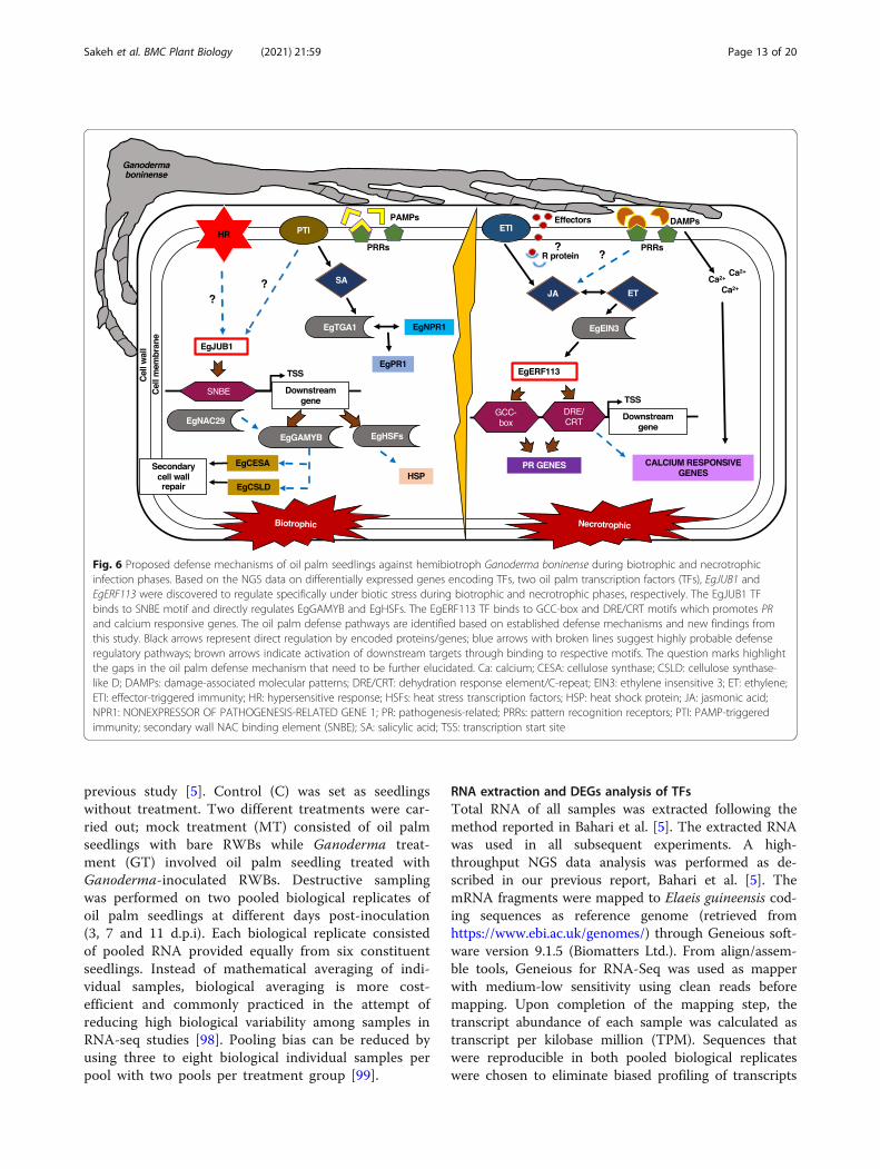
Fig. 6 Proposed defense mechanisms of oil palm seedlings against hemibiotroph Ganoderma boninense during biotrophic and necrotrophicinfection phases. Based on the NGS data on differentially expressed genes encoding TFs, two oil palm transcription factors (TFs), EgJUB1 andEgERF113 were discovered to regulate specifically under biotic stress during biotrophic and necrotrophic phases, respectively. The EgJUB1 TFbinds to SNBE motif and directly regulates EgGAMYB and EgHSFs. The EgERF113 TF binds to GCC-box and DRE/CRT motifs which promotes PRand calcium responsive genes. The oil palm defense pathways are identified based on established defense mechanisms and new findings fromthis study. Black arrows represent direct regulation by encoded proteins/genes; blue arrows with broken lines suggest highly probable defenseregulatory pathways; brown arrows indicate activation of downstream targets through binding to respective motifs. The question marks highlightthe gaps in the oil palm defense mechanism that need to be further elucidated. Ca: calcium; CESA: cellulose synthase; CSLD: cellulose synthase-like D; DAMPs: damage-associated molecular patterns; DRE/CRT: dehydration response element/C-repeat; EIN3: ethylene insensitive 3; ET: ethylene;ETI: effector-triggered immunity; HR: hypersensitive response; HSFs: heat stress transcription factors; HSP: heat shock protein; JA: jasmonic acid;NPR1: NONEXPRESSOR OF PATHOGENESIS-RELATED GENE 1; PR: pathogenesis-related; PRRs: pattern recognition receptors; PTI: PAMP-triggeredimmunity; secondary wall NAC binding element (SNBE); SA: salicylic acid; TSS: transcription start site
Sakeh et al. BMC Plant Biology (2021) 21:59 Page 13 of 20
due to manipulations stages during library construction[100]. Alteration of gene expression profile was analyzedby comparing genes expressed from control with GTsamples. DEGs were further evaluated following strin-gent cut-off values of log2 FC ≥ |1.0| (corresponding to2-fold or more upregulation/downregulation) and P-value < 0.01 [5, 101]. Comparative analysis was con-ducted between two biological replicates of control andGT samples at all time points. DEGs of TFs that met thecut-off values were clustered according to upregulatedand downregulated genes. Transcripts that were identi-fied in both pooled biological replicates were further an-alyzed for identification of DEGs. DEGs of a few stress-related genes involving cell wall modification, ROS pro-duction and PCD were mined from NGS data analysis.
Validation by quantitative real-time PCR (qPCR)qPCR was performed using qPCR Green Master MixLRox (2X) (Biotechrabbit GmbH, Germany). Stability offive endogenous controls; EgGAPDH2, EgNADH5,EgMSD, EgUBQ, and Egß-actin were tested acrosstreated and control samples. The qPCR analysis was per-formed using Bio-Rad CFX Manager™ Software version3.1. Expression levels of all target genes were normalizedwith the expression level of the three most stable refer-ence genes which were EgGAPDH2, EgNADH5 and Egß-actin. Real-time PCR was carried out on control (C),mock-treated (MT) and Ganoderma-treated (GT) oilpalm root samples at 3, 7 and 11 d.p.i. The data includedthree biological replicates of root samples of oil palmseedlings. Comparative analysis of expression levels wasexpressed as fold change ± standard error of mean(SEM) of three individual technical replicates at P-value< 0.01. Significant differences of expression levels be-tween test groups to control were determined usingone-way analysis of variance (ANOVA) followed byTukey’s test for comparison between treatments. Theprimers used for the qPCR are listed in Table 1.
Sequence characterization of EgJUB1 and EgERF113Sequence analysis was carried out using Basic LocalAlignment Search Tool (BLAST 2.9) accessed fromhttp://www.ncbi.nlm.nih.gov. BLAST. A homology searchwas carried out using BLASTX algorithm by comparingthe translated protein sequence of interest (EgJUB1 andEgERF113) with other protein sequences available in theNational Centre of Biotechnology Information (NCBI)database. The nucleotide sequences were translated intoprotein sequences through publicly-accessible website,ExPASy Molecular Biology Server (http://web.expasy.org/translate/). Multiple sequence alignment of protein se-quences from different species were carried out using T-Coffee software version 11.0 (http://tcoffee.crg.cat/apps/tcoffee/do:expresso). NLS was predicted using an access-ible website of NLS Mapper (http://nls-mapper.iab.keio.ac.jp/cgi-bin/NLS_Mapper_form.cgi).
Yeast one-hybrid (Y1H) assayCoding regions of EgJUB1 and EgERF113 (500 ng totalRNA) flanking SMART sequences were amplified fromGT samples at 3 and 11 d.p.i, respectively, using Q5®Hot Start High-Fidelity 2x Master Mix (New EnglandBiolabs). Purified SMART-EgJUB1 and -EgERF113 werefurther amplified by long-distance PCR using Advantage®2 PCR Mix (Takara Bio). Putative tandem repeats of tar-get baits fragments (NACBS, SNBE1, SNBE2, GCC-boxand DRE/CRT) as well as the respective mutated frag-ments were cloned into pAbAi vector and integratedinto the yeast genome. Minimal inhibitory concentration(MIC) of Aureobasidin A (AbA) for each bait- andmutant-reporter yeast strain was determined.Yeast One-Hybrid screening was conducted using
Yeastmaker™ Yeast Transformation System 2 (TakaraBio). Components for yeast co-transformation reactionwere added in given orders as follows; 2 μg of SMART-EgJUB1 or -EgERF113, 1 μg of pGAD7-Rec AD (SmaI-linearized), 50 μg of denatured yeastmaker carrier DNA,50 μL of competent yeast cells Y1HGold [pAbAi-baits]
Table 1 List of primers for quantitative Real-Time PCR (qPCR)
Target Gene Sense sequence(5′-3′)
Anti-sense sequence(5′-3′)
EgJUB1 AATGGAACTCAGTACCTCAGGC ATTATCCTTCCAAGCTCATCCC
EgERF113 AGCAGCACTAAAGTTCAAAGG GAATAAGGTCTGGGTAGGAGG
EgTCP15 GACAAACCCTAACAGCCAAAGTA AAATGTAGCCCACTAGACATGGA
EgEIN3 GGAAGGAGAAGGTGAAGTTTGAT CCATAAGGCTATGCTGAATTTTG
EgMYB122 AGTACCAGACAAGCTTGAAGGC TTACATCCTTAGCTAAACGGGG
Egß-actin GAGAGAGCGTGCTACTCATCTT CGGAAGTGCTTCTGAGATCC
EgNADH5 GCTCCCCTTTATTTGAATACCC AATAGTTAGAGATGCCGCAAGC
EgGAPDH2 GAAGGTCATCATATCTGCTCCC CATCAACAGTCTTCTGAGTGGC
Sakeh et al. BMC Plant Biology (2021) 21:59 Page 14 of 20
or Y1HGold [pAbAi-mutants] and 500 μL of PEG/LiAcsolution. Transformations were plated on selective platesSD/−Leu and SD/−Leu/AbA. Transformation control,p53 was plated on SD/−Leu/AbA200. Plates were incu-bated at 30 °C for 3 days. Confirmation of positive cloneswas performed by colony PCR using Matchmaker InsertCheck PCR Mix 2 (Takara Bio) and sent for sequencingusing T7 promoter primer. A single colony of positiveclone was cultured in synthetic dropout (SD) mediumlacking leucine (SD/−Leu) broth to an optical density(OD600) of 0.1 and diluted in 10-fold dilution series.From each dilution, 10 μL of yeast culture was spottedon SD/−Leu and SD/−Leu/AbA plates. Oligonucleotidesand primers used in the Y1H assay are listed in Table 2.
Isolation of EgJUB1 and EgERF113 nuclear proteinA single colony of positive clones from the Y1H assaywas incubated in 5 mL SD/−Leu broth with overnightshaking at 30 °C. Yeast culture was transferred into 45mL SD/−Leu broth and further grown with overnightshaking (18–20 h) at 30 °C until OD600 reached 0.8–1.0.Yeast cells were pelleted by centrifugation at 1000 g for 5mins at 4 °C. Pellet was washed with 30mL ice-cold ster-ile ultrapure water and centrifuged at 1000 g for 5 minsat 4 °C. Pellet was immediately flash-frozen in liquid ni-trogen and ground into a fine powder. Extraction of nu-clear protein was carried out using NE-PER™ Nuclearand Cytoplasmic Extraction Reagents (Thermo
Scientific). EgJUB1 and EgERF113 nuclear extracts werestored at − 80 °C.
Electrophoretic mobility shift assay (EMSA)Double-stranded of sense and anti-sense oligonucleo-tides were biotin-labelled using Biotin 3′ End DNA La-beling kit (Thermo Scientific). Oligonucleotides used arelisted in Table 2. The binding reaction system of EMSAwas prepared using LightShift EMSA Optimization andControl kit (Thermo Scientific). The binding mixturewas resolved in 6% non-denaturing polyacrylamide gelin 0.5X Tris-borate-EDTA (TBE) buffer and transferredto a positively charged nylon membrane. The membranewas cross-linked at 120 mJ/cm2 for 1 min. The protein-DNA complexes were visualized by LightShift® Chemilu-minescent EMSA kit (Thermo Scientific) according tothe manufacturer’s protocol.
Subcellular localizationThe open reading frames of EgJUB1 and EgERF113 lack-ing stop codon were amplified using KAPA HiFi HotStartReadyMix PCR kit (Thermo Scientific). The gene-specificprimers with CACC flanking at 5’end of forward primerare listed in Table 3. The PCR products were transferredinto the gateway pENTR/D-TOPO entry vector using thepENTR™ Directional Topo Cloning Kit (Thermo FisherScientific). TOPO Cloning Reaction was transformedinto One Shot® TOP10 Chemically Competent cells(Thermo Fisher Scientific) to generate entry clone and
Table 2 List of oligonucleotides for Yeast One-Hybrid (Y1H) assay and electrophoretic mobility shift assay (EMSA)
Target Motifs DNA element sequences (5′ - 3′)
NACBS GATGCCGTGATGCCGTGATGCCGTGATGCCGT
mNACBS GATGACGTGATGACGTGATGACGTGATGACGT
SNBE1 TTGTGTGCTGTGGAAGTTTTTGTGTGCTGTGGAAGTTT
mSNBE1 TTGTGGGCTGTGGAAGTTTTTGTGGGCTGTGGAAGTTT
SNBE2 TGGCTTGTGCAAAAAGTAATGGCTTGTGCAAAAAGTAA
mSNBE2 TGGCTCGTGCAAAAAGTAATGGCTCGTGCAAAAAGTAA
GCC-box TAAGAGCCGCCTAAGAGCCGCCTAAGAGCCGCCTAAGAGCCGCC
mGCC-box TAAGATCCTCCTAAGATCCTCCTAAGATCCTCCTAAGATCCTCC
DRE/CRT TGCCGACATTGCCGACATTGCCGACATTGCCGACAT
mDRE/CRT TATTTACATTATTTACATTATTTACATTATTTACAT
The tandem repeats of DNA elements of each target are bold and underlined. Point mutations are bold and underlined with red
Table 3 List of primers for vector construction of subcellular localization
Target Gene Sense sequence(5′-3′)
Anti-sense sequence(5′-3′)
EgJUB1 CACCATGGAGGAGAAGATGGACAA AGCGTATCTACATTCATGACCGG
EgERF113 CACCATGGAGACCGAGATTAGAATCC CTCTCTTGGTTGGCTAGTTTCTG
Sakeh et al. BMC Plant Biology (2021) 21:59 Page 15 of 20
sequence verified. The expression clones for EgJUB1 andEgERF113 were generated using Gateway LR Clonase IIEnzyme Mix (Thermo Fisher Scientific) by recombin-ation of entry clones into Gateway-compatible destin-ation vector consisting double CaMV 35S promoter (2 x35S) of pMDC85, respectively. The construction ofpMDC85 vector without ccdB gene and insert was usedas a negative control. The LR reaction was transformedinto One Shot™ OmniMAX™ 2 T1® Chemically Compe-tent cells (Thermo Fisher Scientific).The resulting plasmids of 2x35S:GFP (negative con-
trol), 2x35S::EgJUB1-GFP and 2x35S::EgERF113-GFPwere sequence-verified and transformed into compe-tent Agrobacterium tumefaciens strain LBA4404 [102].Agrobacterium-mediated transformation of onion epi-dermal cells was carried out according to Azzemeet al. [103]. Fresh onion scales (1.5 × 1 cm) wereimmersed into 20 mL Agrobacterium suspension har-bouring control and GFP constructs, respectively for16 h at 28 °C. The onion scales were transferred to aMurashige and Skoog (MS) medium pH 5.8 and fur-ther co-cultivated with Agrobacterium for 2 days. Thepeeled onion epidermal cells were rinsed with sterilewater and transferred to glass slides. Fluorescence im-ages were captured using a 20X lens of confocal laserscanning microscope (LSM 5 PASCAL EXCITER,Zeiss, Germany) with excitation at 488 nm and ana-lyzed by LSM 5 Image Browser software (Ver. 4.1).
Statistical analysisFor RNA-seq data analysis, DEGs were determined fol-lowing cut off-values of log2 FC ≥ |1.0| and P-value <0.01. Expression levels of each gene from qPCR analysiswere normalized by three reference genes; EgGAPDH2,EgNADH5 and Egß-actin. Data was presented as mean ±standard error of mean (SEM) of three independenttechnical replicates. Differences of expression level be-tween samples at different time points to control and be-tween group of treatments (MT and GT) weredetermined by using one-way ANOVA analysis followedby Tukey’s test. Significantly different expression levelsas compared to the control were measured according to**P < 0.01, ***P < 0.001 and ****P < 0.0001. ns is definedas not significant. All graphs were generated and ana-lyzed using GraphPad Prism version 5.0 (GraphPadSoft-ware Inc., USA).
Supplementary InformationThe online version contains supplementary material available at https://doi.org/10.1186/s12870-020-02812-7.
Additional file 1:. Electrophoretic mobility shift assay (EMSA) of EgJUB1with SNBE1 probe. Lane 1 to 3 consist of EBNA control system. Lane 4 to8 consist of EgJUB1 test system. Lane 1 and 8 are the blank for EBNA and
EgJUB1 systems, respectively. Lane 2 is the positive control for EMSA.EMSA shows direct binding of EgJUB1 to SNBE1 probe in lane 6. EgJUB1is unable to bind to untransformed yeast and biotinylated mutant SNBE1(mSNBE1) probe in lane 4 and 5, respectively. Successful binding of 200-fold molar excess of unlabelled SNBE1 (competitor) probe is shown inlane 7.
Additional file 2:. Electrophoretic mobility shift assay (EMSA) ofEgERF113 with GCC-box probe. Lane 1 to 3 consist of EBNA control sys-tem. Lanes 4 to 8 consist of EgERF113 test system. Lane 1 and 8 are theblank for EBNA and EgERF113 systems, respectively. Lane 2 is the positivecontrol for EMSA. EMSA shows direct binding of EgERF113 to GCC-boxprobe in lane 6. EgERF113 is unable to bind to untransformed yeast andbiotinylated mutant GCC-box (mGCC-box) probe in lane 4 and 5, respect-ively. Successful binding of 200-fold molar excess of unlabelled GCC-box(competitor) probe is shown in lane 7.
Additional file 3:. Electrophoretic mobility shift assay (EMSA) ofEgERF113 with DRE/CRT probe. Lane 1 to 3 consist of EBNA controlsystem. Lane 4 to 8 consist of EgERF113 test system. Lane 1 and 8 arethe blank for EBNA and EgERF113 systems, respectively. Lane 2 is thepositive control for EMSA. EMSA shows direct binding of EgERF113 toDRE/CRT probe in lane 6. EgERF113 is unable to bind to untransformedyeast and biotinylated mutant DRE/CRT (mDRE/CRT) probe in lane 4 and5, respectively. Successful binding of 200-fold molar excess of unlabelledDRE/CRT (competitor) probe is shown in lane 7.
AbbreviationsA: Alanine; AbA: Aureobasidin A; ABA: Abscisic acid; ANOVA: Analysis ofvariance; AP2/ERF: APETALA2/ethylene responsive factor; Avr: Virulenceeffectors; bHLH: basic helix-loop-helix; BLAST: Basic Local Alignment SearchTool; bZIP: basic leucine zipper; Ca2+: Calcium ion; CAMTA: Calmodulin-binding transcription activator; BSR: Basal stem rot; CWDE: Cell walldegrading enzyme; DAMP: Damage-associated molecular pattern; DBD: DNA-binding domain; DEG: Differentially expressed gene; d.p.i: Days postinoculation; DREB: Dehydration responsive element-binding; DRE/CRT: Dehydration-Responsive Element/C-Repeat;EDTA: Ethylenediaminetetraacetic acid; EIN3: Ethylene-insensitive 3; EIL: EIN3-like; EMSA: Electrophoretic Mobility Shift Assay; ERE: Ethylene-ResponseElement; ERF: Ethylene Responsive Factor; ET: Ethylene; ETI: Effector-triggeredimmunity; F: Phenylalanine; G: Glutamic acid; GAPDH: Glyceraldehyde 3-phosphate dehydrogenase; GT: Ganoderma-treated; HR: Hypersensitiveresponse; HSF: Heat stress transcription factor; HSP: Heat shock protein;Ile: Isoleucine; JA: Jasmonic acid; JAZ: Jasmonic acid-ZIM domain;JUB1: JUNGBRUNNEN 1; Leu: Leucine; LiAc: Lithium acetate; MIC: MinimalInhibitory Concentration; MPOB: Malaysian Palm Oil Board; MT: Mock-treated;MYB: Myeloblastosis; MYC: Myelocytomatosis; NADH: Nicotinamide adeninedinucleotide dehydrogenase; NACBS: NAC binding site; NCBI: National Centreof Biotechnology Information; NGS: Next-Generation Sequencing;NLS: Nuclear localization signals; NPR1: NONEXPRESSOR OF PATHOGENESIS-RELATED GENE 1; ns: Not significant; OD: Optical density; PAMP: Pathogen-associated molecular pattern; PlantTFDB: Plant Transcription Factor Database;PCD: Programmed cell death; PR: Pathogenesis-related; PRR: Patternrecognition receptor; PTI: PAMP-triggered immunity; qPCR: Quantitative Real-Time Polymerase Chain Reaction; RAP2.6: RELATED TO APETALA 2.6; RAP2.6L: RELATED TO APETALA 2.6-LIKE; RBOH: Respiratory burst oxidase homologprotein; ROS: Reactive Oxygen Species; RT: Room temperature; RWB: Rubberwood block; SA: Salicylic acid; SD: Synthetic dropout; SEM: Standard error ofmean; SNBE: Secondary wall NAC binding element; SOD: Superoxidedismutase; TBE: Tris-borate-EDTA; TF: Transcription factor; TPM: Transcript perkilobase million; TSS: Transcription start site; W: Tryptophan; Y1H: Yeast One-Hybrid
AcknowledgementsGanoderma boninense strain PER71 culture was obtained from the MalaysianPalm Oil Board (MPOB). Malaysian Nuclear Agency has provided Cobalt-60gamma radiation service for the research.
Authors’ contributionsSNA conceived and designed the research plan. NMS performed most of theexperiments and prepared the manuscript. MNB contributed to RNA-seqdata analysis. AMA and NAS provided technical assistance in analyzing assays,
Sakeh et al. BMC Plant Biology (2021) 21:59 Page 16 of 20
Y1H and EMSA. IAS provided fungal material and assisted in artificial inocula-tion. SNA complemented in the writing of the manuscript. All authors haveread and approved the final version of the submitted manuscript.
FundingIn this work, the design of the study and collection, analysis andinterpretation of data in writing the manuscript was fully supported by agrant from the Ministry of Education Malaysia, under NanoMalaysia Institutefor Innovative Technology (NanoMITE) Consortium projects 2015–2020(5526302).
Availability of data and materialsThe sequenced mRNA data was deposited at European Nucleotide Archivewith accession number PRJEB27915. https://www.ebi.ac.uk/ena/data/view/PRJEB27915
Ethics approval and consent to participateNot applicable.
Consent for publicationNot applicable.
Competing interestsAll authors declare no competing interests.
Author details1Institute of Plantation Studies, Universiti Putra Malaysia (UPM), 43400Serdang, Selangor, Malaysia. 2Department of Agriculture Technology, Facultyof Agriculture, Universiti Putra Malaysia (UPM), 43400 Serdang, Selangor,Malaysia. 3Department of Biochemistry, Faculty of Biotechnology andBiomolecular Sciences, Universiti Putra Malaysia (UPM), 43400 Serdang,Selangor, Malaysia. 4Ganoderma and Diseases Research for Oil Palm Unit,Malaysian Palm Oil Board, No. 6, Persiaran Institusi, Bandar Baru Bangi, 43000Kajang, Selangor, Malaysia.
Received: 2 June 2020 Accepted: 22 December 2020
References1. Rees RW, Flood J, Hasan Y, Potter U, Cooper RM. Basal stem rot of oil palm
(Elaeis guineensis); mode of root infection and lower stem invasion byGanoderma boninense. Plant Pathol. 2009;58(5):982–9. https://doi.org/10.1111/j.1365-3059.2009.02100.x.
2. Mohammed CL, Rimbawanto A, Page DE. Management of basidiomyceteroot-and stem-rot diseases in oil palm, rubber and tropical hardwoodplantation crops. Forest Pathol. 2014;44(6):428–46. https://doi.org/10.1111/efp.12140.
3. Chong KP, Dayou J, Alexander A. Pathogenic nature of Ganodermaboninense and basal stem rot disease. In: Detection and control ofGanoderma boninense in oil palm crop. Cham: Springer; 2017. p. 5–12.
4. Ho CL, Tan YC. Molecular defense response of oil palm to Ganodermainfection. Phytochemistry. 2014;114:168–77. https://doi.org/10.1016/j.phytochem.2014.10.016.
5. Bahari MNA, Sakeh NM, Abdullah SNA, Ramli RR, Kadkhodaei S.Transciptome profiling at early infection of Elaeis guineensis by Ganodermaboninense provides novel insights on fungal transition from biotrophic tonecrotrophic phase. BMC Plant Biol. 2018;18(1):377. https://doi.org/10.1186/s12870-018-1594-9.
6. Dodds PN, Rafiqi M, Gan PH, Hardham AR, Jones DA, Ellis JG. Effectors ofbiotrophic fungi and oomycetes: pathogenicity factors and triggers of hostresistance. New Phytol. 2009;183(4):993–1000.
7. Catanzariti AM, Dodds PN, Ellis JG. Avirulence proteins from haustoria-forming pathogens. FEMS Microbiol Lett. 2007;269(2):181–8.
8. Zhao Z, Liu H, Wang C, Xu JR. Erratum to: comparative analysis of fungalgenomes reveals different plant cell wall degrading capacity in fungi. BMCGenomics. 2014;4(1):274. https://doi.org/10.1186/1471-2164-15-6.
9. Nusaibah SA, Abdullah SNA, Idris AS, Sariah M, Pauzi ZM. Involvement ofmetabolites in early defense mechanism of oil palm (Elaeis guineensis Jacq.)against Ganoderma disease. Plant Physiol Biochem. 2016;109:156–65. https://doi.org/10.1016/j.plaphy.2016.09.014.
10. Boller T, Felix G. A renaissance of elicitors: perception of microbe-associatedmolecular patterns and danger signals by pattern-recognition receptors.Annu Rev Plant Biol. 2009;60:379–406. https://doi.org/10.1146/annurev.arplant.57.032905.105346.
11. Dodds PN, Rathjen JP. Plant immunity: towards an integrated view of plant–pathogen interactions. Nat Rev Genet. 2010;11(8):539. https://doi.org/10.1038/nrg2812.
12. Cook DE, Mesarich CH, Thomma BP. Understanding plant immunity as asurveillance system to detect invasion. Annu Rev Phytopathol. 2015;53:541–63. https://doi.org/10.1146/annurev-phyto-080614-120114.
13. Pel MJ, Pieterse CM. Microbial recognition and evasion of host immunity. JExp Bot. 2012;64(5):1237–48. https://doi.org/10.1093/jxb/ers262.
14. Zipfel C. Plant pattern-recognition receptors. Trends Immunol. 2014;35(7):345–51. https://doi.org/10.1016/j.it.2014.05.004.
15. Miller RNG, Costa Alves GS, Van Sluys MA. Plant immunity: unravelling thecomplexity of plant responses to biotic stresses. Ann Bot. 2017;119(5):681–7.https://doi.org/10.1093/aob/mcw284.
16. Yi M, Valent B. Communication between filamentous pathogens and plantsat the biotrophic interface. Annu Rev Phytopathol. 2013;51:587–611. https://doi.org/10.1146/annurev-phyto-081211-172916.
17. Rossi FR, Krapp AR, Bisaro F, Maiale SJ, Pieckenstain FL, Carrillo N. Reactiveoxygen species generated in chloroplasts contribute to tobacco leafinfection by the necrotrophic fungus Botrytis cinerea. Plant J. 2017;92(5):761–73. https://doi.org/10.1111/tpj.13718.
18. Vargas WA, Martín JMS, Rech GE, Rivera LP, Benito EP, Díaz-Mínguez JM,Thon MR, Sukno SA. Plant defense mechanisms are activated duringbiotrophic and necrotrophic development of Colletotricum graminicola inmaize. Plant Physiol. 2012;158(3):1342–58. https://doi.org/10.1104/pp.111.190397.
19. Kabbage M, Yarden O, Dickman MB. Pathogenic attributes of Sclerotiniasclerotiorum: switching from a biotrophic to necrotrophic lifestyle. Plant Sci.2015;233:53–60. https://doi.org/10.1016/j.plantsci.2014.12.018.
20. Abdullah SNA, Akhtar MS. Plant and necrotrophic fungal pathogeninteraction: mechanism and mode of action. In: Plant, soil and microbes.Cham: Springer; 2016. p. 29–53.
21. Azizi P, Rafii MY, Abdullah SNA, Nejat N, Maziah M, Hanafi MM, LatifMA, Sahebi M. Toward understanding of rice innate immunity againstMagnaporthe oryzae. Crit Rev Biotechnol.2016;36:165–74.
22. Häffner E, Konietzki S, Diederichsen E. Keeping control: the role ofsenescence and development in plant pathogenesis and defense. Plants.2015;4(3):449–88. https://doi.org/10.3390/plants4030449.
23. Giri MK, Singh N, Banday ZZ, Singh V, Ram H, Singh D, Chattopadhyay S,Nandi AK. GBF 1 differentially regulates CAT2 and PAD4 transcription topromote pathogen defense in Arabidopsis thaliana. Plant J. 2017;91(5):802–15. https://doi.org/10.1111/tpj.13608.
24. Liu B, Ouyang Z, Zhang Y, Li X, Hong Y, Huang L, Liu S, Zhang H, Li D, SongF. Tomato NAC transcription factor SlSRN1 positively regulates defenseresponse against biotic stress but negatively regulates abiotic stressresponse. PLoS One. 2014;9(7):e102067. https://doi.org/10.1371/journal.pone.0102067.
25. Rasmussen S, Barah P, Suarez-Rodriguez MC, Bressendorff S, Friis P,Costantino P, Bones AM, Nielsen HB, Mundy J. Transcriptome responses tocombinations of stresses in Arabidopsis. Plant Physiol. 2013;161(4):1783–94.https://doi.org/10.1104/pp.112.210773.
26. Ng DW, Abeysinghe JK, Kamali M. Regulating the regulators: the control oftranscription factors in plant defense signaling. Int J Mol Sci. 2018;19(12):3737. https://doi.org/10.3390/ijms19123737.
27. Pandey D, Rajendran SRCK, Gaur M, Sajeesh PK, Kumar A. Plant defensesignaling and responses against necrotrophic fungal pathogens. J PlantGrowth Regul. 2016;35(4):1159–74. https://doi.org/10.1007/s00344-016-9600-7.
28. Foyer CH, Rasool B, Davey JW, Hancock RD. Cross-tolerance to biotic andabiotic stresses in plants: a focus on resistance to aphid infestation. J ExpBot. 2016;67(7):2025–37. https://doi.org/10.1093/jxb/erw079.
29. Guo M, Liu JH, Ma X, Luo DX, Gong ZH, Lu MH. The plant heat stresstranscription factors (HSFs): structure, regulation, and function in responseto abiotic stresses. Front Plant Sci. 2016;7:114. https://doi.org/10.3389/fpls.2016.00114.
30. Fragkostefanakis S, Simm S, El-Shershaby A, Hu Y, Bublak D, Mesihovic A,Darm K, Mishra SK, Tschiersch B, Theres K, Scharf C, Schleiff E, Scharf KD. The
Sakeh et al. BMC Plant Biology (2021) 21:59 Page 17 of 20
repressor and co-activator HsfB1 regulates the major heat stresstranscription factors in tomato. Plant Cell Environ. 2019;42(3):874–90. https://doi.org/10.1111/pce.13434.
31. Bechtold U, Albihlal WS, Lawson T, Fryer MJ, Sparrow PAC, Richard F, PersadR, Bowden L, Hickman R, Martin C, Beynon JL, Buchanan-Wollaston V, BakerNR, Morison JIL, Schöffl F, Ott S, Mullineaux PM. Arabidopsis HEAT SHOCKTRANSCRIPTION FACTORA1b overexpression enhances water productivity,resistance to drought, and infection. J Exp Bot. 2013;64(11):3467–81. https://doi.org/10.1093/jxb/ert185.
32. Ramli Z, Abdullah SNA. Functional characterisation of the oil palm type 3metallothionein-like gene (MT3-B) promoter. Plant Mol Biol Rep. 2010;28(3):531–41. https://doi.org/10.1007/s11105-009-0177-1.
33. Hernandez-Garcia CM, Finer JJ. Identification and validation of promotersand cis-acting regulatory elements. Plant Sci. 2014;217–218:109–19. https://doi.org/10.1016/j.plantsci.2013.12.007.
34. Sanchez I, Hernandez-Guerrero R, Mendez-Monroy PE, Martinez-Nuñez MA,Ibarra JA, Pérez-Rueda E. Evaluation of the abundance of DNA-bindingtranscription factors in prokaryotes. Genes. 2020;11(1):52. https://doi.org/10.3390/genes11010052.
35. Huang D, Wang S, Zhang B, Shang-Guan K, Shi Y, Zhang D, Liu X, Wu K, XuZ, Fu X, Zhou YA. Gibberellin-mediated DELLA-NAC signaling cascaderegulates cellulose synthesis in rice. Plant Cell. 2015;27(6):1681–96. https://doi.org/10.1105/tpc.15.00015.
36. Li S. Transcriptional control of flavonoid biosynthesis: fine-tuning of theMYB-bHLH-WD40 (MBW) complex. Plant Signal Behav. 2014;9(1):e27522.https://doi.org/10.4161/psb.27522.
37. Nemesio-Gorriz M, Blair PB, Dalman K, Hammerbacher A, Arnerup J, StenlidJ, Mukhtar SM, Elfstrand M. Identification of Norway spruce MYB-bHLH-WDRtranscription factor complex members linked to regulation of the flavonoidpathway. Front Plant Sci. 2017;8:305. https://doi.org/10.3389/fpls.2017.00305.
38. Sun X, Malhis N, Zhao B, Xue B, Gsponer J, Rikkerink EH. Computationaldisorder analysis in ethylene response factors uncovers binding motifscritical to their diverse functions. Int J Mol Sci. 2020;21(1):74. https://doi.org/10.3390/ijms21010074.
39. Phukan UJ, Jeena GS, Tripathi V, Shukla RK. Regulation of Apetala2/ethyleneresponse factors in plants. Front Plant Sci. 2017;8:150. https://doi.org/10.3389/fpls.2017.00150.
40. Purohit A, Ganguly S, Chaudhuri RK, Chakraborti D. Understanding theInteraction of Molecular Factors During the Crosstalk Between Drought andBiotic Stresses in Plant. In: Molecular Plant Abiotic Stress: Biology andBiotechnology. Hoboken: Wiley; 2019. p. 427–46.
41. Tee SS, Tan YC, Abdullah F, Ong-Abdullah M, Ho CL. Transcriptome of oilpalm (Elaeis guineensis Jacq.) roots treated with Ganoderma boninense. TreeGenet Genomes. 2013;9(2):377–86. https://doi.org/10.1007/s11295-012-0559-7.
42. Ho CL, Tan YC, Yeoh KA, Ghazali AK, Yee WY, Hoh CC. De novotranscriptome analyses of host-fungal interactions in oil palm (Elaeisguineensis Jacq.). BMC Genomics. 2016;17(1):66. https://doi.org/10.1186/s12864-016-2368-0.
43. Yuan P, Du L, Poovaiah B. Ca2+/Calmodulin-dependent AtSR1/CAMTA3 playscritical roles in balancing plant growth and immunity. Int J Mol Sci. 2018;19(6):1764. https://doi.org/10.3390/ijms19061764.
44. Wang Y, Wei F, Zhou H, Liu N, Niu X, Yan C, Zhang L, Han S, Hou C, WangD. TaCAMTA4, a Calmodulin-interacting protein, involved in defenseresponse of wheat to Puccinia triticina. Sci Rep. 2019;9(1):641. https://doi.org/10.1038/s41598-018-36385-1.
45. Viola IL, Camoirano A, Gonzalez DH. Redox-dependent modulation ofanthocyanin biosynthesis by the TCP transcription factor TCP15 duringexposure to high light intensity conditions in Arabidopsis. Plant Physiol.2016;170(1):74–85. https://doi.org/10.1104/pp.15.01016.
46. Li M, Chen H, Chen J, Chang M, Palmer IA, Gassmann W, Liu F, Fu Z. TCPtranscription factors interact with NPR1 and contribute redundantly tosystemic acquired resistance. Front Plant Sci. 2018;9:1153. https://doi.org/10.3389/fpls.2018.01153.
47. Zhang X, Ji Y, Xue C, Ma H, Xi Y, Huang P, Wang H, An F, Li B, Wang Y, GuoH. Integrated regulation of apical hook development by transcriptionalcoupling of EIN3/EIL1 and PIFs in Arabidopsis. Plant Cell. 2018;30(9):1971–88.https://doi.org/10.1105/tpc.18.00018.
48. Ramirez-Prado JS, Latrasse D, Rodriguez-Granados NY, Huang Y, Manza-Mianza D, Brik-Chaouche R, Jaouannet M, Citerne S, Bendahmane A, Hirt H,Raynaud C. The Polycomb protein LHP1 regulates Arabidopsis thaliana
stress responses through the repression of the MYC2-dependent branch ofimmunity. Plant J. 2019;100(6):1118–31. https://doi.org/10.1111/tpj.14502.
49. Zhong R, Lee C, Ye ZH. Global analysis of direct targets of secondary wallNAC master switches in Arabidopsis. Mol Plant. 2010;3(6):1087–103. https://doi.org/10.1093/mp/ssq062.
50. Laluk K, Mengiste T. Necrotroph attacks on plants: wanton destruction orcovert extortion? Arabidopsis Book. 2010;8:e0136. https://doi.org/10.1199/tab.0136.
51. Wu A, Allu AD, Garapati P, Siddiqui H, Dortay H, Zanor MI, Asensi-FabadoMA, Munné-Bosch S, Antonio C, Tohge T, Fernie AR. JUNGBRUNNEN1, areactive oxygen species–responsive NAC transcription factor, regulateslongevity in Arabidopsis. Plant Cell. 2012;24(2):482–506. https://doi.org/10.1105/tpc.111.090894.
52. Ohama N, Sato H, Shinozaki K, Yamaguchi-Shinozaki K. Transcriptionalregulatory network of plant heat stress response. Trends Plant Sci. 2017;22(1):53–65. https://doi.org/10.1016/j.tplants.2016.08.015.
53. Saga H, Ogawa T, Kai K, Suzuki H, Ogata Y, Sakurai N, Shibata D, Ohta D.Identification and characterization of ANAC042, a transcription factor familygene involved in the regulation of camalexin biosynthesis in Arabidopsis.Mol Plant-Microbe Interact. 2012;25(5):684–96. https://doi.org/10.1094/MPMI-09-11-0244.
54. Toth Z, Winterhagen P, Kalapos B, Su Y, Kovacs L, Kiss E. Expression of agrapevine NAC transcription factor gene is induced in response to powderymildew colonization in salicylic acid-independent manner. Sci Rep. 2016;6:30825. https://doi.org/10.1038/srep30825.
55. Lindermayr C, Sell S, Müller B, Leister D, Durner J. Redox regulation of theNPR1-TGA1 system of Arabidopsis thaliana by nitric oxide. Plant Cell. 2010;22(8):2894–907. https://doi.org/10.1105/tpc.109.066464.
56. Qi G, Chen J, Chang M, Chen H, Hall K, Korin J, Liu F, Wang D, Fu ZQ.Pandemonium breaks out: disruption of salicylic acid-mediated defense byplant pathogens. Mol Plant. 2018;11(12):1427–39. https://doi.org/10.1016/j.molp.2018.10.002.
57. McCarthy RL, Zhong R, Ye Z. Secondary wall NAC binding element (SNBE), akey cis-acting element required for target gene activation by secondary wallNAC master switches. Plant Signal Behav. 2011;6(9):1282–5. https://doi.org/10.4161/psb.6.9.16402.
58. Kallamadi PR, Dandu K, Kirti PB, Rao CM, Thakur SS, Mulpuri S. An insightinto powdery mildew–infected, susceptible, resistant, and immunesunflower genotypes. Proteomics. 2018;18(16):1700418. https://doi.org/10.1002/pmic.201700418.
59. Neu E, Domes HS, Menz I, Kaufmann H, Linde M, Debener T. Interaction ofroses with a biotrophic and a hemibiotrophic leaf pathogen leads todifferences in defense transcriptome activation. Plant Mol Biol. 2019;99(4–5):299–316. https://doi.org/10.1007/s11103-018-00818-2.
60. Wei Y, Liu G, Chang Y, He C, Shi H. Heat shock transcription factor 3regulates plant immune response through modulation of salicylic acidaccumulation and signaling in cassava. Mol Plant Pathol. 2018;19(10):2209–20. https://doi.org/10.1111/mpp.12691.
61. Woodger FJ, Millar A, Murray F, Jacobsen JV, Gubler F. The role of GAMYBtranscription factors in GA-regulated gene expression. J Plant Growth Regul.2003;22(2):176–84. https://doi.org/10.1007/s00344-003-0025-8.
62. Wei K, Zhao Y, Zhou H, Jiang C, Zhang B, Zhou Y, Song X, Lu M.PagMYB216 is involved in the regulation of cellulose synthesis in Populus.Mol Breed. 2019;39(5):65. https://doi.org/10.1007/s11032-019-0970-y.
63. Tanaka K, Murata K, Yamazaki M, Onosato K, Miyao A, Hirochika H. Threedistinct rice cellulose synthase catalytic subunit genes required for cellulosesynthesis in the secondary wall. Plant Physiol. 2003;133(1):73–83. https://doi.org/10.1104/pp.103.022442.
64. Després C, Chubak C, Rochon A, Clark R, Bethune T, Desveaux D, Fobert PR.The Arabidopsis NPR1 disease resistance protein is a novel cofactor thatconfers redox regulation of DNA binding activity to the basic domain/leucine zipper transcription factor TGA1. Plant Cell. 2003;15(9):2181–91.https://doi.org/10.1105/tpc.012849.
65. Ndamukong I, Abdallat AA, Thurow C, Fode B, Zander M, Weigel R, Gatz C.SA-inducible Arabidopsis glutaredoxin interacts with TGA factors andsuppresses JA-responsive PDF1. 2 transcription. Plant J. 2007;50(1):128–39.https://doi.org/10.1111/j.1365-313X.2007.03039.x.
66. Wen X, Zhang C, Ji Y, Zhao Q, He W, An F, Jiang L, Guo H. Activation ofethylene signaling is mediated by nuclear translocation of the cleaved EIN2carboxyl terminus. Cell Res. 2012;22:1613–6. https://doi.org/10.1038/cr.2012.145.
Sakeh et al. BMC Plant Biology (2021) 21:59 Page 18 of 20
67. Yoo SD, Cho Y, Sheen J. Emerging connections in the ethylene signalingnetwork. Trends Plant Sci. 2009;14:270–9. https://doi.org/10.1016/j.tplants.2009.02.007.
68. Nieuwenhuizen NJ, Chen X, Wang MY, Matich AJ, Perez RL, Allan AC, GreenSA, Atkinson RG. Natural variation in monoterpene synthesis in kiwifruit:transcriptional regulation of terpene synthases by NAC and ETHYLENE-INSENSITIVE3-like transcription factors. Plant Physiol. 2015;167(4):1243–58. https://doi.org/10.1104/pp.114.254367.
69. Zhu Z, An F, Feng Y, Li P, Xue L, Mu A, Jiang Z, Kim JM, To TK, Li W, ZhangX. Derepression of ethylene-stabilized transcription factors (EIN3/EIL1)mediates jasmonate and ethylene signaling synergy in Arabidopsis. ProcNatl Acad Sci. 2011;108(30):12539–44. https://doi.org/10.1073/pnas.1103959108.
70. Krishnaswamy S, Verma S, Rahman MH, Kav NN. Functional characterizationof four APETALA2-family genes (RAP2. 6, RAP2. 6L, DREB19 and DREB26) inArabidopsis. Plant Mol Biol. 2011;75:107–27. https://doi.org/10.1007/s11103-010-9711-7.
71. Wang Z, Cao G, Wang X, Miao J, Liu X, Chen Z, Qu LJ, Gu H. Identificationand characterization of COI1-dependent transcription factor genes involvedin JA-mediated response to wounding in Arabidopsis plants. Plant Cell Rep.2008;27(1):125–35. https://doi.org/10.1007/s00299-007-0410-z.
72. Ali MA, Abbas A, Kreil DP, Bohlmann H. Overexpression of the transcriptionfactor RAP2. 6 leads to enhanced callose deposition in syncytia andenhanced resistance against the beet cyst nematode Heterodera schachtii inArabidopsis roots. BMC Plant Biol. 2013;13(1):47. https://doi.org/10.1186/1471-2229-13-47.
73. Asahina M, Azuma K, Pitaksaringkarn W, Yamazaki T, Mitsuda N, Ohme-Takagi M, Yamaguchi S, Kamiya Y, Okada K, Nishimura T, Koshiba T, YokotaT, Kamada H, Satoh S. Spatially selective hormonal control of RAP2. 6L andANAC071 transcription factors involved in tissue Reunion in Arabidopsis.Proc Natl Acad Sci. 2011;108(38):16128–32. https://doi.org/10.1073/pnas.1110443108.
74. Matsuoka K, Yanagi R, Yumoto E, Yokota T, Yamane H, Satoh S, Asahina M.RAP2. 6L and jasmonic acid–responsive genes are expressed uponArabidopsis hypocotyl grafting but are not needed for cell proliferationrelated to healing. Plant Mol Biol. 2018;96(6):531–42. https://doi.org/10.1007/s11103-018-0702-4.
75. Wang H, Lin J, Chang Y, Jiang CZ. Comparative transcriptomic analysisreveals that ethylene/H2O2-mediated hypersensitive response andprogrammed cell death determine the compatible interaction of sand pearand Alternaria alternata. Front Plant Sci. 2017;8:195. https://doi.org/10.3389/fpls.2017.00195.
76. Zhao Y, Chang X, Qi D, Dong L, Wang G, Fan S, Jiang L, Cheng Q, Chen X,Han D, Xu P, Zhang S. A novel soybean ERF transcription factor, GmERF113,increases resistance to Phytophthora sojae infection in soybean. Front PlantSci. 2017;8:299. https://doi.org/10.3389/fpls.2017.00299.
77. Franco-Zorrilla JM, López-Vidriero I, Carrasco JL, Godoy M, Vera P, Solano R.DNA-binding specificities of plant transcription factors and their potential todefine target genes. Proc Natl Acad Sci. 2014;111(6):2367–72. https://doi.org/10.1073/pnas.1316278111.
78. Park JM, Park CJ, Lee SB, Ham BK, Shin R, Paek K. Overexpression of thetobacco Tsi1 gene encoding an EREBP/AP2–type transcription factorenhances resistance against pathogen attack and osmotic stress in tobacco.Plant Cell. 2001;13(5):1035–46. https://doi.org/10.1105/tpc.13.5.1035.
79. Lee JH, Hong JP, Oh SK, Lee S, Choi D, Kim W. The ethylene-responsivefactor like protein 1 (CaERFLP1) of hot pepper (Capsicum annuum L.)interacts in vitro with both GCC and DRE/CRT sequences with differentbinding affinities: possible biological roles of CaERFLP1 in response topathogen infection and high salinity conditions in transgenic tobaccoplants. Plant Mol Biol. 2004;55(1):61–81. https://doi.org/10.1007/s11103-004-0417-6.
80. Lee JH, Kim DM, Lee JH, Kim J, Bang JW, Kim WT, Pai HS. Functionalcharacterization of NtCEF1, an AP2/EREBP-type transcriptional activatorhighly expressed in tobacco callus. Planta. 2005;222(2):211–24. https://doi.org/10.1007/s00425-005-1525-5.
81. Zhang G, Chen M, Li L, Xu Z, Chen X, Guo J, Ma Y. Overexpression of thesoybean GmERF3 gene, an AP2/ERF type transcription factor for increasedtolerances to salt, drought, and diseases in transgenic tobacco. J Exp Bot.2009;60(13):3781–96. https://doi.org/10.1093/jxb/erp214.
82. Maruyama Y, Yamoto N, Suzuki Y, Chiba Y, Yamazaki KI, Sato T, Yamaguchi J.The Arabidopsis transcriptional repressor ERF9 participates in resistance
against necrotrophic fungi. Plant Sci. 2013;213:79–87. https://doi.org/10.1016/j.plantsci.2013.08.008.
83. Kaur A, Pati PK, Pati AM, Nagpal AK. In-silico analysis of cis-acting regulatoryelements of pathogenesis-related proteins of Arabidopsis thaliana and Oryzasativa. PLoS One. 2017;12(9):e0184523. https://doi.org/10.1371/journal.pone.0184523.
84. Aldon D, Mbengue M, Mazars C, Galaud JP. Calcium signaling in plant bioticinteractions. Int J Mol Sci. 2018;19(3):665. https://doi.org/10.3390/ijms19030665.
85. Chowdhury S, Basu A, Kundu S. Biotrophy-necrotrophy switch in pathogenevoke differential response in resistant and susceptible sesame involvingmultiple signaling pathways at different phases. Sci Rep. 2017;7(1):17251.https://doi.org/10.1038/s41598-017-17248-7.
86. Frerigmann H, Glawischnig E, Gigolashvili T. The role of MYB34, MYB51 andMYB122 in the regulation of camalexin biosynthesis in Arabidopsis thaliana.Front Plant Sci. 2015;6:654. https://doi.org/10.3389/fpls.2015.00654.
87. Miao H, Cai C, Wei J, Huang J, Chang J, Qian H, Zhang X, Zhao Y, Sun B,Wang B, Wang Q. Glucose enhances indolic glucosinolate biosynthesiswithout reducing primary sulfur assimilation. Sci Rep. 2016;6:31854. https://doi.org/10.1038/srep31854.
88. Kos M, Houshyani B, Achhami BB, Wietsma R, Gols R, Weldegergis BT,Kabouw P, Bouwmeester HJ, Vet LE, Dicke M, van Loon JJ. Herbivore-mediated effects of glucosinolates on different natural enemies of aspecialist aphid. J Chem Ecol. 2012;38(1):100–15. https://doi.org/10.1007/s10886-012-0065-2.
89. Buxdorf K, Yaffe H, Barda O, Levy M. The effects of glucosinolates and theirbreakdown products on necrotrophic fungi. PLoS One. 2013;8(8):e70771.https://doi.org/10.1371/journal.pone.0070771.
90. Cheval C, Aldon D, Galaud JP, Ranty B. Calcium/calmodulin-mediatedregulation of plant immunity. BBA Mol Cell Res. 2013;1833(7):1766–71.https://doi.org/10.1016/j.bbamcr.2013.01.031.
91. Gao X. Cox JrK, He P. functions of calcium-dependent protein kinases inplant innate immunity. Plants. 2014;3(1):160–76. https://doi.org/10.3390/plants3010160.
92. Yang T, Poovaiah BW. An early ethylene up-regulated gene encoding acalmodulin-binding protein involved in plant senescence and death. J BiolChem. 2000;275(49):38467–73. https://doi.org/10.1074/jbc.M003566200.
93. Kakar KU, Nawaz Z, Cui Z, Cao P, Jin J, Shu Q, Ren X. Evolutionary andexpression analysis of CAMTA gene family in Nicotiana tabacum yieldedinsights into their origin, expansion and stress responses. Sci Rep. 2018;8(1):10322. https://doi.org/10.1038/s41598-018-28148-9.
94. Yue R, Lu C, Sun T, Peng T, Han X, Qi J, Yan S, Tie S. Identification andexpression profiling analysis of calmodulin-binding transcription activatorgenes in maize (Zea mays L.) under abiotic and biotic stresses. Front. PlantSci. 2015;6:576. https://doi.org/10.3389/fpls.2015.00576.
95. Kollist H, Zandalinas SI, Sengupta S, Nuhkat M, Kangasjärvi J, Mittler R. Rapidresponses to abiotic stress: priming the landscape for the signaltransduction network. Trends Plant Sci. 2019;24(1):25–37. https://doi.org/10.1016/j.tplants.2018.10.003.
96. Zhang L, Du L, Shen C, Yang Y, Poovaiah BW. Regulation of plant immunitythrough ubiquitin-mediated modulation of Ca2+–calmodulin–AtSR 1/CAMTA 3 signaling. Plant J. 2014;78(2):269–81. https://doi.org/10.1111/tpj.12473.
97. Cheng HQ, Han LB, Yang CL, Wu XM, Zhong NQ, Wu JH, Wang FX, WangHY. Xia, G. X. the cotton MYB108 forms a positive feedback regulation loopwith CML11 and participates in the defense response against Verticilliumdahliae infection. J Exp Bot. 2016;67(6):1935–50. https://doi.org/10.1093/jxb/erw016.
98. Kendziorski C, Irizarry RA, Chen KS, Haag JD, Gould MN. On the utility ofpooling biological samples in microarray experiments. Proc Natl Acad Sci.2005;102(12):4252–7. https://doi.org/10.1073/pnas.0500607102.
99. Rajkumar AP, Qvist P, Lazarus R, Lescai F, Ju J, Nyegaard M, Mors O, BorglumAD, Li Q, Christensen JH. Experimental validation of methods for differentialgene expression analysis and sample pooling in RNA-seq. BMC Genomics.2015;16:548. https://doi.org/10.1186/s12864-015-1767-y.
100. Wang Z, Gerstein M, Snyder M. RNA-Seq: a revolutionary tool fortranscriptomics. Nat Rev Genet. 2009;10(1):57–63. https://doi.org/10.1038/nrg2484.
101. Han Y, Wan H, Cheng T, Wang J, Yang W, Pan H, Zhang Q. ComparativeRNA-seq analysis of transcriptome dynamics during petal development inRosa chinensis. Sci Rep. 2017;7:43382. https://doi.org/10.1038/srep43382.
Sakeh et al. BMC Plant Biology (2021) 21:59 Page 19 of 20
102. Höfgen R, Willmitzer L. Storage of competent cells for Agrobacteriumtransformation. Nucleic Acids Res. 1988;16(20):9877. https://doi.org/10.1093/nar/16.20.9877.
103. Azzeme AM, Abdullah SNA, Aziz MA, Wahab PEM. Oil palm droughtinducible DREB1 induced expression of DRE/CRT-and non-DRE/CRT-containing genes in lowland transgenic tomato under cold and PEGtreatments. Plant Physiol Biochem. 2017;112:129–51. https://doi.org/10.1016/j.plaphy.2016.12.025.
Publisher’s NoteSpringer Nature remains neutral with regard to jurisdictional claims inpublished maps and institutional affiliations.
Sakeh et al. BMC Plant Biology (2021) 21:59 Page 20 of 20