UNIVERSITI PUTRA MALAYSIA
CLONING AND EXPRESSION OF THE HAEMAGGLUTININ- NEURAMINIDASE GENE OF NEWCASTLE DISEASE VIRUS
STRAIN AF2240 lNTO Eschericia coli
SUDANI BT. HJ. SUDIN
FSAS 1997 15
CLONING AND EXPRESSION OF THE HAEMAGGLUTININ
NEURAMINIDASE GENE OF NEWCASTLE DISEASE VIRUS STRAIN AF2240 lNTO Eschericia coli
by
SUDANI BT. HJ. SUDIN
Thesis Submitted in FulfIlment of the Requirements for the Degree of Master of Science in the Faculty of
Science and Enviromental Studies, Universiti Putra Malaysia.
May 1997
ACKNOWLEDGEMENTS
In the name of ALLAH swt, for His most gracefulness and most mercifulness.
I would like to express my deepest appreciation to Assoc. Prof Dr.
Khatijah Mohd. Yusoff for her valuable guidance and knowledge throughout
this project. Thanks a million for being a very patient and understanding
supervisor. I am also indebted to Assoc. Prof Dr. Norani Abd. Samad and
Assoc. Prof Dr. Abdullah Sipat for the access to use all the facilities in Lab
143 and Lab 202.
I would also like to thank Encik Rusin, Pak Pin, Kak Zuridah, Kak Raja
Nor, Najah, Mazidah, Kak Norwat� Aizan, Geok Yong, Ng Ban � Nafizah
and Corina for always coming to my aid when I needed help. Not forgetting,
Mr. Ng Chong Sin and Jullian from Bio-Diagnostic Sdn. Bhd. who were
always ready to solve any technical problems.
To my dearest husband, Khusairi bin Mohamed, my children, Nur
Quraishah and Muhammad Syafiq, and my parents, thanks for all the support,
faith and love. Always being there for me has given me the courage and
11

strength to complete this course.
This study was supported by lRPA grants 1-07-05-027 and 0 1-02-04-
0 1 07.
ill
TABLE OF CONTENTS
Page
ACKN"OWl.EDGEMENTS........................................................................ 11 LIST OF TABLES. ...................................................... ..... .......................... V11 LIST OF FIGURES..................................................................................... Vl11 LIST OF PlATES........ ........ ......... .......................... .................. .................. lX LIST OF ABBREVIATIONS...................................................................... Xl AB S'fRACl'................................................................................................ XlV ABS1RAK.................................................................................................. XVI
CHAPTER
I IN1'R.ODUCTION............................................................................. 1
IT LITERATURE REVIEW.................................................................. 4 Newcastle Disease Virus................................................................... 4
Morphological and Physical Properties........................................ 6 Viral Genome.............................................................................. 9 Membrane Associated Glycoproteins........................................... 9 Virulence ofNDV....................................................................... 13
Polymerase Chain Reaction................................................................ 14 Optimization ofPCR. ........................................................... , ... . . . 15 RNA-PCR.................................................................................. 17 Cloning of the fIN Gene into an Expression Vector.................... 18
ill MA.'TERIALS AND METIIODS................................. ...................... 22 General Procedures............................................................................ 22 Chemicals and Reagents..................................................................... 22 Vrrus .................................................................................................. 22 Bacteria............................................................................................. 23 Virus Cultivation................................................................................. 23
Collection of the Allantoic Fluid......... ... ...................... . . .... . . . ... .. .. 23 Viral Purifica tion......................................................................... 24 Viral RNA Extraction.................................................................. 24
Primer Design..................................................................................... 25
IV
RN"A-PCR.......................... ............... ...................................................... 25 cDNA synthesis ............................ ................................................... ,. 25 Amplification..................................................................................... 27 Detection ofPCR Products........ . . . . . . . . . . . . . . . . . . . . . . . . . . . . . . . . . . . . . . . . . . . . . . . . . . . . . . . . 27
Cloning ofPCR Products.......................................................................... 28 Annealing Protocol. ............................................................................ 28 Transformation Procedure .................................. ................................. 28 Bacterial Colonies Transfer ....... ...................................... .................... 30
Colony Hybridization ....................... ........................................................... 30 Random Primed DNA Labeling.................................... ... ......... . . . . . . . . . . 3 1 Preparation of the Colony Lifts ............................................ ............... 3 1
Processing the Filter ................................... .... ................. ................. .. 32 Hybridization ....................................................................... ............... 32 Stringency Washes ................ .................................. ............................ 33 Colorimetric Detection ....................................................................... 34
Small Scale Preparations of Plasmid. DNA ................................................ 35 Harvesting ................................. .................................. ....................... 35
Plasmid Isolation . . . . . . . . . . . . . . . . . . . . . . . . . . . . . . . . . . . . . . . . . . . . . . . . . . . . . . . . . . . . . . . . . . . . . . . . . . . . . . . . 35 Restriction Enzyme Analysis ..... ......................... ..... ............... ................... 36 Southern Blotting ................................................... ..................... ............. 3 6 Denaturation, Neutralization and Blotting ........................................ ......... 36
Capillary Transfer ..... .... ....... ............................... ................. ............. 37 Hybridization and Colorimetric Detection ...... .................. ................. 37
DNA Sequencing.. .............. ... . . . . . . . . . . . . . . . . . . . . . . . . . . . . . . . . . . . . . . . . . . . . . . . . . . . . . . . . . . . . . . . . . . . 39 Sequencing Reaction Protocols ......................................................... 39 Denaturing Gel for Electrophoresis................................................... 40 Computer Analysis........................................................................... 4 1
Expression and Detection of Fusion Protein.............. . . . . . . . . . . . . . . . . . . . . . . . . . . . . . . . 4 1 Sodium Dodecyl Sulphate Gel Electrophoresis (SDS-PAGE) .............. . . ..................... ...... ............................................. ... 42
Casting of Dis continuos Polyacrylamide Gels ............................ ........ 42 Staining SDS-PAGE with Silver Nitrate ....................... .. ................... 43
Western Blotting . . . . . . . . . . . . . . . . . . . . . . . . . . . . . . . . . . . . . . . . . . . . . . . . . . . . . . . . . . . . . . . . . . . . . . . . . . . . . . . . . . . . . . . 44-
Preparation for Blotting ofSDS-PAGE . . . . . . . . . . . . . . . . . . . . . . . . . . . . . . . . . . . . . . . . . . . . . .44 Assembly of the Trans-blot semi-Dry Transfer Cell ....... ................... . . 44
Immunological Detection of Protein on Nitrocellulose Membrane ............. ....................................................... ................ ..... ........ 45
Staining of Molecular Weight.. .................................... ..................... 45 Detection of Fusion Protein Using NDV anti-Serum ............................................. . ................ . ......................... 45
v
IV RESULTS AND DISCUSSION .............................. ............................ .47
In vitro Amplification of RNA by the Polymerase Chain Reaction (RNA-PCR) ................ ....... ............ .... ......................... .47
RNA-PCR using different RNA concentrations .......................... .4 7 Optimization of the Annealing Temperature (Ta) ....................... .49
Cloning of the lIN" Gene ............................................................ , .......... 52 Plasmid DNA Extraction and Restriction Enzyme Analysis ................................................................................................ 5 8 PCR on the Recombinant Plasmids ....................................................... 60 Sequencing the Recombinant Plasmids ................................................. 63 Expression of the Cloned Gene Product ................................................ 72
VI CONCLUSION ...... , ............................................................................. 77
BmLIOGRAPIIY' .............................................................................................. 79
APPENDICES ................................................................................................... 91
VITA ......................... . . . . ........... . . . . . .................................................................. 102
VI
LIST OF TABLES
Table
1 Classification of Viruses in the Family Paramyxoviridae;
Page
Subfamily Paramyxovirinae ...... ........... ...................... .. .. ............ . . 5
2 Primers Used in This Study........................................................... 26
w
LIST OF FIGURES
Figure
1 Schematic Diagram of the Probable Arrangement of
Page
the Viral Structural Components............................................... 8
2 NDV Genome Organization....................................................... 10
3 Schematic diagram of the Polymerase Chain Reaction..................................................................................... 16
4 Schematic Diagram Depicting the Clone Amp Procedure ................................. . ................................ ................ 29
5 Schematic Diagram of Capillary Transfer.................................... 38
6 The Map of the Recombinant Plasmids ......................................... 68
7 Percentage of Homology for the Inserted DO\Vllstream Sequence................................................................ 69
8 Homology Between the Inserted Upstream Sequence Against the AF2240 Sequence ... ..... ...................... . ........ 70
V1ll
LIST OF PLATES
Plate
1 The Pleomorphic Fonns of Negatively Stained
Page
NDV strain AF2240 ................................................................... 5
2 Amplification of the HN cDNA Using Different Concentrations ofRNA. ............................................................... 48
3 RNA-PCR of the lIN Gene Using Different Annealing Temperatures (Ta) ........................................................ 51
4 Blue Colonies of Trans formants Carrying pAMPI Vector on Medium Containing IPTG and X-gaL..... ................................. 53
5 Recombinants Detected as White Colonies on Medium Containing IPTG and X-gaL........................................................ 54
6a Transformants on a Typical Master Plate ... ......................... ........ 56
6b Positive Clones Detected by Colony Hybridization ................ ....... 57
7 BamHl and EcoRl Digests of Positive Clones ............................. 59
8 Southern Blotting to Determine that Inserts Originated from lIN cDNA ......................................................................... 61
9 PCR on the Recombinant Plasmids Using S I/S2 Primers ............................................................................ 62
10 Autoradiogram of the Single-Stranded Bacteriophage M13mp18 . . . . . . . . . . . . . . . . . . . . . . . . . . . . . . . . . . . . . . . . . . . . . . . . . . . . . . . . . . . . . . . . . . . . . . . . . . . . . . . . . . 65
11 Autoradiogram of the 5' end of the Inserted DNA in the Recombinant Plasmid Using S 1 Primer.................................. 66
12 Nucleotide Sequence of the 3' End of the Inserted cDNA in the Recombinant Plasmid Using S2 primer.. .................. 68
IX
13 Induction of the HN Protein in Eschericia coli DH5u.................. 74
14 Western Blot Analysis of the Induced HN protein ....... . . ....... . ...... 76
x
LIST OF ABBREVIATIONS
cDNA - complementary deoxyribonucleic acid
°C - degrees centrigrade
DNA - deoxyribonucleic acid
dNTP - deoxynucleotide triphosphate
ddNTP - dideoxynucleotide triphosphates
dsDNA - double-stranded DNA
dUMP - deoxyuridine monophosphate
EDTA - Ethylenediaminetetraacetic acid disodium salt
F - fusion protein
g - gram
h - hour
HA - haemagglutinating activity
HN - haemagglutinin-neuraminidase
IPTG - Isoprophylthio-f3-D-galactoside
kb - kilobase
kDa - kilo dalton
kPa - kilopascal
1 - litre
M - Molar
Mab - monoclonal antibody
MDT - mean death time
xi
rum minute
m1 mililitre
ruM milimolar
Mr molecular weight
NA neuraminidase activity
Ncm nitrocellulose membrane
ND Newcastle disease
NDV Newcastle disease virus
ORF open reading frame
PBS phosphate saline buffer
PCR Polymerase chain reaction
pH Puissance hydrogene
RBC red blood cells
RNA ribonucleic acid
RNAsin RNase inhibitor
RNA-PCR reverse transcription of RNA followed by the Polymerase chain reaction
s second
SDS sodium dodecyl sulphate
SDS-PAGE - SDS-polyacrylamide gel electrophoresis
ssDNA single-stranded DNA
T a annealing temperature
T m melting temperature
Taq Thermus aquaticus
xii
TBE Tris-boric-EDTA buffer
TEMED - tetramethylethylenediamine
UPM Universiti Putra Malaysia
U unit
m1 micro litre
V volt
v volume
vRNA viral RNA
v/v volume/volume
W Watt
xg centrifugal force
X-gal 5-bromo-4-chloro-3-indolyl-�-D-gilactoside
xiii
Abstract of thesis submitted to the Senate ofUniversiti Putra Malaysia in fulfilment of the requirements for the degree of Master of Science.
CLONING AND EXPRESSION OF THE HAEMAGGLUTININNEURAMINIDASE GENE OF NEWCASTLE DISEASE VIRUS
STRAIN AF2240 INTO Eschericia coli
By
SUDANI BT. HJ. SUDIN
MAY 1997
Chainnan : Assoc. Prof Dr. Khatijah Mohd. Yusoff
Faculty: Science and Enviromental Studies
A 18 1 5 bp sequence of the open reading frame (ORF) of the
haemagglutinin-neuraminidase (HN) gene of velogenic-viscerotropic Newcastle
disease virus (NDV) strain AF2240 was amplified by the polymerase chain
reaction (PCR) using primers SI (5'CACCAATAGCAGACTCCA3') and S2
(5'CCTTGGCATTGCAGAAG3'). Both primers had dUMP extensions which
allowed direct cloning of the amplified product into the pAMPI vector, which
also has the complementary protruding termini.
xiv
The recombinant plasmid was transformed into competent Eschericia
coli strain DH5u. A total of 174 transformants were obtained and the
transformation efficiency was 3 .8 x 104 CFU/J.1g. The presence of the HN
specific inserts in the transformants were detected by colony hybrisJization,
Southern blotting and finally nucleotide sequence analysis.
The HN protein was shown to be expressed as an unglycosylated
protein of size 63.0 kDa which is comparable to that predicted from the
nucleotide sequence. Thus, this shows that the HN protein was succesfully
expressed from the E. coli and the protein obtained could be used for further
analysis.
xv
Abstrak tesis yang dikemukakan kepada Senat Universiti Putra Malaysia untuk memenuhi keperluan Ijazah Master Sains.
PENGKLONAN DAN PENGEKSPRESAN GEN HAEMAGGLUTININNEURAMINIDASE DARIP ADA VIRUS NEWCASTLE DISEASE
STRAIN AF2240 KE DALAM Eschericia coli.
Oleh
SUDANI BT. HJ. SUDIN
MAY 1997
Pengernsi : Prof Madya Dr. Khatijah Mohd Yusoff
Fakulti : Sains dan Pengajian Alam Sekitar
Sepanjang 1815 nukleotida rangka pembacaan terbuka bagi gen
haemagglutinin-neuraminidase (HN) dari virus Newcastle disease strain
AF2240 telah diamplifikasikan melalui teknik tindakbalas rantai polimerase
(PCR) dengan menggunakan
(5'CACCAATAGCAGACTCCA3')
. .
pnmer-pnmer
dan
SI
S2
(5'CCTTGGCATTGCAAGAAG3'). Kedua-dua primer mempunyai jujukan
tambahan yang terdiri daripada residu-residu dUMP yang membolehkan
pengklonan secara terns hasil-hasil amplifikasi ke dalam vector pAMPl, yang
juga mempunyai hujung-hujung yang berkomplementari dengan hasil-hasil PCR
yang diperolehi.
XIV
Plasmid-plasmid rekombinan yang terbentuk telah ditransformasikan ke
dalam Eschericia coli strain DH5u. Sejumlah 174 transforman telah diperolehi
yang mencatatkan kejayaan transformasi sebanyak 3 .8 x 104 CFU/!!g. Kaedah
kaedah penghibridan kolo� pemblotan Southern dan penjujukan nukleotida
telah dijalankan untuk mengenalpasti kehadiran DNA selitan di dalam vektor
vektor tersebut.
Protein HN yang telah diekspreskan daripada E. coli merupakan protein
yang tidak menjalani proses glikosilasi dan mempunyai berat molekul 63.0 kDa,
sepertimana yang telah dianggarkan daripada jujukan nukleotida gen tersebut.
Maka, ini menunjukkan bahawa protein HN ini telah �eIjaya diekspreskan dan
analisa-analisa selanjutnya boleh dijalankan ke atas protein tersebut.
xvii
CHAPfERl
INTRODUCTION
Newcastle disease (ND) is one of the most important viral disease of
poultry in many parts of the world, including South East Asia and it has a
devastating effect on commercial poultry production. In 1971 an outbreak ofND in
California, US� inflicted US$56 million worth of damage -about US$ 400 million
in today's terms - on the local poultry industry. The outbreak resulted in the
slaughter of 12 million birds. The same disease also costs Indonesia more than
US$100 million per year in control programmes and losses to the local industry. In
Malaysia, this disease is known as 'sampar ayam'. Hashim (1993) reported that the
total losses due to this disease in the village chicken poultry was 15%. The eggs
produced were small in size and with low albumin quality.
The causative agent of this highly infectious disease is the Newcastle
disease virus (NDV)which possesses two glycoprotein spikes : the
haemagglutinin - neuraminidase (lIN) and fusion (F) proteins on its
surface. These glycoproteins play an important role in infection, as well as
elicitation of protective immunity in hosts (Walsh et al., 1 987). The F
protein is synthesized as a precursor, Fo, and is subsequently cleaved into FI
1
2
and F2 components which are held together by disulphide bonds. Generation of
F 1 and F2 is required for the F function in cell-to-cell fusion, hemolysis and
virus penetration (Nagai et aI., 1976, Hsu et aI., 1979; Merz et aI., 198 1). The
HN glycoprotein which carries both haemaggIutinating and neuraminidase
activities (Sugawara et aI., 1982), is involved in virus binding to host cell
receptors (Tozawa et aI., 1973; Shimizu et aI., 1984).
Various vaccination strategies have been used for the prevention and
control of the disease. These include immunization with inactivated virus ;
infection with avirulent strains and infection with naturally occuring mild
lentogenic viruses. One of the main thrusts of molecular biology is the
development of subunit vaccines or vaccines which consist of viral proteins or
peptides free from genomic nucleic acids. This technology , in conjunction
with several others, has allowed the identification and analysis of specific
sites on the virion surface that are important for inducing protective immune
response. These protective antigens may be produced in bulk quantities
by transferring and cloning the corresponding genes into suitable hosts or by
chemical synthesis if the amino acid sequences are known. These genetically
engineered vaccines are potentially purer, safer, cheaper and posseses greater
efficacy than many currently used vaccines.
Velogenic viscerotropic NDV strain AF2240 which is used throughout
this study is one of the most virulent strain, often causing 100% mortality in
3
susceptible chicken flocks (La� 1985). This strain is a local isolate which
has been adapted to the tropical environment and is resistant to 60°C.
Therefore it has the potential to be used in the production of a recombinant
vaccine against ND in Malaysia. The nucleotide sequence of the HN gene of
strain AF2240 was determined by direct RNA sequencing and confirmed by
cycle sequencing (Tan et aI. , 1995). It is 1998 nuc1eotides long with a single
open reading frame that encodes a putative protein of 581 amino acids with a
calculated Mr of63. 8 kDa and five asparagine glycosylation sites. Comparisons
of the AF2240 HN protein sequence with other previously published sequence
showed 88% homology. This HN protein is unique because of its length and
cannot be grouped under the proposed three sizes classes of fIN proteins in
NDV (Sakaguchi et at, 1989).
This study was carried out to clone the HN gene of NDV strain
AF2240 as a preliminary step to an alternative vaccine preparation to live
attenuated strains of NDV and it will also provide further opportunities to
study the structure and function of the HN protein. Thus, the objectives of this
study are :
1. to amplifY the HN gene via the Polymerase Chain Reaction (PCR)
2. to clone the HN gene into an expression vector and transform it into
Eschericia coli strain DH5a; and
3. to express the HN protein
CHAPfERll
LITERATURE REVIEW
Newcastle Disease Virus
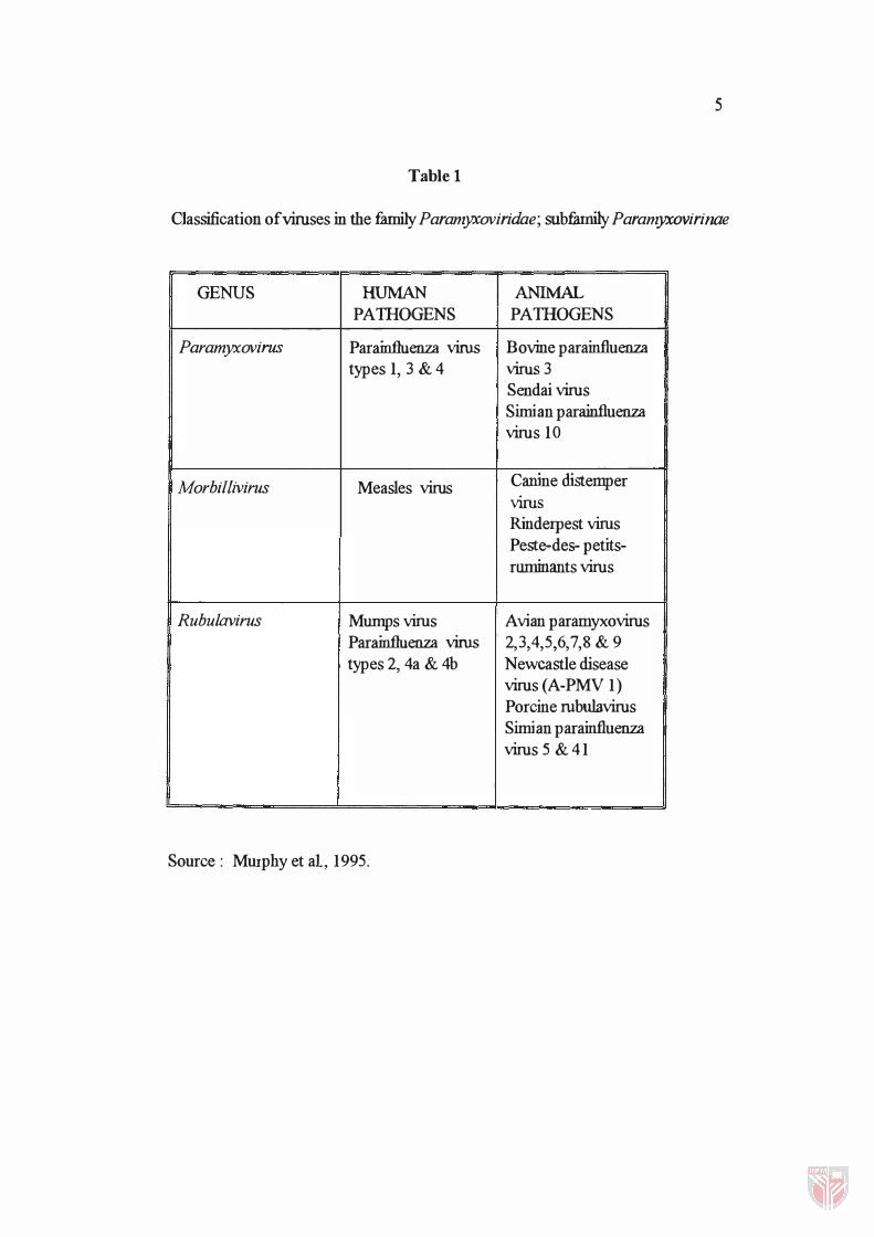
Newcastle disease virus (NDV) is the causative agent of ND. It is
designated as avian paramyxovirus 1 (A-PMV1), formerly the prototype in the
genus Paramyxovims but it is now classified under the genus Rubulavims in the
Paramyxoviridae family (Table 1).
First reported on a farm near Newcastle-up on-Tyne early this centwy
(Alexander, 1988), NDV has since been known to cause three global pandemics :
the first from 1926 to 1934 which spread to most countries in the world, the second
from 1965 to 1972 in the Middle East and the third occurred recently in the 1980's
and related to a mainly neurotropic disease of racing pigeons, caused by an NDV
strain which is distinguishable from other strains by monoclonal antibodies
(Alexander, 1985).
ND occurs in domestic fowls, turkeys, pheasants, pigeons, quail and guinea
fowl Ducks and geese are also susceptible, though less so, as are wild
4
5
Table 1
Classification of viruses in the family Paramyxoviridae; subfamily Paramyxovirinae
GENUS HUMAN ANIMAL PATHOGENS PATHOGENS
Paramyxovirus Parainfluenza virus Bovine parainfluenza types 1, 3 & 4 virus 3
Sendai virus Simian parainfluenza virus 10
Morbillivirus Measles virus Canine distemper VIruS Rinderpest virus Peste-des- petits-ruminants virus
Rubulavirus Mumps virus Avian paramyxovirus Parainfluenza virus 2,3,4,5,6,7 ,8 & 9 types 2, 4a & 4b Newcastle disease
virus ( A-PMV 1) Porcine rubulavirus Simian parainfluenza virus 5 & 41
Source: Murphy et ai, 1995.
6
bird species. The virus can cause mild conjunctivitis in humans although sometimes
it may be severe in poultry farmers, abattoir staB: vaccinators and laboratory
workers. However, there is no evidence of transmission from humans-to-birds
(Alexander, 1988).
Different strains of NDV exhibit a full range of symptoms, from
asymptomic infection to almost 100% mortality of infected birds (Alexander,
1985). Initial symptoms of highly virulent isolates include loss of appetite, drop in
egg production, green dianhoea, swelling of the head and blue-ish comb. Mortality
is close to 100% and many birds die within a day or two; birds th at survive the
initial phase often develop neIVOUS signs. Lower virulence isolates cause coughing,
gasping, wing and leg paralysis, weight loss, drop in egg production, head tremors
and perhaps neIVOUS signs at later stage. Avirulent NDV outbreaks may manifest as
respiratory symptoms, lack of appetite and a drop in egg production.
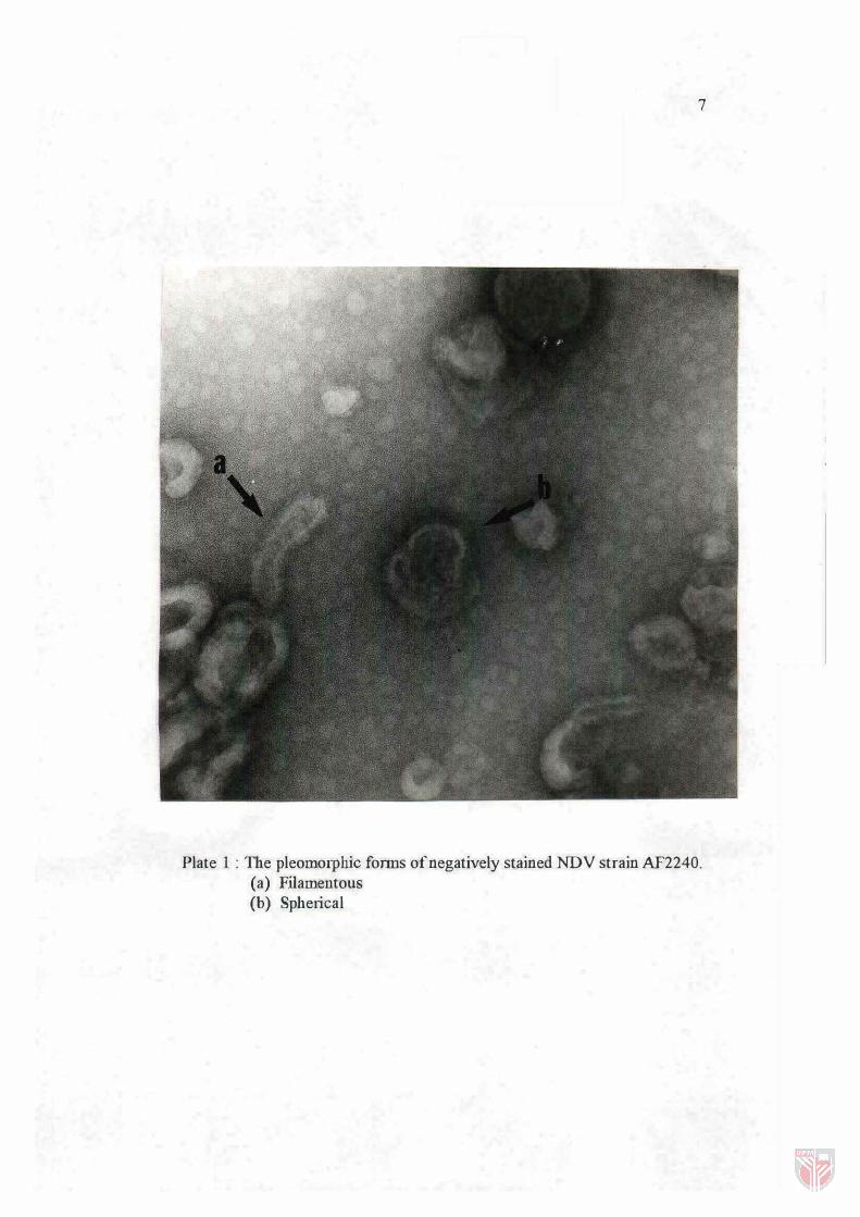
Morphological and Physical Properties
Plate 1 shows negatively stained NDV virions and Fig. 1 shows the
schematic diagram of the probable arrangement of the viral structural components.
The virion can be viewed as consisting of two structural units : i ) the
ribonucleoprotein or the nucleocapsid and ii) the envelope proteins with its
7
Plate I: The pleomorphic fonns of negatively stained NDV strain AF2240. (a) Filamentous (b) Spherical


