dislodged bonded molar tube into wound during orthognathic...
TRANSCRIPT
Case ReportDislodged Bonded Molar Tube into Wound duringOrthognathic Surgery
Tengku Aszraf Tengku Shaeran1 and A. R. Samsudin 1,2
1Oral and Maxillofacial Surgery Unit, School of Dental Sciences, University of Science Malaysia (USM), 16150 Kubang Kerian,Kelantan, Malaysia2Sharjah Institute for Medical Research (SIMR), University of Sharjah, Sharjah, UAE
Correspondence should be addressed to A. R. Samsudin; [email protected]
Received 29 December 2017; Revised 28 March 2018; Accepted 15 April 2018; Published 4 June 2018
Academic Editor: Carla Evans
Copyright © 2018 Tengku Aszraf Tengku Shaeran and A. R. Samsudin. This is an open access article distributed under the CreativeCommons Attribution License, which permits unrestricted use, distribution, and reproduction in any medium, provided theoriginal work is properly cited.
Introduction. Dislodgement of orthodontic appliance into operation wounds may occur while performing orthognathic surgery. Itsoccurrence is commonly associated with bonded upper molar tube. Case Report. A 25-year-old gentleman presented with recurrentupper right vestibular abscess three months following a bimaxillary orthognathic surgery. A bonded molar orthodontic tube haddislodged into the wound during the operation. The clinical presentation initially mimics an odontogenic infection until ourinvestigations revealed that it originated from the dislodged appliance. The abscess was drained, the wound site was explored,and the molar tube and neighbouring rigid fixation plates and screws were removed. The patient recovered well following theprocedure. Conclusion. Dislodged metal orthodontic appliance in oral wound acts as a foreign body that may exert allergicreactions, infection, or inflammation. Pre- and postoperative intraoral examination of fixed orthodontic appliances including itscount should be recorded in orthognathic surgery protocol.
1. Introduction
Patients requiring orthognathic surgery for correction oftheir maxillo-mandibular disharmony will also have toundergo orthodontic treatment during both pre- and post-surgical treatment phases. Tooth alignment and preparationof the future predicted occlusion are required, so that theosteotomized jaws can be easily repositioned in the surgeryin order to achieve stable results. This is followed by a periodof fine-tuning and maintenance of the occlusion afterwards.
Among orthodontists, the use of bonded orthodonticmolar tubes has gained popularity compared to the con-ventional molar banding because the former are easier toplace, without the need for orthodontic separator, morefriendly to the periodontium, and more comfortable tothe patient [1].
Banks and Mcfarlane [2] revealed that failure rates anddisplacement of bonded molar and banded molar are in therange of 33.7% and 18.8%, respectively. This might contrib-ute to the higher percentage of dislodged bonded appliance
in orthognathic surgery as highlighted by Godoy et al. [3].According to them, 76.3% of dislodged orthodontic appli-ance associated with orthognathic are related to involve themaxillary molars, and they were the bonded rather thanbanded-type appliance [2].
2. Case Report
A 25-year-old gentleman presented to our clinic with acomplaint of recurrent pain and swelling on his right cheekof three-month duration. He visited a general practitionereach time, and the condition was resolved with analgesicand antibiotics. However, his symptoms got worse and heattended our Oral Surgery Clinic for consultation.
The patient is a fit and healthy young man with no rele-vant medical history and no known history of allergy. Pastsurgical history revealed that he had underwent bimaxillaryorthognathic surgery one and half year earlier in a localhospital. Although the postoperative period was uneventful,the surgical team informed him that there was a dislodged
HindawiCase Reports in DentistryVolume 2018, Article ID 6540945, 4 pageshttps://doi.org/10.1155/2018/6540945
orthodontic appliance in his right cheek that must haveoccurred during the operation. The team explained to thepatient that this accident was realized later on the next dayafter the surgery when the molar tube from the right maxil-lary second molar was found missing, and its presence wasconfirmed high up in the right maxillary-zygomatic buttressarea shown in the postoperative X-ray image taken on thenext day following the surgery. A series of further postopera-tive radiographs confirmed its location, lying outside theright maxillary antrum. Due to the pronounced postopera-tive facial oedema at that time, no attempt was made toremove the appliance. The absence of sign and symptomsduring further follow-up sessions confirmed the decision toleave it in-situ with continuous clinical observation.
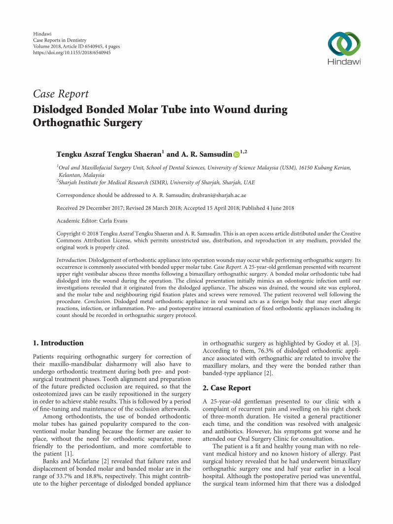
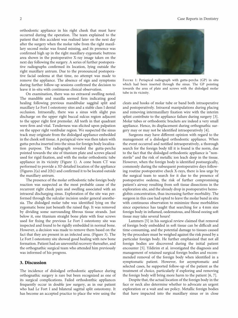
On examination, there was no extraoral swelling noted.The mandible and maxilla seemed firm indicating goodhealing following previous mandibular saggital split andmaxillary Le Fort I osteotomy sites and a stable class I dentalocclusion. Intraorally, there was a sinus with slight pusdischarge on the upper right buccal sulcus region adjacentto the upper right first premolar. All teeth in that quadrantwere firm and vital. Tenderness was elicited upon palpationon the upper right vestibular region. We suspected the sinustrack may originate from the dislodged appliance embeddedin the cheek soft tissue. A periapical view was then taken withgutta-percha inserted into the sinus for foreign body localiza-tion purpose. The radiograph revealed the gutta-perchapointed towards the site of titanium plate and screws placedused for rigid fixation, and with the molar orthodontic tubeappliance in its vicinity (Figure 1). A cone beam CT wasperformed to provide a 3D detailed location of the appliance(Figures 2(a) and 2(b)) and confirmed it to be located outsidethe maxillary antrum.
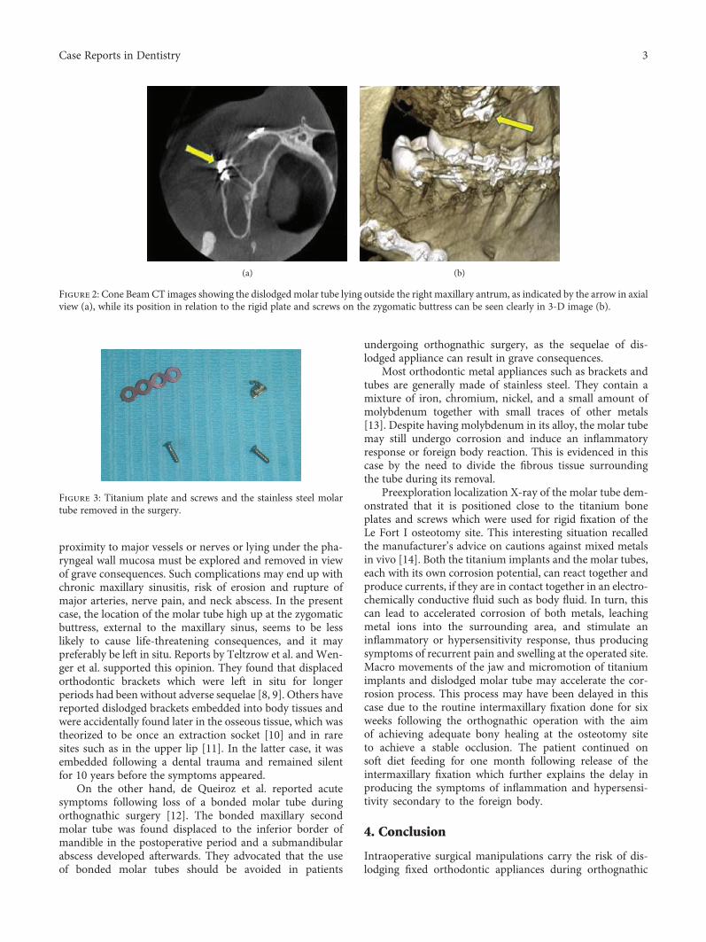
The presence of the molar orthodontic tube foreign bodyreaction was suspected as the most probable cause of therecurrent right cheek pain and swelling associated with anintraoral discharging sinus. Exploration of the site was per-formed through the sulcular incision under general anesthe-sia. The dislodged molar tube was identified lying on thezygomatic bone just beneath the raised flap. It was removedby dividing some surrounding fibrous tissue strands. Justbelow it, one titanium straight bone plate with four screwsused for fixing the previous Le Fort I osteotomy site wasinspected and found to be rigidly embedded in normal bone.However, a decision was made to remove them based on thefact that they are present in an infected area. (Figure 3). TheLe Fort I osteotomy site showed good healing with new boneformation. Patient had an uneventful recovery thereafter, andthe orthognathic surgical team who attended him previouslywas informed of his progress.
3. Discussion
The incidence of dislodged orthodontic appliance duringorthognathic surgery is rare but been recognized as one ofits surgical complications. Failed orthodonthic appliancesfrequently occur in double jaw surgery, as in our patientwho had Le Fort I and bilateral sagittal split osteotomy. Ithas become an accepted practice to place the wire using the
cleats and hooks of molar tube or band both intraoperativeand postoperatively. Intraoral manipulations during placingand removing intermaxillary fixation wire with the interimsplint contribute to the appliance failure during surgery [3].Molar tubes or orthodontic brackets are indeed a very smallappliance. Hence, its displacement during orthognathic sur-gery may or may not be identified intraoperatively [4].
Surgeons may have different opinion with regard to themanagement of a dislodged orthodontic appliance. Whenthe event occurred and notified intraoperatively, a thoroughsearch for the foreign body till it is found is the norm, dueto the fact that the dislodged orthodontic appliance is “non-sterile” and the risk of metallic ion leach deep in the tissue.However, when the foreign body is identified postsurgically,commonly during the subsequent postoperative days follow-ing routine postoperative check X-rays, there is less urge bythe surgical team to search for it due to the presence ofpostoperative oedema, the risk of further compromisingpatient’s airway resulting from soft tissue dissections in theexploration site, and the already drop in postoperative hemo-globin concentration, thus increasing further morbidity. Thesurgeon in this case had opted to leave the molar band in situwith continuous observation to minimize those morbiditiessince experience has taught that searching for a 4mm sizeforeign body in inflamed, oedematous, and blood oozing softtissue may take several hours!
Lammers [5] in his surgical review claimed that removalof foreign body embedded in soft tissue can be difficult andtime-consuming, and the potential damage to tissues causedby the procedure must be weighed against the risk posed by aparticular foreign body. He further emphasized that not allforeign bodies are discovered during the initial patientencounter [5]. Yildirim et al. investigated the diagnosis andmanagement of retained surgical foreign bodies and recom-mended removal of the foreign body when identified in asymptomatic patient. However, for asymptomatic andselected cases, he supported follow-up of the patient as thetreatment of choice, particularly if exploring and removingthe foreign body will bring more harm to the patient [6, 7].
Despite that, the actual location of the foreign body in theface or neck also determine whether to advocate an urgentexploration or a wait and see policy. Metallic foreign bodiesthat have impacted into the maxillary sinus or in close
Figure 1: Periapical radiograph with gutta-percha (GP) in situwhich had been inserted through the sinus. The GP pointingtowards the area of plate and screws with the dislodged molartube in its vicinity.
2 Case Reports in Dentistry
proximity to major vessels or nerves or lying under the pha-ryngeal wall mucosa must be explored and removed in viewof grave consequences. Such complications may end up withchronic maxillary sinusitis, risk of erosion and rupture ofmajor arteries, nerve pain, and neck abscess. In the presentcase, the location of the molar tube high up at the zygomaticbuttress, external to the maxillary sinus, seems to be lesslikely to cause life-threatening consequences, and it maypreferably be left in situ. Reports by Teltzrow et al. and Wen-ger et al. supported this opinion. They found that displacedorthodontic brackets which were left in situ for longerperiods had been without adverse sequelae [8, 9]. Others havereported dislodged brackets embedded into body tissues andwere accidentally found later in the osseous tissue, which wastheorized to be once an extraction socket [10] and in raresites such as in the upper lip [11]. In the latter case, it wasembedded following a dental trauma and remained silentfor 10 years before the symptoms appeared.
On the other hand, de Queiroz et al. reported acutesymptoms following loss of a bonded molar tube duringorthognathic surgery [12]. The bonded maxillary secondmolar tube was found displaced to the inferior border ofmandible in the postoperative period and a submandibularabscess developed afterwards. They advocated that the useof bonded molar tubes should be avoided in patients
undergoing orthognathic surgery, as the sequelae of dis-lodged appliance can result in grave consequences.
Most orthodontic metal appliances such as brackets andtubes are generally made of stainless steel. They contain amixture of iron, chromium, nickel, and a small amount ofmolybdenum together with small traces of other metals[13]. Despite having molybdenum in its alloy, the molar tubemay still undergo corrosion and induce an inflammatoryresponse or foreign body reaction. This is evidenced in thiscase by the need to divide the fibrous tissue surroundingthe tube during its removal.
Preexploration localization X-ray of the molar tube dem-onstrated that it is positioned close to the titanium boneplates and screws which were used for rigid fixation of theLe Fort I osteotomy site. This interesting situation recalledthe manufacturer’s advice on cautions against mixed metalsin vivo [14]. Both the titanium implants and the molar tubes,each with its own corrosion potential, can react together andproduce currents, if they are in contact together in an electro-chemically conductive fluid such as body fluid. In turn, thiscan lead to accelerated corrosion of both metals, leachingmetal ions into the surrounding area, and stimulate aninflammatory or hypersensitivity response, thus producingsymptoms of recurrent pain and swelling at the operated site.Macro movements of the jaw and micromotion of titaniumimplants and dislodged molar tube may accelerate the cor-rosion process. This process may have been delayed in thiscase due to the routine intermaxillary fixation done for sixweeks following the orthognathic operation with the aimof achieving adequate bony healing at the osteotomy siteto achieve a stable occlusion. The patient continued onsoft diet feeding for one month following release of theintermaxillary fixation which further explains the delay inproducing the symptoms of inflammation and hypersensi-tivity secondary to the foreign body.
4. Conclusion
Intraoperative surgical manipulations carry the risk of dis-lodging fixed orthodontic appliances during orthognathic
(a) (b)
Figure 2: Cone Beam CT images showing the dislodged molar tube lying outside the right maxillary antrum, as indicated by the arrow in axialview (a), while its position in relation to the rigid plate and screws on the zygomatic buttress can be seen clearly in 3-D image (b).
Figure 3: Titanium plate and screws and the stainless steel molartube removed in the surgery.
3Case Reports in Dentistry
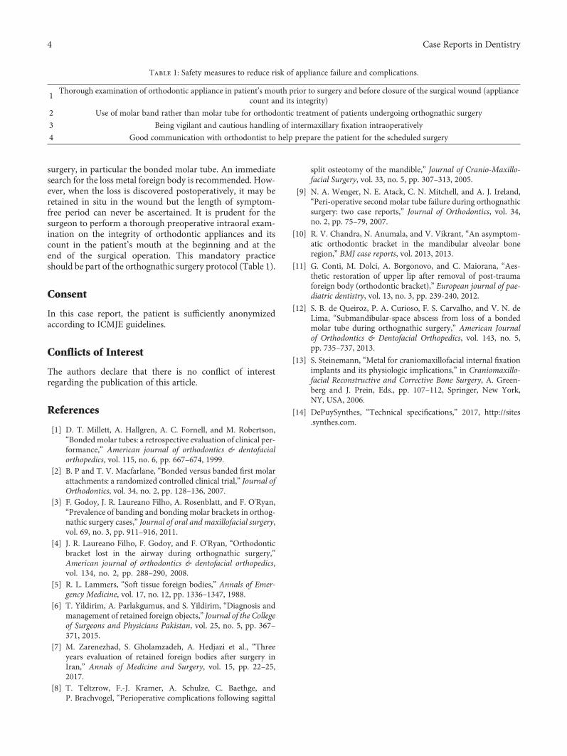
surgery, in particular the bonded molar tube. An immediatesearch for the loss metal foreign body is recommended. How-ever, when the loss is discovered postoperatively, it may beretained in situ in the wound but the length of symptom-free period can never be ascertained. It is prudent for thesurgeon to perform a thorough preoperative intraoral exam-ination on the integrity of orthodontic appliances and itscount in the patient’s mouth at the beginning and at theend of the surgical operation. This mandatory practiceshould be part of the orthognathic surgery protocol (Table 1).
Consent
In this case report, the patient is sufficiently anonymizedaccording to ICMJE guidelines.
Conflicts of Interest
The authors declare that there is no conflict of interestregarding the publication of this article.
References
[1] D. T. Millett, A. Hallgren, A. C. Fornell, and M. Robertson,“Bondedmolar tubes: a retrospective evaluation of clinical per-formance,” American journal of orthodontics & dentofacialorthopedics, vol. 115, no. 6, pp. 667–674, 1999.
[2] B. P and T. V. Macfarlane, “Bonded versus banded first molarattachments: a randomized controlled clinical trial,” Journal ofOrthodontics, vol. 34, no. 2, pp. 128–136, 2007.
[3] F. Godoy, J. R. Laureano Filho, A. Rosenblatt, and F. O'Ryan,“Prevalence of banding and bonding molar brackets in orthog-nathic surgery cases,” Journal of oral and maxillofacial surgery,vol. 69, no. 3, pp. 911–916, 2011.
[4] J. R. Laureano Filho, F. Godoy, and F. O'Ryan, “Orthodonticbracket lost in the airway during orthognathic surgery,”American journal of orthodontics & dentofacial orthopedics,vol. 134, no. 2, pp. 288–290, 2008.
[5] R. L. Lammers, “Soft tissue foreign bodies,” Annals of Emer-gency Medicine, vol. 17, no. 12, pp. 1336–1347, 1988.
[6] T. Yildirim, A. Parlakgumus, and S. Yildirim, “Diagnosis andmanagement of retained foreign objects,” Journal of the Collegeof Surgeons and Physicians Pakistan, vol. 25, no. 5, pp. 367–371, 2015.
[7] M. Zarenezhad, S. Gholamzadeh, A. Hedjazi et al., “Threeyears evaluation of retained foreign bodies after surgery inIran,” Annals of Medicine and Surgery, vol. 15, pp. 22–25,2017.
[8] T. Teltzrow, F.-J. Kramer, A. Schulze, C. Baethge, andP. Brachvogel, “Perioperative complications following sagittal
split osteotomy of the mandible,” Journal of Cranio-Maxillo-facial Surgery, vol. 33, no. 5, pp. 307–313, 2005.
[9] N. A. Wenger, N. E. Atack, C. N. Mitchell, and A. J. Ireland,“Peri-operative second molar tube failure during orthognathicsurgery: two case reports,” Journal of Orthodontics, vol. 34,no. 2, pp. 75–79, 2007.
[10] R. V. Chandra, N. Anumala, and V. Vikrant, “An asymptom-atic orthodontic bracket in the mandibular alveolar boneregion,” BMJ case reports, vol. 2013, 2013.
[11] G. Conti, M. Dolci, A. Borgonovo, and C. Maiorana, “Aes-thetic restoration of upper lip after removal of post-traumaforeign body (orthodontic bracket),” European journal of pae-diatric dentistry, vol. 13, no. 3, pp. 239-240, 2012.
[12] S. B. de Queiroz, P. A. Curioso, F. S. Carvalho, and V. N. deLima, “Submandibular-space abscess from loss of a bondedmolar tube during orthognathic surgery,” American Journalof Orthodontics & Dentofacial Orthopedics, vol. 143, no. 5,pp. 735–737, 2013.
[13] S. Steinemann, “Metal for craniomaxillofacial internal fixationimplants and its physiologic implications,” in Craniomaxillo-facial Reconstructive and Corrective Bone Surgery, A. Green-berg and J. Prein, Eds., pp. 107–112, Springer, New York,NY, USA, 2006.
[14] DePuySynthes, “Technical specifications,” 2017, http://sites.synthes.com.
Table 1: Safety measures to reduce risk of appliance failure and complications.
1Thorough examination of orthodontic appliance in patient’s mouth prior to surgery and before closure of the surgical wound (appliance
count and its integrity)
2 Use of molar band rather than molar tube for orthodontic treatment of patients undergoing orthognathic surgery
3 Being vigilant and cautious handling of intermaxillary fixation intraoperatively
4 Good communication with orthodontist to help prepare the patient for the scheduled surgery
4 Case Reports in Dentistry

DentistryInternational Journal of
Hindawiwww.hindawi.com Volume 2018
Environmental and Public Health
Journal of
Hindawiwww.hindawi.com Volume 2018
Hindawi Publishing Corporation http://www.hindawi.com Volume 2013Hindawiwww.hindawi.com
The Scientific World Journal
Volume 2018Hindawiwww.hindawi.com Volume 2018
Public Health Advances in
Hindawiwww.hindawi.com Volume 2018
Case Reports in Medicine
Hindawiwww.hindawi.com Volume 2018
International Journal of
Biomaterials
Scienti�caHindawiwww.hindawi.com Volume 2018
PainResearch and TreatmentHindawiwww.hindawi.com Volume 2018
Preventive MedicineAdvances in
Hindawiwww.hindawi.com Volume 2018
Hindawiwww.hindawi.com Volume 2018
Case Reports in Dentistry
Hindawiwww.hindawi.com Volume 2018
Surgery Research and Practice
Hindawiwww.hindawi.com Volume 2018
BioMed Research International Medicine
Advances in
Hindawiwww.hindawi.com Volume 2018
Hindawiwww.hindawi.com Volume 2018
Anesthesiology Research and Practice
Hindawiwww.hindawi.com Volume 2018
Radiology Research and Practice
Hindawiwww.hindawi.com Volume 2018
Computational and Mathematical Methods in Medicine
EndocrinologyInternational Journal of
Hindawiwww.hindawi.com Volume 2018
Hindawiwww.hindawi.com Volume 2018
OrthopedicsAdvances in
Drug DeliveryJournal of
Hindawiwww.hindawi.com Volume 2018
Submit your manuscripts atwww.hindawi.com