universiti putra malaysia in vivo and in...
TRANSCRIPT
UNIVERSITI PUTRA MALAYSIA
IN VIVO AND IN VITRO STUDIES OF ANTI-CHOLESTEROL AND ANTI-CARCINOGENIC EFFECTS OF GANODERMA CRUD EXTRACT
CHOONG YEW KEONG
FSAS 2003 47
IN VIVO AND IN VITRO STUDIES OF ANTI-CHOLESTEROL AND ANTICARCINOGENIC EFFECTS OF GANODERMA CRUDE EXTRACT
By
CHOONG YEW KEONG
Thesis Submitted to the School of Graduate Studies, Universiti Putra �alaysia, in Fulfilment of the Requirement for the Degree of Doctor of Philosophy
April 2003
Abstract of thesis presented to the Senate ofUniversiti Putra Malaysia in fulfillment of the requirements for the degree of Doctor of Philosophy
IN VIVO AND IN VITRO STUDIES OF ANTI-CHOLESTEROL AND ANTICARCINOGENIC EFFECTS OF GANODERMA CRUDE EXTRACT
By
CHOONG YEW KEONG
April 2003
Chairman: Associate Professor Dr. Tong Chow Chin
Faculty: Science and Environmental Studies
11
The in vivo and in vitro effect of a local, commercially available Ganoderma fruiting
body powder (GF) and G. lucidum mycelium (GM) grown in soy waste on tumour and
hypercholesterolaemic rats were studied.
Administration of 1 % cholesterol diet in the cholesterol (Chol) group caused a significant
(p<O.05) increase in the serum total cholesterol (TC), triglyceride (TG) and low density
lipoprotein cholesterol (LDL-C) levels while reducing serum high density lipoprotein
cholesterol (HDL-C) level. In the case of the Chol+GF and Chol+GM groups, the initial
serum TC, TG and LDL-C levels showed a much higher levels (p<O.05) compared to the
Chol group. However, the levels gradually decreased towards the end of the experiment.
There was no significant difference in the lipid profiles amongst the Control, GF and GM
groups. Serum alanine transferase (AL T), gamma glutamyltransferase (GGT) and
creatine kinase (CK) in the Chol+GF group as well as the AL T, GGT level in the
iii
Chol+GM group were found to be significantly (p<O.05) lower than those in the Chol
group for both the experiments using GF and GM. Despite the fact that all groups showed
levels of serum urea and creatinine within the normal range, mild degenerative changes in
the glomeruli were seen in the Chol group. In addition, higher level of serum uric acid
was observed in the Chol group compared to the Chol+GF and Chol+GM groups. In term
of the lipid peroxidation and antioxidant enzymes analyses, the Chol+GF and Chol+GM
groups showed a significantly (p<O.05) lower level of malondialdehyde (MDA), catalase
and glutathione peroxidase (GSH-Px) activities but higher vitamin C level compared to
the Chol group.
G. lucidum crude extract exhibited the highest cytotoxic activity (lower ICso) towards
J558 Balb/C mouse myeloma, MDA-MB-435 human breast ductal carcinoma, PN6
leukemia T-cell but not against 3T3 mouse fibroblast normal cell-line as compared to
crude extract from G. tsugae and G. tropicum. Among these cancer cell-lines, JSS8 cells
was the most sensitive to the cytotoxic effects of all the three Ganoderma spp.
Examination by acridine-orange!propidium iodine staining, electron microscopy and
transmission electron microscopy showed that the Ganoderma crude extract caused both
apoptosis and necrosis in the cancer cell-line.
The anti-carcinogenesis test indicated that hypercholesterolaemic rats (the Chol group)
demonstrated a significantly (p<O.05) higher serum MDA level as compared to the GF
and Chol+GF groups. Furthermore, Ganoderma supplemented groups had significantly
(p<O.05) lower levels of serum MDA, catalase, GSH-Px and GGT activities but higher in
1V
the ascorbic acid level as compared to the Chol group. Histologically, the Chol+GF group
showed a much reduced thickened coronary vessel wall as well as normal hair growth in
the anti-carcinogenesis test. The Chol+GM group registered 61.9% normal hepatocytes as
compared to only 5.6% in the Chol group. The presence of epithelisation of lung
epithelium in the Chol group which were not severe in the Chol+GM group indicated the
anti-tumour effect of 10% GM.
Abstrak tesis yang dikemukakan kepada Senat Universiti Putra Malaysia sebagai memenuhi keperluan untuk Ijazah Doktor Falsafah
v
KESAN KAJIAN ANTI-KOLESTEROL DAN ANTI-KARCINOGENIK BAGI EKSTRAK MENTAB GANODERMA SECARA IN VIVO DAN IN VITRO
Oleh
CBOONG YEW KEONG
April 2003
Pengerusi: Profesor Madya Dr. Tong Chow Chin
Fakulti : Sains dan Pengajian Alam Sekitar
Projek ini mengkaji kesan komersial serbuk Ganoderma (GF) dan micelia Ganoderma
(GM) yang tumbuh pada ekstrak kacang soya ke atas barah dan tikus
hiperkloresterolemik secara in vivo dan in vitro.
Penggunaan makanan kolesterol 1 % bagi kumpulan kolesterol (Chol) menyebabkan
signifikan peningkatan paras serum kolesterol keseluruhan (TC), trigliserid (TG),
kolesterol lipoprotin ketumpatan rendah (LDL-C) dan seterusnya menurunkan paras
serum kolesterol lipoprotin ketumpatan tinggi (HDL-C). Dalam hal kumpulan Chol+GF
dan Chol+GM, paras serum TC, TG dan LDL-C lebih tinggi daripada kumpulan Chol
pada mulanya, tetapi parasnya beransur -ansur menurun pada akhir eksperimen. Tidak
terdapat perbezaan yang signifikan bagi paras profil lipid antara kumpulan kawalan, GF
dan GM. Paras serum alanin transferase (AL T), gama glutamil transferase (GGT) dan
VI
kinase (CK) dalam kumpulan Chol+GF dan paras ALT, GGT dalam kumpulan Chol+GM
didapati signifikan (p<O.05) rendah daripada kumpulan Chol dalam eksperimen yang
menggunakan GF dan GM. Walaupun semua kumpulan menunjukkan paras urea dan
creatinine dalam lingkungan yang normal, terdapat juga sedikit degenerasi pada
glomeruli dalam kumpulan Chol setelah diperhatikan melalui kajian histologi. Paras
serum asid urik yang tinggi dalam kumpluan Chol berbanding dengan kumpulan
Chol+GF dan Chol+GM. Dalam analysis peroksidasi lipid dan enzim antioksidan,
kumpuian Chol+GF dan Chol+GM menunjukkan signifikan (p<O.OS) paras
malondeldehid (MDA), aktiviti katalase dan glutation peroksidase (GSH-Px) yang
rendah, tetapi paras vitamin C yang lebih tinggi jika dibandingkan dengan kumpulan
Chol.
Ekstrak mentah G. lucidum menunjukkan aktiviti sitotoksik yang paling berkesan (ICso
yang terendah) terhadap J558 "myeloma" tikus Balb/C, MDA-MB-435 duktul kanser
buah dada manusia, PN6 sel-T leukimia tetapi tidak berkesan terhadap 3T3 sel normal
fibroblast berbanding dengan ekstrak mentah G. tsugae dan G. tropicum. Bagi ketiga-tiga
sel kultur kanser, sel J558 pula yang paling sensitif terhadap kesan sitotoksik. Kajian
dengan menggunakan perwarna tlurasen AOPI dan pemeriksaan dibawah mikroskop
elektron "scanning" dan mikroskop elektron "transmission" menunjukkan bahawa
ekstrak mentah G. lucidum dapat mengakibatkan apoptosis dan neukrosis pada sel kultur
kanser.
Kajian anti-kasinogenik yang dijalankan menunjukkan bahawa tikus hiperkolesterolemik
vii
(kumpulan Chol) menghadapi secara signifikan (p<0.05) paras serum MDA yang tinggi
dibandingkan dengan kumpulan GF, GM, Chol+GF dan Chol+GM. Seterusnya,
kumpulan yang dibekalkan Ganoderma mengandungi paras serum MDA, katalase, GSH
Px dan aktiviti GGT yang rendah secara signifikan dan kandungan serum vitamin C yang
tinggi dibandingkan dengan kumpulan Chol. Dalam kajian histologi, kumpuian Chol+GF
menunjukkan dinding arteri koronari yang kurang tebal dan pertumbuhan buiu yang
normal dalam kajian anti-karsinogenik. Kumpulan Chol+GM mendedahkan 6 1 .9% sel
hepar yang normal berbanding cuma 5.6% dalam kumpulan Chol. Kejadian epitelisasi
dalam epitelium peparu pada kumpulan Chol adalah tidak seteruknya dalam kumpulan
Chol+GM membuktikan kesan anti-barah 1 0% GM.
viii
ACKNOWLEDGEMENTS
I would like to express my deepest appreciation and unqualified gratitude to the
chairman of my supervisory committee Associate Professor Dr. Tong Chow Chin of the
Department of Biochemistry and Microbiology, Universiti Putra Malaysia for his
understanding, patience, invaluable guidance, suggestions and constant encouragement
throughout the planning and execution of the project.
My grateful thanks also to Associate Professor Dr. Noordin Mohamed Mustapha
of Faculty of Veterinary Medicine, Universiti Putra Malaysia for conscientiously serving
as member of my supervisory committee. The support, suggestions and insightful
comments given by other members of my supervisory committee Professor Dr. Suhaila
Mohamed of the Department of Food Science, Faculty of Food Science and
Biotechnology is gatefully acknowledged. My heartfelt appreciation also goes to Dr . Nor
Aini Umar of Department of Chemistry Pathology, Hospital Universiti Kebangsaan
Malaysia, UKM for her generous advice on the clinical chemistry studies.
My thanks and appreciation are also due to the Head of Department Professor
Datin Dr. Kathijah Mohd Yusoff, Department of Biochemistry and Microbiology and
Professor Dr. Abdul Manaf bin Ali, Department of Biotechnology, Faculty of Food
Science and Biotechnology for their kindness in allowing me to pursue pertinent works in
their laboratories.
My sincere gratitude is also extended to Institute Medicinal Research Center, Unit
Pemakanan for providing the materials and methods in vitamin C analysis. I would like
to thank Miss Yang, Miss Khor for their generous advice on this experiment.
IX
I also wish to thank all the staffs of the Department of Biochemistry and
Microbiology, Faculty of Science and Environmental for their kind assistance and co
operation, special thanks are also extended to laboratory assistant Kak Rohaida and Encik
Karim for their support, advice and delightful friendship.
Many thanks are also due to the following people. Mr Zhang, Miss Irine for their
assistance in animal experimental work and antioxidant analysis; Antony, Lim Yen Moi
and Boon Keat for their technical assistance in the cell-culture work and Mr. Siew Kok
Phang for his unfailing support and commitment in this thesis necessitated . The
assistance and support rendered by the staffs of the Hospital Universiti Kebangsaan
Malaysia are duly acknowledged and appreciated.
Last but not least, I am greatly indebted to my beloved wife, son, daughter and my
mother for their loves, patience, supports and sacrifices they have given.
x
I certify that an Examination Committee met on 2nd April 2003 to conduct the final examination of Choong Yew Keong on his Doctor of Philosophy thesis entitled "In Vivo and In Vitro Studies of Anti-cholesterol and Anti-carcinogenic Effects of Ganoderma Crude Extract" in accordance with Universiti Pertanian Malaysia (Higher Degree) Act 1980 and Universiti Pertanian Malaysia (Higher Degree) Regulations 1981. The Committee recommends that the candidate be awarded the relevant degree. Members of the Examination Committee are as follows:
MOHD. NOOR ABDUL WAHAB, Ph.D. Associate Professor Faculty of Science and Environment Studies Universiti Putra Malaysia (Chairman)
TONG CHOW CHIN, Ph.D. Associate Professor Faculty of Science and Environmental Studies Universiti Putra Malaysia (Member)
NOORDIN MOHAMED MUSTAPHA, Ph.D. Associate Professor Faculty of Veterinary Universiti Putra Malaysia (Member)
SUHAILA MOHAMED, Ph.D. Professor Faculty of Food Science and Biotechnology Universiti Putra Malaysia (Member)
NOR AINI UMAR, MD. DCP. Department of Chemistry Pathology HUKM Hospital Universiti Kebangsaan Malaysia (Member)
CHANG YU SHYUN, Ph.D. Pengawai Penyelidik (Tumbuhan Ubatan) Institut Penyelidikan Perhutanan Malaysia (FRIM) (Independent Examiner)
ProfessorlDeputy e , School of Graduate tudies, Universiti Putra M aysia
Date 11 9 JUI" ·(uuJ
xi
This thesis submitted to the Senate of Universiti Putra Malaysia has been accepted as fulfilment of the requirement for the degree of Doctor of Philosophy. The members of the Supervisory Committee are as follows:
Tong Chow Chin, Ph.D. Associate Professor Faculty of Science and Environmental Studies Universiti Putra Malaysia (Chairman)
Noordin Mohamed Mustapha, Ph.D. Associate Professor Faculty of Veterinary Universiti Putra Malaysia (Member)
Suhaila Mohamed, Ph.D. Professor Faculty of Food Science and Biotechnology Universiti Putra Malaysia (Member)
Nor Aini Umar, MD. DCP. Department of Chemistry Pathology HUKM Hospital Universiti Kebangsaan Malaysia (Member)
AINI IDERIS, Ph.D Professor! Dean, School of Graduate Studies, Universiti Putra Malaysia.
Date: r11 JUL 2003
xii
DECLARATION
I hereby declare that the thesis is based on my original work except for quotations and citations which have been duly acknowledged. I also declare that it has not been previously or concurrently submitted for any other degree at UPM or other institutions.
----
� Choong Yew Keong
Date: d-7 - 5'... .1 0"0 3
xiii
TABLE OF CONTENTS
Page
ABSTRACT .............................................................................. ii ABSTRAK .. ......... ........ ............................................... .............. v ACKNOWLEDGEMENTS ........................................ .................... viii APPROVAL............................................................................... x DECLARATION ............... ................................................ ....... ... xii LIST OF TABLES....................................................................... xviii LIST OF FIGURES..................................................................... xix LIST OF ABBREVIATIONS......................................................... xxiii
CHAPTER 1 GENERA.L INTRODUCTION....................... ................... 1
2 �I1r�RA.1r� RE��� .•••••.••••••••••..•••••••••••••.••••••••••••••• 6 2.1 Mycology of G. lucidum............................................. 6 2.2 Bioactive Substances of G. lucidum. . . .. . ... ............ .......... 8
2.2.1 Ganoderma Polysaccharides. . . . . . . . . . . . . . . . . . . . . . . . . . . . . . 8 2.2.2 Triterpenes . . . . . . . . . . . . . . . . . . . . . . . . . . . . . . .. . . . . . . . . . . . . . . . . . 10
2.3 Biomedical Applications. . . . . . . . . . . . . . . . . . . . . . . . . . . . . . . . . . . . . . . . . . . . 11 2.3.1 Immunostimulation with Ganoderma
Polysaccharides. . . . . . . . . ....... ......... .. .................. 12 2.3.2 Ganoderma as a Cardiotonic... . . . . . . . . . . . . . . . . . . . . . . . . . . 13 2.3.3 Other Medicinal Efficacies of Ganoderma spp. . . . . . . . 15
2.4 Concept and Mechanisms of Bioactivity ................ " .. . . . ... 17 2.5 Cholesterol Metabolism. . . . . . . . . . . . . . . . . . . . . ......... ................ 19 2.6 Management of Plasma Cholesterol Levels . . . . . . . . . . . . . . . . . . . ... 19
2.6.1 High Serum Cholesterol and Low Density Lipoprotein ........ , ....... , ......... , .. . .. . .. . . .. ... .. . ... 20
2.6.2 Hypercholesterolaemia. . . .. . . . . . . . . . . . . . . .. .. . . . . .. . .. .. 22 2.6.3 Oxidized LDL and Atherosclerosis ........ , .. , ........ 22 2.6.4 Theories of Atherosclerotic Plaque Development. 25 2.6.5 Arteriosclerosis . . . . . . . . . . . . . . . . . . . . . . . . . . . . . . . . . . . . . . . . . . 26
2.7 Liver Function ......................... ... ,. ...... ... ...... ...... .... 27 2.7.1 Fatty Liver . . . ... ........ ...... .... ......... ...... ..... .... 27
2.8 Enzyme Tests in Liver Disease ............... ........... , .,. ..... 28 2.8.1 Alanin Aminotransferase (ALT).. . . ...... ............. 28 2.8.2 Gamma Glutamyltransferase (GGT) . . . . . . . . . . . . . . . . . . 29
2.9 Renal Function .... ........ .......... ......... . ..................... , 31 2.9.1 Nonprotein Nitrogenous Metabolite Elimination... 32
2.10 Creatine Kinase (CK). . . . .. ...... . ... . ................ ...... ........ 33 2.11 Reduction of Hydrogen Peroxide. . . . . . . . . . . . . . . . . . . . . . . . . . . . . . . . . 35
2.11.1 Antioxidants . . . .. . .. . . .. . .. .. . . .. .. . .. .. . . .. . .. . .. . . . ..... 36
2.11.2 Enzymes that Catal yze Antioxidant Reactions .. . . 2.11.3 Antioxidant Chemicals . . . . . . . . . . . . . . . . . . . . . . . . . . . . . . . . 2.11.4 Malondialdehyde (MDA) . . . . . . . . . . . . . . . . . . . . . . . . . . . . . 2.11.5 Glutathione S-Transferase (BC 2.5.1.18) .......... .
2.12 Skin Cancer . . . . . . . . . . . . . . . . . . .. . . . ... . . . . . . . . . . . . . . . . . . . . . . . . . . . . . .. 2.13 Benzo[a]pyrene (B[a]p) . . . . . . . . . . . . . . . . . . . . . . . . . . . . . . . . . . . . . . . . . . . 2.14 Lung Cancer . . . . . . . . . . . . . . . . . . . . . . . . . . . . . . . . . . . . . . . . . . . . . . . . . . . . . . . . 2.15 Antineoplastic Agents . . . . . . . . . . . . . . . . . . . . . . . . . . . . . . . . . . . . . . . . . . . . .
2.15.1 Natural Product. . . . . . . . . . . . . . . . . ' " . .. . . . . . . . . , . . . . . . . . 2.16 Animal Cell Cultures . . . . . , . . . . .. . . .. . . . ... . . . . . . . . . . . . .. . , . . . . . .. .
2.16.1 Apoptosis and Cellular Senescence . . . . . . . . . . . . . . . .. 2.16.2 Necrosis . . . . . . . , . . . . . . . . . . . . .. . . . .. . . . .. . . . . . . .... . . . . . .
3 GENERAL MATERIALS AND METHODS ••.•••.•••••••••••••.••• 3.1 Materials . . . . . . . . . . . . . . . . . . . . . . . . . . . . . . . . . . . . . . . . . . . . . . . , .. . . . . . . . . . . . 3.2 Methods . . . . . . . . . . . . . . . . . . . . . . . . . . . . . . . . . . . . . . . . . . . . . . . . . . . . . . . . . . . . ..
3.2.1 Procedure of Blood Sampling and Processing . . . . 3.2.2 Cobas Intrega Multiporpose Auto-analyser
Machine ... . . . . . . . . . . . . . . . . . . . . . . . . . . . . . . . . . . . . . . . . . . . . . 3.2.3 Measurement of Serum Malondialdehyde
(MDA) Concentration . . . . . . . . . . .. . . . . . . . . . . . . . . . . . . . . 3.2.4 Measurement of Erythrocytes Catalase activity . . . 3.2.5 Measurement of Whole Blood Glutathione
Peroxidase (GSH-Px) Activity . . . . . . . . . . . . . . . . .. . . . . . 3.2.6 Determination Vitamin C . . . . . . . . . . . . . . . . . . . . . . . . . . . .
4 THE ANTI-CHOLESTEROL EFFECTS OF COMMERCIAL
36 38 39 39 40 42 44 45 46 47 47 48
51 51 54 54
55
56 57
58 S9
GANODERMA FRUITING BODY POWDER (GF) AND SOY WASTE GROWN G. LUCIDUM MYCELIUM(GM) ON HYPERCHOLESTEROLAEMIC RATS • • ••• •••••• •••••••••••••••• 62 4.1 Introduction . . . . . . . . . . . . . .. . . . . . . . . . . . . . . . . . . . . . . . . . . . . ,. ... ...... ... 62 4.2 Materials and Methods. . . . . . . . . . . . . . .. . . . . . . . . . . . . . . . . . . . . . . . . . . . . . 66
4.2.1 Commercial Ganoderma Fruiting Body Powder 66 (GF) . .. .. . . . . . .... . . . .. . . .. . . . . .. . . . .. . . ..... . .. . . . . . . . . ..
4.2.2 G. lucidum Mycelium Grown in Soy Waste. . . . . . . 68 4.2.3 Serum Lipid Profile, Enzyme Antioxidant, MDA
and Vitamin C. . . . . . . . . . . . . . . . . . . . . . . . . . . . . . ... . . . . . . . . . 73 4.2.4 Statistical Analysis. . . . . . . . . . . . . . . . . . . . . . . . . . . . .. . . ...... 74
4.3 Results and Discussion. . . . . . . . . . . . . . . . . . . . . . .. . . . . . . . . . . . . . . . . . . . . 74 4.3.1 The Effect of Feeding 0.1 % GF Powder and 10%
GM on the Body Weight of Rats . . . . . . . .. . . . . . . . . . . . . 75 4.3.2 The Effect of Feeding 0.1 % GF Powder and 10%
GM on the Serum Total Cholesterol (TC) in Rats 77 4.3.3 The Effect of Feeding 0.1 % GF and 10% GM on
the Serum Triglyceride (TG) in Rats . . . ............. 80
xiv
4.3.4 The Effect of Feeding 0.1 % GF and 10% GM on the Serum High Density Lipoprotein (HDL-C) in Rats . . . . . . . . . . . . . . . . . . . . . . . . . . . . . . . . . . . . . . . . . . . . . . . . . . . .....
4.3.5 The Fffect of Feeding 0.1 % GF and 10% GM on the Serum Low Density Lipoprotein (LDL-C) in Rats . . . . . . . . . . . . . . . . . . . . . . . . . . . . . . . . . . . . . . . . . . . . . . . . . . . . . .
4.3.6 The Effect of Feeding 0.1 % GF and 10% GM on the Serum Alanine Aminotransferase (ALT) Level in Rats . . . . . . . . . . . . . . . . . . . . . . . . . . . . . . . . . . . . . . . . . . . .
4.3.7 The Effect of Feeding 0.1 % GF and 10% GM on the Serum Gamma Glutamyltransferase (OOT) level in Rats . . . . . . . . . . . . . . . . . . . . . . . . . . . . . . . . . . . . . . . . . . . . .
4.3.8 The Effect of Feeding 0.1 % GF and 10% GM on the Serum Urea Level in Rats . . . . . . . . . . . . . . . . . . . . . . .
4.3.9 The Effect of Feeding 0.1 % GF and 10% GM on the Serum Creatinine Level in Rats . . . . . . . . . . . . . . . . .
4.3.10 The Effect of Feeding 0.1 % GF and 10% GM on the Serum Uric Acid Level in Rats . . . . . . . . . . . . . . . . .
4.3.11 The Effect of Feeding 0.1 % GF and 10% GM on the Serum Creatine Kinase (CK) Level in Rats . . .
4.3.12 The Effect of Feeding 0.1% GF and 10% GM on the Serum Malondialdehyde (MDA) Level in Rats . . . . . . . . . . . . . . . . . . . . . . . . . . . . . . . . . . . . . . . . . . . . . . . . . . . . . .
4.3.13 The Effect of Feeding 0.1 % GF and 10% GM on the Erythrocyte Catalase Activity in Rats . . . . . . . . . .
4.3.14 The Effect of Feeding 0.1 % GF and 10% GM on the Whole blood Glutathione Peroxidase (GSH-Px) Activity in Rats . . . . . . .. . . . . . .. . . . . . . . . . . . . . . . . . . . .
4.3.15 The Effect of Feeding 0.1 % GF and 10% GM on the Serum Vitamin C Content of Rats . . . . . . . . . . . . . .
4.3.16 Morphology of Heart and Liver . . . . . . . . . . . . . . . . . . . . . 4.4 Conclusion .. . . . . . . .. . . . . . . . . ' " . . ... . . . . . . . . . . . . . . . . . . . . . . . . . .. . . . . .
5 A COMPARATIVE STUDY OF CYTOTOXIC ACTIVITY WITH COMMERCIAL GANODERMA FRUITING BODY POWDER AND THREE GANODERMA SPP. MYCELIUM WATER SOLUBLE CRUDE EXTRACTS ON MOUSE MYELOMA, LEUKAEMIA AND HUMAN BREAST �1l���1t ����-�I� .. . . . . . . . . . . . . . . . . . . . . . . . . . . . . . . . . . . . . . . . . . . . . . . . . . .
5.1 Introduction . . . . . . . . . . . . . . , . . . . . . . . . . . . . . . . . . . . . . . . . . . . . .. . . . . . . . . .. 5.2 Materials and Methods . . . . . . . . . . . . . . . . . . . . . . . . . . . . . . . . . . . . . . . . . . . . .
5.2.1 Cell-Culture .... .. .. . .. . .. .. . . . . . . . . . . . . . . . .. . . . . . . . . . . . . . 5.2.2 Ganoderma Crude Extract . . . . . . . . . .. . . . . . . . . . . . . . . . . . . 5.2.3 Microculture Tetrazolium Assay (MIT) . . . . . . . . . . .. 5.2.4 Scanning Electron Microscopy (SEM) . . . . . . . . . . . . . . 5.2.5 Transmission Electron Microscopy (TEM) . . . . . . . ..
82
86
90
93
96
100
104
106
109
111
113
116 118 124
126 126 128 128 1 31 134 135 1 36
xv
5.2.6 Acridine Orange (AO) and Propidium Iodide (PI) 137 Staining .. . . . . . . . . . . . . . . . . . . . . . .. .... . . . . ......... . ...... .
5 .2.7 DNA Fragmentation Assay. . . . . .... . .. . .. . ..... ..... 137 5.3 Results and Discussion.... . . . . . . . . . .. . . . . . . . . . . . . .. . ..... ...... .... 138
5.3.1 MIT Assay. . . . ..... . ...... .. ... . . . .................... 138 5.3.2 Morphological Assessment of Apoptosis.......... 145
5.4 General Discussion and Conclusion......... ............ ........ 167
6 THE EFFECTS OF COMMERCIAL GANODERMA FRUITING BODY CRUDE EXTRACT ON BENZO[A]PYRENE IN HYPERCHOLESTEROLAEMIC RAT SKIN FOLLOWING TOPICAL APPLICATION.................... 171 6.1 Introduction...... .. . .. . .. . .. ....... . ... . . . .......................... 17 1 6.2 Materials and Methods... . .... .. .. . . . . . ...... .... .............. .... 173
6.2.1 Animal . ... . . .. .... ........................... . ........... 173 6.2.2 Blood Samples Collecting......... ................... 174 6.2.3 Serum Chemistry 174 6.2.4 Clinical Observation.. . ..... ..... 175 6.2.5 Histological Staining Method ................... , . . .. 175 6.2.6 Hepatocytes Counting... . .. . . . ... . . . . .. .. . . .. . . . .. . . .. 177
6.3 . Results and Discussion . . . . . . . . . . . . . . . . . . . . . . . . . . . . . . . . ... . . . . . . . .. 177 6.3.1 Body Weight. ..... . ..... .. .............................. 177 6.3.2 Serum Lipid and Lipoprotein......................... 178 6.3.3 Serum Gamma Glutamyltransferase (GGT) and
Alanine Aminotransferase (ALT) level............. 181 6.3.4 Urea and Creatinine................. ............... .... 183 6.3.5 Uric Acid. . . . . . . . . . . . . .. . . . ..... .... . . . ...... . ........... 184 6.3.6 Malondialdehyle ., . .... . . . . . ...... . .............. ..... . 186 6 .3.7 Catalase.... . . . . . . . .. . . . . . .... ...... ........ ............. 187 6.3.8 Glutathione peroxidase (GSH-Px)................... 188 6.3.9 Vitamin C. . . . . ......... . . . . . .. ....................... .... 190 6.3. 10 Glutathione S-Transferase Activity (GST)......... 191 6.3.1 1 Pathology. . . . . . . . . . . . . . . . . . . . ... . . ..... . . . .... . .. ... ..... 1 93 6.3. 1 1 Histological Examination...... ... ...... ... . .. . ....... 1 95
6.4 Conclusion . . . . . . . . . . . . . . . . . . . .. . . .. .... . . ...................... , .... 1 97
7 EFFECT OF G. LUC/DUM MYCELIUM (GM) ON BENZO[A]PYRENE-INDUCED EARLY ALTERATIONS OF RESPIRA TY EPITHELIUM THE HYPERCHOLESTEROLAEMIC RATS............................. 1 98 7.1 Introduction .. . . . . . . . . .. . . . . . . . . . . . . '" . . . . . . . . . . .. . . . . . . . . . .. .. . . ... 1 98 7.2 Materials and Methods......... . . .. . . . . . . . . . . . . . . .. ..... . . . . . . . . .. 200
7.2. 1 Animals.......... . . . . . . . .. . . . . .. . .. .. ... ... ... . . ..... .... 200 7.2.2 Inoculum......................... .. . . . . . . . .. . . ... . . . .. . . 201 7.2.3 Experimental Design.. . . . . . . . . . . . . ..... ..... . . .. . . . .. . 201 7.2. 4 Intratracheal Administration . .. . . . . . . . . . . . . . . .... . . . . . 201
xvi
7.2.5 Excision of Lung and Histological Staining .. . . . . . . 7.2.6 Detennination of Marker Enzyme Activities .. ... . . 7.2 .7 Detennination of Lipid Peroxidation and
Antioxidant Enzyme .. . . . ... .... ... .... ... ........ ..... . 7.2.8 Glutathione S-Transferase (GST) Estimation in
Lung Tissue . . . .. . . . . . . . . . .. . . .. . . . . . .. . . . . . ....... .. ... . 7.3 Results and Discussion .. . .. . . . . . . . . .. . . . . .. .. . . . . . . . . . . . .. . . .. . ... .
7.3. 1 Body and Lung Weight . . . . . . . . . .. . .. .. .. .. .. ... ... . . . . 7.3.2 Serum Chemistry . . . . . . . .. . . .. . . . . . . . . . . .... .. . . . . . . . .. . 7.3.3 Serum Marker Enzymes .. . . . . . ... . . . . .... . . .... . . . . . . 7.3.4 Antioxidant and Peroxidation Status . .. . . ..... . . . .. . 7.3.5 Clinical Signs .. . . . . .. .. . . . . . . . . . . . .. .... .. . . . . . . .. . . . . . . 7.3.6 Pathology ...... . . . . . . . .. . .. ..... . . .... .. . ..... . . . . . . . . . . .
7.4 Conclusion .. . . .. . . .. ... . ... .. ...... . . .. ... . . . . . ....... . .. . . . . ..... . . .
8 GENERA.L DISCUSSION ............................................... .
202 202
203 203
204 204 205 207 208 217 218 224
225
REFERENCES.............................................................. 228 APPENDICES............................................................... 262 BIOGRAPHICAL SKETCH............................................. 267
xvii
xviii
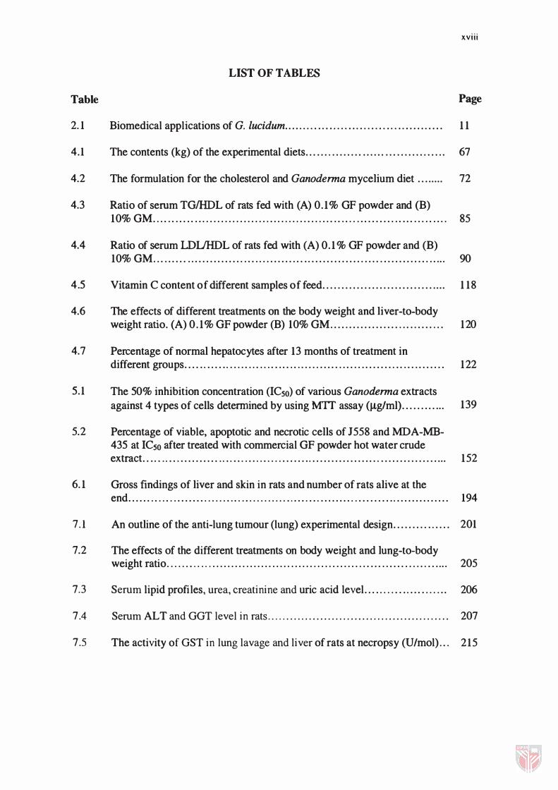
LIST OF TABLES
Table Page
2. 1 Biomedical applications of G. lucidum. . . . ..... . .. . ..... . .. ... .. .. ... .. . . . . .. . . . 1 1
4.1 The contents (kg) of the experimental diets... .... ........ ...... ... ........ ..... 67
4.2 The formulation for the cholesterol and Ganoderma mycelium diet ... ..... 72
4.3 Ratio of serum TGIHDL of rats fed with (A) 0.1 % GF powder and (B) 10%GM .. . . . . . .... . .. . . . . . . . . . . . . . . . . . .. . . . . . . . . . . . . . . . . . . .. . . ......... ..... . . . . . . .. . 85
4.4 Ratio of serum LDUHDL of rats fed with (A) 0.1 % GF powder and (B) 10%GM... ...... . .............. . .. . . . . ....... .... .............. .... . ............ ..... 90
4.5 Vitamin C content of different samples of feed............................. .... 1 18
4.6 The effects of different treatments on the body weight and liver-to-body weight ratio. (A) 0.1 % GF powder (B) 10% GM.................. . .... .... . . . 120
4.7 Percentage of normal hepatocytes after 13 months of treatment in different groups...... .. .... . . . . . . . . . . . . . ... .. . . . . . . ..... . . .. . . . ........ .. . .. ........ 122
5.1 The 50% inhibition concentration (ICso) of various Ganoderma extracts against 4 types of cells determined by using MTT assay (fLglml}............ 139
5.2 Percentage of viable, apoptotic and necrotic cells of J558 and MDA-MB-435 at ICso after treated with commercial GF powder hot water crude extract.... . . . .. . .......... . . .. ... . ... . .. . . . ........ .................. .................. 152
6. 1 Gross findings of liver and skin in rats and number of rats alive at the end...... .. . . . . . ........ . . . . . . .. . . .. . . . . . . . . . ...... ... ......... . .......... .............. 1 94
7.1 An outline of the anti-lung tumour (lung) experimental design........... .... 201
7.2 The effects of the different treatments on body weight and lung-to-body weight ratio. . . . .... . ... . . . . . . . . . . . . . . . . . . . . . . . . . . . . . . . . . . . . . . . . . . ... . . .... . . . . . . ...... 205
7.3 Serum lipid profiles, urea, creatinine and uric acid level..... . . . .... .... . . . . .. 206
7.4 Serum ALT and GGT level in rats................................................ 207
7.5 The activity of GST in lung lavage and liver of rats at necropsy (U/mol) .. . 215
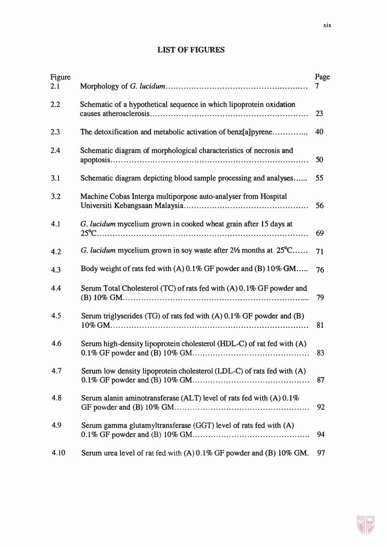
Figure 2.1
2.2
2.3
2.4
3.1
3.2
4.1
4.2
4.3
4.4
4.5
4.6
4.7
4.8
4.9
4.10
xix
LIST OF FIGURES
Page Morphology of G. lucidum... . . .... .. . . . ... . . . . ... . . . . ... . . . .... . . ............... 7
Schematic of a hypothetical sequence in which lipoprotein oxidation causes atherosclerosis... . . . . . . . . . . . . . . . . . . . . . . . . . . . . . . . . . . . . . . . . . . .. . .. . .. . . .. . ... 23
The detoxification and metabolic activation of benz[a]pyrene.............. 40
Schematic diagram of morphological characteristics of necrosis and apoptosis. . . .. . . . . . . . . . . . . . . . . ... . . . . . . .. . . .. ... .. . . . ...... . . .. . ..... . . .............. 50
Schematic diagram depicting blood sample processing and analyses...... 55
Machine Cobas Interga multiporpose auto-analyser from Hospital Universiti Kebangsaan Malaysia .. , ............. , . .. . .. . . .. . . . . .. . ....... . ...... 56
G. lucidum mycelium grown in cooked wheat grain after 15 days at 25°C................................................................................. 69
G. lucidum mycelium grown in soy waste after 21/2 months at 25°C...... 71
Body weight of rats fed with (A) 0.1 % GF powder and (B) 10% OM..... 76
Serum Total Cholesterol (TC) of rats fed with (A) 0.1 % GF powder and (B) 10% GM........................................................................ 79
Serum triglyserides (TO) of rats fed with (A) 0.1 % GF powder and (B) 10%GM ............................................................................ 81
Serum high-density lipoprotein cholesterol (HDL-C) of rat fed with (A) 0.1 % GF powder and (B) 1 0% OM............................................. 83
Serum low density lipoprotein cholesterol (LDL-C) of rats fed with (A) 0.1% GF powder and (B) 1 0%OM ............................................. 87
Serum alanin aminotransferase (ALT) level of rats fed with (A) 0.1 % OF powder and (B) 10% GM...... .............................................. 92
Serum gamma glutamyltransferase (GOT) level of rats fed with (A) 0.1 % OF powder and (B) 10% OM................................. ............ 94
Serum urea level of rat fed with (A) 0.1 % OF powder and (B) 10% GM. 97
4. 1 1 Serum creatinine level of rats fed with (A) 0. 1 % G F powder and (B)
xx
10% GM.. . .. . . . . . . . . . . . . . . . . . . .. ..... . . . . . . . . . . . . . . . . . . . . . . . . . . . . . . . . . . . . . . . . . . . . . . 101
4 .12 Photomicrographs showing the glomerular and tubules in kidney of rats fed with GF powder . . . . . . . . . . . . . . . . . . ... . . . . , . . . . . . . . .... , . . . . ... , . . . . . . . .. . . .. . . . 104
4 . 1 3 Serum uric acid level of rats fed with (A) 0. 1 % GF powder and (B) 10% GM. . . . . . . . . . . . . . . . . . . . . . . . . . . . . . . . . . . . . . .. . . . . . . . . . . . . . . . . . . . . . . . . . . . . . . . . . . . . . .. . . . 105
4. 1 4 Serum creatine kinase (CK) level of rats fed with commercial GP... ...... 107
4 . 1 5 Photomicrographs showing different degree of thickening blood vessel in heart from different groups. . . . . . . . . . . . . . . . . . . . . . . . . . . . . . . . . . . . . . . . . . . .... . . .. 108
4 . 16 Serum Malondialdehyde (MDA) level of rats fed (A) 0. 1 % GF powder and (B) 10% GM. . . . . . . . . . . . . . . . . . . . . . . . . . . . . . . . . . . . . . . . . . . . . . . . . . . . . . . . . .. . . ..... 1 10
4 . 17 Erythrocyte catalase activity of rats fed (A) 0. 1 % GF powder and (B) 10% GM. . . . . . . . . . . . . . . . . . . . . . . . . . . . . . . . . . . . . . . . . .. . . . . . . . . . . . .. . . .. . . . . . . .. . . . ..... 1 12
4 . 18 Whole blood glutathione peroxidase (GSH-Px) of rats fed with (A) 0.1% GF powder and (B) lO%GM.. . . . . . . . . . . . . . . . . . . . . . . . . . . .. . . . . . . . .. . . . ... 1 1 4
4 . 19 Serum vitamin C (ascorbic acid) of rats fed with (A) 0. 1 % GF powder and (B) 10% GM. . . . . . . . . . . . . . . . . . . . . . . . . . . . . . . . . . . . . . .. . . . . . . . .. . . . .. . . . .. . . . . . .. 1 1 6
4 .20 Photographic showing the heart with lung of rat fed with GM in different groups . . . . . . . . . . . . . . . . . . . . . . . . . . . . . . . . . . . . . . . . . . . . . . . . . . . . . . . . . . . . . . . . . . . . 1 19
4.21 Photomicrograph showing liver of rat from different groups. .. . . . . .... . . . . 1 23
5 .1 G. lucidum mycelium mat grown in soy waste milk for 20 days . . . . . . .. . ... 132
5.2 The cytotoxic effect of various Ganoderma extracts against J558. . . . . .. . . 1 4 1
5 .3 The cytotoxic effect of various Ganoderma extracts against MDA-MB-435 . . .. . . . ,. . . . . . . . . . . . . ... . . . . . . . . . . . . .. . . . . . . . . . . . . . . . . . . . . . . . . . . . . . . . . . . . . . . . . . .... 1 42
5 .4 The cytotoxic effect of various Ganoderma extracts against PN6. . . . . . . . . 143
5.5 Photomicrographs showing phase-contrast microscopy examination of J558 with commercial Ganoderma powder hot water crude extract after 72 hours. . . . . . . .. . . . . . . . . . . . . . . . . .. . . ..... . . . . . ... . . . . . . . . . . . . . .. . . . . . . . . . . . . . . . . . . . . 1 47
5.6 Photomicrographs showing phase-contrast microscopy examination of MDA-MB-435 with commercial Ganoderma powder hot water crude
xxi
extract after 72 hours.............................................................. 147
5.7 Photomicrographs showing phase-contrast microscopy examination of PN6 with commercial Ganoderma powder hot water crude extract after 72 hours ................... ,. ... ...... ...... ............... ......... ............ .... 148
5.8 Photomicrographs showing phase-contrast microscopy examination of 3T3 with commercial Ganoderma powder hot water crude extract after 72 hours............................................................................ 148
5.9 Photomirographs showing fluorescence microscopy examination of J558 cells........................................................................... 150
5.10 Photomirographs showing fluorescence microscopy examination of MDA-MB-435 cells............................................................... 151
5.11 Agarose-gel-electrophoretic patterns of DNA isolated from J558 cells after incubation at 37°C for 72 hours with or without commercial Ganoderma powder hot water crude extract ...................................
5.12 Agarose-gel-electrophoretic patterns of DNA isolated from MDA-MB-435 cells after incubation at 37°C for 72 hours with or without commercial Ganoderma powder hot water crude extract ....................
5.13 Eletronmicrographs (SEM) of J558 cells treated with commercial Ganoderma powder hot water crude extract at ICso for 72 hours ...........
5.14 Eletronmicrographs (SEM) of PN6 cells treated with commercial Ganoderma powder hot water crude extract at ICso for 72 hours ...........
5.15 Eletronrnicrographs (SEM) of adherent MDA-MB-43S cells treated with commercial Ganoderma powder hot water crude extract at ICso for 72 hours .................................................................................
5.16 Eletronmicrographs (SEM) of MDA-MB-435 cells treated with commercial Ganoderma powder hot water crude extract at ICso for 72 hours ............... : ................ .................................................
5.17 Eletronmicrographs (TEM) of 1558 cells treated with commercial Ganoderma powder hot water crude extract at IC50 for 72 hours ..........
5.18 Eletronmicrographs (TEM) of MDA-MB-435 cells treated with commercial Ganoderma powder hot water crude extract at IC50 for 72
154
155
157
158
159
160
163
hours... ............ ......... .................. ................ ....................... 165
xxii
6.1 Rat in preparation for topical application of B[a]p........................... 174
6.2 Body weight of rats fed with 0.1 % commercial OF............... ............ 178
6.3 Serum lipid profile in rat. ....... ............... , ............. , .............. ,. '" 179
6.4 Serum (A) gamma glutamyltransferase (OOT) and (B) alanine aminotransferase (ALT) level in rats... ............ ............................ 182
6.5 Serum (A) urea and (B) creatinine level in rats............................... 184
6.6 Serum uric acid level in rats... ......... ... ... .................. ...... ........... 185
6.7 Serum malondialdehyle (MDA) concentration in rats....................... 186
6.8 Erythrocyte catalase activity in rats............................................. 187
6.9 Whole blood glutathione peroxidase (OSH- Px) activity in rats............ 189
6.10 Serum vitamin C content in rats......... ... .................. ................... 191
6.11 Glutathione S-Transferase activity of rats from (A) Liver (B) Lung....... 192
6.12 Photomicrographs showing cross section of skin from different groups after six months. . . . . . . . . . . . . .. . . . . . . . . . . . . . . . . . . . . . . . . . . . . . . .. . .. . . .. . . . .. . . . . . . ... 196
7.1 Serum malondialdehyde concentration in rats... ............ .................. 208
7.2 Erythrocytes catalase activity in rats............................................ 210
7.3 Whole blood glutathione peroxidase (OSH-Px) activity in rats............ 211
7.4 Serum vitamin C content in rats... ....... ................. ...... ............... 214
7.5 Photograph showing the lung and heart of different groups of rats that killed 90 days post instillation................................................... 219
7.6 Photomicrograph of lung tissue from different groups of rats killed 90 days of post instillation........................................................ ... 221
AAT
anti-SRBC
AST
ATCC
Ca
CCl4
Chol
CLL
CTL
em
dL
DMEM
DMSO
EDTA
ELISA
EMEM
FCS
HBSS
g
grp
GF
LIST OF ABBREVIATIONS
Alanine aminotransferase
Anti-Sheep Red Blood Cell
Aspartate aminotransferase
American Type Culture Collection
Calsium
Carbon tetrachloride
Cholesterol
Chronic leukemia
Cytotoxic T lymphocytes
Centrimetre
densilitre
Dulbecco's modification of Eagle's medium
Dimethyl sulfoxide
Ethylene diamine tetraacetic acid
Enzyme linked immunosorbent assay
Eagle's minimum essential medium
Fetal Calf Serum
Hank's Balanced Salt Solution
gram
group
Ganodenna fruiting body
xxiii
xxiv
GGT Gamma glutamyl transferase
GM Ganodenna mycelium
GP Ganodenna polisaccharide
GSH Reduced glutathione
GSH-Px Glutathione peroxidase
GSSG Oxidative glutathione
h hour
H&E Haematoxylin and eosin staining
hGSTPl-l Human GST of class p subunit type 1
H202 Hydrogen peroxide
ICso Inhibition concentration of 50%
IEL Internal Elastic Lamina
KCI Potassium chloride
Kda kilodalton
kg kilograms
L litre
LD Lactate dehydrogenase
M Molar
MDA Malondialdehyde
ml mililitre
mg Milligrams
min minute
mmol Millimolar