universiti putra malaysia molecular and …psasir.upm.edu.my/id/eprint/11690/1/fpv_2003_2_a.pdf ·...
TRANSCRIPT
UNIVERSITI PUTRA MALAYSIA
MOLECULAR AND BIOLOGICAL CHARACTERIZATION OF TWO VERY VIRULENT INFECTIOUS BURSAL DISEASE VIRUS
ISOLATES, UPM94/273 AND UPM97/61
KONG LlH LING
FPV 2003 2
MOLECULAR AND BIOLOGICAL CHARACTERIZATION OF TWO VERY VIRULENT INFECTIOUS BURSAL DISEASE VIRUS ISOLATES,
UPM94/273 AND UPM97/61
By
KONG LlH LING
Thesis Submitted to the School of Graduate Studies, Universiti Putra Malaysia, in Fulfilment of the Requirement for
the Degree of Master of Science
February 2003

Dedicated with love and gratitude to:
My dearest parents, fiance, family and five lovely nieces
11
Abstract of thesis presented to the Senate of Universiti Putra Malaysia in fulfilment of the requirement for the degree of Master of Science
MOLECULAR AND BIOLOGICAL CHARACTERIZATION OF TWO VERY VIRULENT INFECTIOUS BURSAL DISEASE VIRUS ISOLATES,
UPM94/273 AND UPM97/61
By
KONG L1H LING
February 2003
Chairman : Associate Professor Dr. Abdul Rahman Omar, Ph.D.
Faculty: Veterinary Medicine
An atypical very virulent (w) strain (UPM94/273) and typical vv strain
(UPM97/61) of infectious bursal disease virus (IBOV) isolated in
Malaysia, were characterized both in vivo and at the molecular level.
Comparison of the deduced amino acid sequences with other serotype 1
and 2 sequences revealed 16 amino acid residues, which were
conserved only in the vvIBDV. Among the 16 unique amino acid
differences, 8 were in VP1 (146 Asp, 147 Asn, 242 Glu, 390 Met, 393
Asp, 562 Pro, 687 Pro and 695 Arg), 3 were in VP2 (222 Ala, 256 lie and
294 lie), 2 were in VP3 (990 Val and 1005 Ala) and 3 were in VP4 (685
Asn, 715 Ser and 751 Asp). The importance of these unique amino acid
residues is not known but they could affect the virulence of vvIBOV. The
UPM94/273 also demonstrated 6 unique amino acid residues at segment
111
A at positions Ser254, Glu270, Lys588, Ser745, Phe838 and Lys863 and
8 unique amino acid residues at segment B at positions Ala92, Ser1 00,
Va1208, Asp253, Asp560, Asn565, Gly750 and Gly876. In addition, these
amino acid substitutions have not been reported before in wlBDV and
were found only on variant, classical and/or serotype 2 strains. However,
the VP5 region of both wlBDV strains was conserved. The
UPM97/61 demonstrated 7 unique amino acid substitutions at segment A
and 4 unique amino acid substitutions at segment B . However, none of
the amino acids changes have been reported elsewhere in other IBDV
strains. Although the actual functions of the amino acid substitutions are
not know, the unusual amino acid substitutions at segment A and/or B of
both isolates may be important in virus viru lence. Alignments of the
nucleic acid and amino acid sequences of segment A and B followed by
distance analysis al lowed the generation of phylogenetic trees.
Phylogenetic analysis based on segment A and B revealed that all the
wlBDV strains including UPM94/273 isolate can be clustered in a group
that is d istinct from classical , variant, attenuated and serotype 2 strains.
However, the tree branching patterns were qu ite different between
segment A and segment B. in addition , the wlBDV strains showed
several conserved amino acid substitutions at segment B as found in the
Australian 002-73 and serotype 2 strains. These findings indicate that
probably a genetic reassortment may have play an important role in the
emergence of vvIBDV. Flow cytometry and real time peR assays,
indicated that chickens infected with UPM97/61 induced higher
IV

percentages of apoptotic cells but lower level of viral load whereas
UPM94/273 induced lower percentages of apoptotic cel ls but higher level
of viral load, suggesting a negative correlation between viral load and
apoptosis. These results indicated that UPM97/61 was more virulent than
UPM94/273.
v
Abstrak tesis yang dikemukakan kepada Senat Universiti Putra Malaysia sebagai memenuhi keperluan untuk ijazah Master Sains
PENCIRIAN MOLEKUL DAN BIOLOGIK ISOLAT·ISOLAT UPM94/273 DAN UPM97/61 VIRUS PENYAKIT BURSA BERJANGKIT
Oleh
KONG L1H LING
Februari 2003
Pengerusi: Profesor Madya Dr. Abdul Rahman Omar, Ph.D.
Fakulti : Perubatan Veterinar
Virus penyakit bursa berjangkit amat virulen (wi OBV) yang tidak khusus
(UPM94/273) dan yang khusus (UPM97/61 ) d iperolehi dari Malaysia,
telah d icirikan dalam vivo dan berdasarkan molekul . Perbandingan
jujukan asid amino dengan jujukan serotip 1 dan 2 yang lain telah
menunjukkan 1 6 asid amino residu yang cuma terdapat d i wIBOV.
Perbezaan antara 1 6 asid amino yang unik ini , 8 terdapat di VP1 ( 146
Asp, 1 47 Asn, 242 Glu, 390 Met, 393 Asp, 562 Pro, 687 Pro dan 695
Arg), 3 terdapat di VP2 (222 Ala, 256 lie dan 294 l ie), 2 terdapat di VP3
(990 Val dan 1 005 Ala) dan 3 terdapat di VP4 (685 Asn, 71 5 Ser dan 751
Asp). Kepentingan residu-residu asid amino ini adalah tidak diketahui
tetapi ia boleh mempengaruhi viru len vvIBOV. UPM94/273 juga
menunjukkan 6 asid amino yang unik pad a kedudukan Ser254, Glu270,
VI
Lys588, Ser745, Phe838 dan Lys863 mana setiap 2 berada d i VP2, VP3
dan VP4. Tambahan pula, penggantian asid amino pad a kedudukan
Gly254Ser, Ala270Glu, Glu588Lys dan Asp745Ser cuma terdapat di
varian , klasik dan/atau serotip 2. Walaubagaimanapun, bahagian VP5
untuk kedua-dua wlBOV adalah kekal. UPM97/61 menunjukkan 7
penggantian asid amino yang unik d i segmen A dan 4 penggantian asid
amino yang unik d i segmen B. Tetapi, perubahan asid amino in i tidak
pernah dilaporkan pada strain-strain IBOV yang lain. Walaupun fungsi
sebenar penggantian asid amino adalah tidak diketahui, tetapi
penggantian asid amino yang luar biasa ini pada segmen A dan/atau B
untuk kedua-dua isolat mungkin penting dalam virulen virus. Susunan
untuk jujukan asid nukleik dan asid amino untuk segmen A dan B diikuti
oleh anal isis jarak membenarkan pembentukan pokok filogenetik.
Analisis filogenetik berdasarkan segmen A dan B menunjukkan semua
strain wlBOV termasuk isolat UPM94/273 IBOV boleh d ikumpulkan
dalam satu kumpulan d i mana adalah berbeza daripada strain-strain
klasik, varian, akenuat dan serotip 2. Walaupun begitu , corak-corak untuk
cabangan pokok adalah berlainan antara segmen A dan segmen B.
Tambahan pula, strain-strain wlBOV menunjukkan beberapa
penggantian asid amino yang kekal dalam segmen B seperti yang
dijumpai dalam strain-strain Australian 002-73 dan serotip 2. Penemuan
ini menyatakan bahawa kemungkinan penyusunan semula genetic yang
mungkin memainkan peranan yang penting dalam kemunculan wIBOV.
Cara-cara aliran sitometri dan PCR masa sebenar, menyatakan bahawa
VB
ayam-ayam yang dijangkiti dengan UPM97/61 mengakibatkan peratusan
sel-sel apoptosis yang lebih tinggi tetapi takat muatan virus yang lebih
rendah manakala UPM94/273 mengakibatkan peratusan sel-sel
apoptosis yang lebih rendah tetapi takat muatan virus yang lebih tinggi,
mencadangkan satu perhubungan yang negatif wujud d i antara muatan
virus dan apoptosis. Keputusan ini menyatakan bahawa UPM97/61
adalah lebih virulen daripada UPM94/273.
Vlll

ACKNOWLEDGEMENTS
I wou ld l ike to express my heartiest gratitude and appreciation to
Dr. Abdul Rahman Omar, chairman of the supervisory committee for
providing invaluable advice and untiring assistance in this master
research. His constant guidance, participation in laboratory works and
mostly for sharing knowledge on molecular technique that enabled me to
accomplish my work. Moreover, I would l ike to express my sincere thanks
and appreciation to Professor Dr. Aini Ideris, Associate Professor Dr.
Mohd Hair Bejo, Associate Professor Dr. Seow Heng Fong , members of
the supervisory committee for their constructive suggestion, proper
guidance and encouragement throughout my study period .
I am grateful to Maha Abdullah for helping and guiding me in
handling flow cytometry technique; guidance from Dino and Gabrielle in
real time peR approach, and also Siaw Liung and Tzer Miin for their
helpful guidance in SPSS statistical analysis. Special thanks also go to
the staffs in Biologics Lab, Puan Rodiah Husin and Mr. Mohd Redha
Izwan B. Adnani ; and friends Sheau Wei , Siti, Shila, Ain i , Balkis, Lee
Kim, Su Fun, May Ling, Keng Fei, Zul , Thapa, Do Yew, Sok Fang and
every body for always being so willing to render assistance throughout
the course of my study.
IX
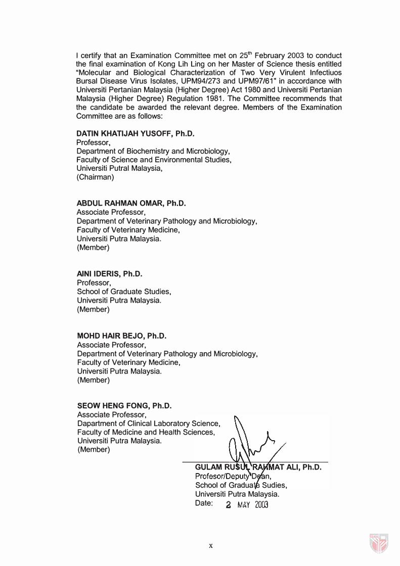
I certify that an Examination Committee met on 25th February 2003 to conduct the final examination of Kong Lih Ling on her Master of Science thesis entitled "Molecular and Biological Characterization of Two Very Virulent Infectiuos Bursal Disease Virus Isolates, UPM94/273 and UPM97/61" in accordance with Universiti Pertanian Malaysia (Higher Degree) Act 1980 and Universiti Pertanian Malaysia (Higher Degree) Regulation 1981. The Committee recommends that the candidate be awarded the relevant degree. Members of the Examination Committee are as follows:
DATIN KHATIJAH YUSOFF, Ph.D. Professor, Department of Biochemistry and Microbiology, Faculty of Science and Environmental Studies, Universiti Putral Malaysia, (Chairman)
ABDUL RAHMAN OMAR, Ph.D. Associate Professor, Department of Veterinary Pathology and Microbiology, Faculty of Veterinary Medicine, Universiti Putra Malaysia. (Member)
AINI IDERIS, Ph.D. Professor, School of Graduate Studies, Universiti Putra Malaysia. (Member)
MOHD HAIR BEJO, Ph.D. Associate Professor, Department of Veterinary Pathology and Microbiology, Faculty of Veterinary Medicine, Universiti Putra Malaysia. (Member)
SEOW HENG FONG, Ph.D. Associate Professor, Dapartment of Clinical Laboratory Science, Faculty of Medicine and Health Sciences, Universiti Putra Malaysia. (Member)
GULAM RU AT ALI, Ph.D. Profesor/Deputy D n, School of Gradua Sudies, Universiti Putra Malaysia. Date: 2 MAY 2003
x
This thesis submitted to the Senate of Universiti Putra Malaysia has been accepted as fulfi lment of the requirement for the degree of Master of Science. The members of the Supervisory Committee are as follows:
ABDUL RAHMAN OMAR, Ph.D. Associate Professor, Department of Veterinary Pathology and Microbiology, Faculty of Veterinary Medicine, Universiti Putra Malaysia. (Chairman)
AINI IDERIS, Ph.D. Professor, School of Graduate Studies, Universiti Putra Malaysia. (Member)
MOHD HAIR BE JO, Ph.D. Associate Professor, Department of Veterinary Pathology and Microbiology, Faculty of Veterinary Medicine, Universiti Putra Malaysia. (Member)
SEOW HENG FONG, Ph.D. Associate Professor, Dapartment of Clinical Laboratory Science, Faculty of Medicine and Health Sciences, Universiti Putra Malaysia. (Member)
Xl
AINI IDERIS, Ph.D. Professor/Dean, School of Graduate Studies, Universiti Putra Malaysia.
Date: 1 2 JUN 2003
DECLARATION
I here declare that the thesis is based on my original work except for quotations and citations which have been duly acknowledged. I also declare that it has not been previously or concurrently submitted for any other degree at UPM or other institutions.
Date: 2/> /2fJ03
xu
DEDICATION ABSTRACT ABSTRAK ACKNOWLEGEMENTS APPROVAL SHEETS DECLARATION FORM LIST OF TABLES LIST OF FIGURES
TABLE OF CONTENTS
LIST OF ABBREVIATIONS
CHAPTER
Page i i i i i vi ix x xii xvi xvii xx
INTRODUCTION.................................................. . . . . . . . . . 1
II LITERATURE REVIEW . . . . . . . . . . . . . . . . . . . . . . . . . . . . . . . . . . . . . . . . . . . . . . . . . 8 I nfectious Bursal Disease . . . . . . . . . . . . . . . . . . . . . . . . . . . . . . . . . . . . . . . . . . . . . . . 8 I nfectious Bursal Disease Virus ( IBDV) . . . . . . . . . . . . . . . . . . . . . . . . . . . . . . 9
Vira l Proteins . . . . . . . . . . . . . . . . . . . . . . . . . . . . . . . . . . . . . . . . . . . . . . . . . . . . . . . 1 0 Antigenic,and Pathotypic Variation . . . . . . . . . . . . . . . . . . . . . . . . . . 1 2 Virulence Markers . . . . . . . . . . . . . . . . . . . . . . . . . . . . . . . . . . . . . . . . . . . . . . . . 1 3
Pathogenesis and Immunosuppression . . . . . . . . . . . . . . . . . . . . . . . . . . . . . 1 5 Diagnosis . . . . . . . . . . . . . . . . . . . . . . . . . . . . . . . . . . . . . . . . . . . . . . . . . . . . . . . . . . . . . . . . . . . . 1 7 Prevention and Control . . . . . . . . . . . . . . . . . . . . . . . . . . . . . . . . . . . . . . . . . . . . . . . . . . 20 Reverse Transcription Polymerase Chain Reaction . . . . . . . . . . . . . 23 Phylogenetic Analysis . . . . . . . . . . . . . . . . . . . . . . . . . . . . . . . . . . . . . . . . . . . . . . . . . . . 24 Flow Cytometric Detection of Apoptosis . . . . . . . . . . . . . . . . . . . . . . . . . . . . 26 Real-Time Quantitative PCR . . . . . . . . . . . . . . . . . . . . . . . . . . . . . . . . . . . . . . . . . . . 28
I I I AMPLIFICATION AND CLONING OF INFECTIOUS BURSAL DISEASE VIRUS BISEGMENTED GENOME....... 31 I ntroduction . . . . . . . . . . . . . . . . . . . . . . . . . . . . . . . . . . . . . . . . . . . . . . . . . . . . . . . . . . . . . . . . . . . . 3 1 Materials and Methods . . . . . . . . . . . . . . . . . . . . . . . . . . . . . . . . . . . . . . . . . . . . . . . . . . . 33 IBDV Isolation . . . . . . . . . . . . . . . . . . . . . . . . . . . . . . . . . . . . . . . . . . . . . . . . . . . . . . . . . . . . . . 33 Propagation of Viruses On Specific Pathogen Free (SPF) Embryonated Chicken Eggs . . . . . . . . . . . . . . . . . . . . . . . . . . . . . . . . . . . . . . . . . . . 33 Extraction of RNA . . . . . . . . . . . . . . . . . . . . . . . . . . . . . . . . . . . . . . . . . . . . . . . . . . . . . . . . . . 34 Determination of RNA Concentration and Purity . . . . . . . . . . . . . . . . . . 35 First-strand Complementary DNA (cDNA) and PCR . . . . . . . . . . . . . 35 Determination of PCR Product Concentration and Purity . . . . . . 38 Agarose Gel Electrophoresis . . . . . . . . . . . . . . . . . . . . . . . . . . . . . . . . . . . . . . . . . . . 38
Xlll
Purification of PCR Products . . . . . . . . . . . . . . . . . . . . . . . . . . . . . . . . . . . . . . . . . . . 39 Cloning of the Full Length of VP1 , VP3, VP4 and VP5 Genes
TOPO® Clon ing Reaction and One Shot® Chemical Transformation . . . . . . . . . . . . . . . . . . . . . . . . . . . . . . . . . . . . . . . . . . . . . . . . . . . 40 Subculture and Analysis of Positive Colonies . . . . . . . . . . . . 4 1
Preparation of Stock Culture . . . . . . . . . . . . . . . . . . . . . . . . . . . . . . . . . . . . . . . . . . . 42 Plasmid Extraction and Purification . . . . . . . . . . . . . . . . . . . . . . . . . . . . . . . . . . . 42 Restriction Enzyme Analysis of the Plasmid . . . . . . . . . . . . . . . . . . . . . . . 44 Results . . . . . . . . . . . . . . . . . . . . . . . . . . . . . . . . . . . . . . . . . . . . . . . . . . .... . . . . . ........... . . . . 45 Amplification of IBDV Genome . . . . . . . . . . . . . . . . . . . . . . . . . . . . . . . . . . . . . . . . 45 Analysis of Recombinant Plasmid . . . . . . . . . . . . . . . . . . . . . . . . . . . . . . . . . . . . . 46 Restriction Enzyme Digestion Analysis . . . . . . . . . . . . . . . . . . . . . . . . . . . . . . 46 Discussion . . . . . . . . . . . . . . . . . . . . . . . . . . . . . . . . . . . . . . . . . . . . . . . . . . . . . . . . . . . . . . . . . . . . . 6 1
IV SEQUENCE AND PHYLOGENETIC ANALYSIS OF IBDV . . . 63
V
I ntroduction . . . . . . . . . . . . . . . . . . . . . . . . . . . . . . . . . . . . . . . . . . . . . . . . . . . . . . . . . . . . . . . . . . . . 63 Materials and Methods . . . . . . . . . . . . . . . . . . . . . . . . . . . . . . . . . . . . . . . . . . . . . . . . . . . 65 Cloning and Sequencing . . . . . . . . . . . . . . . . . . . . . . . . . . . . . . . . . . . . . . . . . . . . . . . . 65 Sequence Assembly and Analysis . . . . . . . . . . . . . . . . . . . . . . . . . . . . . . . . . . . . 65 Construction of Phylogenetic Tree . . . . . . . . . . . . . . . . . . . . . . . . . . . . . . . . . . . . 66 Results . . . . . . . . . . . . . . . . . . . . . . . . . . . . . . . . . . . . . . . . . . . . . . . . . . . . . . . . . . . . . . . . . . . . . . . . . . 69 Sequence Analysis of Segment A . . . . . . . . . . . . . . . . . . . . . . . . . . . . . . . . . . . . 69
VP5 Protein .. . . . . . . . . . . . . . . . . . . . . . . . . . . . . . . . . . . . . . . . . . . . . . . . . . . . . . . 69 Precursor Polyprotein (NH2-VP2-VP4-VP3-COOH) . . . . . 71
Sequence Analysis of Segment B . . . . . . . . . . . . . . . . . . . . . . . . . . . . . . . . . . . . 79 Phylogenetic Analysis . . . . . . . . . . . . . . . . . . . . . . . . . . . . . . . . . . . . . . . . . . . . . . . . . . . 90 Percentage of Homology . . . . . . . . . . . . . . . . . . . . . . . . . . . . . . . . . . . . . . . . . . . . . . . . 93 Discussion . . . . . . . . . . . . . . . . . . . . . . . . . . . . . . . . . . . . . . . . . . . . . . . . . . . . . . . . . . . . . . . . . . . . . 95
FLOW CYTOMETRIC AND REAL TIME QUANTITATIVE PCR ANALYSIS OF IBDV ............................................ .
I ntroduction . . . . . . . . . . . . . . . . . . . . . . . . . . . . . . . . . . . . . . . . . . . . . . . . . . . . . . . . . . . . . . . . . . . .
Materials and Methods . . . . . . . . . . . . . . . . . . . . . . . . . . . . . . . . . . . . . . . . . . . . . . . . . . .
Viruses . . . . . . . . . . . . . . . . . . . . . . . . . . . . . . . . . . . . . . . . . . . . . . . . . . . . . . . . . . . . . . . . . . . . . . . . . .
SPF Embryonated Chicken Eggs . . . . . . . . . . . . . . . . . . . . . . . . . . . . . . . . . . . . . Chorioal lantoic Membrane (CAM) Preparation . . . . . . . . . . .
IBDV I noculation . . . . . . . . . . . . . . . . . . . . . . . . . . . . . . . . . . . . . . . . . . . . . . . ..
Titration of I BDV . . . . . . . . . . . . . . . . . . . . . . . . . . . . . . . . . . . . . . . . . . . . . . . . . . . . . . . . . . ..
SPF Chickens . . . . . . . . . . . . . . . . . . . . . . . . . . . . . . . . . . . . . . . . . . . . . . . . . . . . . . . . . . . . . . ..
Experimental Design . . . . . . . . . . . . . . . . . . . . . . . . . . . . . . . . . . . . . . . . . . . .
Single Cell Suspension Preparation . . . . . . . . . . . . . . . . . . . . . . . . . . . . . . . . .
Viability (Trypan Blue) Test . . . . . . . . . . . . . . . . . . . . . . . . . . . . . . . . . . . . . . . . . . . . .
Propidium Iod ide Staining . . . . . . . . . . . . . . . . . . . . . . . . . . . . . . . . . . . . . . . . . . . . . . .
RNA Extraction . . . . . . . . . . . . . . . . . . . . . . . . . . . . . . . . . . . . . . . . . . . . . . . . . . . . . . . . . . . . . Primer Designing . . . . . . . . . . . . . . . . . . . . . . . . . . . . . . . . . . . . . . . . . . . . . . . . . . . . . . . . . . .
Conventional RT -PCR Amplification . . . . . . . . . . . . . . . . . . . . . . . . . . . . . . . . .
XIV
1 02 1 02 1 04 1 04 1 04 1 04 1 05 1 05 1 05 1 05 1 06 1 07 1 07 1 08 1 08 1 09
SYBR Green 1 Optimization . . . . . . . . . . . . . . . . . . . . . . . . . . . . . . . . . . . . . . . . . . . 1 09 Real-Time Quantitative PCR . . . . . . . . . . . . . . . . . . . . . . . . . . . . . . . . . . . . . . . . . . . 1 09 Statistical Analysis . . . . . . . . . . . . . . . . . . . . . . . . . . . . . . . . . . � . . . . . . . . . . . . . . . . . . . . . . 1 1 1 Results . . . . . . . . . . . . . . . . . '" . . . . . . . . . . . . . . . . . . . . . . . . . . . . . . . . . . . . . . . . . . . . . . . . . . . . . . 1 1 1 Flow Cytometric Analysis . . . . . . . . . . . . . . . . . . . . . . . . . . . . . . . . . . . . . . . . . . . . . . . 1 1 1
Propidium Iodide Analysis . . . . . . . . . . . . . . . . . . . . . . . . . . . . . . . . . . . . . 1 1 1 PCR Amplification . . . . . . . . . . . . . . . . . . . . . . . . . . . . . . . . . . . . . . . . . . . . . . . . . . . . . . . . . 1 1 8 Real-Time Quantitative PCR Analysis . . . . . . . . . . . . . . . . . . . . . . . . . . . . . . . 1 1 8
SYBR Green I Optimization . . . . . . . . . . . . . . . . . . . . . . . . . . . . . . . . . . . 1 1 8 Standard Curve . . . . . . . . . . . . . . . . . . . . . . . . . . . . . . . . . . . . . . . . . . . . . . . . . . . 1 1 8 Comparison of Viral RNA Levels . . . . . . . . . . . . . . . . . . . . . . . . . . . . . 1 20
Discussion . . . . . . . . . . . . . . . . . . . . . . . . . . . . . . . . . . . . . . . . . . . . . . . . . . . . . . . . . . . . . . . . . . . . . 1 26
VI GENERAL DISCUSSION AND CONCLUSION . . . . . . . . . . . . . . . . . . 1 3 1
BIBLIOGRAPHY . . . . . . . . . . . . . . . . . . . . . . . . . . . . . . . . . . . . . . . . . . . . . . . . . . . . . . . . . . . . . . . . . . . . 1 38
APPENDICES . . . . . . . . . . . . . . . . . . . . . . . . . . . . . . . . . . . . . . . . . . . . . . . . . . . . . . . . . . . . . . . . . . . . . . . . 1 62 A. Percent Embryo Lethal Dose . . . . . . . . . . . . . . . . . . . . . . . . . . . . . . . . 1 62 B . Nucleotide and Amino Acid Sequence Alignment. . . . . . 1 63 C. Phylogenetic Tree. . . . . . . . . . . . . . . . . . . . . . . . . . . . . . . . . . . . . . . . . . . . . . . 208 D. Genetic Distances. . . . . . . . . . . . . . . . . . . . . . . . . . . . . . . . . . . . . . . . . . . . . . . 21 0 E. Statistical Analysis for Flow Cytometry and Real Time
Quantitative PCR . . . . . . . . . . . . . . . . . . . . . . . . . . . . . . . . . . . . . . . . . . . . . . . . 2 12
VITA . . . . . . . . . . . . . . . . . . . . . . . . . . . . . . . . . . . . . . . . . . . . . . . . . . . . . . . . . . . . . . . . . . . . . . . . . . . . . . . . . 230
PUBLICATION . . . . . . . . . . . . . . . . . . . . . . . . . . . . . . . . . . . . . . . . . . . . . . . . . . . . . . . . . . . . . . . . . . . 23 1
xv
LIST OF TABLES
Table
3. 1 Primers used to amplify VP3, VP4, VP5 genes and
Page
segment B ... . . . . . . . . . . . . . . . . ... . . . . . . . . . . . . . . . . . . . . . . . . . . . . . . . . . . . . . .. 36
4. 1 Characteristics of IBDV strains used in multiple alignment analysis of segment A sequence. . . . . . . . . . . . . . . . . . 67
4.2 Characteristics of IBDV strains used in multiple alignment analysis of segment B sequence. . . . . . . . . . . . . . . . . . 68
4.3 Comparison of Amino Acid Substitution at Different Position Between UPM94/273 and Other Published Strains in Segment A of IBDV. . . . . . . . . . . . . . . . . . . . . . . . . . . . . . . . . . . . 78
4.4 Comparison of Amino Acid Substitution at Different Position Between Malaysian Strains and Other Published Strains in Segment B of IBDV. . . . . . . . . . . . . . . . . . . . . . 89
5. 1 Percentage of apoptotic cells determined by flow cytometry in IBDV infected and uninfected bursa lymphoid cells. . . . . . . . . . . . . . . . . . . . . . . . . . . . . . . . . . . . . . . . . . . . . . . . . . . . . . . . . 1 1 7
5.2 Quantitation of viral RNA in bursa lymphoid cells from chickens infected with vaccine strain 078, wlBDV strains UPM94/273 and UPM97/61 based on cycle threshold values . . . . . . . . . . . . . . . . . . . . . . . . . . . . . . . . . . . . . . . . . . . . . . . . . . . . . . 1 2 1
5.3 Mortal ity of SPF chickens following infection with different IBDV strains . . . . . . . . . . . . . . . . . . . . . . . . . . . . . . . . . . . . . . . . . . . . . . . 1 23
XVI
LIST OF FIGURES
Figure Page
3 . 1 RT-PCR Product of the Expected UPM94/273 IBDV VP3 Gene Using Specific Primer. . . . . . . . . . . . . . . . . . . . . . . . . . . . . . . . . . . . . . . 47
3.2 RT -PCR Product of the Expected UPM94/273 IBDV VP4 Gene Using Specific Primer . . . . . . . . . . . . . . . . . . . . . . . . . . . . . . . . . . . . . . . 48
3.3 RT- PCR Product of the Expected UPM94/273 IBDV VP5 Gene Using Specific Primer. . . . . . . . . . . . . . . . . . . . . . . . . . . . . . . . . . . . . . . 49
3.4 RT - PCR Product of the Expected 3 Fragments for VP1 Gene of UPM94/273 and UPM97/61 IBDV Using Specific Primers . . . . . . . . . . . . . . . . . . . . . . . . . . . . . . . . . . . . . . . . . . . . . . . . . . . . . . . . . . . . . . . . . . . 50
3 .5 PCR Screening for Recombinant VP3 Gene of UPM94/273 IBDV Isolate . . . . . . . . . . . . . . . . . . . . . . . . . . . . . . . . . . . . . . . . . . . . 51
3 .6 PCR Screening for Recombinant VP4 Gene of UPM94/273 IBDV Isolate . . . . . . . . . . . . . . . . . . . . . . . . . . . . . . . . . . . . . . . . . . . . 52
3 .7 PCR Screening for Recombinant VP5 Gene of UPM94/273 IBDV Isolate . . . . . . . . . . . . . . . . . . . . . . . . . . . . . . . . . . . . . . . . . . . . 53
3.8 PCR Screening for Recombinant Fragment 1 of VP1 Gene of UPM94/273 and UPM97/61 IBDV Isolates . . . . . . . . . . 54
3 .9 PCR Screening for Recombinant Fragment 2 of VP1 Gene of UPm94/273 and UPM97/61 IBDV Isolates . . . . . . . . . . 55
3 . 1 0 PCR Screening for Recombinant Fragment 3 of VP1 Gene of UPM94/273 and UPM97/61 IBDV Isolates . . . . . . . . . . 56
3 . 1 1 Purified Plasmids Digested With EcoR1 Enzyme for VP3 Gene of UPM94/273 IBDV Isolate . . . . . . . . . . . . . . . . . . . . . . . . . . . . . . . . 57
3. 1 2 Purified Plasmids Digested With EcoR1 Enzyme for VP4 Gene of UPM94/273 IBDV Isolate . . . . . . . . . . . . . . . . . . . . . . . . . . . . . . . . 58
3. 1 3 Purified Plasm ids Digested With EcoR1 Enzyme for VP5 Gene of UPM94/273 IBDV Isolate . . . . . . . . . . . . . . . . . . . . . . . . . . . . . . . . 59
3. 1 4 Purified Plasm ids Digested With EcoR1 Enzyme for 3 Fragments of VP1 Gene of UPM94/273 and UPM97/61 IBDV Isolates . . . . . . . . . . . . . . . . . . . . . . . . . . . . . . . . . . . . . . . . . . . . . . . . . . . . . . . . . . . 60
XVll
4 . 1 Nucleotide Sequence and Translation of Amino Acid of VP5 Gene of UPM94/273 IBDV Isolate . . . . . . . . . . . . . . . . . . . . . . . . . . 70
4 .2 Nucleotide Sequence and Translation of Amino Acid of Precursor Polyprotein, VP2-VP4-VP3 Genes of 72 UPM94/273 IBDV Isolate . . . . . . . . . . . . . . . . . . . . . . . . . . . . . . . . . . . . . . . . . . . .
4.3 Nucleotide Sequence and Translation of Amino Acid of Segment B , VP1 Gene of UPM94/273 IBDV Isolate. . . . . . . . . 80
4.4 Nucleotide Sequence and Translation of Amino Acid of Segment B , VP1 Gene of UPM97/61 IBDV Isolate . . . . . . . . . . . 84
4.5 Phylogenetic Tree Construction Based on Amino Acids Sequence of the Segment A IBDV Isolates . . . . . . . . . . . . . . . . . . . . . 91
4 .6 Phylogenetic Tree Construction Based on Amino Acids Sequence of the Segment B IBDV Isolates . . . . . . . . . . . . . . . . . . . . . 92
5 . 1 Detection of apoptosis in bursal lymphocytes by flow cytometry for propidium iodide staining. Bursal lymphocytes were isolated from uninfected control chickens at (A) day 1 , (B) day 2, (C) day 3, (D) day 7 and (E) day 1 4 . . . . . . . . . . . . . . . . . . . . . . . . . . . . . . . . . . . . . . . . . . . . . . . . . . . . . . . . . . . . . . . 1 1 2
5.2 Detection of apoptosis in bursal lymphocytes by flow cytometry for propidium iodide staining. Bursal lymphocytes were isolated from vaccine strain 078 infected chickens at (A) day 1 , (B) day 2, (C) day 3, (D) day 7 and (E) day 1 4 . . . . . . . . . . . . . . . . . . . . . . . . . . . . . . . . . . . . . . . . . . . . . . . . . 1 1 3
5.3 Detection of apoptosis in bursal lymphocytes by flow cytometry for propidium iodide staining. Bursal lymphocytes were isolated from atypical wlBDV strain UPM94/273 infected chickens at (A) day 1 , (B) day 2, (C) day 3, (D) day 7 and (E) day 1 4 . . . . . . . . . . . . . . . . . . . . . . . . . . . . . . . . . . . 1 1 4
5.4 Detection of apoptosis in bursal lymphocytes by flow cytometry for propidium iod ide staining. Bursal lymphocytes were isolated from typical wlBDV strain UPM97/61 infected chickens at (A) day 1 , (B) day 2 , (C) day 3, (D) day 7 and (E) day 1 4 . . . . . . . . . . . . . . . . . . . . . . . . . . . . . . . . . . . 1 1 5
5.5 RT-PCR products of the 1 20 bp VP2 gene. . . . . . . . . . . . . . . . . . . . . 1 1 9
5.6 Standard curve of serial 1 0-fold d ilutions of quantitative standard from 1 0-1 to 1 0-5 mg/ml UPM94/273 IBDV stock virus. . . . . . . . . . . . . . . . . . . . . . . . . . . . . . . . . . . . . . . . . . . . . . . . . . . . . . . . . . . . . . . . . . . . . . . 1 22
XVlll
5.7 A l inear relationship was observed between the amount of input RNA and the CT values for the I BOV infected lymphoid cells, labeled as unknown samples . . . . . . . . . . . . . . . . . . 1 22
5.8 Quantitation of viral RNA in bursa lymphoid cells from chickens infected with vaccine strain 078, wlBOV strains UPM94/273 and UPM97/61 based on log concentration . . . 1 24
5.9 (A) Fluorescence vs. temperature plot of data collected during the melt curve run protocol between 54°C-95°C following amplification of wlBOV strains with SYBR Green I . (B) Melt curve data collected after wlBOV strains amplification with designated primers produced a product peak at 88°C. . . . . . . . . . . . . . . . . . . . . . . . . . . . . . . . . . . . . . . . . . . . . . . . . . . . . . . . . . . . 1 25
XIX
AGPT BLAST bp BSA CAM cDNA °c CE CMX-Ros CT DEPC DH20 DMSO DNA dNTP ds DTT dUTP-FITC E. coli EDTA EIDso ELISA HVT IBD I BDV I FN I PNV kb kDa LB M MC-540 MDA MgS04 ml mM
).1g NaCI Nal NaOH NCBI ng NJ OD ORF
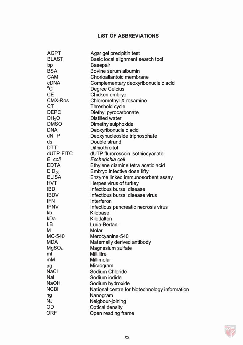
LIST OF ABBREVIATIONS
Agar gel precipitin test Basic local alignment search tool Basepair Bovine serum albumin Chorioallantoic membrane Complementary deoxyribonucleic acid Degree Celcius Chicken embryo Chloromethyl-X-rosamine Threshold cycle Diethyl pyrocarbonate Distilled water Dimethylsulphoxide Deoxyribonucleic acid Deoxynucleoside triphosphate Double strand Dithiothreitol dUTP flurorescein isothiocyanate Escherichia coli Ethylene d iamine tetra acetic acid Embryo infective dose fifty Enzyme linked immunosorbent assay Herpes virus of turkey Infectious bursal d isease Infectious bursal disease virus I nterferon Infectious pancreatic necrosis virus Kilobase Kilodalton Luria-Bertani Molar Merocyanine-540 Maternally derived antibody Magnesium sulfate Mill i l itre Mill imolar Microgram Sodium Chloride Sodium iodide Sodium hydroxide National centre for biotechnology information Nanogram Neigbour-joining Optical density Open reading frame
xx
PBS PCR pi PI pmol PTC QC-PCR QGDPT RdRp RFLP RNA RT-PCR RT SDS SPF SPSS TAE TCVN Tris TUNEL
UPGMA UPM UV VNF VP w (w/v) X-gal
Amino Acid Alanine Arginine Asparagine Aspartic Acid Glutamine Glutamic Acid Glycine Isoleucine Leucine Lycine Methionine Phenylalanine Proline Serine Threonine Tryptophan Valine
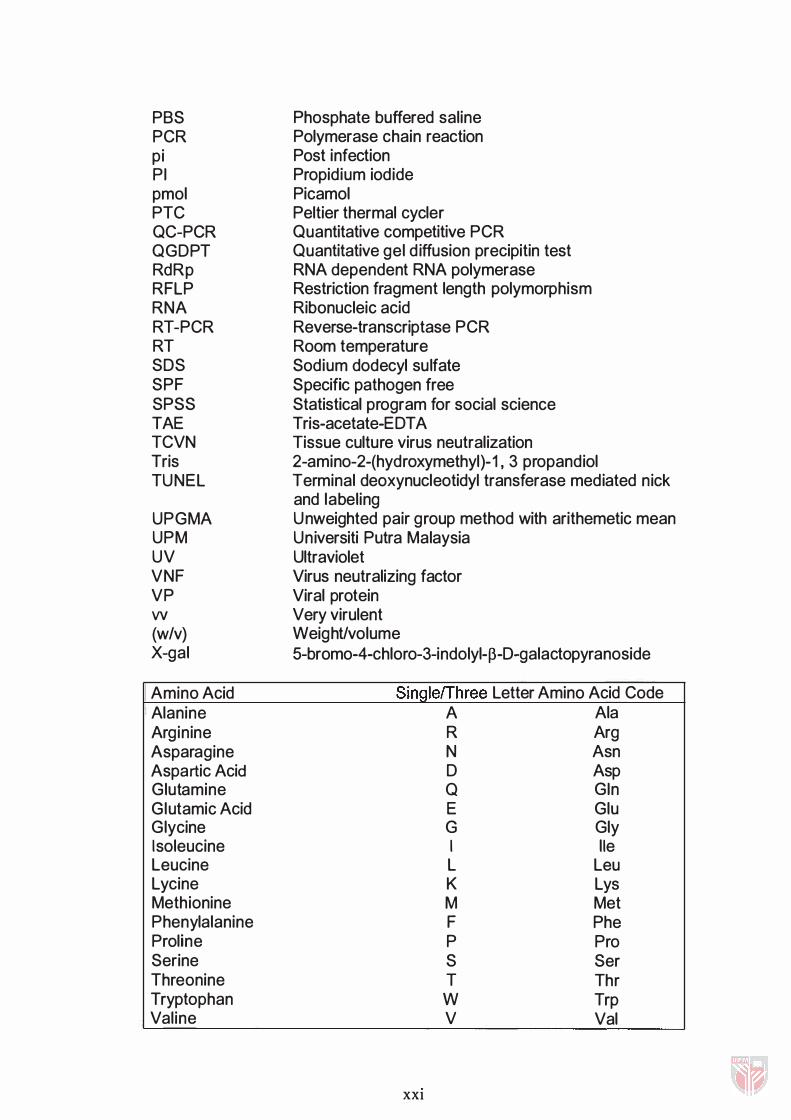
Phosphate buffered saline Polymerase chain reaction Post infection Propidium iodide Picamol Peltier thermal cycler Quantitative competitive PCR Quantitative gel d iffusion precipitin test RNA dependent RNA polymerase Restriction fragment length polymorphism Ribonucleic acid Reverse-transcriptase PCR Room temperature Sodium dodecyl sulfate Specific pathogen free Statistical program for social science Tris-acetate-EDTA Tissue culture virus neutralization 2-amino-2-(hydroxymethyl)-1 , 3 propandiol Terminal deoxynucleotidyl transferase mediated nick and labeling Unweighted pair group method with arithemetic mean Universiti Putra Malaysia Ultraviolet Virus neutralizing factor Viral protein Very virulent Weight/volume 5-bromo-4-chloro-3-indolyl-�-D-galactopyranoside
SinglelThree Letter Amino Acid Code A Ala R Arg N Asn D Asp Q Gin E Glu G Gly I lie L Leu K Lys M Met F Phe P Pro S Ser T Thr W Trp V Val
XXI
CHAPTER I
INTRODUCTION
I nfectious bursal disease (IBD) is an acute contagious viral
disease of young chickens (Kibenge et al. , 1 988; Lasher et al. , 1 994).
The etiological agent, IBD virus ( IBDV), has a predilection for the cells of
the bursa of Fabricius where the virus infects actively d ividing and
differentiating lymphocytes of the B-cell l ineage (Burkhardt et al. , 1 987).
Thus, IBD is a fatal immunosuppressive disease causing heavy losses to
the poultry industry (Eterradossi et al., 1 998).
The first outbreak of IBD was reported in commercial chicken
flocks in Delaware, USA (Cosgrove, 1 962). The IBDV strains, which were
isolated during this outbreak, are now referred to as classical serotype I
isolates. Later on, a second serotype - serotype" of IBDV was identified
(McNulty and Saif, 1 988). These isolates are apathogenic and are
recovered mainly from turkeys ( Ismail et al. , 1 988). Based on antigenic
variation and virulence, serotype I isolates can be divided into several
groups : classical virulent, attenuated, antigenic variant and very viru lent
(w) strains (Cao et al. , 1 998). Since 1 985, antigenic variants of serotype
I I BDV isolates had been recovered from flocks with selection pressure of
field vaccination against classical IBDV serotype I (Snyder, 1 990).
Although being antigenic variant, these isolates have only minor amino
acid changes and do not form any separate serotype. Nevertheless,
these changes occur at the VP2 conformation-dependent antigenic
epitopes that are responsible for stimulating virus neutral izing antibodies
(Bayl iss et al., 1 990). In 1 991 , IBOV isolates, which were able to break
through levels of maternal antibodies that were normally protective, were
reported in Europe (Chettle et al. , 1 989). These isolates, the so cal led
very virulent IBOV (wIBOV), cause more severe clinical signs during an
outbreak with "mortality approaching 1 00% in susceptible flocks, and are
now found almost world-wide (VandenBerg, 2000).
The emergence of highly virulent strains of IBOV has complicated
the immunization programs against the disease. Early vaccination may
result in fai lure due to interference with the maternal antibody, whilst its
delay may cause field virus infections. Therefore, it is important to
characterize the antigenicity and the virulence of IBOV in both vaccine
and field strains in the control of the disease. The effectiveness in the
latter is also dependent on the diagnostic methods used. The d isease
can be d iagnosed based on virus isolation, electron microscopy,
immunofluorescence, virus neutralization, monoclonal antibody assays,
and/or enzyme-linked immunosorbent assay (Lukert and Saif, 1 991 ; Wu
et al. , 1 992; Liu et al., 1 994). However, these methods have one or more
d isadvantages such as being time consuming, labour intensive,
expensive and of low sensitivity (Wu et al. , 1 992).
2
Recently, more sensitive and specific molecular methods have
been used to diagnose and characterize IBOV infections (Jackwood and
Nielsen, 1 997; Moody et a/. , 2000; Boot et al. , 200 1 ). The reverse
transcriptase polymerase chain (RT-PCR) has been widely used to
detect IBOV (Tham et al., 1 995; Jackwood and Nielsen, 1 997). RT-PCR
followed by restriction fragment length polymorphism (RFLP) has also
been used to detect and d ifferentiate IBOV strains (Jackwood and
Sommer, 1 997; Hoque et al., 200 1 ). RT-PCR RFLP profiles of the
amplified hypervariable region of the VP2 gene have been used to
d iagnose and identify molecular differences in the IBOV strains isolated
in different parts of the world (Jackwood and Sommer, 1 999; Hoque et
al., 2001 ). I n these studies, it was found that al l wlBOV isolates have a
conserved Ssp1 and Taq1 sites at the hypervariable region of the VP2
gene. A study has also been carried out to develop a PCR method for the
detection of IBOV based on colorimetric technique (Phong, 2002).
Generally the severity of IBOV infections has been assessed in
terms of mortal ity or the degree of bursal damage, and it has been
difficult to assess viral load because viru lent strains of IBOV do not
repl icate in tissue culture (Moody et al. , 2000). A quantitative competitive
PCR (QC-PCR) assay has been developed to monitor IBOV RNA
extracted from infected bursae (Wu et al. , 1 997). However, this protocol
is labour intensive and the technique has limitations (Souaze et al. ,
1 996). Difficulties in using the RT -PCR technique for quantification
3