studies on some sooty moulds on guava in malaysia · laporkan sebagai laporan terbaru di...
TRANSCRIPT
Pertanika 11(3), 349-355 (1988)
Studies on Some Sooty Moulds on Guava In Malaysia
LIM TONG KWEEDepartment ofPlant Protection
Faculty ofAgriculture,Universiti Pertanian Malaysia
43400 Serdang, Selangor, Malaysia
ABSTRAK
Kulat jelaga membiak dengan banyak di atas daun jambu batu, mendapat makanan dari bahan rnanisan yang dikeluarkan oleh serangga teritip, kutu daun, lalat putih dan koya. Sembi/an spesies, limaspesies dari Ascomyetes iaitu: Phragmocapnias betle, Scorias philippensis, Trichomerium grandisporum,Limacinula musicola dan Aithaloderma clavatisporum, serta empat genus dari Deuteromycetes iaitu:Tripospermum sp., Polychaeton sp., Leptoxyphium sp. dan Conidiocarpus sp. adalah dihuraikan dan dilaporkan sebagai laporan terbaru di Semenanjung Malaysia.
ABSTRACT
Sooty moulds proliferate in abundance on the foliage of guava, subsisting on the honeydew secretedby scale insects aphids, whiteflies, and mealy bugs. Nine species, five Ascomycetes species namely, Phragmocapnias betle, Scorias philippensis, Trichomerium grandisporum, Limacinula musicola and Aithalodermaclavatisporum; and four Deuteromycetes genera viz. Tripospermum sp., Polychaeton sp., Leptoxyphiumsp. and Conidiocarpus sp., are describ~d and reported as new records on guava for Peninsular Malaysia.
INTRODUCTIONSooty moulds are a commonsight on the leaves,twigs and sometimes fruits of many' tropical fruitcrops (Lim and Khoo, 1985). They form thin,black, papery films; black, velutinous growth,black pellicles, or psuedoparenchymatous crusts.Such growths represent unit communities of anassemblage of several diverse species of fungibdonging to the Deuteromycetes and Ascomycetes with dark-coloured somatic and fruit-bodycells, living together saprotrophically in apparentharmonious and mutualistic association. Theirubiquity and abundant proliferation on suchmicro-habitats can be attributed to their adaptability to grow and reproduce in the high stressenvironment of the crop foliage. In accordancewith Grime's ecological terminology (1979), theycan be termed as stress-selected or S-selectedfungi. Such fungi have culminated in a stresstolerant strategy for survival involving the development of adaptations which facilitate endurance ofconditions of continuous environmental stress as
are experienced on the foliage. They can also bedeemed as true foliage residents.
Their diversity, temporal and spatial distribution on such niches depend on the supply ofnutrients, availability of moisture, and thermal andradiation fluctuations. The sooty moulds obtaintheir water from free water films or water dropletson leaves from the rain or dew, atmosphericwater vapour, and water exuded via guttation.They can obtain their nutrition from leaf diffusateor guttation fluid (Tukey, 1971); the cuticle whichproVide a rich and potential source (Baker, 1971,Holloway, 1971); honey dew secretions frominsects such as scale insects, aphids, whiteflies andmealybugs; chemical contaminated rain; organicand inorganic dust particles; pollens; and sporesof other microflora. Thus, they can be found onthe plant foliage with or without the presence ofthe above-mentioned insects. However, on guava(Psidium guajava L.), sooty moulds are usuallyfound in association with insects, subsisting onthe rich, nutritive nabulum of the honey dew.
LIM TONG KWEE
Despite their ubiquity and plenitude, theirsignificance is usually overlooked. Many of thehyphae of the sooty moulds are mucilaginous,absorbing moisture readily and maintaining amoist surface for prolonged periods. This creates avery humid environment within the tree canopy,facilitating the establishment and spread of otherfungal diseases. Extensive growth of the sootymould on the leaves can reduce the photosynthethic activity of the leaves and adversely affects thenormal flowering and fruiting of the tree. Besides,trees heavily colonized by these fungi exhibit adecrease in growth vigour and tend to fruit poorly.On fruits, sooty moulds spoil the cosmetic appearance of the fruits and reduce their marketablevalue (Lim and Khoo, 1985).
Information and studies on sooty mould offruit trees in Malaysia are rather meagre andscanty, particularly on guava. In Peninsular Malaysia, only one sooty mould was listed on guavaTrichopeltis pulchella Speg. (Johnston, 1960).In Sarawak, four species namely : Atichia glomerulosa (Ach.) Flotow, Capnodium moniliformeFraser, Capnophaeum sp. and Phaeochaetiaannonicola (Hansf.) Bat. & Cif. were recorded byTurner (1971). In Sabah, Williams and Uu (1976)recorded three species viz. Aithaloderrna clavatisporum Syd., Caldariomyces fumago Woron anda Chaetothyrium sp.
This paper reports and describes nine previo'usly unrecorded species of sooty mould foundon guava in Peninsular Malaysia. The specimens arekept at the Plant Pathology Herbarium, Department of Plant Protection, Universiti PertanianMalaysia at Serdang, Selangor.
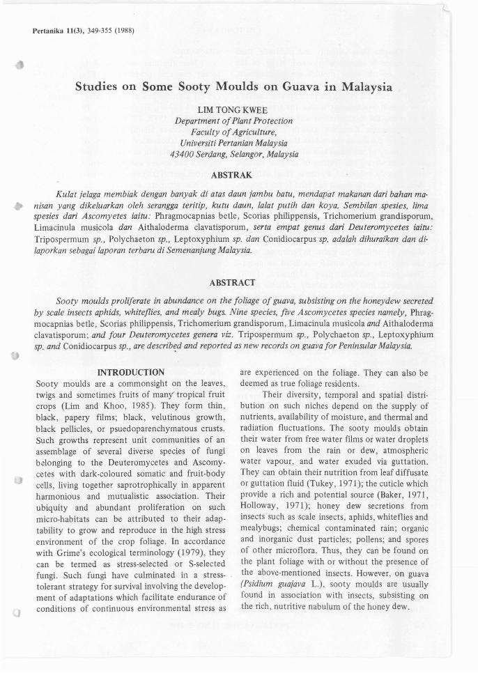
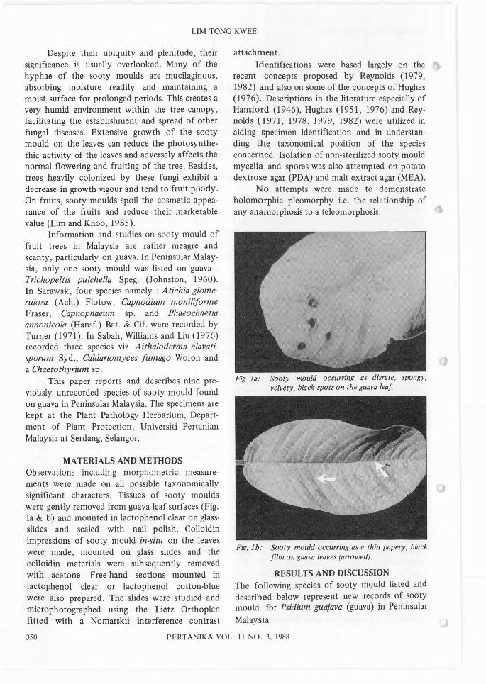
MATERIALS AND METHODSObservations including morphometric measurements were made on all possible taxonomicallysignificant characters. Tissues of sooty mouldswere gently removed from guava leaf surfaces (Fig.la & b) and mounted in lactophenol clear on glassslides and sealed with nail polish. Colloidinimpressions of sooty mould in-situ on the leaveswere made, mounted on glass slides and thecolloidin materials were subsequently removedwith acetone. Free-hand sections mounted inlactophenol clear. or lactophenol cotton-bluewere also prepared. The slides were studied andmicrophotographed using the Lietz Orthoplanfitted with a Nomarskii interference contrast
attachment.Identifications were based largely on the
recent concepts proposed by Reynolds (1979,1982) and also on some of the concepts of Hughes(1976). Descriptions in the literature especially ofHansford (1946), Hughes (1951, 1976) and Reynolds (1971, 1978, 1979, 1982) were utilized inaiding specimen identification and in understanding the taxonomical position of the speciesconcerned. Isolation of non-sterilized sooty mouldmycelia and spores was also attempted on potatodextrose agar (PDA) and malt extract agar (MEA).
No attempts were made to demonstrateholomorphic pleomorphy i.e. the relationship ofany anamorphosis to a teleomorphosis.
Fig. la: Sooty mould occurring as disrete, spongy,velvety, black spots on the guava leaf
Fig. 1b: Sooty mould occu"ing as a thin papery, blackfilm on guava leaves (a"owed).
RESULTS AND DISCUSSIONThe following species of sooty mould listed anddescribed below represent new records of sootymould for Psidium guajava (guava) in Peninsular
Malaysia.
350 PERTANIKA VOL. II NO.3, 1988
STUDIES ON SOME SOOTY MOULDS IN aUAVA IN MALAYSIA.
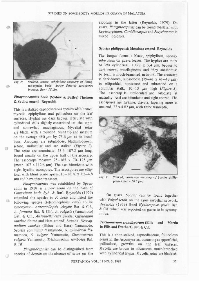
Fig. 2: Stalked, setose, subglobose ascocarp of Phragmocapnias betle. Arrow denotes ascosporesin ascus. Bar =18 ~.
Phragmocapnias betle (Sydow & Butler) Theissen& Sydow emend. Reynolds.
This is a stalked capnodiaceous species with brownmycelia, epiphyllous and pelliculose on the leafsurfaces. Hyphae are dark brown, reticulate withcylindrical cells slightly constricted at the septaand somewhat mucilaginous. Mycelial setaeare black, with a rounded, blunt tip and measureon the average 410 pm by 75.6 pm at its broadbase. Ascocarp are subglobose, blackish-brown,setose, unilocular and are stalked (Figure 2).The setae are acuminate, 53.6-107.2 pm long,found usually on the upper half of the ascocarp.The ascocarps measure 75-165 x 70-125 J.llrI(mean 107 x 112.6 J.IITl). The asci bitunicate witheight hyaline ascospores. The ascospores are elliptical with blunt acute apices, 16-18.76 x 3.2-4.8J,Jm and have three transepta.
Phragmocapnias was established by Spegazinni in 1918 as a new genus on the basis ofCapnodium betle Syd. & But!. Reynolds (1979)emended the species to P. betle and listed thefollowing species (teleomorphosis only) to besynonyms:- Antennellopsis elegans Bat. & Cif.,A. formosa Bat. & Clif., A. vulgaris (Yamamoto)Bat. & Clf., Antennella citri Swada, Capnodiumtanakae Shirae and Hara emend. Sawada, Neocapnodium tanakae (Shirae and Hara) Yamamoto,Scorias communis Yamamoto, S. cylindrical Yamamoto, S. vulgare Yamamoto, Chaetoscoriasvulgaris Yamamoto, TrichQmerium jambosae Bat.&Cif.
Phragmocapnias can be distinguished fromspecies of Scorias on the absence of setae on the
ascocarp in the latter (Reynolds, 1979). Onguava, Phragmocapnias can be found together withLeptoxyphium, Conidiocarpus andPolychaeton inmixed colonies.
Scorias philippensis Mendoza emend. Reynolds
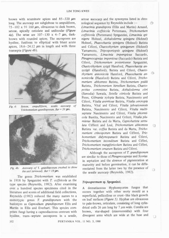
The fungus forms a black, epiphyllous, spongysubiculum on guava leaves. The hyphae are moreor less cylindrical, 10.72 x 5.4 J.llrI, brown todark-brown, mucilaginous and they anastomiseto form a much-branched network. The ascocarpis dark-brown, subglobose (39-41 x 41-43 J.llrI)to ellipsoidal, nonsetose and subtended on acolumnar stalk, 10-15 J.llrI high (Figure 3).The ascocarp is uniloculate and ostiolate atmaturity. Asci are bitunicate and eight-spored. Theascospores are hyaline, clavate, tapering more atone end, 22 x 4.82 J.IITl, with three transepta.
Fig. 3: Stalked, nonsetose ascocarp of Scorias philippenses. Bar =10.5 ~.
On guava, Scorias can be found togetherwith Polychaeton on the same mycelial network.Reynolds (1979) listed Hyalocapnias psidii Bat.& Cif. which was reported on guava to be synonymous.
Trichomerium grandisporum (Ellis and Martinin Ellis and Everhart) Bat. & Cif.
This is a anon-stalked, capnodiaceous, foliicolousgenus in the Ascomycetes, occurring as s!1perficial,pelliculose, growths on the leaf surfaces.Mycelia are brown to olivaceous, much-branchedwith cylindrical hypae. Mycelia setae are blackish-
PERTANlKA VOL. 11 NO.3, 1988 351
LIM TONG KWEE
brown with acuminate apices and 85-320 pmlong. The ascocarp are subglobose to ampuliform,75-102 x 91 160 pm, olivaceous to dark brown,setose, apically ostiolate and unilocular (Figure4a). The setae are 107-130 x 4-7 pm, darkbrown with rounded apices. The ascospores arehyaline, fusiform to elliptical with blunt acuteapices, 18.6-24.12 pm in length and with threetransepta (Figure 4b).
Fig. 4: Setose. ampulliform. sessile ascocarp ofTrichomerium grandisporum. Bar =20 JJm.
Fig.4b: Ascocarp of T. grandisporum crushed to showthe asci (arrowed). Bar =18 JJm.
The genus Trichomerium was establishedin 1918 by Spegazzini with T. coffeicola as thetype species (Reynolds, 1982). After examiningover a hundred species specimens cited in theliterature and scores of additional field collections,Reynolds (1982) reduced the many names to amonotypic genus T. grandisporum with thebasionym as Capnodium grandisporum Ellis andMartin in Ellis and .Everhart. This species comprises fungi having a capnodiaceous centrum withhyaline, trans-septate ascospores in a sessile,
setose ascocarp and the synonyms listed in chronological sequence by Reynolds include ;Limacinia grandispora (Ellis and Martin) Arnaud,Limacinia coffeicola Puttemans, Trichomeriumcoffeicola (Puttemans) Spegazzini, Limacinia spinigera Hohnel, Aithaloderma spinigera (Hohnel)Hohnel, Phaeochaetia spinigera (Hohnel) Batistaand Ciferri, Chaetothyrium spinigerum (Hohnel)Yamamoto, Triposporiopsis spinigera (Hohnel)Yamamoto, Limacinia imperspicua Saccardo,Phragmocapnias impersicua (Saccardo) Batista andCiferri, Trichomerium postericense Spegazzini,Chaetothyrium syzgii Hansford, Phaeochaetia ansyzigii (Hansford), Batista and Ciferri, Chaetothyrium annonicola Hansford, Phaeochaetia annonicola (Hansford) Batista and Ciferri, Trichomerium abhorrens Batista, Trichomerium psidiiBatista, Trichomerium hirtellum Batista, Chaetopotius commistus Batista, Aithaloderma citri(Sawada) Sawada, SeteUa citricola Batista andPeres, Gilmania xylopia Batista, Nascimento andCiferri, Vitalia averrhoae Batista, Vitalia cercropiaBatista, Vital and Ciferri, Vitalia jaboatonensisBatista, Nascimento and Ciferri, Vitalia multiseptata ascimento and Ciferri, Vitalia phormicola Bastita, Nascimento and Ceferri, Vitalia plumierae Batista and da Matta, Capnobatista serrulata Cifferri and Leal, Trichomerium abhorrensBatista vaL coffea Batista and da Matta, Trichomerium crinosporum Batista and Cifferri, Trichomerium didymopanacis Batista and Ciferri,Trichomerium inconditum Batista and Cifferi,Trichomerium mangifericolum Batista and Cifferi,Trichomerium ornatum Batista and Ciferri.
Although the ascospores of T. grandisporumare similar to those of Phragmocapnias and Scoriasin septation and the absence of pigmentation atmaturity and before germination, it can be differentiated from the latter two by the presence ofthe sessile ascocarp (Reynolds, 1982).
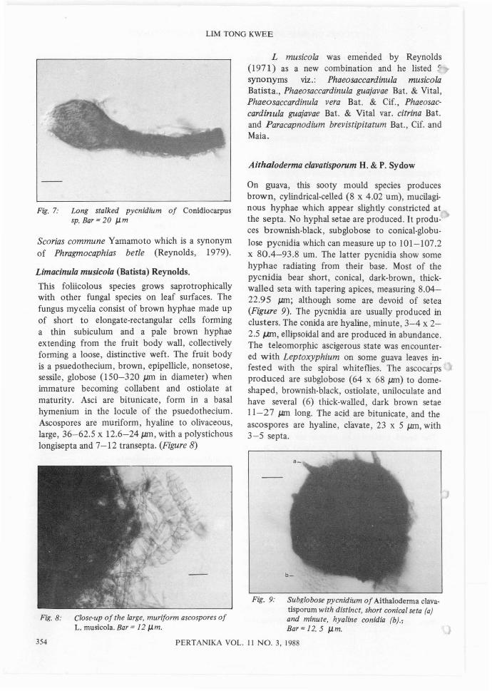
Tripospermum sp. Speganizzi.
A dematiaceous Hyphomycetes fungus thatoccurs together with other sooty mould as asuperficial, pellicullose or crust-like black growthon leaf surfaces (Figure 5). Hyphae are olivaceousto pale-brown, reticulate, consisting of long cylindrical cells 26 ).lm long by 7 ).lm wide. Conidia arebrown, star-shaped (stauroconidia) with fourdivergent arms which are wide at the base and
352 PERTANIKA VOL. II NO.3, 1988
STUDIES ON SOME SOOTY MOULDS IN GUAVA IN MALAYSIA.
tapering to a rounded apex. Each arm can havel-8 cells Le. 3-7 septa and are slightly constric-ted at the septa. Arms with 4-5 septa are the mostcommon (Figure 5). Conidia can also be diradiatealthough triradiate or tetraradiate are the mostcommon. Each conidium arises from a pyriform,stalked cell 5.5-8.8 JJ.ffi long. Each arm measures26-75 JJ.ffi long by 7.65-10.5 JJ.ffi at the base. Theteleomorphosis is uncertain.
The genus Tripospermum was established bySpegazzini in 1918 as a segregate of Triposporiumand T' acerinum was the type species (Hughes,1951 )(Figure 5).
Fig. 5: Tetraradiate stauroconidium of Triposperiumsp. Bar = 15 J,Jm.
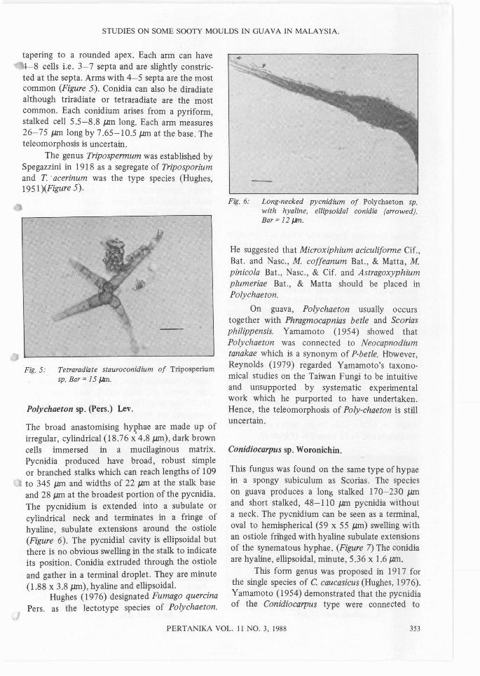
Polychaeton sp. (Pers.) Lev.
The broad anastomising hyphae are made up ofirregular, cylindrical (18.76 x' 4.8 JJ.ffi), dark browncells immersed in a mucilaginous matrix.Pycnidia produced have broad, robust simpleor branched stalks which can reach lengths of 109to 345 JJ.ffi and widths of 22 JJ.ffi at the stalk baseand 28 JJ.ffi at the broadest portion of the pycnidia.The pycnidium is extended into a subulate orcylindrical neck and terminates in a fringe ofhyaline, subulate extensions around the ostiole(Figure 6). The pycnidial cavity is ellipsoidal butthere is no obvious swelling in the stalk to indicateits position. Conidia extruded through the ostiole
and gather in a terminal droplet. They are minute(1.88 x 3.8 JlIl1), hyaline and ellipsoidal.
Hughes (1976) designated Fumago quercinaPers. as the lectotype species of Polychaeton.
Fig. 6: Long·necked pycnidium of Polychaeton sp.with hyaline, ellipsoidal conidia (arrowed).Bar = 12 f.kn.
He suggested that Microxiphium aciculiforme Cif.,Bat. and Nasc., M. coffeanum Bat., & Matta, M.pinicola Bat., Nasc., & Cif. and Astragoxyphiumplumeriae Bat., & Matta should be placed inPolychaeton.
On guava, Polychaeton usually occurstogether with Phragmocapnias betle and Scoriasphilippensis. Yamamoto (1954) showed thatPolychaeton was connected to Neocapnodiumtanakae which is a synonym of P-betle. Hbwever,Reynolds (1979) regarded Yamamoto's taxonomical studies on the Taiwan Fungi to be intuitiveand unsupported by systematic experimentalwork which he purported to have undertaken.Hence, the teleomorphosis of Poly-chaeton is stilluncertain.
Conidiocarpus sp. Woronichin.
This fungus was found on the same type of hypaein a spongy subiculum as Scorias. The specieson guava produces a lon~ stalked 170-230 JJ.ffiand short stalked, 48-11 0 JJ.ffi pycnidia withouta neck. The pycnidium can be seen as a terminaloval to hemispherical (59 x 55 JJ.ffi) swelling withan ostiole fringed with hyaline subulate extensionsof the synematous hyphae. (Figure 7) The conidiaare hyaline, ellipsoidal, minute, 5.36 x 1.6 JlIl1.
This form genus was proposed in 1917 forthe single species of C caucasicus (Hughes, 1976).Yamamoto (1954) demonstrated that the pycnidiaof the Conidiocarpus type were connected to
PERTANIKA VOL. 11 NO.3, 1988 353
LIM TONG KWEE
L musicola was emended by Reynolds(1971) as a new combination and he listed :synonyms viz.: Phaeosaccardinula musicolaBatista., Phaeosaccardinula guajavae Bat. & Vital,Phaeosaccardinula vera Bat. & Cif., Phaeosaccardinula guajavae Bat. & Vital var. citrina Bat.and Paracapnodium brevistipitatum Bat., Cif. andMaia.
Aithaloderma clavatisporum H. & P. Sydow
On guava, this sooty mould species producesbrown, cylindrical-celled (8 x 4.02 urn), mucilaginous hyphae which appear slightly constricted atthe septa. No hyphal setae are produced. It produces brownish-black, subglobose to conical-globulose pycnidia which can measure up to 101-107.2x 80.4-93.8 urn. The latter pycnidia show somehyphae radiating from their base. Most of thepycnidia bear short, conical, dark-brown, thickwalled seta with tapering apices, measuring 8.0422.95 JJ.ffi; although some are devoid of setea(Figure 9). The pycnidia are usually produced inclusters. The conida are hyaline, minute, 3-4 x 22.5 J,mI, ellipsoidal and are produced in abundance.The teleomorphic ascigerous state was encountered with Leptoxyphium on some guava leaves infested with the spiral whiteflies. The ascocarpsproduced are subglobose (64 x 68 JJ.ffi) to domeshaped, brownish-black, ostiolate, uniloculate andhave several (6) thick-walled, dark brown setae11-27 J.l.ffi long. The acid are bitunicate, and theascospores are hyaline, clavate, 23 x 5 JJ.ffi, with3-5 septa.
Fig. 7: Long stalked pycnidium of Conidiocarpussp. Bar = 20 JJ.m
Fig. 9: Subglobose pycnidium of Aithaloderma clavatisporum with distinct, short conical seta (a)
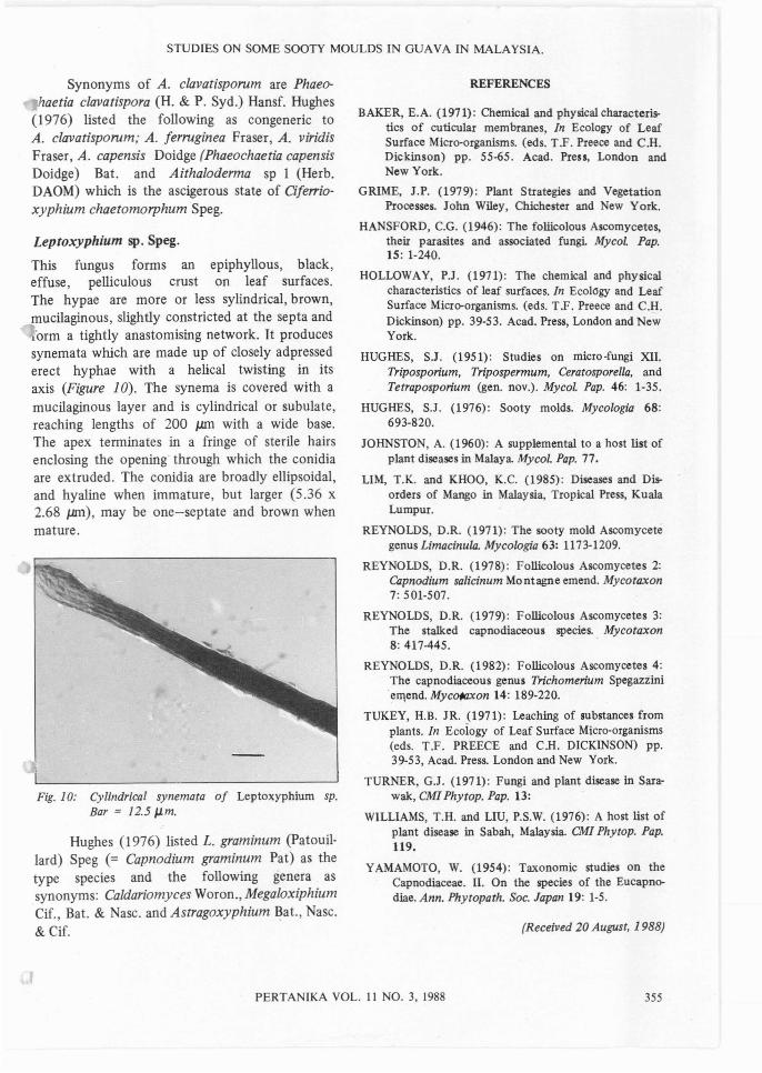
Fig. 8: Close-up of the large, muriform ascospores of and minute, hyaline conidia (b).;L. musicola. Bar =12 JJ.m. Bar =12. 5 JJ.m.
354 PERTANIKA VOL. II NO.3, 1988
Scorias commune Yamamoto which is a synonymof Phragmocaphias betle (Reynolds, 1979).
Limacinula musicola (Batista) Reynolds.
This foliicolous species grows saprotrophicallywith other fungal species on leaf surfaces. Thefungus mycelia consist of brown hyphae made upof short to elongate-rectangular cells forminga thin subiculum and a pale brown hyphaeextending from the fruit body wall, collectivelyforming a loose, distinctive weft. The fruit bodyis a psuedothecium, brown, epipellicle, nonsetose,sessile, globose (150-320 JJ.ffi in diameter) whenimmature becoming collabent and ostiolate atmaturity. Asci are bitunicate, form in a basalhymenium in the locule of the psuedothecium.Ascospores are muriform, hyaline to olivaceous,large, 36-62.5 x 12.6-24 JJ.ffi, with a polystichous10ngisepta and 7-12 transepta. (Figure 8)
STUDIES ON SOME SOOTY MOULDS IN OUAVA IN MALAYSIA.
Synonyms of A. clavatisporum are Phaeo-haetia clavatispora (H. & P. Syd.) Hansf. Hughes
(1976) listed the following as congeneric toA. clavatisporum; A. ferruginea Fraser, A. viridisFraser, A. capensis Doidge (Phaeochaetia capensisDoidge) Bat. and Aithaloderma sp 1 (Herb.DAOM) which is the ascigerous state of Cife"ioxyphium chaetomorphum Speg.
Leptoxyphium sp. Speg.
This fungus forms an epiphyllous, black,effuse, pelliculous crust on leaf surfaces.The hypae are more or less sytindrical, brown,mucilaginous, slightly constricted at the septa and'orm a tightly anastomising network. It produces
synemata which are made up of closely adpressederect hyphae with a helical twisting in itsaxis (Figure 10). The synema is covered with amucilaginous layer and is cylindrical or subulate,reaching lengths of 200 IJ.ITl with a wide base.The apex terminates in a fringe of sterile hairsenclosing the opening' through which the conidiaare extruded. The conidia are broadly ellipsoidal,and hyaline when immature, but larger (5.36 x2.68 J..ln1), may be one-septate and brown whenmature.
Fig. 10: Cylindrical synemata of Leptoxyphium sp.Bar = 12.5 p.m.
Hughes (1976) listed L. graminum (Patouillard) Speg (= Capnodium graminum Pat) as thetype species and the following genera assynonyms: Caldariomyces Woron., MegaloxiphiumCif., Bat. & Nasc. and Astragoxyphium ~at., Nasc.& Cif.
REFERENCES
BAKER, E.A. (1971): Chemical and physical characteristics of cuticular membranes, In Ecology of LeafSurface Micro-organisms. (oos. T.F. Preece and C.H.Dickinson) pp. 55-65. Acad. Press, London andNew York.
GRIME, J.P. (1979): Plant Strategies and VegetationProcesses. John Wiley, Chichester and New York.
HANSFORD, C.G. (1946): The foliicolous Ascomycetes,their parasites and associated fungi. MycoL Pap.15: 1-240.
HOLLOWAY, P.J. (1971): The chemical and physicalcharacteristics of leaf surfaces. In Ecology and LeafSurface Micro-organisms. (eds. T.F. Preece and C.H.Dickinson) pp. 39-53. Acad. Press, London and NewYork.
HUGHES, SJ. (1951): Studies on micro-fungi XII.Triposporium, Tripospermum, Ceratosporella, andTetraposporium (gen. nov.). Mycol. Pap. 46: 1-35.
HUGHES, S.J. (1976): Sooty molds. Mycologia 68:693-820.
JOHNSTON, A. (1960): A supplemental to a host list ofplant diseases in Malaya. Mycol. Pap. 77.
LIM, T.K. and KHOO, K.C. (1985): Diseases and Disorders of Mango in Malaysia, Tropical Press, KualaLumpur.
REYNOLDS, D.R. (1971): The sooty mold Ascomycetegenus Limacinula. Mycologia 63: 1173-1209.
REYNOLDS, D.R. (1978): Follicolous Ascomycetes 2:Capnodium salicinum Montagne emend. Mycotaxon7: 501-507.
REYNOLDS, D.R. (1979): Follicolous Ascomycetes 3:The stalked capnodiaceous species. Mycotaxon8: 417-445. .
REYNOLDS, D.R. (1982): Follicolous Ascomycetes 4:The capnodiaceous genus Trichomerium Spegazzinie1l\end. Myc~on 14: 189-220.
TUKEY, H.B. JR. (1971): Leaching of substances fromplants. In Ecoiogy of Leaf Surface Micro-organisms(eds. T.F. PREECE and C.H. DICKINSON) pp.39-53, Acad. Press. London and New York.
TURNER, G.J. (197l): Fungi and plant disease in Sarawak, CMI Phytop. Pap. 13:
WILLIAMS, T.H. and LIU, P.S.W. (1976): A host list ofplant disease in Sabah, Malaysia. CMI Phytop. Pap.119.
YAMAMOTO, W. (1954): Taxonomic studies on theCapnodiaceae. II. On the species of the Eucapnodiae. Ann. Phytopath. Soc. Japan 19: 1-5.
(Received 20 August, 1988)
PERTANIKA VOL. 11 NO.3, 1988 355