salim et al phytochemistry letters - core · salim et al phytochemistry letters ... chemotaxonomic...
TRANSCRIPT
Salim et al Phytochemistry Letters
Flavan-3-ols from the Leaves of Malaysian Uncaria longiflora var. pteropoda (Miq.)
Ridsd.
Fatimah Salima, Mazatulikhma Mat Zain
a,c, Mohd Syafiq Mohammad Ridzuan
c,
Moses Langatd,e
, Dulcie Mulhollandd,e
, and Rohaya Ahmada,b*
___________________________________________________________________________
aFaculty of Applied Sciences, Universiti Teknologi MARA, 40450 Shah Alam, Selangor,
Malaysia
bAtta-ur-Rahman Institute for Natural Products Discovery (RiND), Universiti Teknologi
MARA, 42300 Bandar Puncak Alam, Selangor, Malaysia
cTissue Culture Research Laboratory, Centre of Synthesis and Chemical Biology, Institute of
Science, Universiti Teknologi MARA, 40450 Shah Alam, Selangor, Malaysia
dNatural Product Research Group, Department of Chemistry, Faculty of Engineering and
Physical Sciences, University of Surrey, Guildford, GU2 7XH, UK.
eSchool of Chemistry and Physics, University of KwaZulu-Natal, Durban, 4041, South Africa
Abstract
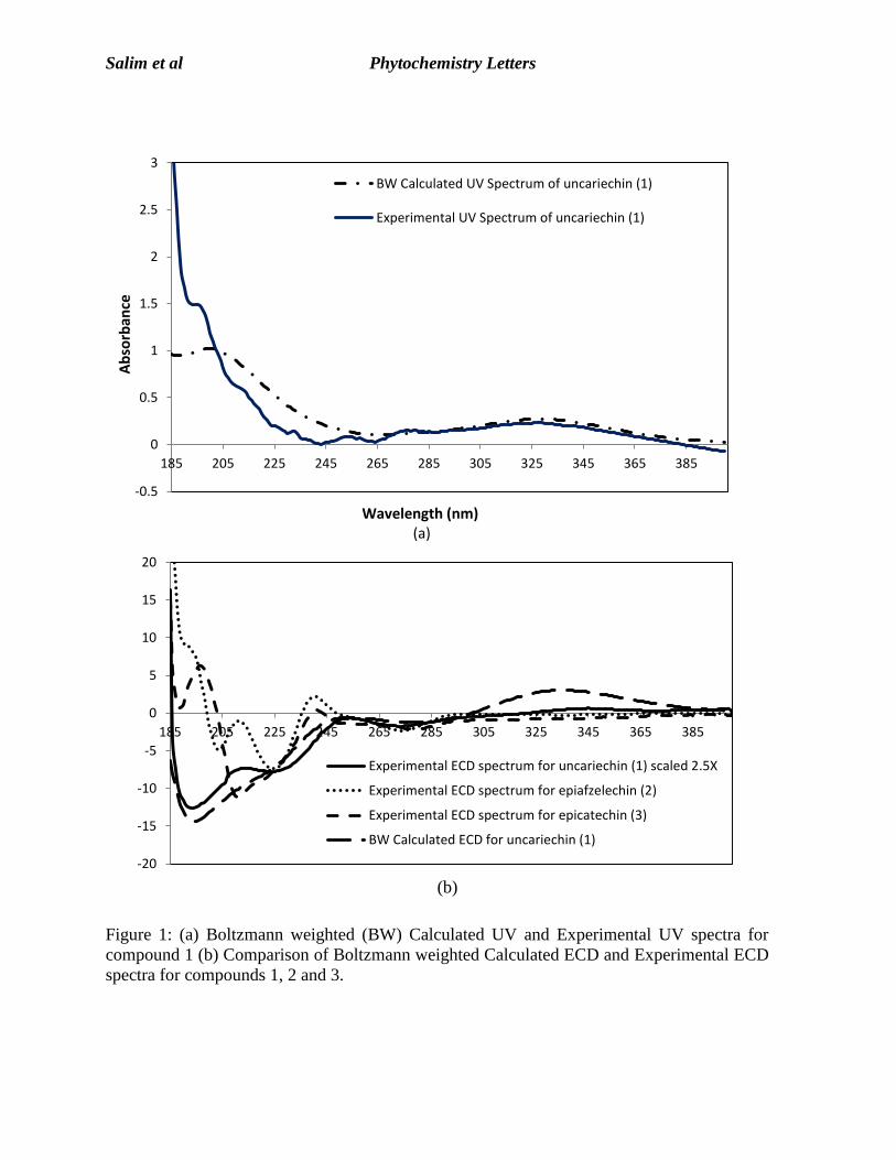
A novel flavonoid, (-)-2R,3R-3,5,4’-trihydoxyflavan-[6,7:5”,6”]-2”-pyranone, named
uncariechin (1), was isolated from the methanolic extract of the leaves of Uncaria longiflora
var. pteropoda (Miq.) Ridsd. along with the known (-)-epiafzelechin (2) and (-)-epicatechin
(3), methyl 4-hydroxybenzoate and 4-hydroxybenzaldehyde, four pentacyclic oxindole
alkaloids, isopteropodine, pteropodine, uncarine F and isopteropodic acid, previously found
in the stems, and two coumarins, scopoletin and 3,4-dihydroxy-7-methoxycoumarin.
Structures of the compounds were elucidated by 1D and 2D NMR, FTIR, UV, MS, and
experimental as well as calculated electronic circular dichroism (ECD) data. Compounds 2
and 3 were evaluated for their neurotoxic and neuroprotective properties against differentiated
SH-SY5Y neuroblastoma cell lines using the MTS assay. Compounds 2 and 3 did not show
any neurotoxic effects but showed strong protective potential against hydrogen peroxide-
induced neurotoxicity with maximum cell viability at a concentration of 1 μM.
*Corresponding author. Tel.: +603-55445592
Email address: [email protected]
Salim et al Phytochemistry Letters
Keywords: Uncaria, uncariechin, flavan-3-ol, Electronic Circular Dichroism, Neurotoxicity,
Neuroprotection
1. Introduction
We have previously reported the biological activity of the genus Uncaria, a genus
belonging to the Rubiaceae family (Ahmad et al., 2011). The genus comprises thirty-four
species, which are mainly shrubby woody climbers distributed in tropical regions, including
Southeast Asia, Africa and Southeast America (Risdale, 1978). U. longiflora var. pteropoda
is one of the fourteen species found in Malaysia (Risdale, 1978). Our previous work on the
woody stem extracts of this plant led to the isolation of two new heteroyohimbine-type
oxindole alkaloids, namely, rauniticine-allo-oxindole B and rauniticinic-allo acid B along
with five of their stereoisomers (Salim et al., 2011). We have also reported on the
chemotaxonomic significance of the pentacyclic oxindole alkaloids in the species (Salim and
Ahmad, 2011). In this paper, we report the isolation and characterization of a novel flavonoid
(-)-2R,3R- 3,5,4’-trihydoxyflavan-[6,7:5”,6”]-2”-pyranone, named uncariechin (1), along with
(-)-epiafzelechin (2) and (-)-epicatechin (3) from the methanol extract of the leaves as well as
methyl 4-hydroxybenzoate and 4-hydroxybenzaldehyde, four pentacyclic oxindole alkaloids,
isopteropodine, pteropodine, uncarine F and isopteropodic acid previously found in the stems
(Salim and Ahmad, 2011) and two coumarins, scopoletin and 3,4-dihydroxy-7-
methoxycoumarin (Abu-Eittah and El-Tawil, 1985) by spectroscopic techniques including
FTIR, UV and 1D and 2D NMR spectroscopy, MS, electronic circular dichroism (ECD)
measurements and calculations. This is the first time the flavonoid composition of this plant
has been described. In view of the potential of catechins as therapeutic cytoprotective agents
for the treatment of neurodegenerative and other diseases (Mandel and Youdim, 2004), we
investigated the neurotoxic and neuroprotection properties of the flavan-3-ols isolated against
differentiated SH-SY5Y neuroblastoma cell line.
2. Results and Discussion
Compound 1 was obtained from the MeOH extract of the leaves of U. longiflora var.
pteropoda as pale yellow crystals (m.p. 249–250 ºC). It was observed on TLC as a bluish
fluorescent spot under UV light (365 nm) and on UV measurement it showed absorption
Salim et al Phytochemistry Letters
maxima at 276, 248, 212 and 195 nm suggesting the presence of a flavan moiety (Merken et
al., 2000). HRESI-MS indicated a molecular formula of C18H14O6 and twelve degrees of
unsaturation.. The IR spectrum showed characteristic absorption bands for free hydroxyl
groups (3435 cm-1
), conjugated lactone carbonyl stretching (1685 cm-1
), a cyclic ether group
(1522 cm-1
) and methylene group bending (1457 cm-1
) .
The aromatic region of the 1H NMR spectrum of compound 1 showed a pair of ortho-
coupled proton resonances at δ 7.44 (2H, d, J = 8.4 Hz, H-2’, H-6’) and at δ 6.88 (2H, d, J =
8.7 Hz, H-3’, H-5’) for a para-substituted ring B of a flavonoid-type compound. A hydroxyl
group was placed at C-4’. These assignments were supported by the COSY spectrum which
showed correlations between the H-2’ and H-3’ and between H-5’ and H-6’ resonances as
well as between the H-6’ and H-2 resonances of ring C. For ring C, the COSY spectrum
showed coupling between resonances at δ 5.17 (H-2), δ 4.38 (H-3), and δ 2.99–2.94 (2H-4).
The chemical shift of H-3 indicated the presence of a hydroxyl group at this position
establishing the presence of a flavan-3-ol. The 2H-4 proton resonances showed correlations in
the HMBC spectrum with the C-5 (δ 114.78), C-9 (δ 160.15) and C-10 (δ 103.89) resonances.
A singlet at δ 6.42 indicated a single proton on ring A. HMBC correlations between this
resonance and the C-9 and C-10 resonances indicated this proton was at C-8 (δ 94.45). The
H-8 resonance also showed HMBC correlations with the oxygenated C-7 resonance at
(154.81) and a resonance at δ 102.08 which was ascribed to C-6.
Three carbon resonances remained to be assigned, a lactone carbonyl resonance (δ
160.55) and two alkene resonance (δ 138.64 and δ 109.71). The corresponding 1H NMR
resonances occurred at δ 8.06 (d, J = 9.6 Hz, H-4”) and δ 6.07 (d, J = 9.6 Hz, H-3”). The
HMBC spectrum was used to determine whether a linear or angular pyranocoumarin was
present. 3J correlations were seen between the H-4” resonance and C-5, C-7 and C-2”
resonances. The molecular formula indicated the need for a fused ring and a pyranone ring
was indicated. In the NOESY experiment run in DMSO, a correlation was observed between
the H-4” and the 5’-OH ( 6.03) proton resonances also confirming that the attachment was
linear.
The relative configuration of compound 1 was established using a NOESY experiment
and a model. The H-2 proton resonance showed correlations in the NOESY spectrum with H-
Salim et al Phytochemistry Letters
3 and one of H-4 proton resonances, allowing for the placement of ring B and the 3-OH on
the same face of the molecule. A J2,3 coupling constant of <1 Hz further confirmed a cis
relationship for H-2 and H-3 in compound 1 (Friedrich and Galensa, 2002). The flavan-3-ols
have two stereocenters and therefore four possible diastereomers, (2R,3S)-trans, (2S,3R)-
trans, (2R,3R)-cis and (2S,3S)-cis are possible. A cis relationship between ring B and the 3-
OH group would support a (2R,3R) or a (2S,3S) configuration (Friedrich and Galensa, 2002).
Firstly, the absolute configuration of the known flavan-3-ols (-)-epiafzalechin (2) and (-)-
epicatechin (3) were confirmed. As for 1, a J2,3 coupling constant of <1 Hz suggested a cis
configuration for both 2 and 3 for a (2R,3R) or a (2S,3S) conformation. The measured optical
rotation for compounds 2 and 3 were -151 and -125, respectively, supporting a (2R,3R)
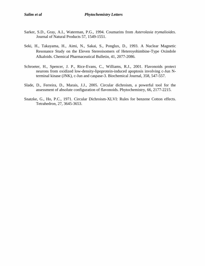
configuration (Nanjo et al., 1996). According to Slade et al., (2005), flavan-3-ols are
characterized by two phenyl chromophores whose UV absorption bands are between 200 and
240 and between 260 and 280 nm giving fingerprint ECD Cotton effects at the respective
wavelengths. A 2R,3R-cis configuration will show two negative Cotton effects at these
wavelengths. To confirm this, experimental ECD analyses for 2 and 3 were carried out in
which two negative Cotton effects at ca. 220 nm and ca. 270 nm were observed, consistent
with a 2R,3R-cis configuration of the compounds. Thus, the absolute configuration of these
compounds were established as (-)-2R,3R-epicatechin and (-)-2R,3R-epiafzalechin.
To determine the absolute configuration of 1 and investigate the effect of the pyranone
moiety on its CD spectrum, both experimental and calculated ECD analyses were carried out.
The latter was done via a systematic conformational search of the (2R,3R) isomer with the
Spartan08 program using molecular mechanics force field (MMFF) calculations. This
generated 12 conformers from which 8 conformers were under an energy cut off of 3
kcal/mol. An ECD analysis was then calculated for each of these conformers using time
dependent density functional theory (TDDFT) at the B3LYP/6-31G (d, f) level built to
Gaussian09 software (Ding et al., 2010). The calculated ECD spectra were Boltzmann
weighted (BW) and compared to the experimental ECD spectrum of 1. As shown in Fig. 1,
the BW-ECD spectrum for the 2R,3R isomer of 1 showed a negative Cotton effect at ca. 220
and a small but distinct negative CE at ca. 270 nm. In addition, the calculated ECD spectrum
also showed a positive CE at ca. 325 nm likely due to a π → π* transition in the extended π-
Salim et al Phytochemistry Letters
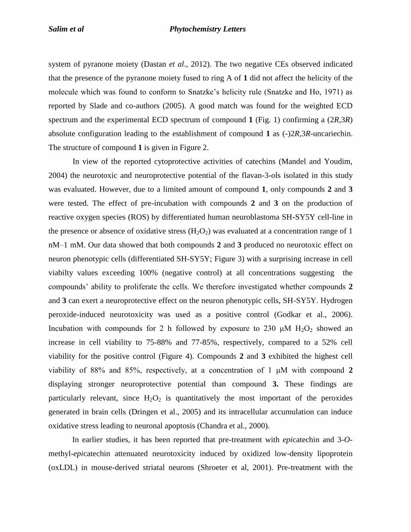
system of pyranone moiety (Dastan et al., 2012). The two negative CEs observed indicated
that the presence of the pyranone moiety fused to ring A of 1 did not affect the helicity of the
molecule which was found to conform to Snatzke’s helicity rule (Snatzke and Ho, 1971) as
reported by Slade and co-authors (2005). A good match was found for the weighted ECD
spectrum and the experimental ECD spectrum of compound 1 (Fig. 1) confirming a (2R,3R)
absolute configuration leading to the establishment of compound 1 as (-)2R,3R-uncariechin.
The structure of compound 1 is given in Figure 2.
In view of the reported cytoprotective activities of catechins (Mandel and Youdim,
2004) the neurotoxic and neuroprotective potential of the flavan-3-ols isolated in this study
was evaluated. However, due to a limited amount of compound 1, only compounds 2 and 3
were tested. The effect of pre-incubation with compounds 2 and 3 on the production of
reactive oxygen species (ROS) by differentiated human neuroblastoma SH-SY5Y cell-line in
the presence or absence of oxidative stress (H2O2) was evaluated at a concentration range of 1
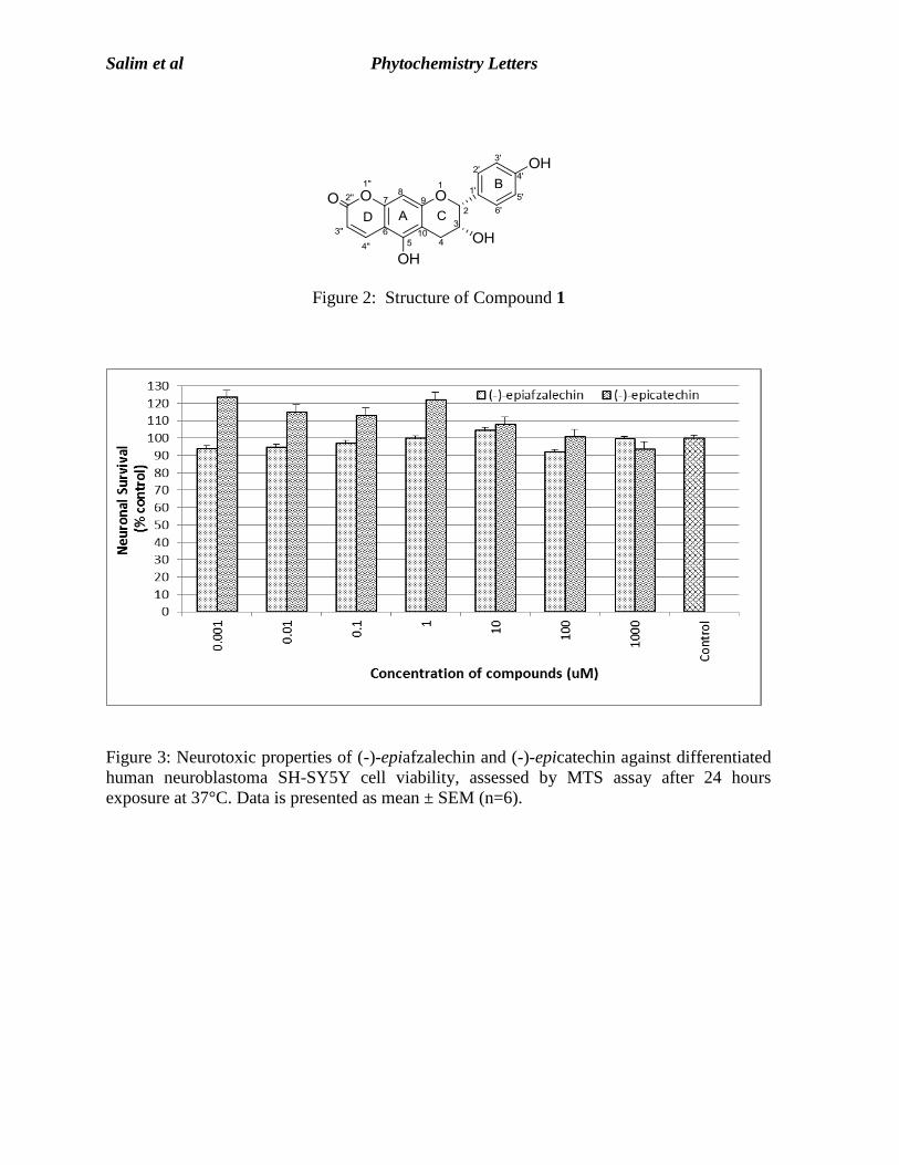
nM–1 mM. Our data showed that both compounds 2 and 3 produced no neurotoxic effect on
neuron phenotypic cells (differentiated SH-SY5Y; Figure 3) with a surprising increase in cell
viabilty values exceeding 100% (negative control) at all concentrations suggesting the
compounds’ ability to proliferate the cells. We therefore investigated whether compounds 2
and 3 can exert a neuroprotective effect on the neuron phenotypic cells, SH-SY5Y. Hydrogen
peroxide-induced neurotoxicity was used as a positive control (Godkar et al., 2006).
Incubation with compounds for 2 h followed by exposure to 230 μM H2O2 showed an
increase in cell viability to 75-88% and 77-85%, respectively, compared to a 52% cell
viability for the positive control (Figure 4). Compounds 2 and 3 exhibited the highest cell
viability of 88% and 85%, respectively, at a concentration of 1 μM with compound 2
displaying stronger neuroprotective potential than compound 3. These findings are
particularly relevant, since H2O2 is quantitatively the most important of the peroxides
generated in brain cells (Dringen et al., 2005) and its intracellular accumulation can induce
oxidative stress leading to neuronal apoptosis (Chandra et al., 2000).
In earlier studies, it has been reported that pre-treatment with epicatechin and 3-O-
methyl-epicatechin attenuated neurotoxicity induced by oxidized low-density lipoprotein
(oxLDL) in mouse-derived striatal neurons (Shroeter et al, 2001). Pre-treatment with the
Salim et al Phytochemistry Letters
compounds at a concentration of 30 µM prior to oxLDL administration led to cell viability of
90-93% as compared with 43% cell viability following treatment with oxLDL alone. In a
related study, epigallocatechin-3-gallate (EGCG) conferred protection against 6-OHDA-
induced human neuroblastoma SH-SY5Y cell damage (Levites et al., 2002) where
pretreatment for 15 min with the compound (0.1–10 µM) conferred significant protection
against 6-OHDA neurotoxicity (38% cell viability at 50 µM). The authors also reported that
EGCG showed maximal cell survival at 1µM (93%) with no effect up to 10 µM and a gradual
decrease in cell viability at higher concentrations. Their results are in good agreement with
our data for compound 3 (epicatechin) as shown in Figure 4 where the highest cell viability
was also observed at 1 µM with a gradual decrease in cell viability at higher concentrations
up to 100 µM. A similar trend was observed for compound 2 (epiafzelechin) for which, to the
best of our knowledge, neurotoxic and neuroprotective potential have not been reported.
3. Experimental
3.1 General
TLC and PTLC were performed using pre-coated aluminium-backed supported silica gel 60
F254 (0.2 mm thickness) and glass supported silica gel 60 F254 (1.0 mm thickness),
respectively. Flavonoids were detected on TLC stained with aluminium chloride (AlCl3)
reagent in which a positive result was indicated by the observation of yellow spots visualized
under UV light at 365 nm. Column chromatography was carried out using silica gel 60, 70-
230 mesh ASTM (Merk 7734) whereas radial chromatography was carried out using glass
plates with Merck’s silica gel Kieselgel 60 PF254 Merk Art 7749. Mass spectra were measured
on an Agilent Technologies 6520 QTOF LC/MS equipped with a dual-ESI source and an
Agilent Technologies LC system 1200 series, where the experiment for compounds 1-3 were
run on negative mode, while the other compounds were run on positive mode. The ultraviolet
(UV) spectra were obtained in methanol on a Shimadzu UV-Vis 160i. The infrared (IR) data
was recorded on a Perkin Elmer model FT-IR spectrometer as KBr disks. Optical rotations
were measured on a JASCO P1020 digital polarimeter. Melting points were determined using
X-4 melting-point apparatus with microscope JM628 digital thermometer. The ECD spectrum
for compounds 1, 2 and 3 were obtained on an Applied Photophysics Chirascan CD
Salim et al Phytochemistry Letters
spectrometer using a 5 mm cell and acetonitrile as the solvent. 1H- and
13C-NMR data for
compound 1 were obtained in acetone-d6 on Bruker 300 Ultrashield NMR spectrometer
measured at 300 and 75 MHz, respectively.
3.2 Plant materials
Uncaria longiflora var. pteropoda stems and leaves were collected from Hutan
Simpan Bangi, Selangor, Malaysia. The leaves and the stems of the plant were separated and
the voucher specimens (HTBP 1336) were deposited at the Herbarium of Taman Botani
Putrajaya, Malaysia.
3.3 Extraction and isolation of compounds
The leaves of U. longiflora var. pteropoda were cut into small pieces, air-dried and
ground into a fine powder. The finely ground plant material was weighed, and extracted
exhaustively with methanol at room temperature for 72 hours. The solvent was removed
under reduced pressure to yield 550 g of crude extract which was successively triturated to
afford 67 g, 72 g, 28 g and 362 g of hexane, chloroform, ethyl acetate and methanol extracts,
respectively. The methanol extract (362 g) was then subjected to liquid-liquid partitioning
between MeOH and Et2O to remove the excess tannins. The dissolved portion was filtered
through cotton wool and the solvent was evaporated to dryness using a rotary evaporator
leaving 120 g of dark solid. Fractionation of the solid with vacuum liquid chromatography
(VLC) using solvents of increasing polarity (DCM, EtOAc and MeOH) yielded five fractions
(100 ml volumes).
Based on the TLC profiles, fractions 3 and 4 were found to contain a high density of
flavonoids and were subsequently combined and subjected to further fractionation via column
chromatography using DCM and MeOH with gradient elution to afford 11 fractions (F1-11)
which was collected based on bands observed on the column. F7 to F9 were pooled and
subjected to preparative thin layer chromatography (PTLC) using a solvent system of
DCM:EtOAc (3:2) resulting in the isolation of pure compound 1 (17 mg). F10 afforded
compound 2 (44 mg) upon purification with PTLC using a solvent system CHCl3:MeOH
(5:1). Employment of a different solvent system [CHCl3:MeOH (33:7)] on the same fraction
Salim et al Phytochemistry Letters
successfully yielded a small amount of 3,4-dihydroxy-7-methoxycoumarin (4 mg). Similarly,
compound 3 (100 mg) was purified from F11 by PTLC using CHCl3: MeOH (62:13) as a
solvent system. Fraction 2 was found to contain two distinct spots on TLC with UV
visualisation at short wave length (254 nm) and upon PTLC development with solvent system
Hexane:EtOAc (4:1) afforded methyl 4-hydroxybenzoate (8 mg) and 4-hydroxybenzaldehyde
(4 mg).
The other five compounds were isolated from the chloroform extract (72 g) which was
subjected to acid-base extraction to afford a crude alkaloid mixture (53 g). This mixture was
chromatographed using VLC with increasing solvent polarity using hexane, DCM, EtOAc
and MeOH to give nine fractions (100 ml volumes) of which those with the same TLC
profiles were combined. Isopteropodine (3552 mg), pteropodine (2481 mg) and scopoletin
(21 mg) were purified with column chromatography using solvent system Hexane:EtOAc
(7:3) from pooled fractions F2 and F3. Preparative TLC of F5 using solvent system
DCM:EtOAc (7:3) yielded uncarine F (43 mg) while centrifugal PTLC on F6 using solvent
system CHCl3:MeOH (20:1) led to the isolation of isopteropodic acid (55 mg). All known
compounds were characterized by NMR spectroscopy and comparison with literature (Seki et
al., 1993, Liu and Feng, 1993, Pouchert and Behnke, 1993, Prasad et al., 2000).
3.4 Characterization of compounds 1-3
The characterization of compound 1 is given below.
3.4.1 (-)-(2R,3R)-uncariechin (1)
Pale yellow amorphous solid, mp 249 – 250 ºC. [α]D20
-312.42º (MeOH, c0.015); MS m/z =
325.0724 [M-H]+, (calcd: [M]
+ 326.0719)) C18H14O6; UV (MeOH) λmax nm: 276, 248, 216;
IR (KBr) υmax cm-1
: 3435, 3239, 1685, 1620, 1602, 1522, 1457; 1
H NMR (Acetone-D,
300MHz) δ ppm : 8.06 (1H, d, J = 9.6 Hz, H-4”), 7.44 (2H, d, J = 8.4 Hz, H-2’, H-6’), 6.88
(2H, d, J = 8.7 Hz, H-3’, H-5’), 6.42 (1H, s, H-8), 6.07 (1H, d, J = 9.6 Hz, H-3’’), 5.17 (1H, s,
H-), 4.38 (1H, m, H-3), 3.00 (3H, br s, 4’-OH, 5-OH, 3-OH), 2.99 (1H, dd, J = 3.0, 15 Hz, H-
4α), 2.94 (1H, dd, J =3.0, 15 Hz, H-4β);13
C NMR (Acetone-D, 75MHz) δ ppm: 160.55 (C-
2’’), 160.15 (C-9), 157.07 (C-4’), 154.81 (C-5), 152.16 (C-7), 138.64 (C-4’’), 129.70 (C-1’),
Salim et al Phytochemistry Letters
128.17 (C-2’), 128.17 (C-6’), 114.78 (C-3’), 114.78 (C-5’), 109.71 (C-3’’), 103.89 (C-10),
102.08 (C-6), 94.45 (C-8), 79.46 (C-2), 64.97 (C-3), 28.43 (C-4).
3.5 Computational method
TDDFT calculations were carried out at 298K in the gas phase with Gaussian 09
(Frisch et al., 2010).
For the conformational search as well as the ECD, the absolute
configuration of 1 (2R,3R) was chosen. The conformational search and geometry optimization
were carried out at the molecular mechanics level of theory employing MMFF force field
incorporated in Spartan08 (Wavefunction, Irvine, CA) software package. The conformers
were selected and further geometry optimized at the modest B3LYP/6-31G (d,f) level of
theory and TDDFT at the B3LYP/6-31(d,f) level of the theory basis set employed to simulate
the ECD spectrum. The predicted wavelengths were used without any scaling. The adequacy
of B3LYP/6-31(d,f) to optimize the geometry and to calculate the ECD spectra of flavan-3-
ols similar to compound 1 have been demonstrated previously (Ding et al., 2010).
3.6 Cell line and culture conditions
The human neuroblastoma cell line, SH-SY5Y were acquired from Dr. Carol Sanfeliu
(Department of Pharmacology and Toxicology, Institute of Biological Research, Barcelona,
Spain). The SH-SY5Y cell line was originally established from a bone marrow biopsy of a
neuroblastoma patient and is a third successive subclone of the parent cell lines SK-N-SH
(Hana et al., 2010). Original studies by Pahlman et al. (1984) reported that SH-SY5Y possess
neuron-like properties, including neurite outgrowth, and morphological changes, and have
been extensively used as an in vitro model for CNS. Neuroblastoma (SH-SY5Y) cells were
adapted to grow in 1:1 of Minimum Essential Medium Eagle (EMEM) (Sigma, USA).
Nutrient mixture F12-Ham (Sigma, USA) supplemented with 1% non-essential amino acids
(PAA Laboratories Gmbh, Austria), 1% L-glutamine (Sigma, USA), 1% 50 μg/ml gentamicin
(PAA Laboratories Gmbh, Austria), and 10% fetal bovine serum (PAA Laboratories Gmbh,
Austria). The cells were maintained in 5% CO2 incubator (Contherm Scientific Ltd, New
Zealand) at 37°C with 95% humidity.
Salim et al Phytochemistry Letters
3.7 Differentiation of cell line by retinoic acid
The SH-SY5Y cells were allowed to achieve 80-100% confluency in a tissue culture
flask with an estimated number of 106 cell/ml. Approximately 2 x 10
4 cells/ml were seeded
onto a 96-well plate and incubated for 24 hours. The cell were then induced to differentiate to
become neuronal-phenotypic cells by adding 10 μM retinoic acid (RA) [Sigma, USA] and
further incubated in a humidified atmosphere containing CO2 at 37°C. The media was
changed after three days with fresh RA and cells were ready to be used on the 6th day.
3.8 Neurotoxic and neuroprotective assay
Neurotoxicity tests were performed by incubating cultured cells (1x10⁴cells/ml) with
ranges of test compound concentrations (1nM – 1mM) overnight in a humidified atmosphere
containing 5% CO2 at 37 ºC. The results were assessed by the MTS assay on the next day. For
the neuroprotection assay, the cultures were incubated with a serial dilution of compounds at
final concentrations ranging from 1 nM to 1mM for 2 h. Cells were subsequently exposed to
230 μM hydrogen peroxide (H2O2, 30%, MERCK, Germany, which caused 52% of cell
viability) before being treated with test compounds. The cultures were further incubated for
24 h, and then cell viability was again determined by the MTS assay. Results were
representative of at least three independent experiments, and expressed as percentage of the
value observed without any treatment (negative control). Cells treated with H2O2 served as a
positive control.
3.9 Statistical analysis
Each experiment was carried out at least in triplicate. Data were reported as mean ±
standard error (SE) of six replicate readings. The significance of differences among different
groups was determined by one-way ANOVA followed by Dunnett’s Multiple Comparison
Test using GraphPad PRISM Version 5.0 whereby positive significance was indicated as
asterisks (* p < 0.05, ** p < 0.01, *** p < 0.001).
Salim et al Phytochemistry Letters
Acknowledgements
The authors wish to thank the Ministry of Higher Education, Technology and Innovation
Malaysia (MOSTI) for FRGS research grant 100-RMI/SF 16/6/2 (23/2012) and En. Ahmad
Zainudin Ibrahim (Taman Botani Putrajaya) for plant collection and identification.
Appreciation also goes to Dr Carol Sanfeliu for SH-SYFY cell line. Rohaya Ahmad thanks
Universiti Teknologi MARA for sabbatical leave approval. Rohaya Ahmad and Fatimah
Salim thank the Department of Chemistry, Faculty of Engineering and Physical Sciences,
University of Surrey, U.K. for ECD training.
References
Abu-Eittah, R.H., El-Tawil, B.A., 1985. The electronic absorption spectra of some coumarins.
A molecular orbital treatment. Canadian Journal of Chemistry 63, 1173-1179.
Ahmad, R., Hashim, M.H., Noor, M.Z., Ismail, N.H., Salim, F., Lajis, N.H., and Shaari, K.,
2011. Antioxidant and Antidiabetic Potential of Malaysian Uncaria. Research Journal
of Medicinal Plant, 5, 587-595.
Chandra, J., Samali, A., Orrenius, S., 2000. Triggering and modulation of apoptosis by
oxidative stress. Free Radical Biology & Medicine. 29, 323-333.
Dastan, D., Salehi, P., Gohari A.R., Zimmerman, S., Kaiser, M., Hamburger, M., Khavasi,
H.R., Ebrahimi, S.N., 2012. Disesquiterpene and sesquiterpene coumarins from
Ferula pseudalliacea, and determination of their absolute configurations.
Phytochemistry 78. 170-178.
Ding, Y., Li, X.,-C. Ferreira, D., 2010. 4-Arylflavan-3-ols as proanthocyanidin models:
Absolute configuration via Density Functional calculation of electronic circular
dichroism. Journal of Natural Products 73, 435-440.
Dringen, R., Pawlowski, P.G., Hirrlinger, J., 2005. Peroxide detoxification by brain cells.
Neuroscience Research, 79, 157-165.
Friedrich, W., Galensa, R., 2002. Identification of a new flavanol glucoside from barley
(Hordeum vulgare L.) and malt. European Food Research and Technology 214, 388-
393.
Frisch, M.J., Trucks, G.W., Schlegel, H.B., Scuseria, G.E., Robb, M.A., Cheeseman, J.R. et
al., 2010. Gaussian, Inc., Wallingford CT, Gaussian 09, Revision B.01.
Salim et al Phytochemistry Letters
Godkar, P.B., Gordon, R.K., Ravindran, A., Doctor, B.P., 2006. Celastrus paniculatus seed
oil and organic extracts attenuate hydrogen peroxide- and glutamate-induced injury in
embryonic rat forebrain neuronal cells. Phytomedicine 13, 29-36.
Hana, N.M.P., Sharil, S., Khairul, A., and Mazatulikhma, M.Z., 2010. Potential
neuroprotective affect of herbal extract (NHA56) on hydrogen peroxide-induced SH-
SY5Y cell lines. Signal Processing and Its Application (CSPA).
Levites, Y., Amit, T., Youdim, M. B. H., Mandel, S., 2002. Involvement of protein kinase C
activation and cell survival/cell cycle genes in green tea polyphenol (-)-
epigallocatechin-3-gallate neuroprotective action. Journal of Biological Chemistry,
277, 30574-30580.
Mandel, S., Youdim, M., 2004. Catechin polyphenols: neurodegeneration and neuro
protection in neurodegenerative diseases. Serial Review: Flavonoids and Isoflavones
(Phytoestrogens): Absorption, Metabolism, and Bioactivity. Free Radical Biology &
Medicine, 37, 304-317.
Merken, H.M., Beecher, G.R., 2000. Measurement of food flavonoids by high-performance
liquid chromatography: a review. Journal of Agricultural Food Chemistry 48, 577-
599.
Nanjo, F., Goto, K., Seto, R., Suzuki, M., Sakai, M., Hara, Y., 1996. Scavenging effects of tea
catechins and their derivatives on 1,1-diphenyl-2-picrylhydrazyl radical. Free Radical
Biology & Medicine 21, 895-902.
Pahlman, S., Ruusala, A.I-., Abrahamsson, L., Mattsson, M.E.K., Esscher, T., 1984. Retinoic
acid-induced differentiation of cultured human neuroblastoma cells: A comparison
with phorbol ester-induced differentiation. Cell Differentiation 14, 135-144.
Pouchert, C.J., Behnke, J., 1993. The Aldrich Library of 13
C and 1H FTNMR spectra, 1
st ed:
Aldrich Chemical Co.: Milwaukee, WI, Vol. II, p 1253.
Prasad, D., Juyal, V., Singh, R., Singh, V., Pant, G., Rawat, M.S.M., 2000. A new secoiridoid
glycoside from Lonicera agustifolia. Fitoterapia 71, 420-424.
Risdale, C.E., 1978. A revision of Mitragyna and Uncaria (Rubiaceae). Blumea, 24, 43-100.
Salim, F., Ahmad, R., 2011. Alkaloids from Malaysian Uncaria longiflora var. pteropoda.
Biochemical Systematics and Ecology, 39, 151-152.
Salim, F., Ismail, N.H., Awang, K., Ahmad, R., 2011. Rauniticine-allo-oxindole B and
Rauniticinic-allo acid B, New Heteroyohimbine-Type Oxindole Alkaloids from the
Stems of Malaysian Uncaria longiflora var. pteropoda. Molecules, 16, 6541- 6548.
Salim et al Phytochemistry Letters
Sarker, S.D., Gray, A.I., Waterman, P.G., 1994. Coumarins from Asterolasia trymalioides.
Journal of Natural Products 57, 1549-1551.
Seki, H., Takayama, H., Aimi, N., Sakai, S., Ponglux, D., 1993. A Nuclear Magnetic
Resonance Study on the Eleven Stereoisomers of Heteroyohimbine-Type Oxindole
Alkaloids. Chemical Pharmaceutical Bulletin, 41, 2077-2086.
Schroeter, H., Spencer, J. P., Rice-Evans, C., Williams, R.J., 2001. Flavonoids protect
neurons from oxidized low-density-lipoprotein-induced apoptosis involving c-Jun N-
terminal kinase (JNK), c-Jun and caspase-3. Biochemical Journal, 358, 547-557.
Slade, D., Ferreira, D., Marais, J.J., 2005. Circular dichroism, a powerful tool for the
assessment of absolute configuration of flavonoids. Phytochemistry, 66, 2177-2215.
Snatzke, G., Ho, P.C., 1971. Circular Dichroism-XLVI: Rules for benzene Cotton effects.
Tetrahedron, 27, 3645-3653.
Salim et al Phytochemistry Letters
Figure 1: (a) Boltzmann weighted (BW) Calculated UV and Experimental UV spectra for
compound 1 (b) Comparison of Boltzmann weighted Calculated ECD and Experimental ECD
spectra for compounds 1, 2 and 3.
-0.5
0
0.5
1
1.5
2
2.5
3
185 205 225 245 265 285 305 325 345 365 385
BW Calculated UV Spectrum of uncariechin (1)
Experimental UV Spectrum of uncariechin (1)
Wavelength (nm) (a)
Ab
sorb
ance
-20
-15
-10
-5
0
5
10
15
20
185 205 225 245 265 285 305 325 345 365 385
Experimental ECD spectrum for uncariechin (1) scaled 2.5X
Experimental ECD spectrum for epiafzelechin (2)
Experimental ECD spectrum for epicatechin (3)
BW Calculated ECD for uncariechin (1)
(b)
Salim et al Phytochemistry Letters
Figure 2: Structure of Compound 1
Figure 3: Neurotoxic properties of (-)-epiafzalechin and (-)-epicatechin against differentiated
human neuroblastoma SH-SY5Y cell viability, assessed by MTS assay after 24 hours
exposure at 37°C. Data is presented as mean ± SEM (n=6).

Salim et al Phytochemistry Letters
Figure 4: Neuroprotective properties of (-)-epiafzalechin and (-)-epicatechin against H2O2-
induced neurotoxicity on differentiated human neuroblastoma SH-SY5Y as assessed by MTS
assay after 24 hours of incubation at 37°C 5% CO2. Data is presented as mean ± SEM (n=6)