pre-processing of automatic skin cancer detection system...
TRANSCRIPT
PRE-PROCESSING OF AUTOMATIC SKIN CANCER
DETECTION SYSTEM: COMPARATIVE STUDY
Azadeh N. Hoshyar¹, Adel .A. Jumaily², Afsaneh N. Hoshyar³
¹°²School of Engineering and Information Technology
University of Technology,
Sydney, Australia
³School of Engineering and Mechatronics
National University of Malaysia,
Selangor, Malaysia
Emails: [email protected], [email protected],
Submitted: Oct 31, 2013 Accepted: July 1, 2014 Published: Sep. 1, 2014
Abstract- Skin cancer is increasing and effect many people in different part of the world. Malignant
melanoma as the deadliest type of skin cancer can be treated successfully if it detected early. Automatic
detection is one of the most challenging research areas that can be used for early detection of such vital
cancer. Over the last few years, many automatic diagnosis systems been suggested by different
researchers targeting increasing of the diagnosis accuracy. This paper presents a quick review on the
design of whole system and focus in preprocessing step of the automatic system. Preprocessing as the
basis of automation system plays a vital role for accurate detection. This paper implements three
techniques of contrast enhancement in the framework of three methodologies to find out the most
effective one for further processing. The quality of resulted images in each methodology has been
found based on testing the skin cancer images database using three image quality measurements.
Index terms: Preprocessing, Skin cancer, Detection, Automatic Systems, Image Processing
INTERNATIONAL JOURNAL ON SMART SENSING AND INTELLIGENT SYSTEMS VOL. 7, NO. 3, SEPTEMBER 2014
1364
I. INTRODUCTION
Melanoma as a cancerous lesion in the pigment is the most dangerous type of skin cancer which
can be cured 100% if it detected early. Therefore, the computerized diagnosis represents the main
stream of detection [1, 2, and 3]. In the design and analysis of computer-aided diagnostic
systems, it is necessary to preprocess the image to get more accurate detection. Preprocessing is
the first and fundamental step which has a direct affect in further operations. It involves different
steps of color-space transformation, removing the noise and unwanted objects, and also contrast
enhancement [4, 5].
Contrast enhancement plays an important role in different medical applications. It is based on the
fact that the visual examination in medical images is vital to specify the different types of
diseases [5]. As most dermoscopic images have the low contrast, therefore, automatic detection
systems require the more effective techniques to achieve the reliable accuracy [4]. Conscious of
the situation, several researchers allocated time and tried to make the process of automatic skin
cancer detection more reliable. Alina et.al 2012, published a paper refers to enhancement and
segmentation techniques in skin lesion diagnosis system. They used adaptive histogram
equalization to improve the low contrast between the nevi region and the surrounding skin area,
and then applied median filtering to remove the noise [4]. Madhankumar and Kuma proposed a
new classification method for their automatic diagnosis system; also they provided a review on
different strategies of preprocessing and segmentation sections. They introduced histogram
equalization as one the common approaches of contrast enhancement in skin cancer detection
which followed by median filter to remove the noise [6]. Sadeghi et.al proposed an approach to
detect the network of pigment structures in dermoscopic images. They performed preprocessing
on their images and applied unsharp masking as the contrast enhancement method followed by
the feature extraction. As the result, they achieved a good accuracy in their classification [7].
While Norton et.al published a paper on developing a method for border detection of dermoscopy
images and could achieve a high precision in detection of both melanocytic and non-melanocytic
lesions. They used adaptive histogram equalization for contrast enhancement and morphological
operation for removing the noise in pre-processing stage [8]. In Lau and Al-Jumaily’s paper, the
histogram equalization algorithm has been applied for the purpose of contrast enhancement in
skin cancer detection [9]. Chung and Sapiro, surveyed the segmentation of skin lesions using
Azadeh N. Hoshyar, Adel .A. Jumaily, Afsaneh N. Hoshyar, PRE-PROCESSING OF AUTOMATIC SKIN CANCER DETECTION SYSTEM: COMPARATIVE STUDY
1365
partial-differential equations (PDE) based system. They applied histogram equalization as their
contrast enhancement technique and anisotropic diffusion as noise removal technique. They
provided a good technique for border detection [10].
Thus, the importance of this detection part leads this paper to present a comparative study
between the most common contrast enhancement techniques from the literature to choose the best
to be used in preprocessing of skin cancer detection system. The paper is organized as follows.
Section 2 has a quick review on the design of computer-aided diagnostic systems. In Section 3,
three contrast enhancement techniques have been compared for determining the most effective
technique in order to apply in the pre-processing step of skin cancer detection systems. Section 4
is the experimental results and analysis, and Section 5 is conclusion and future works.
II. QUICK REVIEW ON THE DESIGN OF AUTOMATIC SKIN CANCER
DETECTION SYSTEM
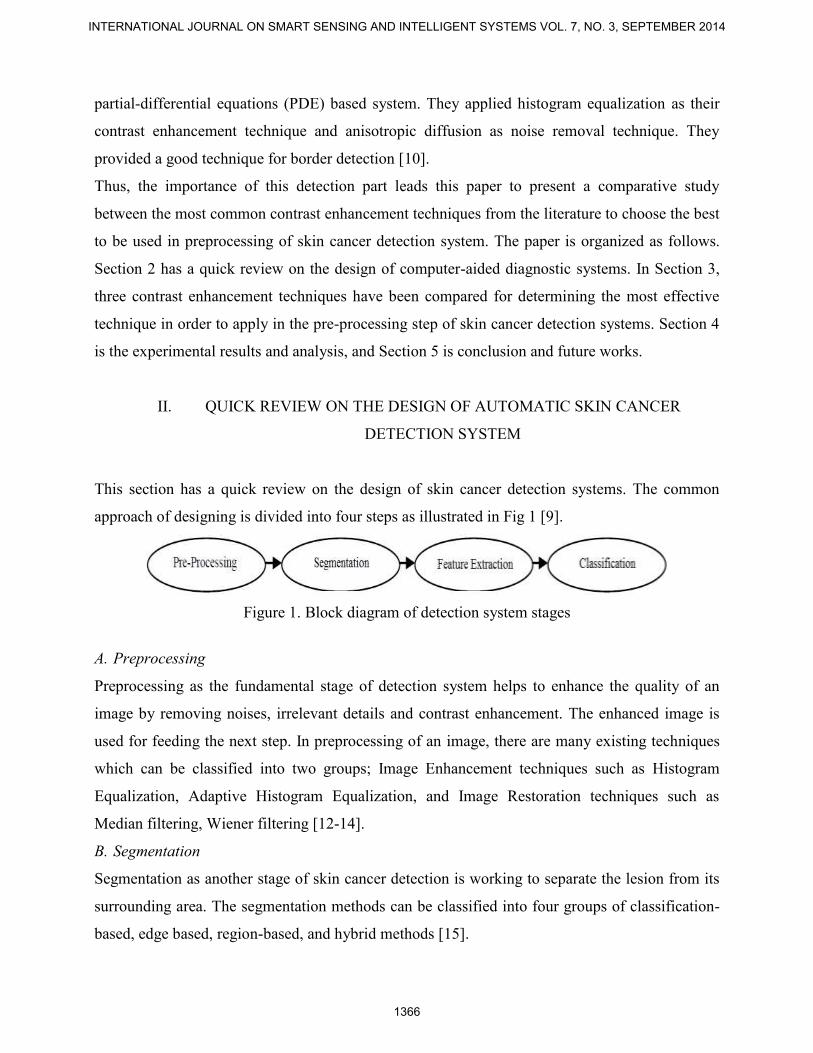
This section has a quick review on the design of skin cancer detection systems. The common
approach of designing is divided into four steps as illustrated in Fig 1 [9].
Figure 1. Block diagram of detection system stages
A. Preprocessing
Preprocessing as the fundamental stage of detection system helps to enhance the quality of an
image by removing noises, irrelevant details and contrast enhancement. The enhanced image is
used for feeding the next step. In preprocessing of an image, there are many existing techniques
which can be classified into two groups; Image Enhancement techniques such as Histogram
Equalization, Adaptive Histogram Equalization, and Image Restoration techniques such as
Median filtering, Wiener filtering [12-14].
B. Segmentation
Segmentation as another stage of skin cancer detection is working to separate the lesion from its
surrounding area. The segmentation methods can be classified into four groups of classification-
based, edge based, region-based, and hybrid methods [15].
INTERNATIONAL JOURNAL ON SMART SENSING AND INTELLIGENT SYSTEMS VOL. 7, NO. 3, SEPTEMBER 2014
1366
C. Feature extraction
Feature extraction is extracting the most reliable, measurable and sensitive features to be supplied
to the classifiers. The most well-known models of feature extraction in skin cancer images are
Pattern analysis, ABCD-rule of dermatoscopy, ELM 7-point checklist, Menzies Method and
Texture Analysis [16].
D. Classification
Classification as the last stage of detection works to classify the lesions into malignant or benign.
The classification methods can be grouped into Global models such as neural networks, Semi–
global models such as radial basis functions, Local models such as k–nearest–neighbors, and
Hybrid models such as projection based radial basis functions network [17].
III. COMPARING THREE CONTRAST ENHANCEMENT TECHNIQUES
Since the main purpose of researchers in automatic skin cancer detection systems is to decrease
the margins of error by choosing the best methods in each stage [18], the idea of this paper is to
solve the basic problem of contrast enhancement in pre-processing of skin cancer detection
systems before proceeding with further image processing techniques. In this section, three
contrast enhancement techniques are compared to figure out the effects of each and guide to
choose the best utilizing technique in the pre-processing step. In other words, the key issue is to
determine which contrast enhancement technique changes the diagnostic content of the image to
be more accurate. From literature, Histogram Equalization, Adaptive Histogram Equalization and
Unsharp Masking which are briefly defined in the following have been chosen as the most
common contrast enhancement techniques to be compared.
1) Histogram Equalization (HE): is identified as one of the most common techniques of contrast
enhancement due to its simplicity and effective performance. It mostly generates the uniform
distribution of pixel values which results in enhanced image with linear cumulative histogram
[19]. The histogram equalization will increase the local contrast of an image without affecting on
global contrast. The histogram of an image is defined as a discrete function
p(rk) = nk / n (1)
Azadeh N. Hoshyar, Adel .A. Jumaily, Afsaneh N. Hoshyar, PRE-PROCESSING OF AUTOMATIC SKIN CANCER DETECTION SYSTEM: COMPARATIVE STUDY
1367
Where rk , nk , n and k are defined as the kth gray level, the number of pixels in an image with
that gray level , the total number of pixels in whole of image and k =0, 1, 2, …, L-1. P(rk) is an
probability estimation of the occurrence of gray level rk [20].
2) Adaptive Histogram Equalization (AHE): as another recognized technique of contrast
enhancement considers the local contextual region of an image. In other word, the value of each
pixel is computed based on the rank in local contextual region instead of entire image. It
computes several histograms for each section of an image and employs that for redistribution [19,
21].
3) Unsharp Masking (UM): is the widely used approach of contrast enhancement which is simple
in concept and computation. This technique emphasizes on high-frequency components of image
to enhance the edges and details [22]. The sharpened image is obtained by adding high pass
image to the original image. High pass image as the result of unsharp masking is created using
the subtraction of the low-pass filtered version of image from the input image. However, for more
efficient result, the larger kernel size is employed [23].
ƒunsharp (x, y)= ƒ (x, y)+ k * ƒhighpass (x,y) (2)
Where ƒ (x, y), k, ƒhighpass (x,y) and ƒunsharp (x, y) are the original image, kernel size, high pass
image and the sharpened image, respectively. The performance result is the smooth image
modification spread over a larger area.
However, each of above three techniques offers very good results for improving the quality of an
image [24]. The paper is proposing to comparison of contrast enhancement of these techniques.
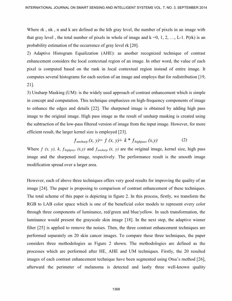
The total scheme of this paper is depicting in figure 2. In this process, firstly, we transform the
RGB to LAB color space which is one of the beneficial color models to represent every color
through three components of luminance, red/green and blue/yellow. In such transformation, the
luminance would present the grayscale skin image [18]. In the next step, the adaptive wiener
filter [25] is applied to remove the noises. Then, the three contrast enhancement techniques are
performed separately on 20 skin cancer images. To compare these three techniques, the paper
considers three methodologies as Figure 2 shown. The methodologies are defined as the
processes which are performed after HE, AHE and UM techniques. Firstly, the 20 resulted
images of each contrast enhancement technique have been segmented using Otsu’s method [26],
afterward the perimeter of melanoma is detected and lastly three well-known quality
INTERNATIONAL JOURNAL ON SMART SENSING AND INTELLIGENT SYSTEMS VOL. 7, NO. 3, SEPTEMBER 2014
1368
measurements of modified Hausdorff Distance [27], Euclidean distance [28] and Correlation [29]
are used to estimate the similarity between the resulted images of each methodology with their
template patterns. For each image, the results of these three methodologies are compared to get
the best and thereupon the most effective contrast enhancement technique. This process is
performed by three above quality measurements to get the accurate results. All the operations are
performed in Matlab 7.12.0 (R2011a).
Figure 2. The total scheme for comparison of contrast enhancement
Azadeh N. Hoshyar, Adel .A. Jumaily, Afsaneh N. Hoshyar, PRE-PROCESSING OF AUTOMATIC SKIN CANCER DETECTION SYSTEM: COMPARATIVE STUDY
1369
IV. EXPERIMENT RESULTS AND ANALYSIS
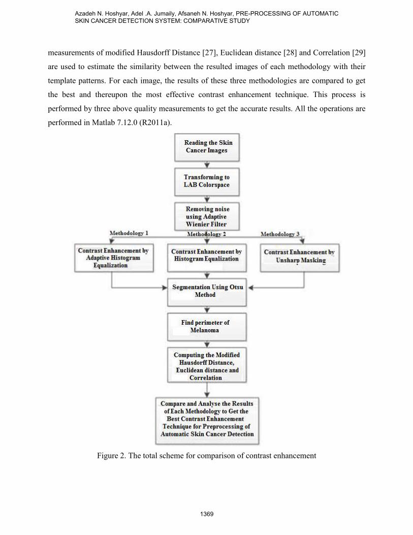
To evaluate the performance of contrast enhancement techniques, AHE, HE and UM, we
examine them on 20 skin cancer images which have been also traced manually by a dermatologist
to determine the boundaries and called patterns. These pattern images are used to compare with
our resulted images. The sample of pattern image is shown in figure 3 and the resulted images
from each step of the three methodologies are presented in figure 4.
Figure 3. Pattern image
Figure 4. a) Original image b) Greyscale image c) Removing the noise d) Contrast Enhancement
using AHE, HE, UM e) Segmentation f) perimeter of melanoma g) outlined original image
h) Overlaying the result on the image pattern
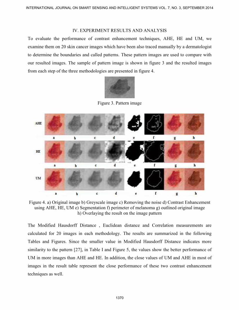
The Modified Hausdorff Distance , Euclidean distance and Correlation measurements are
calculated for 20 images in each methodology. The results are summarized in the following
Tables and Figures. Since the smaller value in Modified Hausdorff Distance indicates more
similarity to the pattern [27], in Table I and Figure 5, the values show the better performance of
UM in more images than AHE and HE. In addition, the close values of UM and AHE in most of
images in the result table represent the close performance of these two contrast enhancement
techniques as well.
INTERNATIONAL JOURNAL ON SMART SENSING AND INTELLIGENT SYSTEMS VOL. 7, NO. 3, SEPTEMBER 2014
1370
Table 1: Result table of Modified Hausdorff Distance
Figure 5. Modified Hausdorff Distance
In Euclidean Distance measurement, the smaller value shows the degree of mismatch between the
resulted image and pattern [28]. Therefore, the values in Table II and Figure 6 depict the better
performance of AHE in more images than UM and HE. Moreover, the more similar values in the
Azadeh N. Hoshyar, Adel .A. Jumaily, Afsaneh N. Hoshyar, PRE-PROCESSING OF AUTOMATIC SKIN CANCER DETECTION SYSTEM: COMPARATIVE STUDY
1371
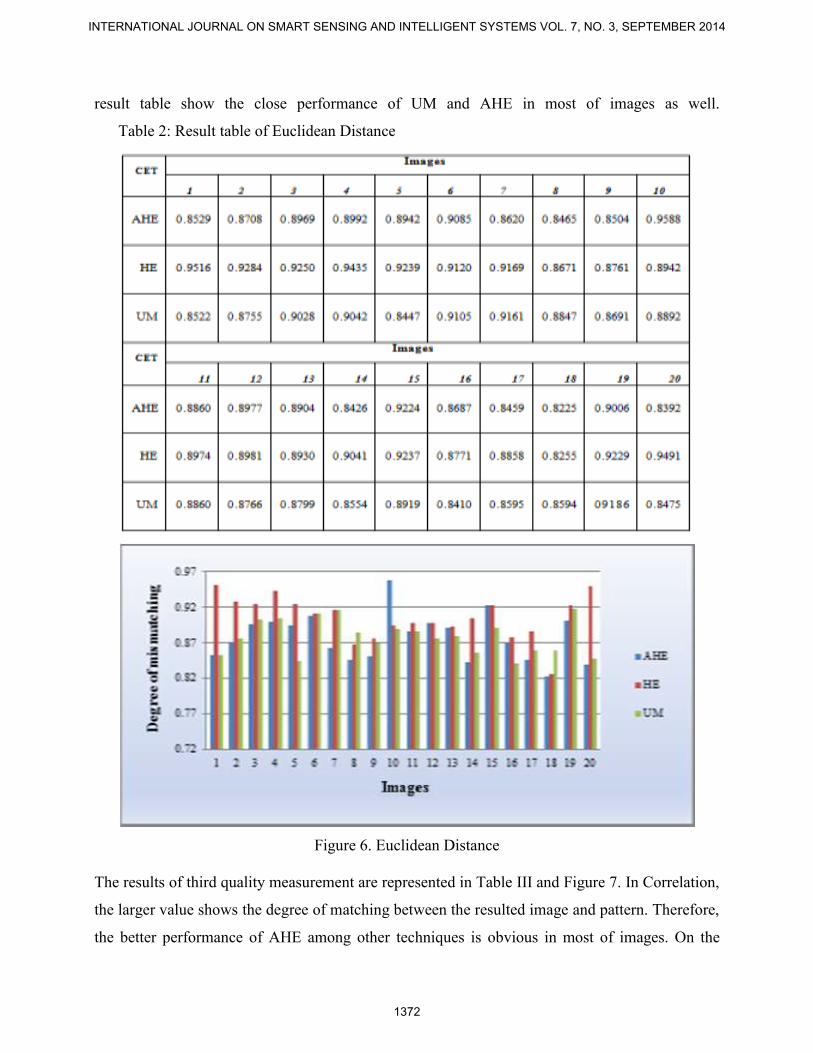
result table show the close performance of UM and AHE in most of images as well.
Table 2: Result table of Euclidean Distance
Figure 6. Euclidean Distance
The results of third quality measurement are represented in Table III and Figure 7. In Correlation,
the larger value shows the degree of matching between the resulted image and pattern. Therefore,
the better performance of AHE among other techniques is obvious in most of images. On the
INTERNATIONAL JOURNAL ON SMART SENSING AND INTELLIGENT SYSTEMS VOL. 7, NO. 3, SEPTEMBER 2014
1372
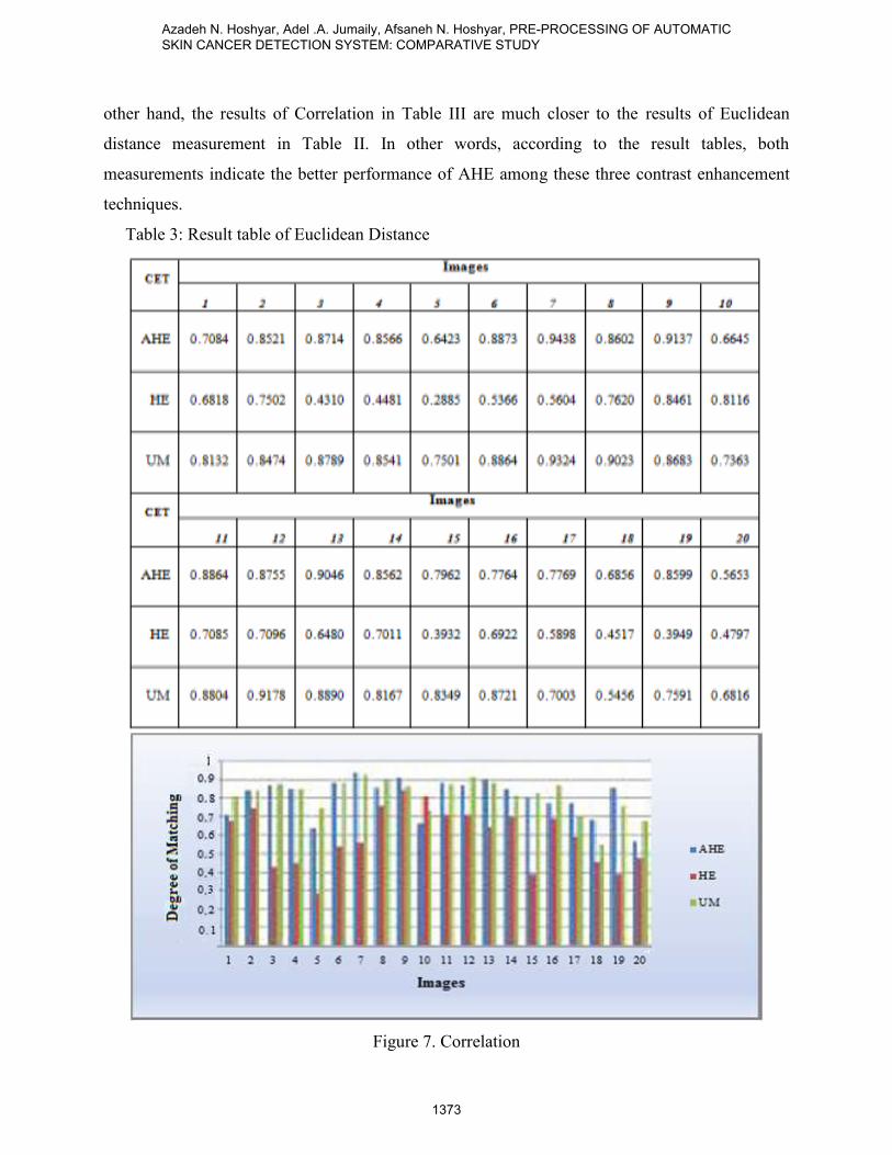
other hand, the results of Correlation in Table III are much closer to the results of Euclidean
distance measurement in Table II. In other words, according to the result tables, both
measurements indicate the better performance of AHE among these three contrast enhancement
techniques.
Table 3: Result table of Euclidean Distance
Figure 7. Correlation
Azadeh N. Hoshyar, Adel .A. Jumaily, Afsaneh N. Hoshyar, PRE-PROCESSING OF AUTOMATIC SKIN CANCER DETECTION SYSTEM: COMPARATIVE STUDY
1373
The above three Tables show the good performance of AHE and UM, and the worse performance
of HE as a contrast enhancement technique. Although AHE and UM have a close performance,
two measurements of Euclidean Distance and Correlation among three applied measurements
show the better performance of AHE in most of images.
V. CONCLUSION AND FUTURE WORK
Three Contrast Enhancement techniques, namely, Adaptive Histogram Equalization, Histogram
Equalization and Unsharp masking have been implemented to compare the most effective one in
preprocessing stage of skin cancer detection system. After applying the preprocessing techniques,
the image segmentation is performed. The resulted images of each methodology are compared
with its patterns using three measurements of Modified Hausdorff distance , Euclidean distance
and Correlation to estimate the more similar one to the pattern for determining the best contrast
enhancement technique. Experimental results on skin cancer images shown although the
performance of UM and AHE are very close, the AHE is more effective than UM in most of
images.
While the present study performed the comparison between contrast enhancement techniques in
the preprocessing of skin cancer detection systems as described earlier, more improved images
can be obtained by applying more effective results for further processing.
REFERENCES
[1] S. Maryam, R. Majid, T.K. Lee, M.Stella Atkins, “A novel method for detection of pigment
network in dermoscopic images using graphs”, Computerized Medical Imaging and Graphics,
35(2):137-43, 2011.
[2] S. Muhamad Isa, M. Eka Suryana, M. Ali Akbar, A. Noviyanto, W. Jatmiko and A. Murni
Arymurthy, “Performance Analysis of ECG Signal Compression using SPIHT”, International
Journal On Smart Sensing And Intelligent Systems Vol. 6, No. 5, 2013.
[3] Y. LUO, P. LIU, M. LIAO,” An artificial immune network clustering algorithm for
mangroves remote sensing image”, International Journal On Smart Sensing And Intelligent
Systems Vol. 7, No. 1, 2014.
INTERNATIONAL JOURNAL ON SMART SENSING AND INTELLIGENT SYSTEMS VOL. 7, NO. 3, SEPTEMBER 2014
1374
[4] S. Alina, C. Mihai Ciuc, T. Radulescu, L. Wanyu, D.Petrache, “Preliminary Work on
Dermatoscopic Lesion Segmentation,” 20th European Signal Processing Conference (EUSIPCO
2012), August 21-27, Romania, 2012.
[5] D. Chang, W. Wu, “Image Contrast Enhancement Based on a Histogram Transformation of
Local Standard Deviation”, IEEE Transactions on Medical Imaging, Vol. 17, No. 4, 1998.
[6] K. Madhankuma, and P. Kumar, “Characterization of Skin Lesions”, Proceedings of the
International Conference on Pattern Recognition, Informatics and Medical Engineering, March
21-23, India, 2012
[7] M.Sadeghi, M.Razmara, T.K. Lee, M.S. Atkins,” A novel method for detection of pigment
network in dermoscopic images using graphs”, Computerized Medical Imaging and Graphics 35,
137–143, 2011.
[8] K.A. Norton, H. Iyatomi, M. Emre Celebi, G. Schaefer, M. Tanaka, and K. Ogawa,
“Development of a Novel Border Detection Method for Melanocytic and Non-Melanocytic
Dermoscopy Images”, 32nd Annual International Conference of the IEEE EMBS Buenos Aires,
Argentina, August 31 - September 4, 2010.
[9] H.T. Lau, and A. Al-Jumaily, “Automatically Early Detection of Skin Cancer: Study Based
on Neural Network Classification”, International Conference of Soft Computing and Pattern
Recognition, 4-7 December, Malaysia, 2009.
[10] D.H. Chung, G. and Sapiro, “Segmenting Skin Lesions with Partial-Differential-Equations-
Based Image Processing Algorithms”, IEEE Transactions on Medical Imaging, Vol. 19, No. 7,
2000.
[11] A.N. Hoshyar, A. Al-Jumaily, and R. Sulaiman, “Review on Automatic Early Skin Cancer
Detection”, International Conference on Computer Science and Service System (CSSS), 27-29
June, China, 2011.
[12] S.S. Al-amri, N.V. Kalyankar, and S.D. Khamitkar, “Linear and Non-linear Contrast
Enhancement Image”, IJCSNS International Journal of Computer Science and Network Security,
Vol.10, No.2, pp. 139, 2010.
[13] S.S. Agaian, K.P. Lentz, and A.M. Grigoryan, “A New Measure of Image Enhancement”,
International Conference on Signal Processing & Communication, 5-9 June, 2000.
[14] L. R. Lagendijk, and J. Biemond, “Basic Methods for Image Restoration and Identification”,
Handbook of Image and Video Processing 2nd edition, Elsevier Academic Press, 167-181, 2005.
Azadeh N. Hoshyar, Adel .A. Jumaily, Afsaneh N. Hoshyar, PRE-PROCESSING OF AUTOMATIC SKIN CANCER DETECTION SYSTEM: COMPARATIVE STUDY
1375
[15] R.C. Gonzalez, and R.E. Woods, “Digital Image Processing”, 3rd Edition, Prentice- all, Inc.,
New Jersey, ISBN: 10: 013168728x, pp: 594, 2008.
[16] I. Maglogiannis, and C.N. Doukas, “Overview of Advanced Computer Vision Systems for
Skin Lesions Characterization”, IEEE Transactions on Information Technology in Biomedicine,
Vol. 13, N. 5, 2009
[17] M.J. Ogorzaáek, G. Surówka, L. Nowak, and C. Merkwirth, “New Approaches for
Computer-Assisted Skin Cancer Diagnosis”, The Third International Symposium on
Optimization and Systems Biology (OSB’09), 20-22 September, China, 2009
[18] J. Mesquita, “Classification of Skin Tumours through the Analysis of Unconstrained
Images”, De Montfort University Leicester, Faculty of Computer Science and Engineering, UK,
2008.
[19] H. Yeganeh, A. Ziaei, A.H. Rezaie, “A Novel Approach for Contrast Enhancement Based on
Histogram Equalization”, Proceedings of the International Conference on Computer and
Communication Engineering, 13-15 May, Malaysia, 2008.
[20] V.P. Vishwakarma, S. Pandey and M. N. Gupta,” Adaptive Histogram Equalization and
Logarithm Transform with Rescaled Low Frequency DCT Coefficients for Illumination
Normalization”, International Journal of Recent Trends in Engineering, Vol.1, No. 1, 2009.
[21] C.W. Kurak, “Adaptive histogram equalization: a parallel implementation”, Proceedings of
the Fourth Annual IEEE Symposium in Computer-Based Medical Systems, Univ. of North
Florida, Jacksonville, FL, USA ,1991.
[22] X. Xiaoling, and X. Zhang, “An Improved Unsharp Masking Method for Borehole Image
Enhancement”, 2nd International Conference on Industrial Mechatronics and Automation,30-31
May, China, 2010.
[23] D. Prasanna, P.Neelamegam, S.Sriram, N.Raju ,” Enhancement of vein patterns in hand
image for biometric and biomedical application using various image enhancement techniques”,
International Conference On Modeling Optimization And Computing, Procedia Engineering, Vol
38, pp. 1174 – 1185, 2012.
[24] S.H. Rubin, R. Kountchev, V. Todorov, and R. Kountcheva, “Contrast Enhancement with
Histogram-Adaptive Image Segmentation”, IEEE International Conference on Information Reuse
and Integration, 16-18 Sep, USA, 2006.
INTERNATIONAL JOURNAL ON SMART SENSING AND INTELLIGENT SYSTEMS VOL. 7, NO. 3, SEPTEMBER 2014
1376
[25] H. Zhang, “Spatially Adaptive Wiener Filtering For Image Denoising Using Undecimated
Wavelet Transform”, ELEC 590 project report, Deaprtment of Electrical and Computer
Engineering, USA, 1999.
[26] N. Otsu, “A Threshold Selection Method from Gray Level Histograms,” IEEE Trans SMC,
Vol. 9, pp. 62–66, 1979.
[27] M.P. Dubuisson, and A.K. Jain , “A modified Hausdorff distance for object matching”,
Proceedings of the 12th IAPR International Conference on Computer Vision & Image
Processing, Vol. 1, 9-13 Oct, Jerusalem, 1994.
[28] L. Wang, Y. Zhang, and J. Feng, “On the Euclidean Distance of Images”, Center for
Information Sciences, School of Electronics Engineering and Computer Sciences, Peking
University, China, 2002.
[29] S.Varshney, N. Rajpa, and R. Purwar, “Comparative Study of Image Segmentation
Techniques and Object Matching using Segmentation”, International Conference on Methods and
Models in Computer Science, 14-15 Dec, India, 2009.
Azadeh N. Hoshyar, Adel .A. Jumaily, Afsaneh N. Hoshyar, PRE-PROCESSING OF AUTOMATIC SKIN CANCER DETECTION SYSTEM: COMPARATIVE STUDY
1377