no less than a woman improving breast cancer detection
TRANSCRIPT

Prof. Datin Dr. Rozi Mahmud
Professor Datin D
r. Rozi M
ahmud
NO LESS THAN A WOMAN
IMPROVING BREAST CANCER DETECTION & DIAGNOSIS
NO LESS THAN A WOMAN
IMPROVING BREAST CANCER
DETECTION & DIAGNOSIS

NO LESS THAN A WOMAN
IMPROVING BREAST CANCER
DETECTION & DIAGNOSIS
PROFESSOR DATIN DR. ROZI MAHMUD
Professor Datin Dr. Rozi Mahmud MBBS UM, MMed Rad, UKM
Universiti Putra Malaysia PressSerdang • 2017
http://www.penerbit.upm.edu.myPROFESSOR DATIN DR. ROZI MAHMUD
17 MARCH 2017
Dewan Kuliah UtamaFakulti Perubatan dan Sains Kesihatan
Universiti Putra Malaysia
NO LESS THAN A WOMAN
IMPROVING BREAST CANCER
DETECTION & DIAGNOSIS
© Penerbit Universiti Putra Malaysia 2017Cetakan Pertama 2017
Hak cipta terpelihara. Mana-mana bahagian penerbitan ini tidak boleh dihasilkan semula, disimpan dalam sistem simpanan kekal, atau dipindahkan dalam sebarang bentuk atau sebarang cara elektronik, mekanik, penggambaran semula, rakaman dan sebagainya tanpa terlebih dahulu mendapat izin daripada pihak Penerbit Universiti Putra Malaysia.
Penerbit UPM adalah anggota Persatuan Penerbit Buku Malaysia (MABOPA) No. Ahli: 9802
ISBN 978-967-344-695-7
Reka letak teks : Sahariah Abdol Rahim @ IbrahimReka bentuk kulit : Md Fairus Ahmad
Reka bentuk, reka letak dan dicetak olehPenerbit Universiti Putra Malaysia43400 UPM, SerdangSelangor Darul EhsanTel: 03-89468851/8854/8429Faks: 03-89416172E-mel: [email protected] web: http://penerbit.upm.edu.my
Contents
Abstract 1
Introduction 3
Symptoms of Breast Cancer 9
Diagnosing Breast Cancer 9
Imaging Modalities in Detecting Breast Cancer 11
Future of Breast Imaging 53
Other Studies on Diseases Related to Women 55
Research Grants 57
International Collaborations 61
Concluding Remarks and Personal Statement 62
References 63
Biography 73
Acknowledgements 77
List of Inaugural Lectures 81

1 ❘❘❚
Rozi Mahmud
ABSTRACT
Breasts, being the ultimate symbol of femininity, make breast cancer one of the most traumatic events any woman could ever face. Perhaps it is this sense of pride in these attributes that makes many women reluctant to discuss and share their experiences with breast cancer. Many may feel that their absolute core identity has been shaken, making them less than a woman. The fear and stigma attached to this disease are currently among the major difficulties faced by healthcare providers in convincing women to effectively manage their breast disease. It may leave women feeling isolated and as a result, withdrawing from society and even life- making them feel less than a woman. Beyond the stigma and mental anguish there is also the tremendous stress of going through a number of surgeries, chemotherapies and radiation therapies, with the risk of treatment failure and recurrence always at the back of their minds. Fortunately various studies confirm that early breast cancer detection saves lives, reduces medical treatments and costs, and ultimately, gives one hope for a better future. The availability of effective screening reduces the mortality from breast cancer by up to 50%. Most women will be lucky enough to never develop breast cancer, but for the many of those who do, their lives may be saved by advanced detection. Currently, breast cancer detected at an early stage can be treated appropriately, with most being cured. The role of a health care provider is therefore extremely important, in counselling and motivating women to overcome their fears and come forward for regular examinations. The role of a radiologist is equally important in synergizing imaging modalities towards achieving the best of medical care for the public. These are some of the ways to help and
❚❘❘ 2
No Less Than a Woman: Improving Breast Cancer Detection and Diagnosis
support in the management of the disease and in making the ladies feel no less than a woman. In order to reach a superior level in early detection and diagnosis of breast cancer, our research team studied various methods to overcome some of the limitations in breast imaging. These methods include Computer Aided Diagnosis techniques involving various existing imaging modalities such as mammogram, tomosynthesis, breast ultrasound, computed tomography laser mammography (CTLM) and thermography of the breast. More rewarding research on newer imaging devices includes the ultra-wide band (UWB) imaging of the breast. Recent usage of a computational model involving Monte Carlo Simulation for early breast cancer detection using wire mesh collimator gamma camera in scintimammography is also gaining interest amongst clinicians.
3 ❘❘❚
Rozi Mahmud
INTRODUCTION
General Information on Breast Cancer
Breast cancer occurs when the cells in the lobules (milk producing glands) or the ducts become abnormal and divide uncontrollably (Bleyer, A., Welch, H. G. 2012). These abnormal cells then begin to invade the surrounding breast tissue (Figure 1) and may eventually spread via blood vessels and lymphatic channels to the lymph nodes and other parts of the body.
Facts:
1. Breast cancer is the second leading cause of cancer related deaths in women, exceeded only by lung cancer. The 5-year survival rate is largely dependent on the disease stage (Lee, S.C., et al., 2014), so earlier detection and treatment results in a better prognosis.
2. Over recent decades, the burden of breast cancer has been increasing at an alarming rate in Malaysia, making breast cancer the most common form of cancer affecting Malaysian women (Yip, C.H.,et al., 2014). Based on the Malaysian National Cancer Registry Report 2007-2011, the prevalence of breast cancer among women is 32.1%.

❚❘❘ 4
No Less Than a Woman: Improving Breast Cancer Detection and Diagnosis
Figure 1 Late stage breast cancer(Adapted from www.drclark.net)
Risk Factors
The exact cause of breast cancer is unknown. Many studies have found some associated risk factors to be as listed in Tables 1, 2 and 3: (American Cancer Society, 2015)
Table 1 Known risk factors of breast cancer
Risk Additional information
BEING A WOMANJust being a woman is the biggest risk factor for developing breast cancer.
AGEAs with many other diseases, risk of breast cancer goes up with age. About two out of three invasive breast cancers are found in women aged 55 or older.
FAMILY HISTORY
Women with close relatives who have been diagnosed with breast cancer have a higher risk of developing the disease. This includes one first-degree female relatives (sister, mother, daughter) diagnosed with breast cancer, which doubles the risk.
5 ❘❘❚
Rozi Mahmud
GENETICS
About 5% to 10% of breast cancers are thought to be hereditary, caused by abnormal genes passed from parent to child. Carriers of the BRCA I and BRCA II genes have at least a 40 to 85 per cent risk.
RACE/ETHNICITY
White women are slightly more likely to develop breast cancer than African American, Hispanic and Asian women. African American women are however more likely to develop more aggressive, more advanced-stage breast cancer that is diagnosed at a young age.
EXPOSURE TO RADIATION
Radiation to Chest or Face before age 30. Radiation to the chest to treat another cancer (not breast cancer), such as Hodgkin’s disease or non-Hodgkin’s lymphoma, poses a higher-than-average risk of breast cancer. Radiation to the face of an adolescent to treat acne also increases the risk of developing breast cancer later in life.
PERSONAL HISTORY OF BREAST CANCER
Once diagnosed with breast cancer, there is 3 to 4 times higher likelihood of developing a new cancer in the other breast or a different part of the same breast.
CERTAIN BREAST CHANGES
Benign breast conditions may increase risk of breast cancer.
BEING OVERWEIGHT
Overweight and obese women have a higher risk of being diagnosed with breast cancer compared to women who maintain a healthy weight, especially after menopause. Being overweight can also increase the risk of the breast cancer coming back (recurrence) in women who have had the disease.
PREGNANCY HISTORY
Women who have not had a full-term pregnancy or who have their first child after age 30 have a higher risk of breast cancer compared to women who give birth before age 30.
BREASTFEEDING HISTORY
Breastfeeding can lower breast cancer risk, especially if a woman breastfeeds for longer than 1 year.
❚❘❘ 6
No Less Than a Woman: Improving Breast Cancer Detection and Diagnosis
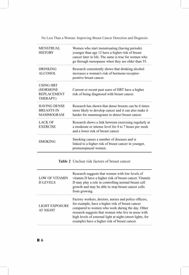
MENSTRUAL HISTORY
Women who start menstruating (having periods) younger than age 12 have a higher risk of breast cancer later in life. The same is true for women who go through menopause when they are older than 55.
DRINKING ALCOHOL
Research consistently shows that drinking alcohol increases a woman’s risk of hormone-receptor-positive breast cancer.
USING HRT (HORMONE REPLACEMENT THERAPY)
Current or recent past users of HRT have a higher risk of being diagnosed with breast cancer.
HAVING DENSE BREASTS IN MAMMOGRAM
Research has shown that dense breasts can be 6 times more likely to develop cancer and it can also make it harder for mammograms to detect breast cancer.
LACK OF EXERCISE
Research shows a link between exercising regularly at a moderate or intense level for 4 to 7 hours per week and a lower risk of breast cancer
SMOKINGSmoking causes a number of diseases and is linked to a higher risk of breast cancer in younger, premenopausal women.
Table 2 Unclear risk factors of breast cancer
LOW OF VITAMIN D LEVELS
Research suggests that women with low levels of vitamin D have a higher risk of breast cancer. Vitamin D may play a role in controlling normal breast cell growth and may be able to stop breast cancer cells from growing.
LIGHT EXPOSURE AT NIGHT
Factory workers, doctors, nurses and police officers, for example, have a higher risk of breast cancer compared to women who work during the day. Other research suggests that women who live in areas with high levels of external light at night (street lights, for example) have a higher risk of breast cancer.
7 ❘❘❚
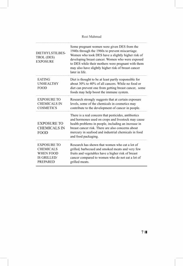
Rozi Mahmud
DIETHYLSTILBES-TROL (DES) EXPOSURE
Some pregnant women were given DES from the 1940s through the 1960s to prevent miscarriage. Women who took DES have a slightly higher risk of developing breast cancer. Women who were exposed to DES while their mothers were pregnant with them may also have slightly higher risk of breast cancer later in life.
EATING UNHEALTHY FOOD
Diet is thought to be at least partly responsible for about 30% to 40% of all cancers. While no food or diet can prevent one from getting breast cancer, some foods may help boost the immune system.
EXPOSURE TO CHEMICALS IN COSMETICS
Research strongly suggests that at certain exposure levels, some of the chemicals in cosmetics may contribute to the development of cancer in people.
EXPOSURE TO CHEMICALS IN FOOD
There is a real concern that pesticides, antibiotics and hormones used on crops and livestock may cause health problems in people, including an increase in breast cancer risk. There are also concerns about mercury in seafood and industrial chemicals in food and food packaging.
EXPOSURE TO CHEMICALS WHEN FOOD IS GRILLED/PREPARED
Research has shown that women who eat a lot of grilled, barbecued and smoked meats and very few fruits and vegetables have a higher risk of breast cancer compared to women who do not eat a lot of grilled meats.
❚❘❘ 8
No Less Than a Woman: Improving Breast Cancer Detection and Diagnosis
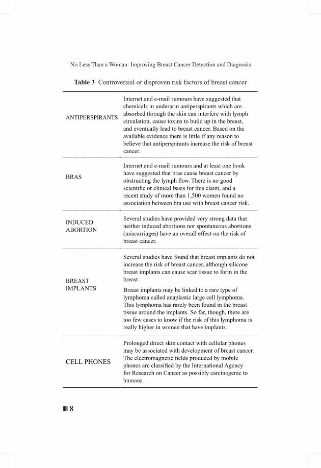
Table 3 Controversial or disproven risk factors of breast cancer
ANTIPERSPIRANTS
Internet and e-mail rumours have suggested that chemicals in underarm antiperspirants which are absorbed through the skin can interfere with lymph circulation, cause toxins to build up in the breast, and eventually lead to breast cancer. Based on the available evidence there is little if any reason to believe that antiperspirants increase the risk of breast cancer.
BRAS
Internet and e-mail rumours and at least one book have suggested that bras cause breast cancer by obstructing the lymph flow. There is no good scientific or clinical basis for this claim, and a recent study of more than 1,500 women found no association between bra use with breast cancer risk.
INDUCED ABORTION
Several studies have provided very strong data that neither induced abortions nor spontaneous abortions (miscarriages) have an overall effect on the risk of breast cancer.
BREAST IMPLANTS
Several studies have found that breast implants do not increase the risk of breast cancer, although silicone breast implants can cause scar tissue to form in the breast.
Breast implants may be linked to a rare type of lymphoma called anaplastic large cell lymphoma. This lymphoma has rarely been found in the breast tissue around the implants. So far, though, there are too few cases to know if the risk of this lymphoma is really higher in women that have implants.
CELL PHONES
Prolonged direct skin contact with cellular phones may be associated with development of breast cancer.The electromagnetic fields produced by mobile phones are classified by the International Agency for Research on Cancer as possibly carcinogenic to humans.
9 ❘❘❚
Rozi Mahmud
SYMPTOMS OF BREAST CANCER
In most women in Malaysia symptoms of breast cancer is presented by a lump in the breast. The lump is usually painless, grows slowly and may alter the contour or size of the breast. It may also cause skin changes, an inverted nipple or bloodstained nipple discharge. The lymph gland in the armpit will also be swollen if affected by the cancer cells. In late stages, the growth may ulcerate through the skin and become infected. Bone pain, tenderness over the liver, severe headaches, shortness of breath and a chronic persistent cough may be an indication of the cancer spreading to the other organs in the body. The most common signs of possible breast cancer: • a swelling of part of the breast
• thickening in or near the breast or underarm area
• skin irritation, dimpling or distortion
• redness or scaling of the breast skin or nipple
• nipple pain, inversion, rash or tenderness
• nipple discharge other than breast milk
• non-cyclical breast pain
DIAGNOSING BREAST CANCER
Breast Self-Examination (BSE)/Breast Self-Awareness (familiar with own breast)
Numerous professional organizations, including the United States Preventive Services Task Force, American Academy of Family Physicians and Canadian Task Force on Preventive Health Care, recommend against clinicians teaching women how to perform BSE, concluding that adequate evidence suggests that teaching BSE does not reduce breast cancer mortality. The American Cancer
❚❘❘ 10
No Less Than a Woman: Improving Breast Cancer Detection and Diagnosis
Society (ACS) leaves the decision about BSE to a woman’s personal choice, recommending clinical instruction for women who choose to perform a structured self-examination. Both ACS and the American College of Obstetricians and Gynecologists (ACOG) encourage all women to become familiar with how their breasts normally look and feel so that anything out of the ordinary can be promptly brought to the attention of their healthcare providers (American Cancer Society, 2015).
Clinical Breast Examination (CBE).
Another strategy for improving screening is to consider greater reliance on the CBE, performed by physicians, as opposed to the self-breast examination done by women on themselves. Furthermore; CBE detects cancers that share similar prognostic features with tumours detected by mammography. Thus, it is likely that CBE does detect clinically significant breast cancers. Given the convenience of CBE, it should be part of the routine screening for women (American Cancer Society, 2015).
Role of Radiologist in Early Diagnosis and Further Management of The Disease
Ascertaining the correct diagnosis and stage of breast cancer can be challenging, and the importance of the radiologist’s role has increased over the years. It is challenging as no medical equipment or machine is 100% perfect and radiologists need to synergize their vast knowledge to match the findings and make proper request for further examinations (Lee, S.C., et al., 2014). The American Joint Committee on Cancer (AJCC) staging systems for breast cancer (7th edition) provides a tumour-node-metastasis (TNM) classification scheme for breast cancer that is important for determining prognosis and treatment.
11 ❘❘❚
Rozi Mahmud
Radiologic information through medical equipment may alter stage, prognosis or treatment, that includes tumour size; number of tumour lesions; total span of disease; regional nodal status, local regional invasion and distant metastases to other anatomic structures. Currently the only way to detect asymptomatic early breast cancer is through screening mammography. This particular examination however has its own weaknesses and limitations. Other methods used are considered as adjunct modalities, which in combination with a mammogram, may increase the level of sensitivity or specificity in detecting the disease.
IMAGING MODALITIES IN DETECTING BREAST CANCER
There are various modalities and techniques in detecting breast cancer, mainly involving a varied range of electromagnetic waves, either ionizing or non-ionizing radiation. These include:
• Mammogram (analog or digital)
• Tomosynthesis
• Ultrasound*
• Magnetic Resonance Imaging (MRI)*
• Computer Tomography laser mammography*
• Thermography*
• Ultra-wide band imaging*
• Scintigraphy* uses non ionizing radiation
Ultrasound, magnetic resonance imaging (MRI), computed tomography laser mammography, thermography, ultra-wide band imaging and scintigraphy are considered as adjunct examinations.

❚❘❘ 12
No Less Than a Woman: Improving Breast Cancer Detection and Diagnosis
Mammogram (Analog Or Digital)
Currently the mammogram is the gold standard imaging modality for detecting breast cancer (Figure 2). The widespread implementation of screening mammography has decreased the mortality of breast cancer by as much as 50%. (American Cancer Society, 2015).
Figure 2 Mammography equipment, technique(Adapted from commons.wikimedia.org)
Mammography is the process of using low-energy X-rays (usually around 30 kVp) to examine the breast, which is used as a diagnostic and screening tool. The goal of screening mammography is for early detection of breast cancer, typically through detection of characteristic masses and or micro-calcifications. Up to 50% of breast cancers can be associated with calcification while 15-30% of calcifications biopsied for various reasons tend to be malignant in asymptomatic patients. (Paredes, E. S., Atlas of mammography, 2007)

13 ❘❘❚
Rozi Mahmud
For screening mammogram in an asymptomatic person, mammography may be performed biennially (every two years) in women of 50 – 74 years of age. In low and intermediate risk women aged 40 – 49 years, mammograms should not be offered routinely. However, women aged 40 – 49 years should not be denied screening mammography if they desire it. For the high risk group, screening should be done from the age of 30 years with both MRI and mammography as it is more effective than mammography alone. In young women (< 35 years old), ultrasound should be used as the initial imaging modality (Clinical Practice Guidelines: Management of Breast Cancer Second Edition, 2010)
Signs of Cancer in a Mammogram
i. Presence of MassCommon malignant features of invasive cancers include irregular shape and indistinct or spiculated margins of a mass. There may be associated architectural distortion or asymmetrical density (Figure 3 and 4).
Figure 3 Asymmetrical densityFigure 4 Irregular shape and
indistinct or spiculated margins of a mass
❚❘❘ 14
No Less Than a Woman: Improving Breast Cancer Detection and Diagnosis
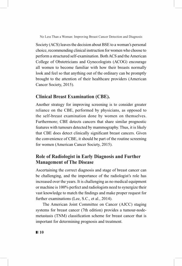
ii. Presence of microcalcificationsMicrocalcifications are tiny mineral deposits within the breast tissue. There are various types of calcifications but findings of segmental distribution and pleomorphic shape are the features with the highest predictive value of malignancy (Figure 5).
Figure 5 Malignant calcifications in mammogram image
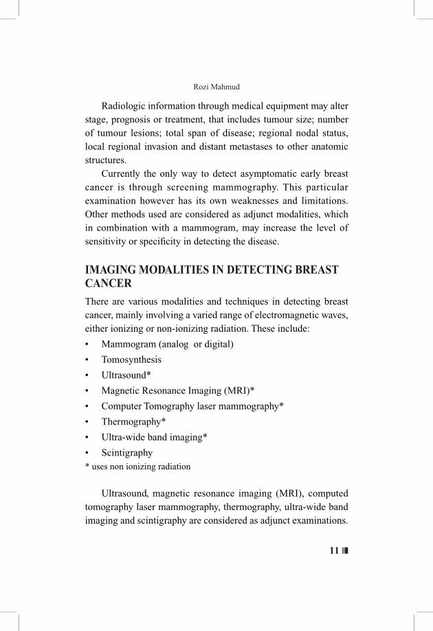
iii. Nipple retraction. This raises suspicion of an underlying cancer.
Figure 6 Nipple retraction in mammogram image
15 ❘❘❚
Rozi Mahmud
iv. Enlarged axillary lymph nodes. Involvement of the nodes indicates worsening prognosis.
Understanding Mammogram ReportsBreast Imaging Reporting and Data System (BI-RADS) ACR BI-RADS® ATLAS — MAMMOGRAPHY
Mammogram reports include the type of breast density and categories for type of radiological pathology or abnormalities (Table 4 and 5). This ensures a standard way of reporting mammogram results with standard words and terms and as a guide in managing suspicious findings. The type of breast density needs to be mentioned in the report as dense breasts may obscure the potential mass or calcifications as these abnormalities may also appear dense or white because of the lack of contrast between them and a dense breast background. Many states in the United States call for mandatory reporting of breast density and women with dense breasts are also informed that their mammograms are hard to read.Upon knowing that they have dense breasts; women can be guided to undergo additional screening, such as by breast ultrasound, MRI or other newer methods.

❚❘❘ 16
No Less Than a Woman: Improving Breast Cancer Detection and Diagnosis
Table 4 BI-RADS breast density
Density Group (Level) Description Explanation
IThe breasts are almost entirely fatty
The breasts contain little fibrous and glandular tissue, which means the mammogram would likely detect anything abnormal.
There are scattered areas of fibro glandular density
There are a few areas of fibrous and glandular tissue in the breast.
The breasts are heterogeneously dense
This may obscure small masses. The breast has more areas of fibrous and glandular tissue that are found throughout the breast. This can make it hard to see small masses.
17 ❘❘❚
Rozi Mahmud
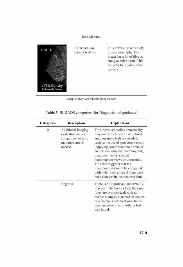
The breasts are extremely dense
This lowers the sensitivity of mammography. The breast has a lot of fibrous and glandular tissue. This can lead to missing some cancers.
(Adapted from www.huffingtonpost.com)
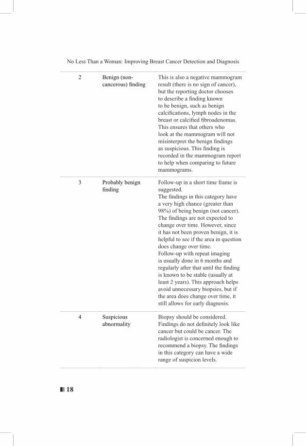
Table 5 BI-RADS categories (for Diagnosis and guidance)
Categories Description Explanation
0 Additional imaging evaluation and/or comparison to prior mammograms is needed.
This means a possible abnormality may not be clearly seen or defined and thus more tests are needed, such as the use of spot compression (applying compression to a smaller area when doing the mammogram), magnified views, special mammogram views or ultrasound. This also suggests that the mammogram should be compared with older ones to see if there have been changes in the area over time.
1 Negative There is no significant abnormality to report. The breasts look the same (they are symmetrical) with no masses (lumps), distorted structures or suspicious calcifications. In this case, negative means nothing bad was found.
❚❘❘ 18
No Less Than a Woman: Improving Breast Cancer Detection and Diagnosis
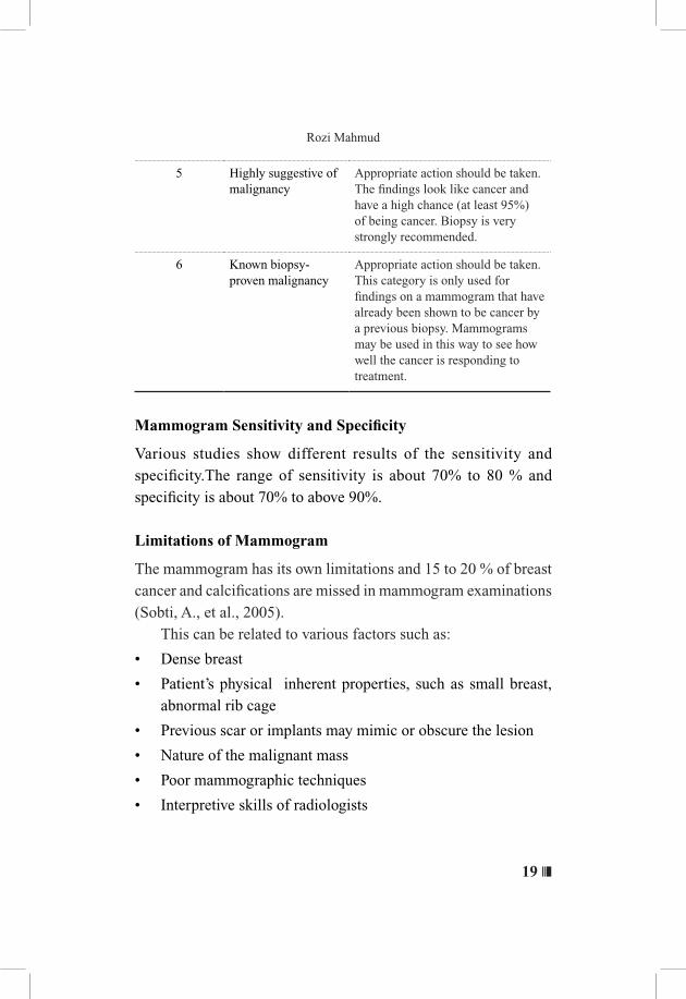
2 Benign (non-cancerous) finding
This is also a negative mammogram result (there is no sign of cancer), but the reporting doctor chooses to describe a finding known to be benign, such as benign calcifications, lymph nodes in the breast or calcified fibroadenomas. This ensures that others who look at the mammogram will not misinterpret the benign findings as suspicious. This finding is recorded in the mammogram report to help when comparing to future mammograms.
3 Probably benign finding
Follow-up in a short time frame is suggested.The findings in this category have a very high chance (greater than 98%) of being benign (not cancer). The findings are not expected to change over time. However, since it has not been proven benign, it is helpful to see if the area in question does change over time. Follow-up with repeat imaging is usually done in 6 months and regularly after that until the finding is known to be stable (usually at least 2 years). This approach helps avoid unnecessary biopsies, but if the area does change over time, it still allows for early diagnosis.
4 Suspicious abnormality
Biopsy should be considered.Findings do not definitely look like cancer but could be cancer. The radiologist is concerned enough to recommend a biopsy. The findings in this category can have a wide range of suspicion levels.
19 ❘❘❚
Rozi Mahmud
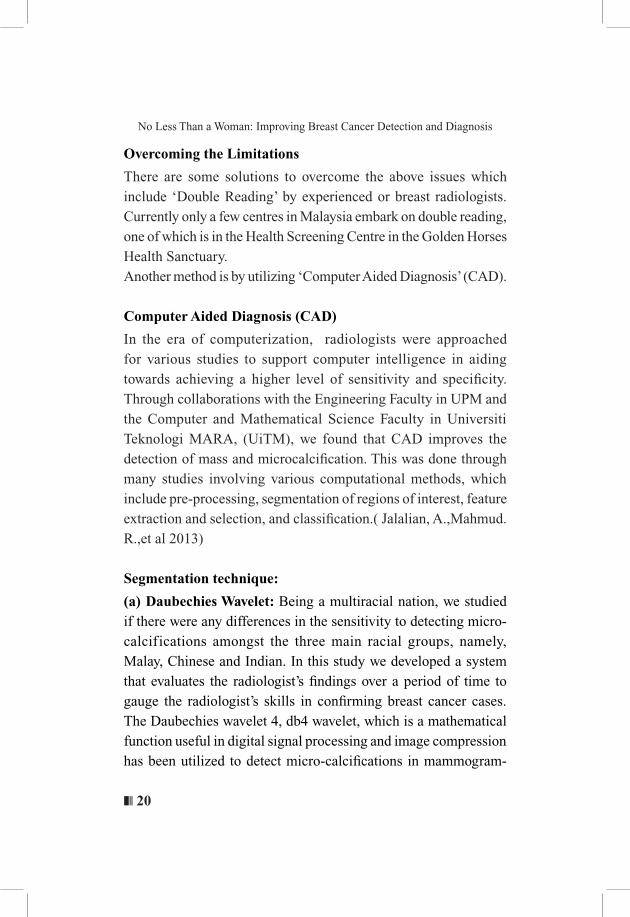
5 Highly suggestive of malignancy
Appropriate action should be taken.The findings look like cancer and have a high chance (at least 95%) of being cancer. Biopsy is very strongly recommended.
6 Known biopsy-proven malignancy
Appropriate action should be taken.This category is only used for findings on a mammogram that have already been shown to be cancer by a previous biopsy. Mammograms may be used in this way to see how well the cancer is responding to treatment.
Mammogram Sensitivity and Specificity
Various studies show different results of the sensitivity and specificity.The range of sensitivity is about 70% to 80 % and specificity is about 70% to above 90%.
Limitations of Mammogram
The mammogram has its own limitations and 15 to 20 % of breast cancer and calcifications are missed in mammogram examinations (Sobti, A., et al., 2005). This can be related to various factors such as:
• Dense breast
• Patient’s physical inherent properties, such as small breast, abnormal rib cage
• Previous scar or implants may mimic or obscure the lesion
• Nature of the malignant mass
• Poor mammographic techniques
• Interpretive skills of radiologists
❚❘❘ 20
No Less Than a Woman: Improving Breast Cancer Detection and Diagnosis
Overcoming the Limitations
There are some solutions to overcome the above issues which include ‘Double Reading’ by experienced or breast radiologists. Currently only a few centres in Malaysia embark on double reading, one of which is in the Health Screening Centre in the Golden Horses Health Sanctuary. Another method is by utilizing ‘Computer Aided Diagnosis’ (CAD).
Computer Aided Diagnosis (CAD)
In the era of computerization, radiologists were approached for various studies to support computer intelligence in aiding towards achieving a higher level of sensitivity and specificity. Through collaborations with the Engineering Faculty in UPM and the Computer and Mathematical Science Faculty in Universiti Teknologi MARA, (UiTM), we found that CAD improves the detection of mass and microcalcification. This was done through many studies involving various computational methods, which include pre-processing, segmentation of regions of interest, feature extraction and selection, and classification.( Jalalian, A.,Mahmud. R.,et al 2013)
Segmentation technique:
(a) Daubechies Wavelet: Being a multiracial nation, we studied if there were any differences in the sensitivity to detecting micro-calcifications amongst the three main racial groups, namely, Malay, Chinese and Indian. In this study we developed a system that evaluates the radiologist’s findings over a period of time to gauge the radiologist’s skills in confirming breast cancer cases. The Daubechies wavelet 4, db4 wavelet, which is a mathematical function useful in digital signal processing and image compression has been utilized to detect micro-calcifications in mammogram-
21 ❘❘❚
Rozi Mahmud
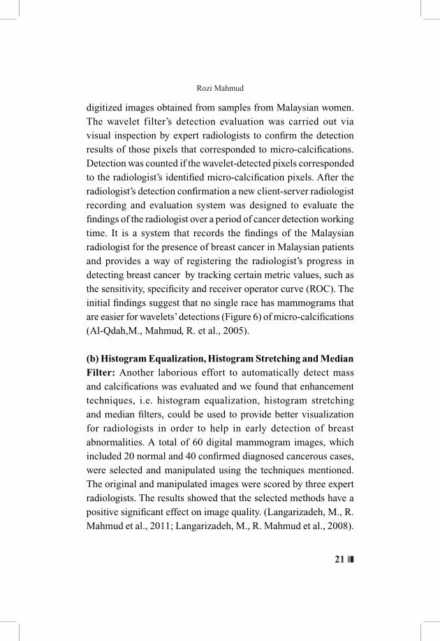
digitized images obtained from samples from Malaysian women. The wavelet filter’s detection evaluation was carried out via visual inspection by expert radiologists to confirm the detection results of those pixels that corresponded to micro-calcifications. Detection was counted if the wavelet-detected pixels corresponded to the radiologist’s identified micro-calcification pixels. After the radiologist’s detection confirmation a new client-server radiologist recording and evaluation system was designed to evaluate the findings of the radiologist over a period of cancer detection working time. It is a system that records the findings of the Malaysian radiologist for the presence of breast cancer in Malaysian patients and provides a way of registering the radiologist’s progress in detecting breast cancer by tracking certain metric values, such as the sensitivity, specificity and receiver operator curve (ROC). The initial findings suggest that no single race has mammograms that are easier for wavelets’ detections (Figure 6) of micro-calcifications (Al-Qdah,M., Mahmud, R. et al., 2005).
(b) Histogram Equalization, Histogram Stretching and Median Filter: Another laborious effort to automatically detect mass and calcifications was evaluated and we found that enhancement techniques, i.e. histogram equalization, histogram stretching and median filters, could be used to provide better visualization for radiologists in order to help in early detection of breast abnormalities. A total of 60 digital mammogram images, which included 20 normal and 40 confirmed diagnosed cancerous cases, were selected and manipulated using the techniques mentioned. The original and manipulated images were scored by three expert radiologists. The results showed that the selected methods have a positive significant effect on image quality. (Langarizadeh, M., R. Mahmud et al., 2011; Langarizadeh, M., R. Mahmud et al., 2008).
❚❘❘ 22
No Less Than a Woman: Improving Breast Cancer Detection and Diagnosis
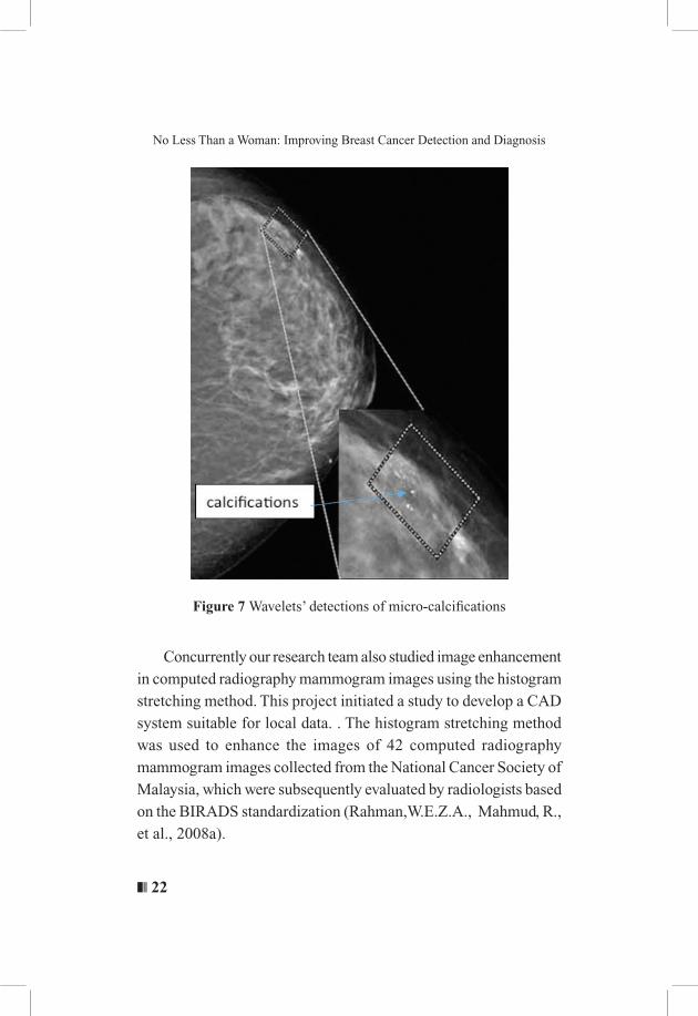
Figure 7 Wavelets’ detections of micro-calcifications
Concurrently our research team also studied image enhancement in computed radiography mammogram images using the histogram stretching method. This project initiated a study to develop a CAD system suitable for local data. . The histogram stretching method was used to enhance the images of 42 computed radiography mammogram images collected from the National Cancer Society of Malaysia, which were subsequently evaluated by radiologists based on the BIRADS standardization (Rahman,W.E.Z.A., Mahmud, R., et al., 2008a).
23 ❘❘❚
Rozi Mahmud
(c) Seed-Based Region Growing and Mathematical Morphology
Combining intellectual resources with University Teknologi MARA, we studied various mathematical aspects to increase the level of detection of microcalcification via intelligent pathways which include automated seed point selection for seed based region growing in segmenting microcalcifications (Malek, A, ., Mahmud, R., et al., 2010a) and Region and Boundary Segmentation of Microcalcifications using Seed-Based Region Growing and Mathematical Morphology (Malek, A, ., Mahmud, R., et al., 2010b). The experimental results showed that the algorithm successfully segmented the microcalcifications with a small average error. Other mechanisms that we explored include Image Segmentation of Mammography Images Using Fuzzy C-means Clustering Algorithm and Pixel area validation of segmented malignant tumors in digital mammographic images (Embong, R.,Mahmud, R., et al., 2008; Embong, R., Mahmud, R., et al., 2009) which also gave some promising results in detecting the calcifications automatically.
(d) Active Contour
Due to the promising results in automatic detection of microcalcifications in mammograms, we furthered our studies using the active contour model, also called snakes, which is a framework in computer vision for delineating an object’s outline from a possibly noisy 2D image. It comprises computer generated curves that move within images to locate the object’s boundaries. Basically, this technique has the ability to converge to boundaries of images which are long, thin and have irregular shapes. Prior to evaluating the real mammogram images, images from a standard breast phantom MRI-156 for image quality calibration were used. In this study only the breast phantom images at the
❚❘❘ 24
No Less Than a Woman: Improving Breast Cancer Detection and Diagnosis
exposures of 25 kV, 28 kV and 35 kV with milliampere (mAs) of 0.5 were considered. It was found that when the image was divided into four equal regions the detection of abnormalities in the image was more effective as compared to when the image was processed as a whole. We studied 30 actual mammograms with presence of micro calcifications, and when the snake algorithm was applied on these images it was found that this method could successfully trace the boundaries of the microcalcifications. The final results were then presented to radiologists for scoring (Yasiran, S., Mahmud, R., et al., 2008b) Another equally important finding was from a study that involved comparison between, Gradient Vector Flow (GVF) snake and Enhanced Distance (ED) snake in segmenting microcalcifications. (Yasiran, S., Mahmud, R., et al., 2011a). The performance was measured based on actual area of the average percentage difference traced by expert radiologists. The results obtained showed that the values of average percentage difference for the GVF and ED snakes were 4.3% and 6.68%, respectively. These results indicate that the GVF snake has better performance with 95.7% (Yasiran, S., Mahmud, R., et al., 2011a). Similar studies and almost equivalent outcomes were also seen using the three edge detection techniques and the Distance Active Contour (DAC) method (Yasiran, S., Mahmud, R., et al., 2012). Efficiency and accuracy of the Enhanced Distance Active Contour (EDAC) method for microcalcifications segmentation was also studied. (Yasiran, S. S., Mahmud, R., et al 2011b; Yasiran, S. S., Mahmud, R., et al 2011c)
25 ❘❘❚
Rozi Mahmud
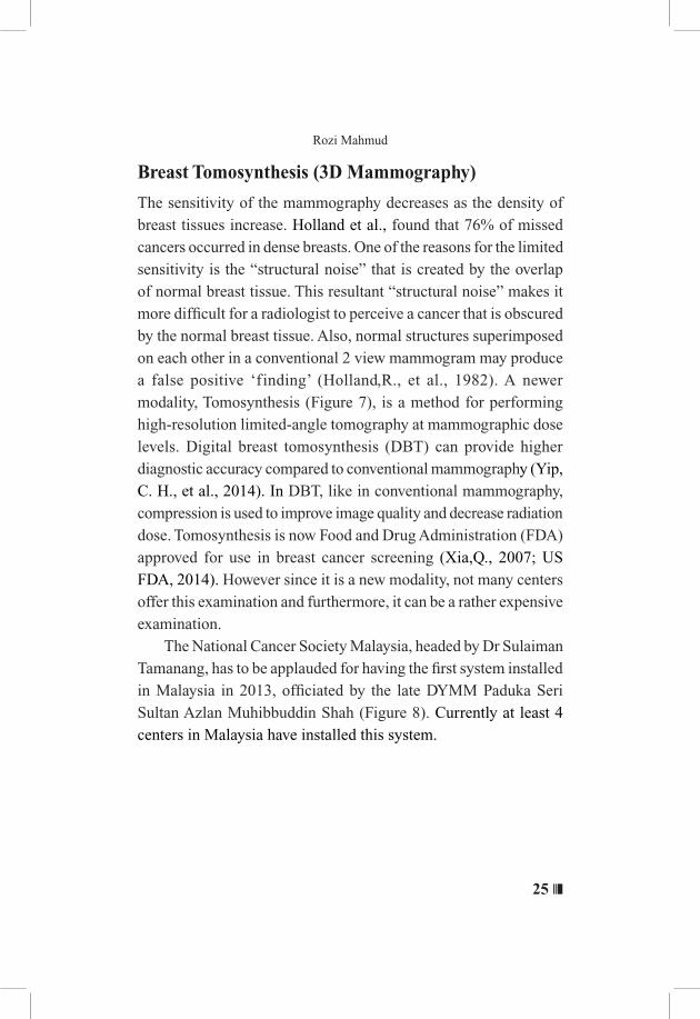
Breast Tomosynthesis (3D Mammography)
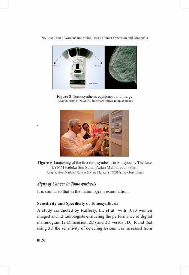
The sensitivity of the mammography decreases as the density of breast tissues increase. Holland et al., found that 76% of missed cancers occurred in dense breasts. One of the reasons for the limited sensitivity is the “structural noise” that is created by the overlap of normal breast tissue. This resultant “structural noise” makes it more difficult for a radiologist to perceive a cancer that is obscured by the normal breast tissue. Also, normal structures superimposed on each other in a conventional 2 view mammogram may produce a false positive ‘finding’ (Holland,R., et al., 1982). A newer modality, Tomosynthesis (Figure 7), is a method for performing high-resolution limited-angle tomography at mammographic dose levels. Digital breast tomosynthesis (DBT) can provide higher diagnostic accuracy compared to conventional mammography (Yip, C. H., et al., 2014). In DBT, like in conventional mammography, compression is used to improve image quality and decrease radiation dose. Tomosynthesis is now Food and Drug Administration (FDA) approved for use in breast cancer screening (Xia,Q., 2007; US FDA, 2014). However since it is a new modality, not many centers offer this examination and furthermore, it can be a rather expensive examination. The National Cancer Society Malaysia, headed by Dr Sulaiman Tamanang, has to be applauded for having the first system installed in Malaysia in 2013, officiated by the late DYMM Paduka Seri Sultan Azlan Muhibbuddin Shah (Figure 8). Currently at least 4 centers in Malaysia have installed this system.
❚❘❘ 26
No Less Than a Woman: Improving Breast Cancer Detection and Diagnosis
Figure 8 Tomosynthesis equipment and image(Adapted from HOLOGIC: http://www.breasttomo.com.au)
.
Figure 9 Launching of the first tomosynthesis in Malaysia by The Late DYMM Paduka Seri Sultan Azlan Muhibbuddin Shah
(Adapted from National Cancer Society Malaysia (NCSM):www.barco.com)
Signs of Cancer in Tomosynthesis
It is similar to that in the mammogram examination.
Sensitivity and Specificity of Tomosynthesis
A study conducted by Rafferty. E., et al with 1083 women imaged and 12 radiologists evaluating the performance of digital mammogram (2 Dimension, 2D) and 2D versus 3D, found that using 3D the sensitivity of detecting lesions was increased from
27 ❘❘❚
Rozi Mahmud
66% to 76% and the specificity increased from 81% to 89% while the recall rate was reduced by 43% (Rafferty, E., et al., 2013).
Limitations of Tomosynthesis
Several studies show that tomosynthesis has a lower sensitivity for the detection of calcifications compared with digital mammography (Spangler, M. L., et al., 2011; Timberg. P. et al., 2012). This limitation is based on the inability of multiple cross sectional images to depict the distribution of calcifications, i.e., a true cluster of calcifications is only detected on a volumetric summation image, such as those produced with conventional imaging.
Overcoming the Limitations
The solution to overcome the above issue is by integrating a slab, where multiple slices are combined to produce a composite image with better resolution. Currently, together with the Engineering
Faculty of UPM, Computer Science Faculty of UITM and National
Cancer Society Malaysia, we are developing a computer aided
diagnosis system using a 3 dimensional approach. Research is
still in progress.
❚❘❘ 28
No Less Than a Woman: Improving Breast Cancer Detection and Diagnosis
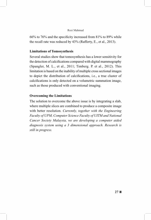
Figure 10 Improved low contrast visibility using tomosynthesis vs digital mammogram
(Adapted from www.aapm.org/meetings/amos2/pdf/34-9727-64244-513.pdf
The image above (Figure 10) shows improved low contrast visibility using tomosynthesis as compared to digital mammogram at lower radiation dose.This is still at the experimental stage.
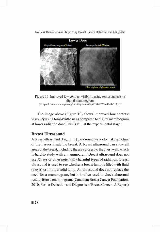
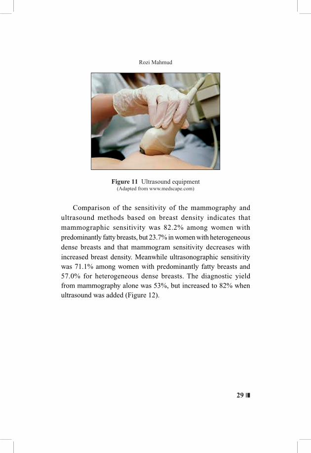
Breast Ultrasound A breast ultrasound (Figure 11) uses sound waves to make a picture of the tissues inside the breast. A breast ultrasound can show all areas of the breast, including the area closest to the chest wall, which is hard to study with a mammogram. Breast ultrasound does not use X-rays or other potentially harmful types of radiation. Breast ultrasound is used to see whether a breast lump is filled with fluid (a cyst) or if it is a solid lump. An ultrasound does not replace the need for a mammogram, but it is often used to check abnormal results from a mammogram. (Canadian Breast Cancer Foundation. 2010, Earlier Detection and Diagnosis of Breast Cancer - A Report)
29 ❘❘❚
Rozi Mahmud
Figure 11 Ultrasound equipment(Adapted from www.medscape.com)
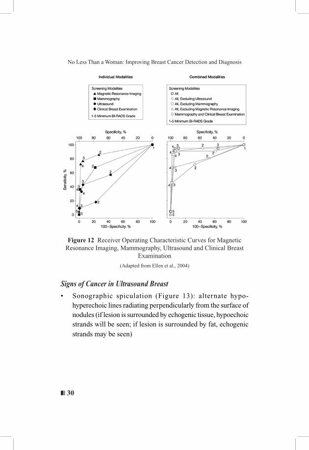
Comparison of the sensitivity of the mammography and ultrasound methods based on breast density indicates that mammographic sensitivity was 82.2% among women with predominantly fatty breasts, but 23.7% in women with heterogeneous dense breasts and that mammogram sensitivity decreases with increased breast density. Meanwhile ultrasonographic sensitivity was 71.1% among women with predominantly fatty breasts and 57.0% for heterogeneous dense breasts. The diagnostic yield from mammography alone was 53%, but increased to 82% when ultrasound was added (Figure 12).
❚❘❘ 30
No Less Than a Woman: Improving Breast Cancer Detection and Diagnosis
Figure 12 Receiver Operating Characteristic Curves for Magnetic Resonance Imaging, Mammography, Ultrasound and Clinical Breast
Examination
(Adapted from Ellen et al., 2004)
Signs of Cancer in Ultrasound Breast
• Sonographic spiculation (Figure 13): alternate hypo-hyperechoic lines radiating perpendicularly from the surface of nodules (if lesion is surrounded by echogenic tissue, hypoechoic strands will be seen; if lesion is surrounded by fat, echogenic strands may be seen)

31 ❘❘❚
Rozi Mahmud
Figure 13 Spiculation and abnormal flow
• Deeper (taller) than wide (Figure 14), usually indicates a malignant feature.
Figure 14 Malignant breast lesion, deeper (taller) than wide
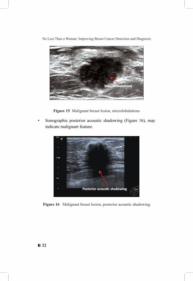
• Microlobulations (Figure 15): small lobulations 1-2 mm on the surface; risk of malignancy rises with increasing numbers
❚❘❘ 32
No Less Than a Woman: Improving Breast Cancer Detection and Diagnosis
Figure 15 Malignant breast lesion, microlobulations
• Sonographic posterior acoustic shadowing (Figure 16), may indicate malignant feature.
Figure 16 Malignant breast lesion, posterior acoustic shadowing

33 ❘❘❚
Rozi Mahmud
• Duct extension (Figure 17) is seen as a projection from a nodule which extends radially within or around a duct towards the nipple
Figure17 Malignant breast lesion, ductal extension
• Heterogeneous (Figure 18) echotexture may indicate a malignant feature.
Figure 18 Malignant breast lesion, heterogeneous echotexture
❚❘❘ 34
No Less Than a Woman: Improving Breast Cancer Detection and Diagnosis
Ultrasound Sensitivity and Specificity
Ultrasound’s sensitivity was found to be within 95.7% (in the good hands of a well-trained breast radiologist), while mammography’s sensitivity was 60.9%. Mammography proved to be the leader in specificity at 94.4% compared with 89.2% for ultrasound (Hardy, K., 2013).
Limitations of Ultrasound
As with mammography, ultrasound examination has its own set of limitations which include:
1. Operator dependable and needs a trained radiologist or breast radiologists to do the investigation.
2. Ultrasound may miss micro-calcification which can be an early sign of breast cancer.
3. Although ultrasound has excellent contrast resolution, it has low spatial resolution and therefore cannot provide as much detail as a mammogram image.
Though most true breast lumps will be found by mammography or ultrasound, some abnormalities escape detection on both imaging tests. For example, a lump may be able to be felt but does not appear on mammography or ultrasound images. If this is the case, then fine needle aspiration biopsy (FNA) is often performed (Devolli-Disha, E., et al., 2009).
Overcoming the Limitations
In order to overcome the above limitations, we have studied various aspects of ultrasound computation and to a certain extent we have managed to increase the radiologist’s efficiency in interpreting the results of the ultrasound machine using Computer-Aided Detection (CAD) algorithms which include:
35 ❘❘❚
Rozi Mahmud
Segmentation techniques:
(a) Fast Histogram Equalization for Medical Image Enhancement Some of the studies involved Automatic Mass Detection in Ultrasound Phantom Images where this work is concentrated on extraction of mass in Ultrasound breast images to help radiologists interpret such images efficiently using Computer-Assisted Detection. A set of six popular ultrasound machines were selected and images were acquired by sweeping: modes of operation, transducer, frequency and contrast. To make a complete set of ultrasound images in B-Mode a multi-purpose multi tissue Ultrasound Phantom was used. Gamma corrections, contrast stretching and filtering accompanied by morphological Image Processing were among the steps applied to produce the final image. We are able to automatically extract cystic masses from ultrasound phantom images and improve the eff iciency of interpretation using Computer-Aided Detection. The following parameters were swept, modes of operation, transducer, frequency and contrast, while producing the phantom images. Ultrasound images were acquired using a quality multi tissue Ultrasound Phantom in B-Mode. Gamma corrections, contrast stretching, filtering and morphological image processing were among the steps applied to find the output image utilising Fast Histogram Equalization for Medical Image Enhancement. Two experienced radiologists marked the final images. Statistical analysis of the results showed a sensitivity of 99% and accuracy of 98% for the proposed framework. As a side result, based on the actual depth of each image, processing time was also decreased (Khatib, F.,Mahmud, R., et al., 2012a; Khatib, F.,Mahmud, R., et al.,2012b; Khatib, F.,Mahmud, R., et al., 2012c; Khatib, F.,Mahmud, R., et al., 2012d; Khatib, F.,Mahmud, R., et al., 2012 e)

❚❘❘ 36
No Less Than a Woman: Improving Breast Cancer Detection and Diagnosis
(b) Active Contour Algorithm Similar to the mammogram study, we applied the same technique, Active Contour Algorithm, in ultrasound of the breast.This includes Segmentation of Masses from Breast Ultrasound Images using Parametric Active Contour Algorithm where the boundaries of the masses identified were used in classification of cancerous or non-cancerous masses. Specifically, the Balloon Snake was applied in segmenting the masses in the breast ultrasound images. Comparison of the masses segmented by the Balloon Snake against the areas traced by the radiologist was done. It was found that from forty-five masses tested, the average percentage area difference for the Balloon Snake was 4.47%. This implies that the accuracy of the segmentation results for the Balloon Snake was 95.53% (Jumaat, A.K., Mahmud, R., et al., 2010a). In order to improve the efficiency of interpretation of the ultrasound images, we studied various ways to enhance the images to an acceptable level using Computer-Aided Detection (CAD) algorithms. We used MATLAB, an on-line system providing machine aid for the mechanical symbolic processes encountered in analysis and an image processing tool box and applied factors such as signal to noise ratio(SNR), peak signal to noise ratio(PSNR) and mean square error(MSE) parameters to check the output image. Analysis of the results showed that our proposed framework enhanced visualization and decreased interpreting time while increasing SNR (Khatib, F.,Mahmud, R., et al., 2012f). Subsequently we also went into depth segmenting and characterizing of the masses in breast ultrasound images using active contour, where the active contour or Snake, which is a computer generated curve was used to trace the boundaries of images. We studied segmentation of masses on breast ultrasound images and the characterization of the segmented masses as

37 ❘❘❚
Rozi Mahmud
malignant or benign. Initially, the Balloon Snake was chosen to segment the masses. Comparison of the masses areas segmented by the Balloon Snake against the areas traced by the radiologist was done. The experimental results showed that from fifty masses tested, the Balloon Snake successfully segmented the masses with accuracy of 95.71%. Then, the masses were characterized as benign or malignant using a proposed method, namely, the semi-automated characterization (SAC) method. This method is based on the segmented masses produced by the Balloon Snake. The criterion of angular margin was considered in characterizing the masses as malignant or benign using the SAC method. The characterization reading of a mass using the SAC method was compared with thirty sets of characterization readings of a mass by different radiologists. The comparison was made in terms of sensitivity and specificity values. Based on the values, the receiver operating characteristics (ROC) curve was plotted for each set of comparisons. From the thirty sets of comparisons, it was found that the area under the curve for all the thirty ROC curves were greater than 0.7. The value implies that the SAC method gives high accuracy in differentiating benign from malignant masses. Since the method is based on the segmented masses by the Balloon Snake, the value also implies that the accuracy of the Balloon Snake in segmenting images is high (95.71%) (Jumaat, A.K., Mahmud, R., et al., 2011).
(c) Gradient Vector Flow snakes GVF is an extension of the well-known method snakes or active contours. The difference between traditional snakes and GVF snakes lies in that the latter’s convergence of boundary concavities where they do not need to be initialized close to the boundary. In another study, we compared the Balloon Snake and Gradient Vector Flow (GVF) Snake, in Segmenting Masses from Breast Ultrasound Images (Jumaat, A.K., Mahmud, R., et al., 2010b).
❚❘❘ 38
No Less Than a Woman: Improving Breast Cancer Detection and Diagnosis
Comparisons were done on the masses areas segmented by the Balloon Snake and the GVF Snake against actual masses areas. The better method with smaller values of average percentage area difference was chosen to be applied in segmenting masses in real breast ultrasound images. The performance was measured in terms of average percentage area difference traced by the chosen method against the area traced by expert radiologists. It was found that from eighty breast ultrasound phantom images (40 cyst and 40 solid masses) tested, the values of average percentage area difference for cyst and solid masses in the Balloon Snake were 3.07% and 9.17%, respectively, while for the GVF Snake it was 13.43% and 48.37%, respectively. Hence the Balloon Snake was chosen to segment the real breast ultrasound images whereby fifty images were tested. Segmentation of the images showed that the average percentage area difference of the Balloon Snake was 4.29%, which means 95.71% accuracy.
(d) Angular Margin Measurement, active contourFor an additional method to aid the radiologists in classifying the lesions in breast ultrasounds as benign or malignant we studied characterization of masses based on angular margin measurement and active contour. (Jumaat, A.K., Mahmud, R., et al., 2012a; Jumaat, A.K., Mahmud, R., et al., 2012b) Comparison was done of the masses areas segmented by the Balloon Snake against the areas traced by radiologists. Experimental results showed that from fifty masses tested, the Balloon Snake successfully segmented the masses with accuracy of 95.71%. Then, a Balloon Snake segmented mass was characterized as benign or malignant based on the measurement of angular margin. A mass is considered as malignant if it has at least one acute angular margin, otherwise it is benign. The characterization of a mass by the Balloon
39 ❘❘❚
Rozi Mahmud
Snake based on the angular margin criterion was compared with the actual type of a mass as characterized by an expert radiologist. The comparison was made in terms of sensitivity and specificity values. Based on the values, the receiver operating characteristics (ROC) curve was plotted. It was found that the area under the curve was 0.8455. The value implies that the Balloon Snake gives high accuracy in characterizing masses as benign from malignant based on the criterion used.
Breast Magnetic Resonance Imaging (MRI)
MRI is a diagnostic exam that uses a combination of a large magnet, radio waves and a computer to produce detailed images of organs and structures within the body. Breast magnetic resonance imaging (MRI) is an imaging test that creates detailed images of the internal structures of the breast.
Benefits of Breast MRI
It is able to:1. Detect small abnormalities2. Effective for dense breasts3. Image of breast implants/ruptures4. Evaluate inverted nipples5. Locate primary tumour in women whose cancer has spread to
armpit lymph nodes6. Detect residual cancer after lumpectomy7. Determine what type of surgery is indicated: lumpectomy or
mastectomy8. Detect cancer recurrence after lumpectomy9. May be useful to screen women at high breast cancer risk

❚❘❘ 40
No Less Than a Woman: Improving Breast Cancer Detection and Diagnosis
Signs of Cancer in Breast MRI
Masses which are spiculated (Figure 19), irregular and have abnormal enhancement patterns as well as ductal enhancement, axillary nodes enlargement with abnormal pattern of enhancement may have high correlation with cancer (The women’s health resources. (2015). Breast Cancer MRI.).
Figure 19 Right breast cancerAdapted from: www.aboutcancer.com
Limitations of Breast MRI
1. Non-specific; often cannot distinguish cancerous and non-cancerous tumors
2. May lead to unnecessary, difficult to perform biopsies3. Cannot image calcifications, tiny calcium deposits that can
indicate early breast cancers4. Expensive and not widely available5. Some patients who are claustrophobic may not tolerate MRI6. Requires use of contrast agent7. More time-consuming than mammography8. MRI centres cannot always produce results cited in research
studies
41 ❘❘❚
Rozi Mahmud
Overcoming the Limitations
Many researchers are currently studying various ways to overcome the limitations.
Computed Tomography Laser Mammography (CTLM)
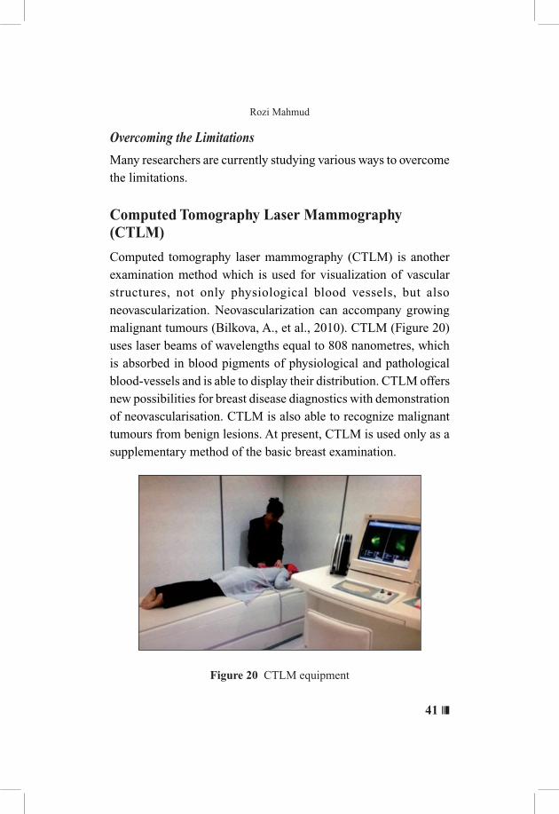
Computed tomography laser mammography (CTLM) is another examination method which is used for visualization of vascular structures, not only physiological blood vessels, but also neovascularization. Neovascularization can accompany growing malignant tumours (Bilkova, A., et al., 2010). CTLM (Figure 20) uses laser beams of wavelengths equal to 808 nanometres, which is absorbed in blood pigments of physiological and pathological blood-vessels and is able to display their distribution. CTLM offers new possibilities for breast disease diagnostics with demonstration of neovascularisation. CTLM is also able to recognize malignant tumours from benign lesions. At present, CTLM is used only as a supplementary method of the basic breast examination.
Figure 20 CTLM equipment

❚❘❘ 42
No Less Than a Woman: Improving Breast Cancer Detection and Diagnosis
Signs of Cancer in CTLM
The image below (Figure 21) shows presence of neovascularization:
Figure 21 Presence of neovascularization seen as specific characteristic in its distribution and pattern
(Adapted from: www. eurolifecare.com)
CTLM Sensitivity and Specificity
Positive lesions were observed more significantly in malignant than in benign lesions. The sensitivity of mammography versus mammography plus CTLM was 34.4% versus 81.57% among extremely dense breasts and 68.29% versus 95.34% among heterogeneously dense breasts (Qi, J. & Ye,Z., 2013).
Limitations of CTLM
For detection of abnormal flow, the radiologist sometimes needs to manipulate and change the range of the window width and level multiple times. There are also many appearances of angiogenesis which can be easily missed.
43 ❘❘❚
Rozi Mahmud
Overcoming the Limitations
Our research on CTLM is still at the final stage of analysis. However at the preliminary stage, we have found that overall results indicate superiority of the 3DFuzzy C Means (FCM) technique compared to other methods. One of our papers, with the title ‘Computed Automatic 3D Segmentation Methods in Computed Tomography Laser mammography’, was also accepted for presentation at the 3rd International Conference on Advances in Computing, Electronics and Communications, 10 - 11 October 2015 (Jalalian, A., Mahmud, R., et al., 2015).
Breast Thermography
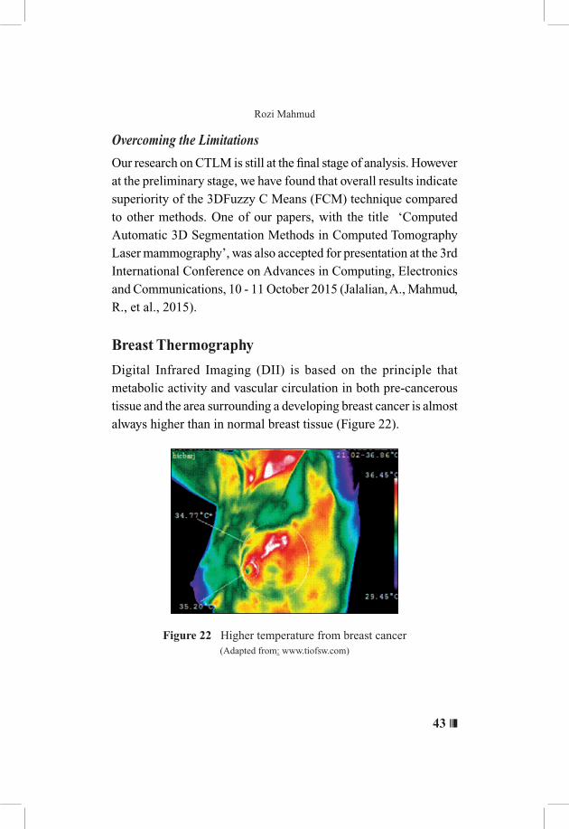
Digital Infrared Imaging (DII) is based on the principle that metabolic activity and vascular circulation in both pre-cancerous tissue and the area surrounding a developing breast cancer is almost always higher than in normal breast tissue (Figure 22).
Figure 22 Higher temperature from breast cancer(Adapted from: www.tiofsw.com)
❚❘❘ 44
No Less Than a Woman: Improving Breast Cancer Detection and Diagnosis
Signs of Cancer in Breast Thermography
DII is based on detecting the heat produced by increased blood vessel circulation and metabolic changes associated with a tumor’s genesis and growth. By detecting minute variations in normal blood vessel activity, infrared imaging may find thermal signs suggesting a cancerous state.
Breast Thermography Sensitivity and Specificity
The Breast Thermography sensitivity was 25% with specificity of 85%. Despite being non-invasive and painless, because of the low sensitivity for breast cancer, DII is not recommended for the primary evaluation of symptomatic patients nor should it be used on a routine basis as a screening test for breast cancer. (Kontos, M., et al, 2011)
Limitations of Breast Thermography
Interpreting breast thermography images can be quite challenging, as the colour code may overlap thus missing the diagnosis.
Overcoming the Limitations
In order to aid the radiologists in detecting breast lesions, we have studied the role of image segmentation leading towards computer aided diagnosis. In breast thermography diagnostics, proper detection and segmentation of the areola area as well as detection of breast boundaries present the biggest challenge. As the boundaries of breasts, especially in the upper quadrants, are usually not present, this poses a great challenge to segment breasts automatically. Many approaches have been developed to segment the breasts in the past, such as Snakes, Active Contours and Circular Hough Transforms, but these methods fail to detect the boundaries of the breast with the
45 ❘❘❚
Rozi Mahmud
required level of accuracy, especially the upper boundaries of the breast. The most recent segmentation method is Random Walkers, where the breast can be segmented accurately, which increases the accuracy and reliability of computer aided detection/diagnosis systems (Moghbel, M., Mahmud, R., et al., 2012).
Breast Ultra Wide Band (UWB)
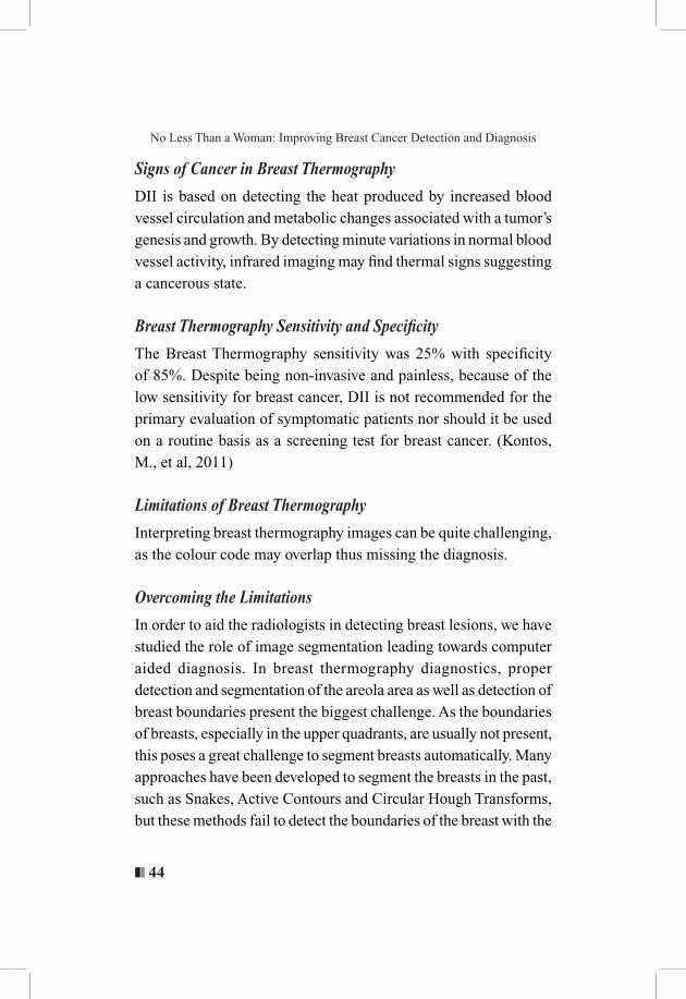
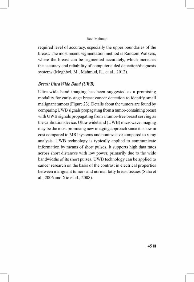
Ultra-wide band imaging has been suggested as a promising modality for early-stage breast cancer detection to identify small malignant tumors (Figure 23). Details about the tumors are found by comparing UWB signals propagating from a tumor-containing breast with UWB signals propagating from a tumor-free breast serving as the calibration device. Ultra-wideband (UWB) microwave imaging may be the most promising new imaging approach since it is low in cost compared to MRI systems and noninvasive compared to x-ray analysis. UWB technology is typically applied to communicate information by means of short pulses. It supports high data rates across short distances with low power, primarily due to the wide bandwidths of its short pulses. UWB technology can be applied to cancer research on the basis of the contrast in electrical properties between malignant tumors and normal fatty breast tissues (Saha et al., 2006 and Xio et al., 2008).
❚❘❘ 46
No Less Than a Woman: Improving Breast Cancer Detection and Diagnosis
Figure 23 UWB experimental setup
Some of my research related to ultra-wide band in general reveals the possibility of diagnosing breast cancer using UWB.
Neural Network (NN) based Pattern Recognition
We studied breast tumor detection using neural network nn-based UWB imaging as an experimental complement to simulation work for early breast tumor detection. The experiments were conducted using commercial Ultrawide-Band (UWB) transceivers and Neural Network (NN) based Pattern Recognition (PR) software for imaging and proposed breast phantoms for homogenous and heterogeneous tissues. The proposed breast phantoms (homogeneous and heterogeneous) and tumor were constructed using available low cost materials and their mixtures with minimal effort. A specific glass was used as skin. All the materials and their mixtures were considered according to the ratio of the dielectric properties of the breast tissues. Experiments to detect tumors were performed in a regular noisy room environment. The UWB signals were transmitted from one side of the breast phantom (for both cases) and received from the opposite side diagonally, repeatedly. Using
47 ❘❘❚
Rozi Mahmud
discrete cosine transform (DCT) of these received signals, a Neural Network (NN) module was developed, trained and tested. The tumor existence, size and location detection rates for both cases were highly satisfactory, which were approximately: (i) 100%, 95.8% and 94.3% for homogeneous; and (ii) 100%, 93.4% and 93.1% for heterogeneous cases, respectively. This gives assurance of early detection and the practical usefulness of the developed system in the near future (Alshehri, S., Mahmud, R.,et al., 2011a). We then proceeded to another level which included discriminating benign and malignant changes where both simulation and experimental studies were conducted to detect and locate breast tumors along with their classification as malignant and/or benign in three dimensional (3D) breast models. The contrast between the dielectric properties of these two tumor types was the main key. These dielectric properties are mainly controlled by the water and blood content of tumors. For simulation, electromagnetic simulator software was used. The experiment was conducted using Commercial Ultra-wide Band (UWB) transceivers, Neural Network (NN) based Pattern Recognition (PR) software for imaging and homogenous breast phantom. The 3D homogeneous breast phantom and tumors were fabricated using pure petroleum jelly and a mixture of wheat flour and water, respectively. The simulation and experimental setups were performed by transmitting the UWB signals from one side of the breast model and receiving from the opposite side, diagonally. Using discrete cosine transform (DCT) of received signals, we trained and tested the developed experimental Neural Network model. In the 3D breast model, the achieved detection accuracy of tumor existence was around 100%, while the locating accuracy in terms of (x, y, z) position of a tumor within the breast reached approximately 89.2% and 86.6% in the simulation and experimental works, respectively. For classification,
❚❘❘ 48
No Less Than a Woman: Improving Breast Cancer Detection and Diagnosis
the permittivity and conductivity detection accuracy were 98.0% and 99.1% in simulation, and 98.6% and 99.5% in experimental works, respectively. Tumor detection and type specification in 3D may lead to successful clinical implementation followed by saving of precious human lives in the near future (Alshehri, S., Mahmud, R.,et al., 2011a; Alshehri, S., Mahmud, R.,et al., 2011b)
UWB Sensitivity and Specificity
This method is still under research using phantoms where the sensitivity, specificity and limitations are yet to be proven.
Scintimammography
Scintimammography, also known as nuclear medicine breast imaging, is an examination that may be used to investigate a breast abnormality that has been discovered on mammography. Scintimammography is also known as Breast Specific Gamma Imaging (BSGI) or Molecular Breast Imaging (MBI). Nuclear medicine is a branch of medical imaging that uses small amounts of radioactive material to diagnose and determine the severity of or to treat a variety of diseases, including many types of cancers, heart disease, gastrointestinal, endocrine, neurological disorders and other abnormalities within the body. Since nuclear medicine procedures are able to pinpoint molecular activity within the body, they offer the potential to identify diseases at the earliest stages as well as a patient’s immediate response to therapeutic interventions. The procedure is noninvasive and involves the injection of a radiotracer, or drug that emits radioactivity, into the patient. As the radiotracer accumulates differently in different kinds of tissues, it can help physicians determine whether cancer could be present,
49 ❘❘❚
Rozi Mahmud
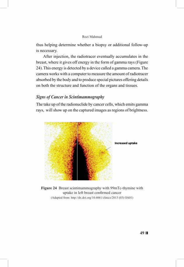
thus helping determine whether a biopsy or additional follow-up is necessary. After injection, the radiotracer eventually accumulates in the breast, where it gives off energy in the form of gamma rays (Figure 24). This energy is detected by a device called a gamma camera. The camera works with a computer to measure the amount of radiotracer absorbed by the body and to produce special pictures offering details on both the structure and function of the organs and tissues.
Signs of Cancer in Scintimammography
The take up of the radionuclide by cancer cells, which emits gamma rays, will show up on the captured images as regions of brightness.
Figure 24 Breast scintimammography with 99mTc-thymine with uptake in left breast confirmed cancer
(Adapted from: http://dx.doi.org/10.6061/clinics/2013 (03) OA01)
❚❘❘ 50
No Less Than a Woman: Improving Breast Cancer Detection and Diagnosis
Scintimammography Sensitivity and Specificity
Several multi centric studies with blinded image interpretation have established the sensitivity and specificity of scintimammography to be above 85 and 90 %, respectively, as compared to 89 and 14 %, respectively, for X-ray mammography (B, K, Das. et al., 2006).
Limitations of Scintimammography
Scintimammography is not a primary screening procedure for breast cancer. It should not be considered a replacement for mammography or ultrasound. Additionally, nuclear medicine procedures can be time-consuming. The image resolution of structures of the body obtained with nuclear medicine procedures may not be as clear as with other imaging techniques, such as mammography or MRI. If an abnormality is detected on scintimammography, it may be difficult to find the lesion using other imaging modalities, thus making it difficult to perform a biopsy (Radiology information for patients Scintimammography. RSNA, 2015).
Overcoming the Limitations
There are some solutions to overcome the above issues which includes increasing the detection of small cancers in Scintimammography. In order to further increase the detection of small cancers, we studied a computational model involving Monte Carlo Simulation on Breast Cancer Detection Using Wire Mesh Collimator Gamma Camera (Saripan, M. I.,Mahmud, R., et al., 2009). This paper presents the preliminary results of the new low energy high resolution wire-mesh collimator gamma camera in mapping breast cancer cells, by employing 140 keV photons of Technetium-99 m radionuclide tracer. The complete model for photons propagation and detection as well as the human cells’
51 ❘❘❚
Rozi Mahmud
activities was simulated using Monte Carlo N-Particle code. Abnormal cells of different tumors to background values were investigated, and the results from the conventional collimator and wire-mesh collimator were compared. The results were evaluated in terms of the collimator sensitivity and the contrast to the background ratio. In our assessment, the wire mesh collimator gamma camera yields slightly better results than the multihole collimator for sensitivity, but produces insignificant performance in the contrast to background evaluation. We also studied the characteristics of a newly developed wire mesh collimator (WMC) in Single Photon Emission Computed tomography (SPECT) and Characterization of Sinogram Optimum Filter in Tc-99m Breast SPECT Images. The characterization was based on a Butterworth filter with a trade-off between noise suppression and spatial resolution degradation. We performed Monte Carlo simulation studies in order to quantify lesion detectability and to determine the parameters involved in the relationship between optimum cut-off frequency, total image counts and tumor size which were revealed for a breast SPECT imaging system equipped with a WMC. Characteristics of the Sinogram for the Wire Mesh Collimator in Tc-99m breast SPECT imaging was investigated in this research and the optimum cut-off frequency for the Butterworth filter was found to be not only related to the count density, but also linked to the change in tumor diameter, which may help radiologists to predict the cut-off frequency from these two parameters. (Dong, X., R. Mahmud., et al 2015)
❚❘❘ 52
No Less Than a Woman: Improving Breast Cancer Detection and Diagnosis
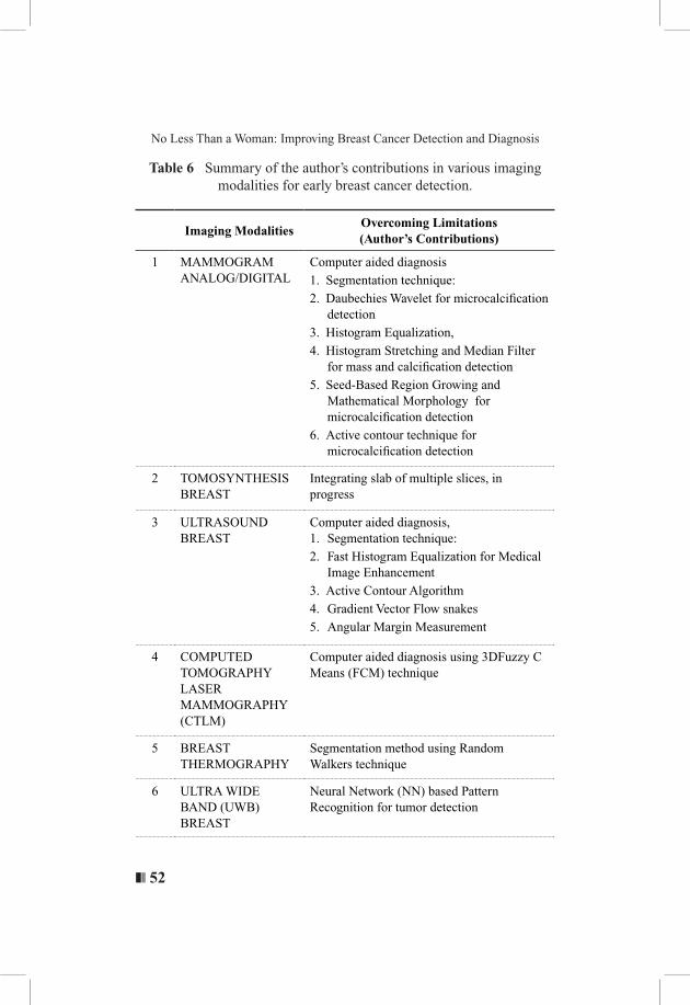
Table 6 Summary of the author’s contributions in various imaging modalities for early breast cancer detection.
Imaging ModalitiesOvercoming Limitations(Author’s Contributions)
1 MAMMOGRAM ANALOG/DIGITAL
Computer aided diagnosis
1. Segmentation technique:
2. Daubechies Wavelet for microcalcification detection
3. Histogram Equalization,
4. Histogram Stretching and Median Filter for mass and calcification detection
5. Seed-Based Region Growing and Mathematical Morphology for microcalcification detection
6. Active contour technique for microcalcification detection
2 TOMOSYNTHESIS BREAST
Integrating slab of multiple slices, in progress
3 ULTRASOUND BREAST
Computer aided diagnosis, 1. Segmentation technique:
2. Fast Histogram Equalization for Medical Image Enhancement
3. Active Contour Algorithm
4. Gradient Vector Flow snakes
5. Angular Margin Measurement
4 COMPUTED TOMOGRAPHY LASER MAMMOGRAPHY (CTLM)
Computer aided diagnosis using 3DFuzzy C Means (FCM) technique
5 BREAST THERMOGRAPHY
Segmentation method using Random Walkers technique
6 ULTRA WIDE BAND (UWB) BREAST
Neural Network (NN) based Pattern Recognition for tumor detection
53 ❘❘❚
Rozi Mahmud
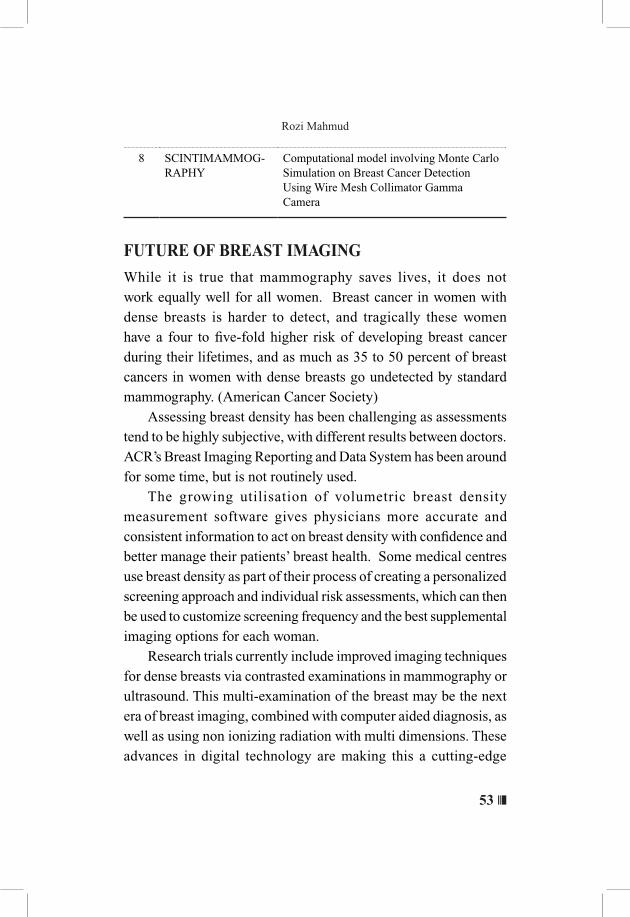
8 SCINTIMAMMOG-RAPHY
Computational model involving Monte Carlo Simulation on Breast Cancer Detection Using Wire Mesh Collimator Gamma Camera
FUTURE OF BREAST IMAGING
While it is true that mammography saves lives, it does not work equally well for all women. Breast cancer in women with dense breasts is harder to detect, and tragically these women have a four to five-fold higher risk of developing breast cancer during their lifetimes, and as much as 35 to 50 percent of breast cancers in women with dense breasts go undetected by standard mammography. (American Cancer Society) Assessing breast density has been challenging as assessments tend to be highly subjective, with different results between doctors. ACR’s Breast Imaging Reporting and Data System has been around for some time, but is not routinely used. The growing utilisation of volumetric breast density measurement software gives physicians more accurate and consistent information to act on breast density with confidence and better manage their patients’ breast health. Some medical centres use breast density as part of their process of creating a personalized screening approach and individual risk assessments, which can then be used to customize screening frequency and the best supplemental imaging options for each woman. Research trials currently include improved imaging techniques for dense breasts via contrasted examinations in mammography or ultrasound. This multi-examination of the breast may be the next era of breast imaging, combined with computer aided diagnosis, as well as using non ionizing radiation with multi dimensions. These advances in digital technology are making this a cutting-edge
❚❘❘ 54
No Less Than a Woman: Improving Breast Cancer Detection and Diagnosis
discipline, with the possibility of a home monitoring device for early detection at our finger tips. Via whatever means, the fact remains that early detection can prevent the need for extensive treatments and improve the chances of breast conservation, thus making those affected with the disease no less than a women.
Personalized Screening in Breast Disease
Currently, expanding personalized risk profiles of women via the use of molecular profiling is a trend. When used in combination, the overall effect of these advances provides better imaging options for women with dense breasts and a new gold standard in breast cancer screening and diagnosis for all women. (Warner, E.,et al 2004) Genetic screening would provide an approach where care and treatment are based on both f ixed and modifiable risk factors unique to each patient, as well as the pathological and molecular characteristics that make each breast carcinoma unique. Unfortunately, this type of screening is not as yet widely available in Malaysia. Our research related to Gene Expression of Cancer:
Multiplex Gene Expression of Cancer-related Genes in Peripheral Blood of Malaysian Breast Cancer Patients
The objective was to identify the cancer gene expression profiles in eight breast cancer patients. RNA was extracted from blood of eight normal respondents and eight breast cancer patients. Nine genes were multiplexed, AKT1, AR, BRCA2, ESR2, KRT8, KRT18, PTGS2, SLC39A6 and TFF3, to screen for their expression levels using fragment analysis approach. The mean relative expressions of BRCA2, PTGS2 and SLC39A6 were found to be up-regulated while that for AKT1, AR, ESR2, KRT8, KRT18 and TFF3 were
55 ❘❘❚
Rozi Mahmud
down-regulated in the normal respondents compared to the breast cancer patients. Independent-samples T-test showed gene expression profiles for all the nine genes were not significantly different between the groups (p>0.05). It is suggested that more genes be adopted for screening to identify cancer genes that are overexpressed in Malaysian cancer patients. (Yow, W. K., Mahmud, R., et al., 2015)
OTHER STUDIES ON DISEASE RELATED TO WOMEN
Our passion for learning about diseases that involves women is immense and we have engaged in various research which include:
• Effects of germinated brown rice andBonemass densityand uterus (Muhammad, S. I.,Mahmud,R., et al., 2013a;Muhammad,S.I.,Mahmud,R.,etal.,2013b;Muhammad,S.I.,Mahmud,R.,etal.,2013c)
• TheProtectiveEffectsofNigellasativaonMetabolicSyndromeand its hypolipidemic inMenopausalWomen (Ibrahim,R.,Mahmud,R.,etal.,2014a;Ibrahim,R.,Mahmud,R., etal.,2014b)
• Analysis of brain image segmentation and atrophy inAlzheimer’sdiseasewhichcommonlyaffectsthefemalegender(Roslan,R.,Mahmud,R.,etal.,2010;Balafar,M.,Mahmud,R.,etal.,2008a;Balafar,M.,Mahmud,R.,etal.,2008b,Balafar,M.,Mahmud,R.,etal.,2008c,Balafar,M.,Mahmud,R.,etal.,2008d,Balafar,M.,Mahmud,R.,et al., 2008e;Balafar,M.,Mahmud,R.,etal.,2010a;Balafar,M.,Mahmud,R.,etal.,2010b;Farzan,A.,Mahmud,R.,etal.,2010,Farzan,A.,Mahmud,R.,etal.,2011;Farzan,A.,Mahmud,R.,etal.,2015)
❚❘❘ 56
No Less Than a Woman: Improving Breast Cancer Detection and Diagnosis
• Various detailed studies on Birds nest which includenutrigenomiceffectsofediblebird’snestoninsulinsignallingin ovariectomized rats, howLactoferrin and ovotransferrincontribute toward antioxidative effects of Edible Bird’sNest against hydrogen peroxide-induced oxidative stress inhumanSH-SY5YcellsandEffectsofEdibleBird’sNestonhippocampalandcorticalneurodegenerationinovariectomizedrats (Zhiping, H., Mahmud, R.,et al., 2015a; Zhiping, H., Mahmud, R.,et al., 2015b; Zhiping, H., Mahmud, R.,et al., 2015c)

57 ❘❘❚
Rozi MahmudR
ESE
AR
CH
GR
AN
TS
Tab
le 7
Res
earc
h gr
ants
No.
Pro
ject
Tit
leR
ole
Yea
rSo
urce
of
fund
Stat
us
1.
A
nti-
inva
sive
and
ant
i-m
etas
tati
c ac
tivit
ies
of n
ew a
ndro
grap
holi
de d
eriv
ativ
esC
o-in
vest
igat
or20
07S
CIE
NC
EF
UN
D,
MO
ST
IC
ompl
eted
2.
E
arly
bre
ast c
ance
r de
tect
ion
usin
g w
ire-
mes
h co
llim
ator
Co-
inve
stig
ator
2007
RU
GS
, UP
MC
ompl
eted
3.
V
irtu
al a
nim
al a
utop
sy
Co-
inve
stig
ator
2008
SC
IEN
CE
FU
ND
, M
OS
TI
Com
plet
ed
4.
C
ompu
ter
aide
d di
agno
sis
in b
reas
t can
cer
dete
ctio
n in
mam
mog
raph
y us
ing
wav
elet
an
d se
gmen
tati
on m
etho
d.C
o-in
vest
igat
or20
08S
CIE
NC
EF
UN
D,
MO
ST
IC
ompl
eted
5.
T
he e
ffec
t of
past
use
of
Dep
ot M
edro
x P
roge
stro
n A
ceta
te o
n bo
ne m
iner
al d
ensi
ty
in p
ostm
enop
ausa
l wom
en
Pri
ncip
al
inve
stig
ator
2009
RU
GS
, UP
MC
ompl
eted
6.
A
com
pari
son
stud
y of
CT
LM
and
m
amm
ogra
m s
tudy
for
se
nsit
ivit
y an
d sp
ecifi
city
Pri
ncip
al
inve
stig
ator
2009
Abl
e G
loba
l Sdn
. B
erha
d, M
alay
sia
Com
plet
ed
❚❘❘ 58
No Less Than a Woman: Improving Breast Cancer Detection and Diagnosis
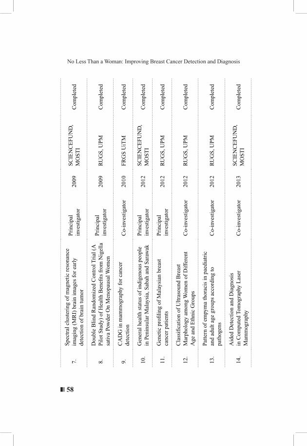
7.
S
pect
ral c
lust
erin
g of
mag
neti
c re
sona
nce
imag
ing
(MR
I) b
rain
imag
es f
or e
arly
de
tect
ion
of b
rain
tum
or
Pri
ncip
al
inve
stig
ator
2009
SC
IEN
CE
FU
ND
, M
OS
TI
Com
plet
ed
8.
D
oubl
e B
lind
Ran
dom
ized
Con
trol
Tri
al (
A
Pil
ot S
tudy
) of
Hea
lth
Ben
efits
fro
m N
igel
la
sativ
a Po
wde
r O
n M
enop
ausa
l Wom
en
Pri
ncip
al
inve
stig
ator
2009
RU
GS
, UP
MC
ompl
eted
9.
C
AD
G in
mam
mog
raph
y fo
r ca
ncer
de
tect
ion
Co-
inve
stig
ator
2010
FR
GS
UiT
MC
ompl
eted
10.
G
ener
al h
ealt
h st
atus
of
indi
geno
us p
eopl
e in
Pen
insu
lar
Mal
aysi
a, S
abah
and
Sar
awak
Pri
ncip
al
inve
stig
ator
2012
SC
IEN
CE
FU
ND
, M
OS
TI
Com
plet
ed
11.
G
enet
ic p
rofi
ling
of
Mal
aysi
an b
reas
t ca
ncer
pat
ient
sP
rinc
ipal
in
vest
igat
or20
12R
UG
S, U
PM
Com
plet
ed
12.
C
lass
ifica
tion
of
Ult
raso
und
Bre
ast
Mar
phol
ogy
amon
g W
omen
of
Dif
fere
nt
Age
and
Eth
nic
Gro
ups
Co-
inve
stig
ator
2012
RU
GS
, UP
MC
ompl
eted
13.
Pa
tter
n of
em
pym
a th
orac
is in
pae
diat
ric
and
adul
t age
gro
ups
acco
rdin
g to
pa
thog
ens
Co-
inve
stig
ator
2012
RU
GS
, UP
MC
ompl
eted
14.
A
ided
Det
ecti
on a
nd D
iagn
osis
in
Com
pute
d To
mog
raph
y L
aser
M
amm
ogra
phy
Co-
inve
stig
ator
2013
SC
IEN
CE
FU
ND
, M
OS
TI
Com
plet
ed
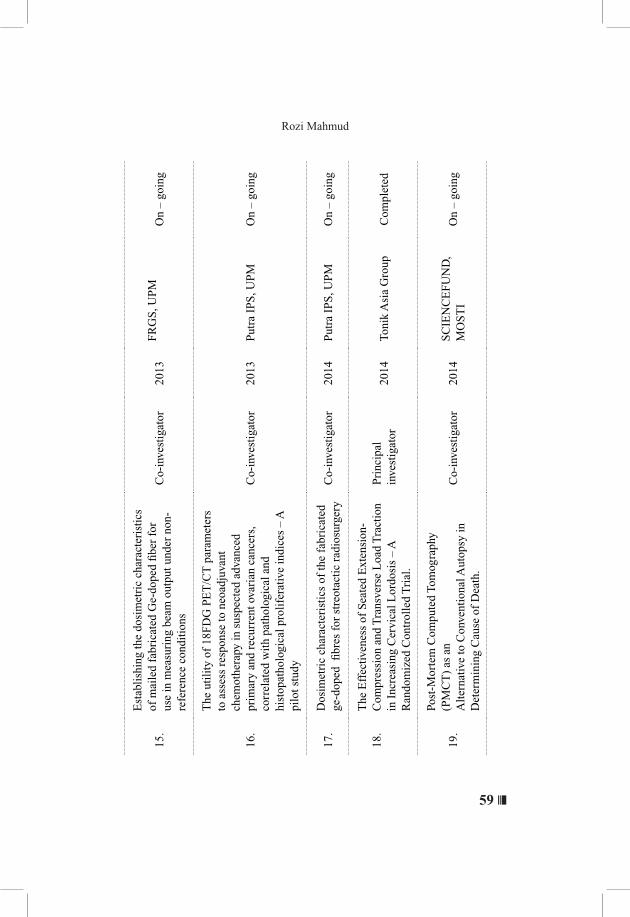
59 ❘❘❚
Rozi Mahmud
15.
Est
abli
shin
g th
e do
sim
etri
c ch
arac
teri
stic
s of
mai
led
fabr
icat
ed G
e-do
ped
fibe
r fo
r us
e in
mea
suri
ng b
eam
out
put u
nder
non
-re
fere
nce
cond
itio
ns
Co-
inve
stig
ator
2013
FR
GS
, UP
M
On
– go
ing
16.
The
uti
lity
of
18F
DG
PE
T/C
T p
aram
eter
s to
ass
ess
resp
onse
to n
eoad
juva
nt
chem
othe
rapy
in s
uspe
cted
adv
ance
d pr
imar
y an
d re
curr
ent o
vari
an c
ance
rs,
corr
elat
ed w
ith
path
olog
ical
and
hi
stop
atho
logi
cal p
roli
fera
tive
indi
ces
– A
pi
lot s
tudy
Co-
inve
stig
ator
2013
Put
ra I
PS
, UP
MO
n –
goin
g
17.
D
osim
etri
c ch
arac
teri
stic
s of
the
fabr
icat
ed
ge-d
oped
fibr
es f
or s
treo
tact
ic r
adio
surg
ery
Co-
inve
stig
ator
2014
Put
ra I
PS
, UP
MO
n –
goin
g
18.
The
Eff
ectiv
enes
s of
Sea
ted
Ext
ensi
on-
Com
pres
sion
and
Tra
nsve
rse
Loa
d T
ract
ion
in I
ncre
asin
g C
ervi
cal L
ordo
sis
– A
Ran
dom
ized
Con
trol
led
Tri
al.
Pri
ncip
al
inve
stig
ator
2014
Toni
k A
sia
Gro
upC
ompl
eted
19.
Post
-Mor
tem
Com
pute
d To
mog
raph
y (P
MC
T)
as a
nA
lter
nativ
e to
Con
vent
iona
l Aut
opsy
in
Det
erm
inin
g C
ause
of
Dea
th.
Co-
inve
stig
ator
2014
SC
IEN
CE
FU
ND
, M
OS
TI
On
– go
ing
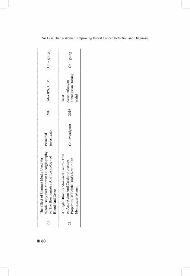
❚❘❘ 60
No Less Than a Woman: Improving Breast Cancer Detection and Diagnosis
20.
The
Eff
ect o
f C
ontr
ast M
edia
Use
d Fo
r W
hole
Bod
y Po
st-M
orte
m C
t-A
ngio
grap
hy
on T
he B
ioch
emis
try
And
Tox
icol
ogy
of
Blo
od A
nd U
rine
Pri
ncip
al
inve
stig
ator
2016
Put
ra I
PS
, UP
MO
n –
goin
g
21.
A S
ingl
e B
lind
Ran
dom
ized
Con
trol
Tri
al
on A
nti-
Agi
ng A
nd C
ardi
o-pr
otec
tive
Pro
pert
ies
Of
Edi
ble
Bir
d’s
Nes
t In
Pre
-M
enop
ause
Wom
en
Co-
inve
stig
ator
2016
Pus
at
Kec
emer
lang
an
Keb
angs
aan
Bur
ung
Wal
id
On
– go
ing
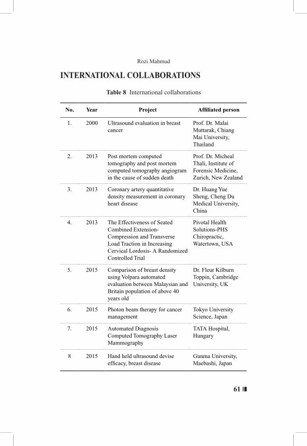
61 ❘❘❚
Rozi Mahmud
INTERNATIONAL COLLABORATIONS
Table 8 International collaborations
No. Year Project Affiliated person
1. 2000 Ultrasound evaluation in breast cancer
Prof. Dr. Malai Muttarak, Chiang Mai University, Thailand
2. 2013 Post mortem computed tomography and post mortem computed tomography angiogram in the cause of sudden death
Prof. Dr. Micheal Thali, Institute of Forensic Medicine, Zurich, New Zealand
3. 2013 Coronary artery quantitative density measurement in coronary heart disease
Dr. Huang Yue Sheng, Cheng Du Medical University, China
4. 2013 The Effectiveness of Seated Combined Extension-Compression and Transverse Load Traction in Increasing Cervical Lordosis- A Randomized Controlled Trial
Pivotal Health Solutions-PHS Chiropractic, Watertown, USA
5. 2015 Comparison of breast density using Volpara automated evaluation between Malaysian and Britain population of above 40 years old
Dr. Fleur Kilburn Toppin, Cambridge University, UK
6. 2015 Photon beam therapy for cancer management
Tokyo University Science, Japan
7. 2015 Automated Diagnosis Computed Tomography Laser Mammography
TATA Hospital, Hungary
8 2015 Hand held ultrasound devise efficacy, breast disease
Gunma University, Maebashi, Japan
❚❘❘ 62
No Less Than a Woman: Improving Breast Cancer Detection and Diagnosis
CONCLUDING REMARKS AND PERSONAL STATEMENT
Currently, there are no clear findings as to the causes of breast cancer, but we as researchers need to continue to strive to find the most appropriate diagnostic and treatment solutions. Beyond the technology, early detection of breast cancer is what saves lives. This early detection however, can only be achieved through body awareness and awareness of any potential changes. Women need to come forward for screening using the various imaging facilities available under the guidance of health professionals. Women should not allow themselves to feel less than a woman
by delaying the process of early detection of breast cancer. They need to know that breast cancer can be treated and there are ways to overcome the perceived loss of one’s femininity. By choosing the healthiest lifestyle options possible, women can empower themselves and take inspired action to make sure their breast cancer risk is as low as possible.
Only God’s knowledge is complete and inexhaustible.
63 ❘❘❚
Rozi Mahmud
REFERENCESAl-Qdah, M., Ramli, A. R., & Mahmud, R. (2005). A system of
microcalcifications detection and evaluation of the radiologist: comparative study of the three main races in Malaysia. Computers in Biology and Medicine, 35(10), 905-914.
Al-Qdah, M., Ramli, A. R., & Mahmud, R. (2003). A new tool to record and evaluate the radiologist’s findings with specific design for malaysian women . The Internet Journal of Medical Technology, 2(1), 1-7.
Al-Qdah M., Ramli A.R. , Wirza R. , Mahmud R., Taysir M. (2003). Detection of calcifications in mammography using wavelets. Student Conference on Research and Development (SCORED) Proceedings.
AlShehri, S. A., Khatun, S., Jantan, A. B., Raja Abdullah, R. S. A., Mahmud, R., & Awang, Z. (2011a). Experimental breast tumor detection using NN-based UWB imaging. Progress In Electromagnetics Research, 111, 447-465.
AlShehri, S. A., Khatun, S., Jantan, A. B., Raja Abdullah, R. S. A., Mahmud, R., & Awang, Z. (2011b). 3D experimental detection and discrimination of malignant and benign breast tumor using NN-based UWB imaging system. Progress In Electromagnetics Research, 116, 221-237.
American Cancer Society (2015). Causes, Risk Factors, and Prevention: what are the risk factors for breast cancer? Retrieved from: http://www.cancer.org/cancer/breastcancer/detailedguide/breast-cancer-risk-factors
Balafar, M., Ramli, A. R., Saripan, M. I., Mahmud, R., Mashohor, S., & Balafar, H. MRI segmentation of medical images using FCM with initialized class centers via genetic algorithm. Paper presented at International Symposium on Information Technology (ITSim), Kuala Lumpur, Malaysia. 2008a.
Balafar, M., Ramli, A. R., Saripan, M. I., Mahmud, R., & Mashohor, S. (2008b). Medical image segmentation using fuzzy C-mean (FCM) and dominant grey levels of image. International Conference on Visual Information Engineering Xian, China

❚❘❘ 64
No Less Than a Woman: Improving Breast Cancer Detection and Diagnosis
Balafar, M., Ramli, A. R., Saripan, M. I., Mahmud, R., Mashohor, S., & Balafar, M. New multi-scale medical image segmentation based on fuzzy c-mean (FCM). Paper presented at the Innovative Technologies in Intelligent Systems and Industrial Applications, IEEE Conference (2008c)
Balafar, M., Ramli, A. R., Saripan, M. I., Mahmud, R., & Mashohor, S. (2008d). Medical image segmentation using fuzzy C-mean (FCM), learning vector quantization (LVQ) and user interaction. Paper presented at the International Conference on Intelligent Computing.
Balafar, M., Ramli, A. R., Saripan, M. I., Mahmud, R., & Mashohor, S. Medical image segmentation using anisotropic filter, user interaction and fuzzy C-mean (FCM). Paper presented at the International Conference on Intelligent Computing. 2008e.
Balafar, M., RAHMAN RAMLI, A., IQBAL SARIPAN, M., Mashohor, S., & Mahmud, R. (2010a). Improved fast fuzzy C-mean and its application in medical image segmentation. Journal of Circuits, Systems, and Computers, 19(1), 203-214.
Balafar, M. A., RAHMAN RAMLI, A., Saripan, M. I., Mashohor, S., & Mahmud, R. (2010b). Medical image segmentation using fuzzy c-mean (FCM) and user specified data. Journal of Circuits, Systems, and Computers, 19(1), 112-118
Bílková, A., Janík, V., & Svoboda, B. (2009). Computed tomography laser mammography. Casopis lekaru ceskych, 149(2), 61-65.
Bleyer, A., & Welch, H. G. (2012). Effect of three decades of screening mammography on breast-cancer incidence. New England Journal of Medicine, 367(21), 1998-2005.
Clinical Practice Guidelines (2010). Management of breast cancer. Retrieved from: https://www.google.com.my/#q=CPG+%E2%80%93+Management+of+breast+cancer+-+2nd+edition:+Nov+2010
Canadian Breast Cancer Foundation (2010). Earlier Detection and Diagnosis of Breast Cancer A Report. ISBN: 978-0-9782900-4-7
Das, B. K., Biswal, B. M., & Bhavaraju, M. (2006). Role of scintimammography in the diagnosis of breast cancer. The Malaysian journal of medical sciences: MJMS, 13(1), 52-57.
65 ❘❘❚
Rozi Mahmud
Department of Health Care Services (DHCS), Every Woman Counts (EWC) program (2013). Detection and Screening of Mammogram. Retrieved from: https://qap.sdsu.edu/education/bcrl/Bcrl_detectscreen/bcrl_detectscreen_index.html.
Devolli-Disha, E., Manxhuka-Kërliu, S., Ymeri, H., & Kutllovci, A. (2009). Comparative accuracy of mammography and ultrasound in women with breast symptoms according to age and breast density. Bosnian journal of basic medical sciences/Udruzenje basicnih mediciniskih znanosti= Association of Basic Medical Sciences, 9(2), 131-136.
Dong, X., M.I. Saripan, R. Mahmud, S. Mashohor, S.Hashim, & Bradley D.A. (2015). Characterization of Sinogram Optimum Filter for Wire Mesh Collimator in Tc-99m Breast SPECT Images. 13th International Symposium on Radiation Physics (ISRP-13). Beijing, China.
Embong, R., Rahman, W. E. W. A., Ahmed, T., Mahmud, R., Ibrahim, A., Abu Bakar, Z., & Ismail, N. A. (2008). Image segmentation of mammography images using fuzzy C-means clustering algorithm. Seminar Kebangsaan Aplikasi Sains dan Matematik (SKASM08), Batu Pahat, Johor, Malaysia.
Embong, R., Rahman, W. E. W. A., Ahmed, T., Mahmud, R., Ibrahim, A., Abu Bakar, Z., & Salikin, M. (2009). Pixel area validation of segmented malignant tumors in digital mammographic images. The fifth IMT-GT International Conference on Mathematics, Statistics and Their Applications (ICMSA09), Bukit Tinggi, West Sumatera, Indonesia.
Farzan, A., Mashohor, S., Ramli, R., & Mahmud, R. (2011). Discriminant analysis of intermediate brain atrophy rates in longitudinal diagnosis of alzheimer’s disease. Diagnostic pathology, 6(1), 105.
Farzan, A., Mashohor, S., Ramli, A. R., & Mahmud, R. (2015). Boosting diagnosis accuracy of Alzheimer’s disease using high dimensional recognition of longitudinal brain atrophy patterns. Behavioural brain research, 29, 124-130.
Farzan, A., Ramli, A. R., Mashohor, S., & Mahmud, R. Fuzzy modeling of brain tissues in Bayesian segmentation of brain MR images. Paper presented at the Biomedical Engineering and Sciences (IECBES), IEEE EMBS Conference (2010).
❚❘❘ 66
No Less Than a Woman: Improving Breast Cancer Detection and Diagnosis
Hardy, K. (2013). Early ultrasound - breast cancer diagnosis in younger women.:http://www.radiologytoday.net/archive/rt0113p26.shtml#sthash.ICMdMX8q.dpuf
Holland, R., Mravunac, M., Hendriks, J. H., & Bekker, B. V. (1982). So‐called interval cancers of the breast: Pathologic and radiologic analysis of sixty‐four cases. Cancer, 49(12), 2527-2533.
Ibrahim, R. M., Hamdan, N. S., Ismail, M., Saini, S. M., Abd Rashid, S., Abd Latiff, L., & Mahmud, R. (2014a). Protective effects of Nigella sativa on metabolic syndrome in menopausal women. Adv Pharm Bull, 4(1), 29-33.
Ibrahim, R. M., Hamdan, N. S., Mahmud, R., Imam, M. U., Saini, S. M., Rashid, S. N. A., . . Ismail, M. (2014b). A randomised controlled trial on hypolipidemic effects of Nigella Sativa seeds powder in menopausal women. Journal of translational medicine, 12(1), 82-87.
Jalalian, A., Mashohor, S. B., Mahmud, H. R., Saripan, M. I. B., Ramli, A. R. B., & Karasfi, B. (2013). Computer-aided detection/diagnosis of breast cancer in mammography and ultrasound: a review. Clinical imaging, 37(3), 420-426.
Jalalian, A., Mashohor, S. B., Mahmud, H. R., Saripan, M. I. B., Ramli, A. R. B., & Karasfi, B. (2013). Computed automatic 3D segmentation methods in computed tomography laser mammography. Third International conference advances in computing, electronics and Communication, Zurich, Switzerland.
Jumaat, A. K., Rahman, W. E. Z. W. A., Ibrahim, A., & Mahmud, R. (2010a). Segmentation of masses from breast ultrasound images using parametric active contour algorithm. Procedia-Social and Behavioral Sciences, 8, 640-647.
Jumaat, A. K., Rahman, W. E. Z. W., Ibrahim, A., & Mahmud, R. Comparison of balloon snake and GVF snake in segmenting masses from breast ultrasound images. Paper presented at the second International Conference on Computer Research and Development, IEEE Computer Society Washington, DC, USA. (2010b).
Jumaat, A. K., Rahman, W. E. Z. W., Ibrahim, A., & Mahmud, R. Segmentation and characterization of masses in breast ultrasound images using active contour. Paper presented at the Signal and Image Processing Applications (ICSIPA), IEEE International Conference (2011).
67 ❘❘❚
Rozi Mahmud
Jumaat, A. K., Rahman, W. E., Ibrahim, A., Yasiran, S. S., Mahmud, R., & Malek, A. A. (2012a). Masses Characterization Based on Angular Margin Measurement. Paper presented at the 2012 Fourth International Conference on Computational Intelligence, Modelling and Simulation.
Jumaat, A. K., Rahman, W. E. Z. W., Ibrahim, A., Yasiran, S. S., & Mahmud, R. (2012b). Masses Characterization Using Active Contour. Journal of Communication and Computer, 9, 1358-1363.
Khatib, F., Mahmud, R., Mashohor, S., Saripan, M. I., & Abdullah, R. S. A. R. (2012a). Enhancing Automatic Mass Detection in Ultrasound Phantom Images. First IEEE Healthcare Technology Conference: Translational Engineering in Health & Medicine, Houston, Texas, USA.
Khatib, F., Mahmud, R., Mashohor, S., Saripan, M. I., & Abdullah, R. S. A. R. (2012b). Automatic Tumor Detection in Ultrasound Breast Images: A Phantom Study. Paper presented at the Computational Intelligence, Communication Systems and Networks (CICSyN). Fourth International Conference on Computational Intelligence, Communication Systems and Networks (CICSyN), Phuket, Thailand.
Khatib, F., Mahmud, R., Mashohor, S., Saripan, M. I., & Abdullah, R. S. A. R. (2012c). Automatic Mass Detection in Ultrasound Phantom Images. 34th Annual International Conference of IEEE Engineering in Medicine and Biology Society, San Diego, California.
Khatib, F., Mahmud, R., Mashohor, S., Saripan, M. I., & Abdullah, R. S. A. R. (2012d) Automatic Detection of Masses in Ultrasound Phantom Images. Image Electronics and Visual Computing Workshop the Institute of Image Electronics Engineers , Japan. Nov.
Khatib, F., Mahmud, R., Mashohor, S., Saripan, M. I., & Abdullah, R. S. A. R. (2012e). Automated cystic mass extraction from ultrasound phantom images. Iranian Journal of Medical Information,1 (2), 1-5.
Khatib, F., Mahmud, R., Saripan, M. I., Mashohor, S., & Abdullah, R. S. A. R. (2012f). Enhancing Breast Ultrasound Images: A Phantom Study. Biophysical Journal, 102(3), 186-191
Kolb, T. M., Lichy, J., & Newhouse, J. H. (2002). Comparison of the performance of screening mammography, physical examination, and breast us and evaluation of factors that influence them: An analysis of 27,825 patient evaluations 1. Radiology, 225(1), 165-175.
❚❘❘ 68
No Less Than a Woman: Improving Breast Cancer Detection and Diagnosis
Kontos, M., Wilson, R., & Fentiman, I. (2011). Digital infrared thermal imaging (DITI) of breast lesions: sensitivity and specificity of detection of primary breast cancers. Clinical radiology, 66(6), 536-539.
Langarizadeh, M., Mahmud, R., Ramli, A., Napis, S., Beikzadeh, M., & Rahman, W. (2011). Improvement of digital mammogram images using histogram equalization, histogram stretching and median filter. Journal of medical engineering & technology, 35(2), 103-108.
Langarizadeh, M., & Mahmud, R. (2008). Detection of Lesions in Digital Mammogram Images. Germany, Saarbrücken: Lap Lambert Academic Publishing.
Lee, S. C., Jain, P. A., Jethwa, S. C., Tripathy, D., & Yamashita, M. W. (2014). Radiologist’s role in breast cancer staging: providing key information for clinicians. Radiographics, 34(2), 330-342.
Malaysia National Cancer Registry Report (2007-2011)
Malek, A. A., Rahman, W. E. Z. W. A., Ibrahim, A., & Mahmud, R. (2010a). An automated seed point selection for seed based region growing in segmenting microcalcifications. International Congress on Computer Applications and Computational Science (CACS), Singapore.
Malek, A. A., Rahman, W. E. Z. W. A., Ibrahim, A., Mahmud, R., Yasiran, S. S., & Jumaat, A. K. (2010b). Region and boundary segmentation of microcalcifications using seed-based region growing and mathematical morphology. Procedia-Social and Behavioral Sciences, 8, 634-639.
Moghbel, M., Mahmud, H. R., Mashohor, S., & Saripan, M. I. B. Random walkers based segmentation method for breast thermography. Paper presented at International Conference on Biomedical Engineering and Sciences I, Langkawi, Malaysia. December 2012.
Muhammad, S. I., Maznah, I., Mahmud, R., Zuki, A. B. Z., & Imam, M. U. (2013aUpregulation of genes related to bone formation by γ-amino butyric acid and γ-oryzanol in germinated brown rice is via the activation of GABA B-receptors and reduction of serum IL-6 in rats. Clin Interv Aging, 8, 1259-1271.
69 ❘❘❚
Rozi Mahmud
Muhammad, S. I., Ismail, M., Mahmud, R. B., Salisu, A. M., & Zakaria, Z. A. (2013b). Germinated brown rice and its bioactives modulate the activity of uterine cells in oophorectomised rats as evidenced by gross cytohistological and immunohistochemical changes. BMC complementary and alternative medicine, 13(1), 198.
Muhammad, S. I., Maznah, I., Mahmud, R. B., Saeed, M. I., Imam, M. U., & Ishaka, A. (2013c). Estrogen receptor modulatory effects of germinated brown rice bioactives in the uterus of rats through the regulation of estrogen-induced genes. Drug design, development and therapy, 7, 1409-1420
National Breast Cancer Foundation, Inc. (2015). Clinical Breast Exam. Retrieved from: www.nationalbreastcancer.org/clinical-breast-exam.
Paredes, E. S., Atlas of mammography. Lippincott Williams & Wilkins. (2007) ISBN:0781764335.
Qi, J., & Ye, Z. (2013). CTLM as an adjunct to mammography in the diagnosis of patients with dense breast. Clinical imaging, 37(2), 289-294.
Rafferty, E. A., Park, J. M., Philpotts, L. E., Poplack, S. P., Sumkin, J. H., Halpern, E. F., & Niklason, L. T. (2013). Assessing radiologist performance using combined digital mammography and breast tomosynthesis compared with digital mammography alone: results of a multicenter, multireader trial. Radiology, 266(1), 104-113.
Rahman, W. E. Z. W. A., Ibrahim, A., Bakar, Z. A., Mahmud, R., Salikin, M. S., & Manaf, M. Post processing of breast phantom MRI-156 images using snake algorithm. Paper presented at the the Fifth International Conference on Computer Graphics, Penang, Malaysia. August 2008.
Rahman, W. W. A., Ibrahim, A., Bakar, Z. A., Mahmud, R., Salikin, M., & Manaf, M. (2008). A pilot study in image enhancement in computed radiography mammogram images using histogram stretching method. Paper presented at the 4th Kuala Lumpur International Conference on Biomedical Engineering 2008.
Roslan, R., Jamil, N., & Mahmud, R. Skull stripping of MRI brain images using mathematical morphology. Paper presented at the Biomedical Engineering and Sciences (IECBES), IEEE. (2010)bala
❚❘❘ 70
No Less Than a Woman: Improving Breast Cancer Detection and Diagnosis
RSNA, Radiology information for patients. (2015). Scintimammography. Retrieved from: http://www.radiologyinfo.org/en/pdf/Scintimammo.pdf.
Saha, P., Sasaki, N., & Kikkawa, T. (2006). A Single-Chip Gaussian Monocycle Pulse Transmitter Using 0.18/spl m/m CMOS Technology for Intra/Interchip UWB Communication. In 2006 Symposium on VLSI Circuits, 2006. Digest of Technical Papers. (pp. 204-205). IEEE.
Salih, Q. A., Ramli, A. R., Mahmud, R., & Wirza, R. (2005). Brain white and gray matter anatomy of MRI segmentation based on tissue evaluation. Medscape General Medicine, 7(2), 1.
Saripan, M. I., Saad, W. H. M., Hashim, S., Mahmud, R., Nordin, A. J., & Mahdi, M. A. (2009). Monte carlo simulation on breast cancer detection using wire mesh collimator gamma camera. IEEE Transactions on Nuclear Science, 56(3), 1321-1324.
Sobti, A., Sobti, P., & Keith, L. G. (2004). Screening and diagnostic mammograms: why the gold standard does not shine more brightly. International journal of fertility and women’s medicine, 50(5 Pt 1), 199-206.
Spangler, M. L., Zuley, M. L., Sumkin, J. H., Abrams, G., Ganott, M. A., Hakim, C., . . . Gur, D. (2011). Detection and classification of calcifications on digital breast tomosynthesis and 2D digital mammography: a comparison. American Journal of Roentgenology, 196(2), 320-324.
The women’s health resources. (2015). Breast Cancer MRI - Magnetic Resonance Imaging. Retrieved from: http://www.imaginis.com/mri-scan/magnetic-resonance-breast-imaging-mri-mr-2.
Thomas M. Kolb, MD, Jacob Lichy. , et al. (2002) Comparison of the Performance of Screening Mammography, Physical Examination, and Breast US and Evaluation of Factors that Influence Them: An Analysis of 27,825 Patient Evaluations. Radiology,225 (1)
Timberg P., Bath M., Andersson I., et al. (2012). Visibility of microcalcification clusters and masses in breast tomosynthesis image volumes and digital mammography: a 4AFC human observer study.
Med Phys, 39, 2431-2437.
71 ❘❘❚
Rozi Mahmud
Tubesha, Z., Imam, M. U., Mahmud, R., & Ismail, M. (2013). Study on the potential toxicity of a thymoquinone-rich fraction nanoemulsion in sprague dawley rats. Molecules, 18(7), 7460-7472.
US Food and Drug Administration (FDA). Selenia Dimensions 3D System–P080003. (2014 October 16. Retrieved from: http://www.fda.gov/medicaldevices/productsandmedicalprocedures/deviceapprovalsandclearances/recently-approveddevices/ucm246400.htm.
Warner, E., Plewes, D. B., Hill, K. A., Causer, P. A., Zubovits, J. T., Jong, R. A., . . . Messner, S. J. (2004). Surveillance of BRCA1 and BRCA2 mutation carriers with magnetic resonance imaging, ultrasound, mammography, and clinical breast examination. Jama, 292(11), 1317-1325.
Xia, Q. (2007). Dedicated computed tomography of the breast: Image processing and its impact on breast mass detectability: ProQuest.
Yasiran, S., Rahman, W., Ibrahim, A., Abu Bakar, Z., Manaf, M., & Mahmud, R. (2008a). A pilot study in segmenting microcalcifications in computed radiography mammogram images using distance snake. Second International Conference on Science & Technology (ISCTIE), Pinang, Malaysia.
Yasiran, S., Rahman, W., Ibrahim, A., Abu Bakar, Z., Manaf, M., Mahmud, R. & Salikin, S. (2008b). A pilot study in segmenting microcalcifications in computed radiography mammogram images using snake algorithm. International Conference on Science and Technology: Application in Industry and Education, Penang, Malaysia.
Yasiran, S. S., Jumaat, A. K., Manaf, M., Ibrahim, A., Zarina, W. R. W. E., Malek, A., . . . Mahmud, R. Comparison between GVF snake and ED snake in segmenting microcalcifications. Paper presented at the Computer Applications and Industrial Electronics (ICCAIE), IEEE, Penang, Malaysia. (December 2011a).
Yasiran, S. S., Ibrahim, A., Rahman, W. E. Z. W. A., & Mahmud, R. (2011b). Efficiency of Enhanced Distance Active Contour (EDAC) for Microcalcifications Segmentation. Fifth Kuala Lumpur International Conference on Biomedical Engineering, Malaysia.
❚❘❘ 72
No Less Than a Woman: Improving Breast Cancer Detection and Diagnosis
Yasiran, S. S., Rahman, W. E. Z. W. A., Ibrahim, A., Kadir, A., & Mahmud, R. Accuracy of Enhanced Distance Active Contour (EDAC) for Microcalcifications Segmentation. Paper presented at International Conference on Biomedical Engineering and Technology IPCBEE, Kuala Lumpur, Malaysia. (June 2011c).
Yasiran, S. S., Jumaat, A. K., Malek, A. A., Hashim, F. H., Nasrir, N. D., Hassan, S. N. A. S., . . . Mahmud, R. (2012). Microcalcifications segmentation using three edge detection techniques. Paper presented at the Electronics Design, Systems and Applications (ICEDSA), 2012 IEEE International Conference on.
Yip, C.H., Bhoo, P. N., & Teo, S. (2014). A review of breast cancer research in Malaysia. Med J Malaysia, 69, 8-12.
Yow, W.K., M. U. Imam, R. Mahmud, K. H. Ling, K. Thilakavathy. (2015). Multiplex Gene Expression of Cancer-Related Genes in Peripheral Blood of Malaysian Breast Cancer Patients. 11th Malaysia Genetics Congress. Selangor, Malaysia.
Zhiping, H., Imam, M. U., Ismail, M., Ismail, N., Yida, Z., Ideris, A., Mahmud, R. (2015a). Effects of edible bird’s nest on hippocampal and cortical neurodegeneration in ovariectomized rats. Food & function, 6(5), 1701-1711.
Zhiping, H., Imam MU, Ismail M, Azmi NH, Ismail N, Ideris A, and Mahmud R. (2015b). Lactoferrin and ovotransferrin contribute toward antioxidative effects of Edible Bird’s Nest against hydrogen peroxide-induced oxidative stress in human SH-SY5Y cells. Biosci Biotechnol Biochem;79(10), 1570-8.
Zhiping, H., Imam MU, Ismail M, Ooi DJ, Ideris A, and Mahmud R. Nutrigenomic effects of edible bird’s nest on insulin signaling in ovariectomized rats. Drug Des Devel Ther (2015c);9: 4115-25.

73 ❘❘❚
Rozi Mahmud
BIOGRAPHY
Professor Datin Dr. Rozi Mahmud was born in Butterworth, Pulau Pinang. She, however, spent her schooling years in Petaling Jaya, Selangor, attending Sekolah Rendah Sultan Alam Shah, during her primary school years, and Sekolah Menengah Assunta for her secondary school education, where she obtained the best Malay student results for the Malaysian Certificate of Education in 1980. After completing her schooling, she joined Pusat Asasi Sains Universiti Malaya where she obtained the Best Student Award for Asasi Sains in 1981 and was awarded the Professor Ungku Aziz award by YM Prof. Diraja Ungku Abdul Aziz bin Ungku Abdul Hamid. A year later, along with her Jabatan Perkhidmatan Awam (JPA) scholarship, she received the Petronas hadiahsiswa award. While pursuing her medical degree in University of Malaya, from 1983 till 1988, Professor Datin Dr. Rozi Mahmud received many best student awards for various medical subjects. She subsequently went on to pursue her Masters in Radiology and obtained her MMed Rad from Universiti Kebangsaan Malaysia in April 1995 and subsequently joined the Universiti Malaya Medical Center . In 1996 Professor Datin Dr. Rozi Mahmud trained under Professor Dr. Robert A. Zimmerman, chief of the Division of Neuroradiology and MRI at the Children’s Hospital of Philadelphia followed by further updates at the New York University Hospital. In year 2000, she decided to focus on female diseases and trained for mammogram and breast biopsy under Professor E. Zanon from L’Istituto Nazionale di Fisica Nucleare INFN, Sezione in Torino Italy. She joined Universiti Putra Malaysia in year 2000 where she held many administrative positions along with serving as a radiologist in both Hospital Besar Kuala Lumpur and Hospital Serdang, Puchong.
❚❘❘ 74
No Less Than a Woman: Improving Breast Cancer Detection and Diagnosis
Professor Datin Dr. Rozi Mahmud was chairperson for the Continuous Medical Education committee in the Faculty of Medicine and Health Sciences for many years. She was also a member of various committees which included; Continuous Medical Education in the Faculty of Medicine and Health Sciences, Continuous Medical Education and Continuous Professional Development in Hospital Kuala Lumpur and the Steering Committee for E-Clinic University Health Centre. She was also the evaluator for JPA Students’ Overseas Scholarship, JPA, evaluator for the development of the Center of Excellence for Magnetic Resonance Imaging ( MRI ) in the Ministry of Science, Technology and Innovation (MOSTI) and Ministry of Higher Education ( MOHE ), evaluator for Project Manager and Consultant to Intech Resources Sdn. Bhd. for Infectious Diseases Early Warning as well as for System Telemedicine and THIS (Total Hospital Information System) (THIS) Malaysia in UPM and the Ministry of Health (MOH). She was also an active evaluator for Pameran Reka Cipta, Penyelidikan dan Inovasi (PRPI) and for University grants under the clinical cluster. With her vast experience in academics, she was later elected as a Committee Member for the Lembaga Akreditasi Negara (Radiology Studies) and as a member of the International Organization for Standardization (ISO), Faculty of Medicine and Health Sciences - MS ISO 9001:2008. As an active information and communications technology (ICT) user in the field of Radiology and hospital information systems, she was also appointed as the ICT Working Committee Officer for UPM. Active in research, she was also appointed as a research associate for the Institute of Advance Technology (ITMA), Institute of Bioscience, Cancer Resource and Education Center (CaRE) as well as the Institute of Software and Multimedia.
75 ❘❘❚
Rozi Mahmud
Professor Datin Dr. Rozi Mahmud has reviewed many scientific papers for the Malaysian Journal of Medicine & Health Sciences in UPM and was also a regular referee for the Conference on Intelligent Systems and Robotics (CISAR) as well as for the Journal of Visual Communication and Image Representation. During her tenure as the head of the Imaging Department, Professor Datin Dr. Rozi Mahmud, put a lot of effort into the curriculum development and was the coordinator for the master’s program in radiology as well as for curriculum development for the medical degree. Professor Datin Dr. Rozi Mahmud is one of the very few accredited Radiologists for Auditing X-Ray Films under the H- Class License by the Ministry of Health. Her administrative positions include the Head of Imaging Department, Laboratory Head for Applied Informatics Laboratory, Institute of Software and Multimedia and currently as the Director of the Cancer Resource and Education Center (CaRE). Prof. Datin Dr. Rozi Mahmud is well known for her vast knowledge on breast diseases. She has numerous research publications in this field, and has also engaged herself in a number of screening programmes, both locally and internationally. Little did she know that she would herself be challenged with the disease which she encountered almost every day in her career. In her quest to search for answers on the various aspects of breast cancer, she herself was given a special gift (Anugerah) from the Creator when she was diagnosed with early stage breast cancer in May 2014. After personally experiencing the whole turmoil of this disease and the roller coaster events she had to go through, she finally reached a level of acceptance and found that the Creator had given her a beautiful pathway of knowing the Creator Himself,
❚❘❘ 76
No Less Than a Woman: Improving Breast Cancer Detection and Diagnosis
understanding what life is all about and turning her energy to positive channels by embarking on more breast cancer research and community works. Family and friends are her pillars of strength, especially her husband Datuk Hamzah Bachee, her two loving sons, Muhammad Khaidir and Muhammad Hurairah, and her beautiful daughter, Hanis Radhiah, who were with her throughout her initial stage of surgical management. As a secondary support at home, she is thankful to have Bibik Haryati, her domestic helper, who helped her tremendously during her recovery period. They have all been very encouraging, emotionally, mentally and above all, spiritually, supportive of her condition and helping her despite the unexpected obstacles. All these experiences have inspired her to further elevate herself towards greater achievements in life.
77 ❘❘❚
Rozi Mahmud
ACKNOWLEDGEMENT
In the name of Allah, the Most Gracious, the Most Merciful, the
Most Knowledgeable…
Alhamdulillah, all praises to Allah for the strength and His blessings in completing this book. This inaugural book and lecture would not have been possible without the guidance and help of several individuals who in one way or another contributed and extended their valuable assistance in its preparation and completion. First and foremost, my utmost gratitude to:
• Prof. Datin Dr. Aini Ideris, the Vice Chancellor of Universiti Putra Malaysia
• The late Dato’ Ir. Dr Radin Umar Radin Sohadi, the former Vice Chancellor of Universiti Putra Malaysia
• Prof. Datuk Dr. Abdul Jalil Nordin, the Dean of Faculty of Medicine and Health Sciences
• Prof. Dr. Norlijah Othman, the former Dean of Faculty of Medicine and Health Sciences
for their continuous support in my academic field. To all staff members in the Imaging Department and Cancer Resource and Education Center (CaRE), both academic and non-academic, thank you for your cooperation and for always being there for me. Your true spirit of teamwork and a pinch of ‘sehati sejiwa’ sense never failed to amaze me. Million thanks to all other colleagues from other departments in the faculty and other faculties, Faculty of Engineering, Faculty of Veterinary Medicine, Faculty of Computer Science and Information Technology, Institute of Bioscience, and other institutions,
❚❘❘ 78
No Less Than a Woman: Improving Breast Cancer Detection and Diagnosis
Universiti Teknologi MARA, Universiti Kebangsaan Malaysia, Universiti Teknologi Malaysia, Golden Horses Health Sanctuary, National Cancer Council Malaysia (MAKNA) and National Cancer Society Malaysia (NCSM), for their immense collaborations, shared knowledge and contributions. Special thanks to Prof. Dr. Maznah Ismail and Assoc. Prof Suhaimi Napis for being my mentors and inspiration in many of my research projects and undertakings. My gratitude also goes to all my former and current students who have contributed greatly towards my research over the years. Very special thanks to my dear research assistants and students; Cik Ramlah Mohd Ibrahim, Puan Nurul Nadiah Mohd Nasir, Mr Abdul Satta Arif Khammas and Miss Tamara Gien Pooke, who have tirelessly and wholeheartedly helped me in editing this book and preparing for the inaugural lecture, despite their tight schedules in pursuing their own PhD studies. This inaugural lecture is especially dedicated to the Life Free from Cancer (LFFC) group, managed by the lovely Puan Norlida Idris. This social media group consists of almost two hundred breast cancer patients, helping and supporting each other. A million thanks to this group for being there with me during my lowest points and helping me in combatting breast cancer, as well as contributing as subjects in my research. My heartfelt appreciation for the love, understanding and continuous encouragement from my dear husband, Datuk Hamzah Bachee. Thank you for always being there for me, through thick and thin. Thank you for the love and endurance which I received from my children; Muhammad Khaidir Noor Hisham, Hanis Radhiah, Muhammad Hazrin and Muhammad Hurairah. Mama loves all of
79 ❘❘❚
Rozi Mahmud
you immensely, and I hope I have been a good mother to all of you despite my challenges in life. To my loving mum, Hajjah Saodah Ebon, I love you dearly Mak Ning. Mohon ampun dan maaf atas segala kesilapan…
To my late father Haji Mahmud Bin Bakar, my academic achievements are truly due to your encouragement, “aging is no
hindrance in making yourself better”, I will always remember that Abah… may Allah bless you always.. To my dear sisters, Norizan and Mastura, thanks for being there with me, near or far… Above all, my syukur is to the one and only, the Omnipresent ALLAH, for answering my prayers, for giving me the strength to go through the varied turmoil and challenges in life that has made me an unsung Hero within myself… And to all who read this book, I have never put myself as less
than a woman …


81 ❘❘❚
Rozi Mahmud
LIST OF INAUGURAL LECTURES
1. Prof. Dr. Sulaiman M. Yassin The Challenge to Communication
Research in Extension 22 July 1989
2. Prof. Ir. Abang Abdullah Abang Ali Indigenous Materials and Technology
for Low Cost Housing 30 August 1990
3. Prof. Dr. Abdul Rahman Abdul Razak Plant Parasitic Nematodes, Lesser
Known Pests of Agricultural Crops 30 January 1993
4. Prof. Dr. Mohamed Suleiman Numerical Solution of Ordinary
Differential Equations: A Historical Perspective
11 December 1993
5. Prof. Dr. Mohd. Ariff Hussein Changing Roles of Agricultural
Economics 5 March 1994
6. Prof. Dr. Mohd. Ismail Ahmad Marketing Management: Prospects
and Challenges for Agriculture 6 April 1994
7. Prof. Dr. Mohamed Mahyuddin Mohd. Dahan
The Changing Demand for Livestock Products
20 April 1994
8. Prof. Dr. Ruth Kiew Plant Taxonomy, Biodiversity and
Conservation 11 May 1994
9. Prof. Ir. Dr. Mohd. Zohadie Bardaie Engineering Technological
Developments Propelling Agriculture into the 21st Century
28 May 1994
10. Prof. Dr. Shamsuddin Jusop Rock, Mineral and Soil 18 June 1994
11. Prof. Dr. Abdul Salam Abdullah Natural Toxicants Affecting Animal
Health and Production 29 June 1994
12. Prof. Dr. Mohd. Yusof Hussein Pest Control: A Challenge in Applied
Ecology 9 July 1994
13. Prof. Dr. Kapt. Mohd. Ibrahim Haji Mohamed
Managing Challenges in Fisheries Development through Science and Technology
23 July 1994
14. Prof. Dr. Hj. Amat Juhari Moain Sejarah Keagungan Bahasa Melayu 6 August 1994
15. Prof. Dr. Law Ah Theem Oil Pollution in the Malaysian Seas 24 September 1994
16. Prof. Dr. Md. Nordin Hj. Lajis Fine Chemicals from Biological
Resources: The Wealth from Nature 21 January 1995
17. Prof. Dr. Sheikh Omar Abdul Rahman Health, Disease and Death in
Creatures Great and Small 25 February 1995

❚❘❘ 82
No Less Than a Woman: Improving Breast Cancer Detection and Diagnosis
18. Prof. Dr. Mohamed Shariff Mohamed Din
Fish Health: An Odyssey through the Asia - Pacific Region
25 March 1995
19. Prof. Dr. Tengku Azmi Tengku Ibrahim Chromosome Distribution and
Production Performance of Water Buffaloes
6 May 1995
20. Prof. Dr. Abdul Hamid Mahmood Bahasa Melayu sebagai Bahasa Ilmu-
Cabaran dan Harapan 10 June 1995
21. Prof. Dr. Rahim Md. Sail Extension Education for
Industrialising Malaysia: Trends, Priorities and Emerging Issues
22 July 1995
22. Prof. Dr. Nik Muhammad Nik Abd. Majid
The Diminishing Tropical Rain Forest: Causes, Symptoms and Cure
19 August 1995
23. Prof. Dr. Ang Kok Jee The Evolution of an Environmentally
Friendly Hatchery Technology for Udang Galah, the King of Freshwater Prawns and a Glimpse into the Future of Aquaculture in the 21st Century
14 October 1995
24. Prof. Dr. Sharifuddin Haji Abdul Hamid
Management of Highly Weathered Acid Soils for Sustainable Crop Production
28 October 1995
25. Prof. Dr. Yu Swee Yean Fish Processing and Preservation:
Recent Advances and Future Directions
9 December 1995
26. Prof. Dr. Rosli Mohamad Pesticide Usage: Concern and Options 10 February 1996
27. Prof. Dr. Mohamed Ismail Abdul Karim
Microbial Fermentation and Utilization of Agricultural Bioresources and Wastes in Malaysia
2 March 1996
28. Prof. Dr. Wan Sulaiman Wan Harun Soil Physics: From Glass Beads to
Precision Agriculture 16 March 1996
29. Prof. Dr. Abdul Aziz Abdul Rahman Sustained Growth and Sustainable
Development: Is there a Trade-Off 1 or Malaysia
13 April 1996
30. Prof. Dr. Chew Tek Ann Sharecropping in Perfectly
Competitive Markets: A Contradiction in Terms
27 April 1996
31. Prof. Dr. Mohd. Yusuf Sulaiman Back to the Future with the Sun 18 May 1996
32. Prof. Dr. Abu Bakar Salleh Enzyme Technology: The Basis for
Biotechnological Development 8 June 1996
33. Prof. Dr. Kamel Ariffin Mohd. Atan The Fascinating Numbers 29 June 1996
34. Prof. Dr. Ho Yin Wan Fungi: Friends or Foes 27 July 1996
35. Prof. Dr. Tan Soon Guan Genetic Diversity of Some Southeast
Asian Animals: Of Buffaloes and Goats and Fishes Too
10 August 1996
83 ❘❘❚
Rozi Mahmud
36. Prof. Dr. Nazaruddin Mohd. Jali Will Rural Sociology Remain Relevant
in the 21st Century? 21 September 1996
37. Prof. Dr. Abdul Rani Bahaman Leptospirosis-A Model for
Epidemiology, Diagnosis and Control of Infectious Diseases
16 November 1996
38. Prof. Dr. Marziah Mahmood Plant Biotechnology - Strategies for
Commercialization 21 December 1996
39. Prof. Dr. Ishak Hj. Omar Market Relationships in the Malaysian
Fish Trade: Theory and Application 22 March 1997
40. Prof. Dr. Suhaila Mohamad Food and Its Healing Power 12 April 1997
41. Prof. Dr. Malay Raj Mukerjee A Distributed Collaborative
Environment for Distance Learning Applications
17 June 1998
42. Prof. Dr. Wong Kai Choo Advancing the Fruit Industry in
Malaysia: A Need to Shift Research Emphasis
15 May 1999
43. Prof. Dr. Aini Ideris Avian Respiratory and
Immunosuppressive Diseases- A Fatal Attraction
10 July 1999
44. Prof. Dr. Sariah Meon Biological Control of Plant Pathogens:
Harnessing the Richness of Microbial Diversity
14 August 1999
45. Prof. Dr. Azizah Hashim The Endomycorrhiza: A Futile
Investment? 23 October 1999
46. Prof. Dr. Noraini Abdul Samad Molecular Plant Virology: The Way
Forward 2 February 2000
47. Prof. Dr. Muhamad Awang Do We Have Enough Clean Air to
Breathe? 7 April 2000
48. Prof. Dr. Lee Chnoong Kheng Green Environment, Clean Power 24 June 2000
49. Prof. Dr. Mohd. Ghazali Mohayidin Managing Change in the Agriculture
Sector: The Need for Innovative Educational Initiatives
12 January 2002
50. Prof. Dr. Fatimah Mohd. Arshad Analisis Pemasaran Pertanian
di Malaysia: Keperluan Agenda Pembaharuan
26 January 2002
51. Prof. Dr. Nik Mustapha R. Abdullah Fisheries Co-Management: An
Institutional Innovation Towards Sustainable Fisheries Industry
28 February 2002
52. Prof. Dr. Gulam Rusul Rahmat Ali Food Safety: Perspectives and
Challenges 23 March 2002
53. Prof. Dr. Zaharah A. Rahman Nutrient Management Strategies for
Sustainable Crop Production in Acid Soils: The Role of Research Using Isotopes
13 April 2002
❚❘❘ 84
No Less Than a Woman: Improving Breast Cancer Detection and Diagnosis
54. Prof. Dr. Maisom Abdullah Productivity Driven Growth: Problems
& Possibilities 27 April 2002
55. Prof. Dr. Wan Omar Abdullah Immunodiagnosis and Vaccination for
Brugian Filariasis: Direct Rewards from Research Investments
6 June 2002
56. Prof. Dr. Syed Tajuddin Syed Hassan Agro-ento Bioinformation: Towards
the Edge of Reality 22 June 2002
57. Prof. Dr. Dahlan Ismail Sustainability of Tropical Animal-
Agricultural Production Systems: Integration of Dynamic Complex Systems
27 June 2002
58. Prof. Dr. Ahmad Zubaidi Baharumshah
The Economics of Exchange Rates in the East Asian Countries
26 October 2002
59. Prof. Dr. Shaik Md. Noor Alam S.M. Hussain
Contractual Justice in Asean: A Comparative View of Coercion
31 October 2002
60. Prof. Dr. Wan Md. Zin Wan Yunus Chemical Modification of Polymers:
Current and Future Routes for Synthesizing New Polymeric Compounds
9 November 2002
61. Prof. Dr. Annuar Md. Nassir Is the KLSE Efficient? Efficient Market
Hypothesis vs Behavioural Finance 23 November 2002
62. Prof. Ir. Dr. Radin Umar Radin Sohadi Road Safety Interventions in Malaysia:
How Effective Are They? 21 February 2003
63. Prof. Dr. Shamsher Mohamad The New Shares Market: Regulatory
Intervention, Forecast Errors and Challenges
26 April 2003
64. Prof. Dr. Han Chun Kwong Blueprint for Transformation or
Business as Usual? A Structurational Perspective of the Knowledge-Based Economy in Malaysia
31 May 2003
65. Prof. Dr. Mawardi Rahmani Chemical Diversity of Malaysian
Flora: Potential Source of Rich Therapeutic Chemicals
26 July 2003
66. Prof. Dr. Fatimah Md. Yusoff An Ecological Approach: A Viable
Option for Aquaculture Industry in Malaysia
9 August 2003
67. Prof. Dr. Mohamed Ali Rajion The Essential Fatty Acids-Revisited 23 August 2003
68. Prof. Dr. Azhar Md. Zain Psychotheraphy for Rural Malays -
Does it Work? 13 September 2003
69. Prof. Dr. Mohd. Zamri Saad Respiratory Tract Infection:
Establishment and Control 27 September 2003
70. Prof. Dr. Jinap Selamat Cocoa-Wonders for Chocolate Lovers 14 February 2004

85 ❘❘❚
Rozi Mahmud
71. Prof. Dr. Abdul Halim Shaari High Temperature Superconductivity:
Puzzle & Promises 13 March 2004
72. Prof. Dr. Yaakob Che Man Oils and Fats Analysis - Recent
Advances and Future Prospects 27 March 2004
73. Prof. Dr. Kaida Khalid Microwave Aquametry: A Growing
Technology 24 April 2004
74. Prof. Dr. Hasanah Mohd. Ghazali Tapping the Power of Enzymes-
Greening the Food Industry 11 May 2004
75. Prof. Dr. Yusof Ibrahim The Spider Mite Saga: Quest for
Biorational Management Strategies 22 May 2004
76. Prof. Datin Dr. Sharifah Md. Nor The Education of At-Risk Children:
The Challenges Ahead 26 June 2004
77. Prof. Dr. Ir. Wan Ishak Wan Ismail Agricultural Robot: A New Technology
Development for Agro-Based Industry 14 August 2004
78. Prof. Dr. Ahmad Said Sajap Insect Diseases: Resources for
Biopesticide Development 28 August 2004
79. Prof. Dr. Aminah Ahmad The Interface of Work and Family
Roles: A Quest for Balanced Lives 11 March 2005
80. Prof. Dr. Abdul Razak Alimon Challenges in Feeding Livestock:
From Wastes to Feed 23 April 2005
81. Prof. Dr. Haji Azimi Hj. Hamzah Helping Malaysian Youth Move
Forward: Unleashing the Prime Enablers
29 April 2005
82. Prof. Dr. Rasedee Abdullah In Search of An Early Indicator of
Kidney Disease 27 May 2005
83. Prof. Dr. Zulkifli Hj. Shamsuddin Smart Partnership: Plant-
Rhizobacteria Associations 17 June 2005
84. Prof. Dr. Mohd Khanif Yusop From the Soil to the Table 1 July 2005
85. Prof. Dr. Annuar Kassim Materials Science and Technology:
Past, Present and the Future 8 July 2005
86. Prof. Dr. Othman Mohamed Enhancing Career Development
Counselling and the Beauty of Career Games
12 August 2005
87. Prof. Ir. Dr. Mohd Amin Mohd Soom Engineering Agricultural Water
Management Towards Precision Framing
26 August 2005
88. Prof. Dr. Mohd Arif Syed Bioremediation-A Hope Yet for the
Environment? 9 September 2005
89. Prof. Dr. Abdul Hamid Abdul Rashid The Wonder of Our Neuromotor
System and the Technological Challenges They Pose
23 December 2005
❚❘❘ 86
No Less Than a Woman: Improving Breast Cancer Detection and Diagnosis
90. Prof. Dr. Norhani Abdullah Rumen Microbes and Some of Their
Biotechnological Applications 27 January 2006
91. Prof. Dr. Abdul Aziz Saharee Haemorrhagic Septicaemia in Cattle
and Buffaloes: Are We Ready for Freedom?
24 February 2006
92. Prof. Dr. Kamariah Abu Bakar Activating Teachers’ Knowledge and
Lifelong Journey in Their Professional Development
3 March 2006
93. Prof. Dr. Borhanuddin Mohd. Ali Internet Unwired 24 March 2006
94. Prof. Dr. Sundararajan Thilagar Development and Innovation in the
Fracture Management of Animals 31 March 2006
95. Prof. Dr. Zainal Aznam Md. Jelan Strategic Feeding for a Sustainable
Ruminant Farming 19 May 2006
96. Prof. Dr. Mahiran Basri Green Organic Chemistry: Enzyme at
Work 14 July 2006
97. Prof. Dr. Malik Hj. Abu Hassan Towards Large Scale Unconstrained
Optimization 20 April 2007
98. Prof. Dr. Khalid Abdul Rahim Trade and Sustainable Development:
Lessons from Malaysia’s Experience 22 June 2007
99. Prof. Dr. Mad Nasir Shamsudin Econometric Modelling for
Agricultural Policy Analysis and Forecasting: Between Theory and Reality
13 July 2007
100. Prof. Dr. Zainal Abidin Mohamed Managing Change - The Fads
and The Realities: A Look at Process Reengineering, Knowledge Management and Blue Ocean Strategy
9 November 2007
101. Prof. Ir. Dr. Mohamed Daud Expert Systems for Environmental
Impacts and Ecotourism Assessments 23 November 2007
102. Prof. Dr. Saleha Abdul Aziz Pathogens and Residues; How Safe
is Our Meat? 30 November 2007
103. Prof. Dr. Jayum A. Jawan Hubungan Sesama Manusia 7 December 2007
104. Prof. Dr. Zakariah Abdul Rashid Planning for Equal Income
Distribution in Malaysia: A General Equilibrium Approach
28 December 2007
105. Prof. Datin Paduka Dr. Khatijah Yusoff
Newcastle Disease virus: A Journey from Poultry to Cancer
11 January 2008
106. Prof. Dr. Dzulkefly Kuang Abdullah Palm Oil: Still the Best Choice 1 February 2008
107. Prof. Dr. Elias Saion Probing the Microscopic Worlds by
Lonizing Radiation 22 February 2008

87 ❘❘❚
Rozi Mahmud
108. Prof. Dr. Mohd Ali Hassan Waste-to-Wealth Through
Biotechnology: For Profit, People and Planet
28 March 2008
109. Prof. Dr. Mohd Maarof H. A. Moksin Metrology at Nanoscale: Thermal
Wave Probe Made It Simple 11 April 2008
110. Prof. Dr. Dzolkhifli Omar The Future of Pesticides Technology
in Agriculture: Maximum Target Kill with Minimum Collateral Damage
25 April 2008
111. Prof. Dr. Mohd. Yazid Abd. Manap Probiotics: Your Friendly Gut
Bacteria 9 May 2008
112. Prof. Dr. Hamami Sahri Sustainable Supply of Wood and
Fibre: Does Malaysia have Enough? 23 May 2008
113. Prof. Dato’ Dr. Makhdzir Mardan Connecting the Bee Dots 20 June 2008
114. Prof. Dr. Maimunah Ismail Gender & Career: Realities and
Challenges 25 July 2008
115. Prof. Dr. Nor Aripin Shamaan Biochemistry of Xenobiotics:
Towards a Healthy Lifestyle and Safe Environment
1 August 2008
116. Prof. Dr. Mohd Yunus Abdullah Penjagaan Kesihatan Primer di
Malaysia: Cabaran Prospek dan Implikasi dalam Latihan dan Penyelidikan Perubatan serta Sains Kesihatan di Universiti Putra Malaysia
8 August 2008
117. Prof. Dr. Musa Abu Hassan Memanfaatkan Teknologi Maklumat
& Komunikasi ICT untuk Semua 15 August 2008
118. Prof. Dr. Md. Salleh Hj. Hassan Role of Media in Development:
Strategies, Issues & Challenges 22 August 2008
119. Prof. Dr. Jariah Masud Gender in Everyday Life 10 October 2008
120 Prof. Dr. Mohd Shahwahid Haji Othman
Mainstreaming Environment: Incorporating Economic Valuation and Market-Based Instruments in Decision Making
24 October 2008
121. Prof. Dr. Son Radu Big Questions Small Worlds:
Following Diverse Vistas 31 October 2008
122. Prof. Dr. Russly Abdul Rahman Responding to Changing Lifestyles:
Engineering the Convenience Foods 28 November 2008
123. Prof. Dr. Mustafa Kamal Mohd Shariff
Aesthetics in the Environment an Exploration of Environmental: Perception Through Landscape Preference
9 January 2009
124. Prof. Dr. Abu Daud Silong Leadership Theories, Research
& Practices: Farming Future Leadership Thinking
16 January 2009
❚❘❘ 88
No Less Than a Woman: Improving Breast Cancer Detection and Diagnosis
125. Prof. Dr. Azni Idris Waste Management, What is the
Choice: Land Disposal or Biofuel? 23 January 2009
126. Prof. Dr. Jamilah Bakar Freshwater Fish: The Overlooked
Alternative 30 January 2009
127. Prof. Dr. Mohd. Zobir Hussein The Chemistry of Nanomaterial and
Nanobiomaterial 6 February 2009
128. Prof. Ir. Dr. Lee Teang Shui Engineering Agricultural: Water
Resources 20 February 2009
129. Prof. Dr. Ghizan Saleh Crop Breeding: Exploiting Genes for
Food and Feed 6 March 2009
130. Prof. Dr. Muzafar Shah Habibullah Money Demand 27 March 2009
131. Prof. Dr. Karen Anne Crouse In Search of Small Active Molecules 3 April 2009
132. Prof. Dr. Turiman Suandi Volunteerism: Expanding the
Frontiers of Youth Development 17 April 2009
133. Prof. Dr. Arbakariya Ariff Industrializing Biotechnology: Roles
of Fermentation and Bioprocess Technology
8 May 2009
134. Prof. Ir. Dr. Desa Ahmad Mechanics of Tillage Implements 12 June 2009
135. Prof. Dr. W. Mahmood Mat Yunus Photothermal and Photoacoustic:
From Basic Research to Industrial Applications
10 July 2009
136. Prof. Dr. Taufiq Yap Yun Hin Catalysis for a Sustainable World 7 August 2009
137 Prof. Dr. Raja Noor Zaliha Raja Abd. Rahman
Microbial Enzymes: From Earth to Space
9 October 2009
138 Prof. Ir. Dr. Barkawi Sahari Materials, Energy and CNGDI
Vehicle Engineering 6 November 2009
139. Prof. Dr. Zulkifli Idrus Poultry Welfare in Modern
Agriculture: Opportunity or Threat? 13 November 2009
140. Prof. Dr. Mohamed Hanafi Musa Managing Phosphorus: Under Acid
Soils Environment 8 January 2010
141. Prof. Dr. Abdul Manan Mat Jais Haruan Channa striatus a Drug
Discovery in an Agro-Industry Setting
12 March 2010
142. Prof. Dr. Bujang bin Kim Huat Problematic Soils: In Search for
Solution 19 March 2010
143. Prof. Dr. Samsinar Md Sidin Family Purchase Decision Making:
Current Issues & Future Challenges 16 April 2010
89 ❘❘❚
Rozi Mahmud
144. Prof. Dr. Mohd Adzir Mahdi Lightspeed: Catch Me If You Can 4 June 2010
145. Prof. Dr. Raha Hj. Abdul Rahim Designer Genes: Fashioning Mission
Purposed Microbes 18 June 2010
146. Prof. Dr. Hj. Hamidon Hj. Basri A Stroke of Hope, A New Beginning 2 July 2010
147. Prof. Dr. Hj. Kamaruzaman Jusoff Going Hyperspectral: The "Unseen"
Captured? 16 July 2010
148. Prof. Dr. Mohd Sapuan Salit Concurrent Engineering for
Composites 30 July 2010
149. Prof. Dr. Shattri Mansor Google the Earth: What's Next? 15 October 2010
150. Prof. Dr. Mohd Basyaruddin Abdul Rahman
Haute Couture: Molecules & Biocatalysts
29 October 2010
151. Prof. Dr. Mohd. Hair Bejo Poultry Vaccines: An Innovation for
Food Safety and Security 12 November 2010
152. Prof. Dr. Umi Kalsom Yusuf Fern of Malaysian Rain Forest 3 December 2010
153. Prof. Dr. Ab. Rahim Bakar Preparing Malaysian Youths for The
World of Work: Roles of Technical and Vocational Education and
Training (TVET) 14 January 2011
154. Prof. Dr. Seow Heng Fong Are there "Magic Bullets" for
Cancer Therapy? 11 February 2011
155. Prof. Dr. Mohd Azmi Mohd Lila Biopharmaceuticals: Protection,
Cure and the Real Winner 18 February 2011
156. Prof. Dr. Siti Shapor Siraj Genetic Manipulation in Farmed
Fish: Enhancing Aquaculture Production
25 March 2011
157. Prof. Dr. Ahmad Ismail Coastal Biodiversity and Pollution:
A Continuous Conflict 22 April 2011
158. Prof. Ir. Dr. Norman Mariun Energy Crisis 2050? Global
Scenario and Way Forward for Malaysia
10 June 2011
159. Prof. Dr. Mohd Razi Ismail Managing Plant Under Stress: A
Challenge for Food Security 15 July 2011
160. Prof. Dr. Patimah Ismail Does Genetic Polymorphisms Affect
Health? 23 September 2011
161. Prof. Dr. Sidek Ab. Aziz Wonders of Glass: Synthesis,
Elasticity and Application 7 October 2011
162. Prof. Dr. Azizah Osman Fruits: Nutritious, Colourful, Yet
Fragile Gifts of Nature 14 October 2011
❚❘❘ 90
No Less Than a Woman: Improving Breast Cancer Detection and Diagnosis
163. Prof. Dr. Mohd. Fauzi Ramlan Climate Change: Crop Performance
and Potential 11 November 2011
164. Prof. Dr. Adem Kiliçman Mathematical Modeling with
Generalized Function 25 November 2011
165. Prof. Dr. Fauziah Othman My Small World: In Biomedical
Research 23 December 2011
166. Prof. Dr. Japar Sidik Bujang The Marine Angiosperms, Seagrass 23 March 2012
167. Prof. Dr. Zailina Hashim Air Quality and Children's
Environmental Health: Is Our Future Generation at Risk?
30 March 2012
168. Prof. Dr. Zainal Abidin Mohamed Where is the Beef? Vantage Point
form the Livestock Supply Chain 27 April 2012
169. Prof. Dr. Jothi Malar Panandam Genetic Characterisation of Animal
Genetic Resources for Sustaninable Utilisation and Development
30 November 2012
170. Prof. Dr. Fatimah Abu Bakar The Good The Bad & Ugly of Food
Safety: From Molecules to Microbes 7 December 2012
171. Prof. Dr. Abdul Jalil Nordin My Colourful Sketches from Scratch:
Molecular Imaging 5 April 2013
172. Prof. Dr. Norlijah Othman Lower Respiratory Infections in
Children: New Pathogens, Old Pathogens and The Way Forward
19 April 2013
173. Prof. Dr. Jayakaran Mukundan Steroid-like Prescriptions English
Language Teaching Can Ill-afford 26 April 2013
174. Prof. Dr. Azmi Zakaria Photothermals Affect Our Lives 7 June 2013
175. Prof. Dr. Rahinah Ibrahim Design Informatics 21 June 2013
176. Prof. Dr. Gwendoline Ee Cheng Natural Products from Malaysian
Rainforests 1 November 2013
177. Prof. Dr. Noor Akma Ibrahim The Many Facets of Statistical
Modeling 22 November 2013
178. Prof. Dr. Paridah Md. Tahir Bonding with Natural Fibres 6 December 2013
179. Prof. Dr. Abd. Wahid Haron Livestock Breeding: The Past, The
Present and The Future 9 December 2013
180. Prof. Dr. Aziz Arshad Exploring Biodiversity & Fisheries
Biology: A Fundamental Knowledge for Sustainabale Fish Production
24 January 2014
181. Prof. Dr. Mohd Mansor Ismail Competitiveness of Beekeeping
Industry in Malaysia 21 March 2014
91 ❘❘❚
Rozi Mahmud
182. Prof. Dato' Dr. Tai Shzee Yew Food and Wealth from the Seas:
Health Check for the Marine Fisheries of Malaysia
25 April 2014
183. Prof. Datin Dr. Rosenani Abu Bakar Waste to Health: Organic Waste
Management for Sustainable Soil Management and Crop Production
9 May 2014
184. Prof. Dr. Abdul Rahman Omar Poultry Viruses: From Threat to
Therapy 23 May 2014
185. Prof. Dr. Mohamad Pauzi Zakaria Tracing the Untraceable:
Fingerprinting Pollutants through Environmental Forensics
13 June 2014
186. Prof. Dr. -Ing. Ir. Renuganth Varatharajoo
Space System Trade-offs: Towards Spacecraft Synergisms
15 August 2014
187. Prof. Dr. Latiffah A. Latiff Tranformasi Kesihatan Wanita ke
Arah Kesejahteraan Komuniti 7 November 2014
188. Prof. Dr. Tan Chin Ping Fat and Oils for a Healthier Future: Makro, Micro and Nanoscales 21 November 2014
189. Prof. Dr. Suraini Abd. Aziz Lignocellulosic Biofuel: A Way
Forward 28 November 2014
190. Prof. Dr. Robiah Yunus Biobased Lubricants: Harnessing
the Richness of Agriculture Resources
30 January 2015
191. Prof. Dr. Khozirah Shaari Discovering Future Cures from
Phytochemistry to Metabolomics 13 February 2015
192. Prof. Dr. Tengku Aizan Tengku Abdul Hamid
Population Ageing in Malaysia: A Mosaic of Issues, Challenges and Prospects
13 March 2015
193. Prof. Datin Dr. Faridah Hanum Ibrahim
Forest Biodiversity: Importance of Species Composition Studies
27 March 2015
194. Prof. Dr. Mohd Salleh Kamarudin Feeding & Nutritional Requirements of Young Fish
10 April 2015
195. Prof. Dato' Dr. Mohammad Shatar Sabran
Money Boy: Masalah Sosial Era Generasi Y
8 Mei 2015
196. Prof. Dr. Aida Suraya Md. Yunus Developing Students' Mathematical
Thinking: How Far Have We Come? 5 June 2015
197. Prof. Dr. Amin Ismail Malaysian Cocoa or Chocolates: A
Story of Antioxidants and More... 14 August 2015
198. Prof. Dr. Shamsuddin Sulaiman Casting Technology: Sustainable
Metal Forming Process 21 August 2015
199. Prof. Dr. Rozita Rosli Journey into Genetic: Taking the
Twist and Turns of Life 23 October 2015
❚❘❘ 92
No Less Than a Woman: Improving Breast Cancer Detection and Diagnosis
200. Prof. Dr. Nor Aini Ab Shukor The Un(Straight) Truth About Trees 6 November 2015
201. Prof. Dato' Dr. Ir Mohd Saleh Jaafar Advancing Concrete Materials and
Systems: The Search Continues 13 November 2015
202. Prof. Dr. Maznah Ismail Germinated Brown Rice and
Bioactive Rich Fractions: On Going Journey form R&D to Commercialisation
29 April 2016
203. Prof. Dr. Habshah Midi Amazing Journey to Robust Statistics
Discovering Outliers for Efficient Prediction
6 May 2016
204. Prof. Dr. Mansor Ahmad @ Ayob Going Green with Bionanocomposites
27 May 2016
205. Prof. Dr. Fudziah Ismail Exploring Efficient Numerical Methods
for Differental Equations 23 September 2016
206. Prof. Dr. Noordin Mohamed Mustapha Meandering Through the Superb
Scientific World of Pathology: Exploring Intrapolations
30 September 2016
207. Prof. Dr. Mohd. Majid Konting Teaching for Quality Learning: A
Leadership Challenge 21 October 2016
208. Prof. Dr. Ezhar Tamam Are University Students Getting Enough
Interethnic Communication and Diversity Engagement Experiences? Concerns and Considerations
11 November 2016
208. Prof. Dr. Bahaman Abu Samah Enhancing Extension Research using
Structural Equation Modeling 18 November 2016
209. Prof. Dr. Wen Siang Tan Fighting the Hepatitis B Virus: Past,
Present & Future 9 December 2016
210. Prof. Dr. Mahmud Tengku Muda Mohamed
Postharvest: An Unsung Solution for Food Security
20 January 2017
211. Prof. Dr. Sherina Mohd Sidik Mental Health in the Community-
Malaysia: A 20-Year Journey of a Family Medicine Consultant
27 January 2017
212. Prof. Dr. Zaidon Ashaari Low Density Wood: From Poor to
Excellent 10 Februari 2017
213. Prof. Ir. Dr. Mohd Zainal Ab. Kadir Lightning: A Bolt from the Blue 17 February 2017