malaysian dental journal - mda.org.my · dr. siti adibah othman dr. siti mazlipah ismail assoc....
TRANSCRIPT
�
Editor: AssociateProfessorDr.SeowLiangLin BDS(Mal),MSc.(London),FDSRCS(England),PhD(Mal),FICD SchoolofDentistry InternationalMedicalUniversity 126,Jalan19/155B,BukitJalil 57000,KualaLumpur,Malaysia E-mail:[email protected]
AssistantEditor: Dr. Shahida Mohd SaidSecretary: Dr. Wey Mang ChekTreasurer: Dr. Lee Soon BoonEx-Officio: Dr. S. Sivanesan
EditorialAdvisoryBoard: Professor Dr. Ong Siew Tin Professor Dr. Phrabhakaran Nambiar Dr. Elise Monerasinghe Dr. Lam Jac Meng Dr. Mohamad Muzafar Hamirudin Associate Professor Dr. Roslan Saub
The Editor of the Malaysian Dental Association wishes to acknowledge the tireless efforts of the following referees to ensure that the manuscripts submitted are of high standard.
Prof. Dr. Toh Chooi Gait Prof. Dr. Ong Siew Tin Dato’ Prof. Dr. Hashim b. Yaacob Prof. Dr. Lui Joo Loon Prof. Zubaidah Abdul Rahim Prof. Dr. Phrabhakaran NambiarDr. Zamros Yuzadi Prof. Dr. David Wilson Prof. Dr. Tara Bai Taiyeb AliDr. Elise Monorasinghe Assoc. Prof. Dr. Theunis Oberholzer Prof. Dr. Rahimah Abdul KadirDr. Lau Shin Hin Dr. Fathilah Abdul Razak Prof. Dr. Nik Noriah Nik HusseinDr. Loke Shuet Toh Dr. Lam Jac Meng Assoc. Prof. Dr. Datin Rashidah EsaDr. Shahida Said Dr. Nor Himazian Mohamed Assoc. Prof. Dr. Norsiah YunusDr. Zamri Radzi Dr. Norliza Ibrahim Assoc. Prof. Dr. Tuti Ningseh Mohd DomDr. Norintan Ab. Murat Dr. Mohd Fadhli Khamis Assoc. Prof. Dr. Roszalina RamliDr. Siti Adibah Othman Dr. Siti Mazlipah Ismail Assoc. Prof. Dr. Roslan Abdul Rahman Prof. DR. Khoo Suan Phaik Prof. Dr. Siar Chong Huat Dr. Mohd Muzafar HamirudinDr. Dalia Abdullah Dr. Wong Mei Ling Dr. Zeti Adura Che Abd. AzizDr. Wey Mang Chek Assoc. Prof. Dr. Shanmuhasuntharam Dr. Rohaya Megat Abdul Wahab
Malaysian Dental Journal (2008) 29(1) 1-2© 2008 The Malaysian Dental Association
MALAYSIANDENTALJOURNAL
�
MalaysianDentalAssociationCouncil2007-2008
President: Dr. Sivanesan Sivalingam Immediate Past President: Dr. Wong Foot MeowHon. General Secretary: Dr. Xavier JayakumarAsst. Hon. Gen. Secretary: Dr. Sorayah SidekHon. Financial Secretary: Dr. Lee Soon BoonAsst. Hon. Financial Secretary: Dr. Mohd Muzaffar HaminudinHon. Publication Secretary: Dr. Seow Liang LinChairman, Northern Zone: Dr. Neoh Gim BokSecretary, Northern Zone: Dr. Teh Tat BengChairman, Southern Zone: Dr. Steven Phun Tzy ChiehSecretary, Southern Zone: Dr. Leong Chee SanElected Council Member: Dr. Haja BadrudeenElected Council Member: Dr. Abu Razali bin SainiNominated Council Member: Dr. V. NedunchelianNominated Council Member: Dr. Hj. Marusah JamaludinNominated Council Member: Dr. Chia Ah ChikInvited Council Member: Dr. Raymond Chai
ThePublisherThe Malaysian Dental Association is the official Publication of the Malaysian Dental Association. Please address all correspondence to:
Editor,MalaysianDentalJournal
MalaysianDentalAssociation54-�, (�nd Floor), Medan Setia �, Plaza Damansara,
Bukit Damansara, 50490 Kuala LumpurTel: 603-�095�53�, �0947606, Fax: 603-�0944670
Website address: http://mda.org.myE-mail: [email protected] / [email protected]
Cover page : Clinical appearance of oral squamous cell carcinoma and photomicrograph of indirect immunofluorescence of oral squamour cell carcinoma. Picture courtesy of Prof. Dr. Ong Siew and Prof. Dr. Nirmala Rao.
3
AimAndScopeThe Malaysian Dental Journal covers all aspects of work in Dentistry and supporting aspects of Medicine. Interaction with other disciplines is encouraged. The contents of the journal will include invited editorials, original scientific articles, case reports, technical innovations. A section on back to the basics which will contain articles covering basic sciences, book reviews, product review from time to time, letter to the editors and calendar of events. The mission is to promote and elevate the quality of patient care and to promote the advancement of practice, education and scientific research in Malaysia.
PublicationThe Malaysian Dental Journal is an official publication of the Malaysian Dental Association and is published half yearly (KDN PP4069/��/98)
SubscriptionMembers are reminded that if their subscription are out of date, then unfortunately the journal cannot be supplied. Send notice of change of address to the publishers and allow at least 6 - 8 weeks for the new address to take effect. Kindly use the change of address form provided and include both old and new address. Subscription rate: Ringgit Malaysia 60/- for each issue, postage included. Payment in the form of Crossed Cheques, Bank drafts / Postal orders, payable to Malaysian Dental Association. For further information please contact :
ThePublicationSecretaryMalaysianDentalAssociation
54-2,(2ndFloor),MedanSetia2,PlazaDamansara,BukitDamansara,50490KualaLumpur
BackissuesBack issues of the journal can be obtained by putting in a written request and by paying the appropriate fee. Kindly send Ringgit Malaysia 50/- for each issue, postage included. Payment in the form of Crossed Cheques, Bank drafts / Postal orders, payable to Malaysian Dental Association. For further information please contact:
ThePublicationSecretaryMalaysianDentalAssociation
54-2,(2ndFloor),MedanSetia2,PlazaDamansara,BukitDamansara,50490KualaLumpur
Copyright© �007 The Malaysian Dental Association. All rights reserved. No part of this publication may be reproduced, stored in a retrieval system or transmitted in any form or by means of electronic, mechanical photocopying, recording or otherwise without the prior written permission of the editor.
MembershipandchangeofaddressAll matters relating to the membership of the Malaysian Dental Association including application for new member-ship and notification for change of address to and queries regarding subscription to the Association should be sent to Hon General Secretary, Malaysian Dental Association, 54-� (�nd Floor) Medan Setia �, Plaza Damansara, Bukit Damansara, 50490 Kuala Lumpur. Tel: 603-�095�53�, �095�495, �0947606, Fax: 603-�0944670, Website Address: http://www.mda.org.my. Email: [email protected] or [email protected]
DisclaimerStatements and opinions expressed in the articles and communications herein are those of the author(s) and not necessarily those of the editor(s), publishers or the Malaysian Dental Association. The editor(s), publisher and the Malaysian Dental Association disclaim any responsibility or liability for such material and do not guarantee, warrant or endorse any product or service advertised in this publication nor do they guarantee any claim made by the manufacturer of such product or service.
Malaysian Dental Journal (2008) 29(1) 3© 2008 The Malaysian Dental Association
MALAYSIANDENTALJOURNAL
4
Malaysian Dental Journal (2008) 29(1) 4© 2008 The Malaysian Dental Association
MALAYSIANDENTALJOURNAL
CONTENT
MDJ : The Changing Trends in Dentistry 5 Seow LL
Estimation of Calcium, Phosphate and Alpha Amylase Concentrations in Stimulated Whole Saliva of Children with Different Caries Status: A Comparative Study 6 Prabhakar A.R, Shubha A.B, Mahantesh T
Radiological Features Of Different Histopathological Variants Of Ameloblastomas �4 Chen YN, Nambiar P
Psychological Impacts Of Dental Fluorosis Among Malaysian School Children �0 Mohd Nor M, Sheiham A, Tsakos G
Evaluation of Amorphous Calcium Phosphate (ACP) as an alternative liner- An in vivo study �5 Prabhakar A.R, Shrirang S, Sugandhan S, Ameet J. K
The Expert Says….How to diagnose pulpal status? 3� Safura A B
Effect Of Beverages And Food Source On Wear Resistance Of Composite Resins 34 Seow LL, Chong SY, Lau MN, Tiong SG, Yew CC
Essentials of Clinical Periodontology and Periodontics. 40
Relationship between patients’ perceptions and clinical indications for dental extraction 4� in Mukah Division, Sarawak Ling XF, Chan JA
Case Series Analysis of Oral Cancer and Their Risk Factors 46 Khan AR, Anwar N, Manan AHB, Narayan KA
An Indirect Immunofluorescence Study in Different Grades of Oral Squamous Cell Carcinoma 5� Prakash S, Rao MN, Kotian MN
Collaborative Learning: A Peer Group Teaching In Oral Hygiene Instructions Activity 55 Yahya NA
Abstracts Of Scientific Papers Presented At The �5th Fdi/mda Scientific Convention And Trade Exhibition, 59 �5th - �7th January �008
Continuing Professional Development Quiz 74
Instructions to contributors 76
5
Malaysian Dental Journal (2008) 29(1) 5© 2008 The Malaysian Dental Association
MALAYSIANDENTALJOURNAL
EDITORIAL:ThEChANgINgTRENDSINDENTISTRY
Warmest Greetings to all of you.
First of all, I wish to share some good news with all of you. With regards to the indexing of MDJ with EBSCO Publishing as mentioned by Dr. Ngeow in the previous issue of MDJ, I am glad to inform that we have been successful in renewing the contract for further three years (�008-�0�0). A good working relationship has been established owing to the prompt provision of the content of MDJ for the past three years. The next task is to obtain the index status with Index Medicus, of which I have been in contact with Scopus International with regards to this matter.
I would like to take this opportunity to briefly mention about integrating the current knowledge of the caries process into everyday clinical practice in the provision of oral health care. A Commission of the FDI has reported a decline in caries rates in nine countries i.e. in many developed countries, caries is no longer a pandemic disease. The clinicians were seeing fewer patients with active caries than two decades ago. It is recommended therefore that treatment of caries should ideally be a combination of community based and also patient centred, that is, treatment should be designed to meet the specific needs of the individual.
Caries is a slowly progressing disease controlled by numerous interacting factors and the ultimate consequence is the cavitation in the tooth. The dental practitioners, therefore is faced with the tasks of identifying caries prone individuals, diagnosing the level of caries activity and eventually to design appropriate management programme that meet the needs of the individual. Therefore, routine dental examination ideally should include caries risk assessment to provide patient with an idea of his/her own caries risk profile and advice can be given accordingly. Saliva flow rate, saliva pH, plaque pH and diet are amongst the few basic areas in the risk assessment and can be conducted at minimum cost and time, the dental surgery assistant can be trained to get involve in the procedure. The assessment can provide the patients with invaluable information to alter the risk profile.
Traditionally, dental practitioners replaced the tooth structure destroyed by caries process with various restorative materials. The caries process has largely being viewed as an irreversible process from the point of restoration. With the changing philosophy and advancement in dental materials, there has been a paradigm shift in this aspect. Emphasis has been placed on attempts to remineralise eg. internal remineralisation of caries affected tooth tissues. Clinical trials have shown promising results in the remineralisation procedures.
Last but not least, I wish to extend my heartfelt thanks to all for giving me the opportunity to serve MDA and be the Editor of MDJ. I would also like to record my sincerest thanks to Dr. Ngeow Wei Cheong, the ex-editor of MDJ, for his guidance and support. I have the opportunity to work with him in the previous few issues of MDJ.
Thank you.
Associate Professor Dr. Seow Liang Lin
Editor
Malaysian Dental Journal
Estimation of Calcium, Phosphate and Alpha Amylase Concentrations in Stimulated Whole Saliva of Children with Different Caries Status: A Comparative Study Prabhakar A.R. M.D.S. Professor and Head, Dept. of Pedodontics and Preventive Dentistry, Bapuji Dental College and Hospital, Davangere, Karnataka,India.
Shubha A.B. M.D.S. Assistant professor, Dept. of Pedodontics and Preventive Dentistry, Bapuji Dental College and Hospital, Davangere, Karnataka,India.
Mahantesh T. M.D.S. Reader, Dept. of Pedodontics and Preventive Dentistry, Bapuji Dental College and Hospital, Davangere , Karnataka, India
ABSTRACT Saliva being one of the important host factors, along with its components influences the process of dental caries. The aim of the present study was to compare the salivary calcium, phosphate and alpha-amylase concentrations in stimulated whole saliva of children with different caries status. Sixty 9-10 year-old children were grouped into caries free group (DMFS+dfs=0), low to moderate caries group (DMFS+dfs= 3-8) and high caries group (DMFS + dfs ≥ 9). Three saliva samples were collected from each child at seven days interval and analyzed for calcium, phosphate and alpha-amylase concentrations using an autoanalyzer. From the results obtained, it was found that, with the increase in the calcium, phosphate and alpha-amylase concentrations in the saliva, the caries status of the individual decreases. Hence it could be concluded that the above mentioned factors play a significant role in influencing the caries status of the individual.
Key words Caries, Salivary Calcium, Phosphate and Alpha-Amylase.
�
Malaysian Dental Journal (2008) 29(1) 6-13© 2008 The Malaysian Dental Association
InTRoDuCTIon
The oral cavity is a distinctive ecosystem, which performs a wide range of functions, harbours a plethora of microorganisms and is unique in accommodating exposed mineralized tissues. In spite of this it has its own inbuilt defensive mechanisms to fight against oral diseases1. Dental caries is the most common chronic disease affecting the human race. It affects individuals of both sexes and is independent of age, race and socio-economic status. It has been reported that factors like dietary habits, oral hygiene and structure of the tooth and saliva have profound effect on dental caries.2, 3 Saliva has a profound influence in the prevention of dental caries. Its proper secretion and composition provide a better quality of life 4, 5. To a large extent it fosters oral health; whereas lack of its secretion contributes to the disease process. No other etiological factor can influence the outcome of a disease such as dental caries, as much as the saliva could. Saliva is involved in the maintenance and protection of the tooth hard tissues by providing a source of calcium
and phosphate ions 2, 4, 5. These ions influence the driving force for the precipitation or dissolution of calcium hydroxyapatite (HAP), the principal inorganic component of dental hard tissues �. These ions play a key role in the post-eruptive maturation of the enamel and facilitate the remineralization of incipient carious lesions 5, 7. Another important role of the saliva is in the maintenance of oral hygiene. Among the enzymes of saliva, α-amylase is important in the catabolism of starch and glycogen. This is a hydrolytic enzyme which cleaves α (1-4) glycosidic linkage with in the chemical structure. This way starch containing food debris retained around teeth and the oral mucosa are degraded and removed from the oral cavity 5, 8, 9. One of the striking features of this enzyme is that it is exclusively of salivary origin when compared to other enzymes of saliva which are of both salivary and bacterial origin. This enzyme was found to bind with various bacteria. This close relation of α-amylase with carbohydrate digestion and oral microbial flora complicates its action in the dental caries process.
MALAYSIAn DEnTAL JouRnAL
7
Phabhakar / Shubha / Mahantesh
The normal concentrations of these components of saliva found to vary from person to person and from place to place. According to Jenkins the concentration of salivary calcium in a healthy individual is 5.8mg/dl (2.2-11.3mg/dl) in resting saliva and �mg/dl in stimulated saliva. Phosphate concentration is 1�.8mg/dl (�.1-71mg/dl) in resting saliva and 12mg/dl in stimulated saliva. Amylase concentration is �0U/ml in resting saliva and 120U/ml in stimulated saliva4
This study holds its uniqueness by making an attempt to explore the possible nature of the relation between the most abundant inorganic components of saliva like calcium, phosphate and the enzyme α-amylase of stimulated whole saliva with the severity of caries among children. Hence the present study was conducted with the aim of estimating and correlating the concentrations of salivary calcium, phosphate and α-amylase with the caries status of 9 – 10 year old children. METHoDS
The present study was conducted in the Department of Pedodontics and Preventive Dentistry, Bapuji Dental College and Hospital, Davangere, in collaboration with the Central Laboratory, Bapuji Hospital, Davangere, Karnataka, India.
Sample selection
Sixty children (31 boys and 29 girls) aged 9-10 years from four schools in Davangere city were selected for the study. Informed written consent was obtained from parents. Ethical clearance was obtained from the institutional review board.The criteria for inclusion were the child should be: a) Free from systemic or local diseases which affect salivary secretion. b) A permanent resident of Davangere city and consuming only municipal water. Those children who fulfilled the above criteria were screened for dental caries status under the natural light using mouth mirror and explorer. The caries status of each child was scored by using DMFS and dfs indices and categorized into three groups depending on their caries status 10. 1. Group I: Control group- Caries free group (DMFS+dfs = 0) 2. Group II: Low to moderate caries group (DMFS +dfs = 3-8) 3. Group III: High caries group (DMFS+dfs ≥ 9) The study sample of �0 children (31-boys and 29-girls) was selected employing multistage stratified random sampling procedure from the group of children screened. Each group had 20 children.
Collection of Salivary Samples
On the day of collection, the participating children were instructed not to eat or drink anything for at least 1 hour before the collection of saliva samples. This was to avoid the influence of immediate food consumption and contamination on the composition of saliva.�, 11, 12. The circadian rhythm can change composition of saliva in the same individual at different times of the same day. To control the circadian variation, all the three samples from all the children were collected between 10 am -11.30 am 11-
13. The children were asked to rinse their mouth thoroughly 10 minutes before collection to avoid any residual food debris. Then they were made to sit in a well-ventilated and well-lit room11-13. Each child was given a piece of approximately 2 gms of paraffin wax and asked to chew it on both sides of jaw�. Children were asked to spit out the initial saliva collected in the mouth as it might contain food debris. Collection was done by allowing the children to drool or gently expectorate into clean, sterile, ice chilled test tubes 4,�,13. 2-3 ml of saliva was collected from each child, the quantity which is sufficient for analysis of all the three components 12. Immediately after collection the lid of the test tubes were closed and transferred to laboratory within 30 minutes of collection. These samples were stored at 40ºC, until analysis on the same day12. Three samples were collected from each child at weekly interval with a gap of seven days between collections. Salivary Analysis:
Analysis of the saliva samples was carried out on the same day of collection. Samples were centrifuged at 5000 rpm for 5 minutes to remove debris �. Then each sample was estimated for calcium (O-Cresolphthalein reagent) �, phosphate (phosphomolybdate reagent) � and alpha-amylase (CNPG3 method using 2 chloro-4 nitro alpha-maltotrioside reagent)14 concentrations. Estimation of these parameters was done using an autoanalyzer which works on the principle of atomic absorption spectrophotometry3, � (Ciba Corning, USA). The values obtained were tabulated and subjected to statistical analysis. Statistical Analysis:
Descriptive statistics that included mean, standard deviation and minimum and maximum values were determined for each of the test groups. One-way ANOVA was used for simultaneous multiple group comparisons followed by Mann-Whitney Test for pair wise comparisons. Pearson’s correlation coefficient was used to assess the relationship between caries status and various salivary parameters. Significance for all the statistical tests was predetermined at a p-value of 0.05 or less.
8
Estimation of Calcium, Phosphate and Alpha Amylase Concentrations in Stimulated Whole Saliva of Children with Different Caries Status: A Comparative Study
RESuLTS
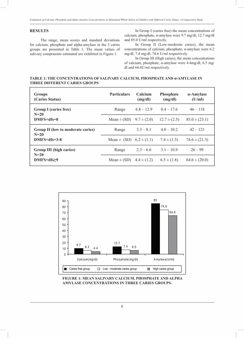
The range, mean scores and standard deviations for calcium, phosphate and alpha-amylase in the 3 caries groups are presented in Table 1. The mean values of salivary components estimated are exhibited in Figure 1.
In Group I (caries free) the mean concentrations of calcium, phosphate, α-amylase were 9.7 mg/dl, 12.7 mg/dl and 85.0 U/ml respectively. In Group II (Low-moderate caries), the mean concentrations of calcium, phosphate, α-amylase were �.2 mg/dl, 7.4 mg/dl, 74.� U/ml respectively In Group III (High caries), the mean concentrations of calcium, phosphate, α-amylase were 4.4mg/dl, �.5 mg/dl and �4.�U/ml respectively.
TABLE 1: THE ConCEnTRATIonS oF SALIVARY CALCIuM, PHoSPHATE AnD α-AMYLASE In THREE DIFFEREnT CARIES GRouPS
Groups Particulars Calcium Phosphate α-Amylase(Caries Status) (mg/dl) (mg/dl) (u/ml)
Group I (caries free) Range �.8 – 12.9 8.4 – 17.� 4� – 118n=20DMFS+dfs=0 Mean ± (SD) 9.7 ± (2.0) 12.7 ± (2.5) 85.0 ± (23.1)
Group II (low to moderate caries) Range 3.3 – 8.1 4.0 – 10.2 42 – 121n=20 DMFS+dfs=3-8 Mean ± (SD) �.2 ± (1.1) 7.4 ± (1.5) 74.� ± (21.3)
Group III (high caries) Range 2.3 – �.� 3.1 – 10.9 2� – 99n=20DMFS+dfs≥9 Mean ± (SD) 4.4 ± (1.2) �.5 ± (1.8) �4.� ± (20.0)
FIGuRE 1: MEAn SALIVARY CALCIuM, PHoSPHATE AnD ALPHA AMYLASE ConCEnTRATIonS In THREE CARIES GRouPS.
9.7 12.7
85
6.2 7.4
74.6
4.4 6.5
64.6
0
10
20
30
40
50
60
70
80
90
Calc ium(mg/d l) Phos phate(mg/d l) A my las e(U/ml)
High caries groupLow - moderate caries groupCaries free group
9
Phabhakar / Shubha / Mahantesh
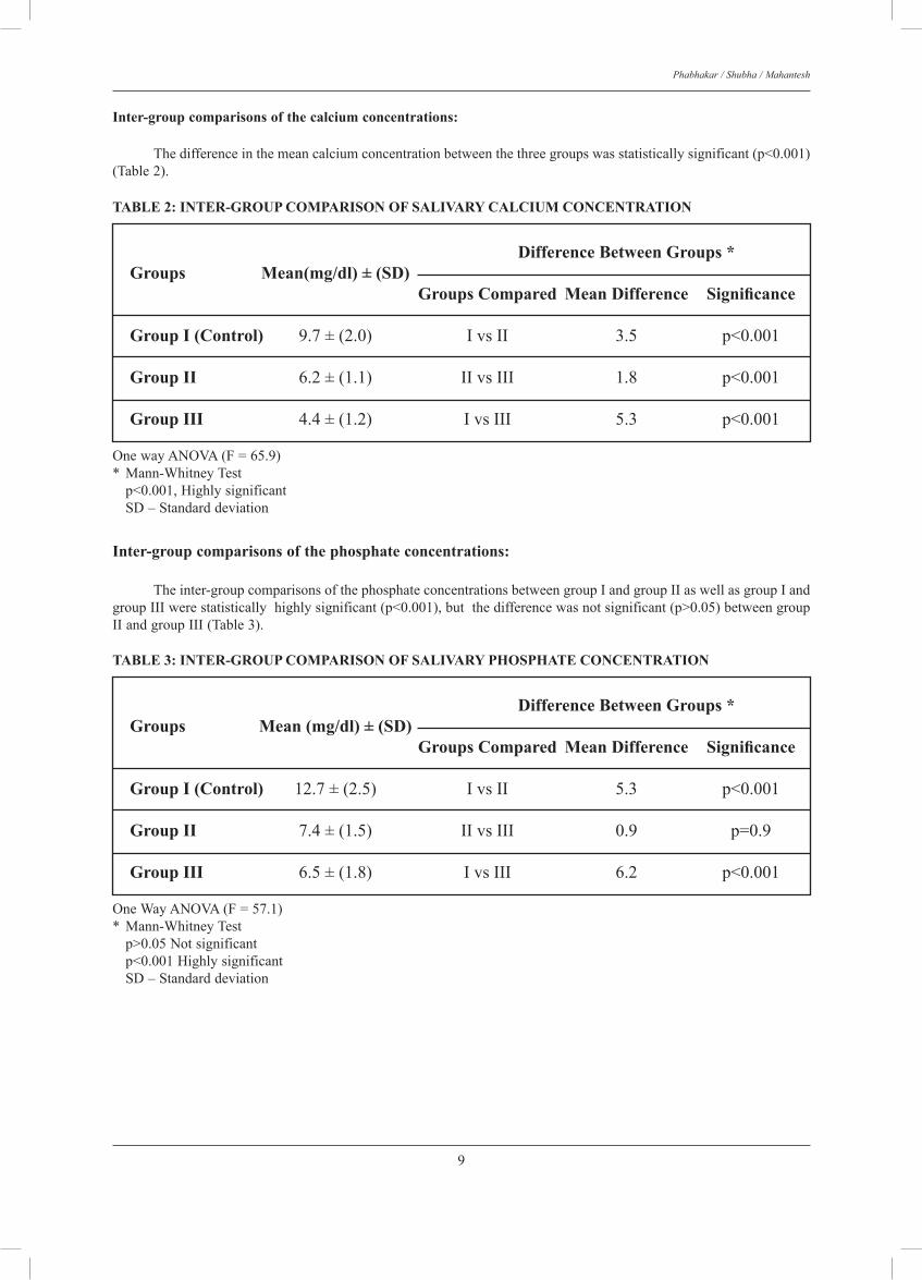
Inter-group comparisons of the calcium concentrations:
The difference in the mean calcium concentration between the three groups was statistically significant (p<0.001) (Table 2).
TABLE 2: InTER-GRouP CoMPARISon oF SALIVARY CALCIuM ConCEnTRATIon
Difference Between Groups *Groups Mean(mg/dl) ± (SD) Groups Compared Mean Difference Significance
Group I (Control) 9.7 ± (2.0) I vs II 3.5 p<0.001
Group II �.2 ± (1.1) II vs III 1.8 p<0.001
Group III 4.4 ± (1.2) I vs III 5.3 p<0.001
One way ANOVA (F = �5.9) * Mann-Whitney Test p<0.001, Highly significant SD – Standard deviation
TABLE 3: InTER-GRouP CoMPARISon oF SALIVARY PHoSPHATE ConCEnTRATIon
Difference Between Groups *Groups Mean (mg/dl) ± (SD) Groups Compared Mean Difference Significance
Group I (Control) 12.7 ± (2.5) I vs II 5.3 p<0.001
Group II 7.4 ± (1.5) II vs III 0.9 p=0.9
Group III �.5 ± (1.8) I vs III �.2 p<0.001
One Way ANOVA (F = 57.1) * Mann-Whitney Test p>0.05 Not significant p<0.001 Highly significant SD – Standard deviation
Inter-group comparisons of the phosphate concentrations: The inter-group comparisons of the phosphate concentrations between group I and group II as well as group I and group III were statistically highly significant (p<0.001), but the difference was not significant (p>0.05) between group II and group III (Table 3).
10
Estimation of Calcium, Phosphate and Alpha Amylase Concentrations in Stimulated Whole Saliva of Children with Different Caries Status: A Comparative Study
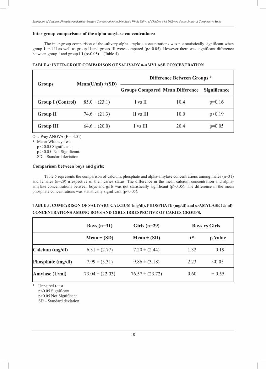
Inter-group comparisons of the alpha-amylase concentrations: The inter-group comparison of the salivary alpha-amylase concentrations was not statistically significant when group I and II as well as group II and group III were compared (p> 0.05). However there was significant difference between group I and group III (p<0.05) (Table 4).
TABLE 4: InTER-GRouP CoMPARISon oF SALIVARY α-AMYLASE ConCEnTRATIon
Difference Between Groups *Groups Mean(u/ml) ±(SD) Groups Compared Mean Difference Significance
Group I (Control) 85.0 ± (23.1) I vs II 10.4 p=0.1�
Group II 74.� ± (21.3) II vs III 10.0 p<0.19
Group III �4.� ± (20.0) I vs III 20.4 p=0.05
One Way ANOVA (F = 4.51) * Mann-Whitney Test p < 0.05 Significant. p > 0.05 Not Significant. SD – Standard deviation Comparison between boys and girls: Table 5 represents the comparison of calcium, phosphate and alpha-amylase concentrations among males (n=31) and females (n=29) irrespective of their caries status. The difference in the mean calcium concentration and alpha-amylase concentrations between boys and girls was not statistically significant (p>0.05). The difference in the mean phosphate concentrations was statistically significant (p<0.05).
TABLE 5: CoMPARISon oF SALIVARY CALCIuM (mg/dl), PHoSPHATE (mg/dl) and α-AMYLASE (u/ml)
ConCEnTRATIonS AMonG BoYS AnD GIRLS IRRESPECTIVE oF CARIES GRouPS.
Boys (n=31) Girls (n=29) Boys vs Girls Mean ± (SD) Mean ± (SD) t* p Value
Calcium (mg/dl) �.31 ± (2.77) 7.20 ± (2.44) 1.32 = 0.19
Phosphate (mg/dl) 7.99 ± (3.31) 9.8� ± (3.18) 2.23 <0.05
Amylase (u/ml) 73.04 ± (22.03) 7�.57 ± (23.72) 0.�0 = 0.55
* Unpaired t-test p<0.05 Significant p>0.05 Not Significant SD – Standard deviation
11
Phabhakar / Shubha / Mahantesh
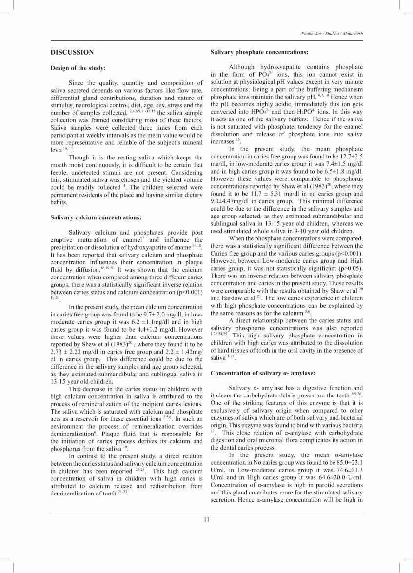
DISCuSSIon
Design of the study: Since the quality, quantity and composition of saliva secreted depends on various factors like flow rate, differential gland contributions, duration and nature of stimulus, neurological control, diet, age, sex, stress and the number of samples collected, 2,4,�,9,11-13,15 the saliva sample collection was framed considering most of these factors. Saliva samples were collected three times from each participant at weekly intervals as the mean value would be more representative and reliable of the subject’s mineral level1�, 17. Though it is the resting saliva which keeps the mouth moist continuously, it is difficult to be certain that feeble, undetected stimuli are not present. Considering this, stimulated saliva was chosen and the yielded volume could be readily collected 4. The children selected were permanent residents of the place and having similar dietary habits.
Salivary calcium concentrations: Salivary calcium and phosphates provide post eruptive maturation of enamel7 and influence the precipitation or dissolution of hydroxyapatite of ename l �,18. It has been reported that salivary calcium and phosphate concentration influences their concentration in plaque fluid by diffusion.1�,19,20 It was shown that the calcium concentration when compared among three different caries groups, there was a statistically significant inverse relation between caries status and calcium concentration (p<0.001) 19,20. In the present study, the mean calcium concentration in caries free group was found to be 9.7± 2.0 mg/dl, in low-moderate caries group it was �.2 ±1.1mg/dl and in high caries group it was found to be 4.4±1.2 mg/dl. However these values were higher than calcium concentrations reported by Shaw et al (1983)20 , where they found it to be 2.73 ± 2.23 mg/dl in caries free group and 2.2 ± 1.42mg/dl in caries group. This difference could be due to the difference in the salivary samples and age group selected, as they estimated submandibular and sublingual saliva in 13-15 year old children. This decrease in the caries status in children with high calcium concentration in saliva is attributed to the process of remineralization of the incipient caries lesions. The saliva which is saturated with calcium and phosphate acts as a reservoir for these essential ions 2,5,�. In such an environment the process of remineralization overrides demineralization�. Plaque fluid that is responsible for the initiation of caries process derives its calcium and phosphorus from the saliva 19. In contrast to the present study, a direct relation between the caries status and salivary calcium concentration in children has been reported 21-23. This high calcium concentration of saliva in children with high caries is attributed to calcium release and redistribution from demineralization of tooth 21-23.
Salivary phosphate concentrations:
Although hydroxyapatite contains phosphate in the form of PO4
3- ions, this ion cannot exist in solution at physiological pH values except in very minute concentrations. Being a part of the buffering mechanism phosphate ions maintain the salivary pH. �,7, 18 Hence when the pH becomes highly acidic, immediately this ion gets converted into HPO4
2- and then H2PO4- ions. In this way it acts as one of the salivary buffers. Hence if the saliva is not saturated with phosphate, tendency for the enamel dissolution and release of phosphate ions into saliva increases 15. In the present study, the mean phosphate concentration in caries free group was found to be 12.7±2.5 mg/dl, in low-moderate caries group it was 7.4±1.5 mg/dl and in high caries group it was found to be �.5±1.8 mg/dl. However these values were comparable to phosphorus concentrations reported by Shaw et al (1983)20, where they found it to be 11.7 ± 5.31 mg/dl in no caries group and 9.0±4.47mg/dl in caries group. This minimal difference could be due to the difference in the salivary samples and age group selected, as they estimated submandibular and sublingual saliva in 13-15 year old children, whereas we used stimulated whole saliva in 9-10 year old children. When the phosphate concentrations were compared, there was a statistically significant difference between the Caries free group and the various caries groups (p<0.001). However, between Low-moderate caries group and High caries group, it was not statistically significant (p>0.05). There was an inverse relation between salivary phosphate concentration and caries in the present study. These results were comparable with the results obtained by Shaw et al 20 and Bardow et al 21. The low caries experience in children with high phosphate concentrations can be explained by the same reasons as for the calcium 5,�. A direct relationship between the caries status and salivary phosphorus concentrations was also reported 1,22,24,25. This high salivary phosphate concentration in children with high caries was attributed to the dissolution of hard tissues of tooth in the oral cavity in the presence of saliva 1,24.
Concentration of salivary α- amylase: Salivary α- amylase has a digestive function and it clears the carbohydrate debris present on the teeth 8,9,2�. One of the striking features of this enzyme is that it is exclusively of salivary origin when compared to other enzymes of saliva which are of both salivary and bacterial origin. This enzyme was found to bind with various bacteria 27. This close relation of α-amylase with carbohydrate digestion and oral microbial flora complicates its action in the dental caries process. In the present study, the mean α-amylase concentration in No caries group was found to be 85.0±23.1 U/ml, in Low-moderate caries group it was 74.�±21.3 U/ml and in High caries group it was �4.�±20.0 U/ml. Concentration of α-amylase is high in parotid secretions and this gland contributes more for the stimulated salivary secretion. Hence α-amylase concentration will be high in
12
Estimation of Calcium, Phosphate and Alpha Amylase Concentrations in Stimulated Whole Saliva of Children with Different Caries Status: A Comparative Study
stimulated saliva compared to unstimulated saliva8. In this study, when the enzyme concentrations compared among three different caries groups showed an inverse relationship between caries status and α-amylase concentrations of saliva. There was a statistically significant difference between Caries free group and High caries group (p<0.05). These results were comparable with the results obtained by Ziegler et al.28 and Bardow et al.21. The low caries status in individuals with high α-amylase concentration could be due to the starch clearance action of α-amylase 5,8,9,2�.
Comparison between girls and boys irrespective of caries groups: When comparisons were done between girls and boys irrespective of their caries status the concentrations of phosphate were significantly higher in girls than in boys (p<0.05). These results were comparable to the results of Mazengo et al 13. However, on comparison between boys and girls, the concentrations of calcium and α-amylase were not statistically significant (p>0.05).
ConCLuSIon
Salivary calcium, phosphate and alpha-amylase concentrations were highest in Caries free group and lowest in high caries group. The concentrations of these ions were moderate in low-moderate caries group. Salivary calcium, phosphate and alpha-amylase concentrations increased with the decrease in the caries status of the individual. The outcome of the study elicits the fact that calcium, phosphate and alpha-amylase concentrations in saliva definitely influence the dental caries process. However, clinical interpretation of the results obtained in the present study should be made carefully as it involved only one of the host factor components of the multifactorial etiology of dental caries.
This article is of importance to paediatric dentist as: • Major oral disease among children is dental caries. This paper evaluates the role played by a host protective factor, ‘saliva’, in the etiology of dental caries. • The salivary factors evaluated here may prove to be useful measures of caries experience in children and allow paediatric dentists to target preventive measures appropriately.
REFEREnCES
1. Gandhy M, Damle SG. Relation of salivary inorganic phosphorus and alkaline phosphatase to the dental caries status in children. J Indian Soc Pedod Prev Dent 2003, 21: 135-138.
2. Shafer WG, Hine MK, Levy BM. A text book of Oral Pathology. 4th Ed. Philadelphia: W.B. Saunders Co. 1993, 40�-478.
3. Kedjarune U, Migasena P, Changbumrung S, Pongpaew P, Tungtrongchitr R. Flow rate and composition of whole saliva in children from rural and urban Thailand with different
caries prevalence and dietary intake. Caries Res 1997, 31: 148-154.
4. Jenkins GN. Physiology and Biochemistry of the Mouth, 4th ed. Oxford: Blackwell Scientific Pub. 1978, 284-359.
5. Nikiforuk G. Understanding dental caries - Etiology and mechanisms, Basic and Clinical Aspects. Vol. 1: 1st ed. New York: Karger. 1985, 23�-2�0.
�. Anderson P, Hector MP, Rampersad MA. Critical pH in resting and stimulated whole saliva in groups of children and adults. Int J Pediatr Dent 2001, 11: 2��-273.
7. Mandel ID. The functions of saliva. J Dent Res �� (Spec Iss) 1987, �23-�27.
8. Jacobsen K, Lyche Melvaer K, Hensten-Pettersen A. Some properties of salivary amylase: A survey of the literature and some observations. J Dent Res 1972, 51 (2): 381-388.
9. Edgar WM. Saliva: Its secretion, composition and functions. Br Dent J 1992, 172: 305-312.
10. Frohlich S, Lettow A, Kruger J, Gocke R. Salivary composition of children in relation to different caries group models. Caries Res 1997, 31: 305, Abstr.No.75.
11. Dezan CC, Nicolau J, Souza DN, Walter LRF. Flow rate, amylase activity and protein and sialic acid concentrations of saliva from children aged 18, 30 and 42 months attending a baby clinic. Arch Oral Biol 2002, 47: 423-427.
12. Soderling E. Practical aspects of salivary analyses chapter 1: in “Human Saliva: Clinical Chemistry and Microbiology”. Tenovuo JO. Vol.1, C.R.C. Press, Florida: 1989, 1-24.
13. Mazengo MC, Soderling E, Alakuijala P, Tiekso J, Tenovuo J, Simell O, Hausen H. Flow rate and composition of whole saliva in rural and urban Tanzania with special reference to diet, age and gender. Caries Res 1994, 28: 4�8-47�.
14. Winn Deen ES, David H, Sigler G, Chavez R. Development of a direct assay for alpha-amylase. Clin Chem 1988, 34: 2005-2008.
15. Mandel ID. Relation of saliva and plaque to caries. J Dent Res 1974, Suppl. 2: 53: 24�-2��.
1�. Ashley FP. Calcium and phosphorus concentrations of dental plaque related to dental caries in 11 to 14 year old male subjects. Caries Res 1975, 9: 351-3�2.
17. Larsen MJ, Jensen AF, Madsen DM, Pearce EIF. Individual variations of pH, buffer capacity and concentrations of calcium and phosphate in unstimulated saliva. Caries Res 1997, 31: 30�, Abstr.No.77.
18. ten Cate B. The role of saliva in mineral Equilibra-Caries and Calculus Formation. Chapter 9. In: “Saliva and Oral Health” 2nd Ed. Edgar WM, O’Mullane D.M., London: British Dental Association; 199�, 123-13�.
19. Ashley FP. Relationship of diet, saliva, plaque and caries. J Dent Res 1972, 51: 1234.
20. Shaw L, Murray JJ, Burchell CK, Best JS. Calcium and phosphorus content of plaque and saliva in relation to dental caries. Caries Res 1983, 17: 543-548.
21. Bardow A, Hofer E, Nyvad B, ten Cate JM, Kirkeyby S, Moe D, Nauntofte B: Effect of saliva composition on experimental root caries. Caries Res 2005, 39: 71-77.
22. Bowen WH, Velez H, Aguila M, Velasquez H, Sierra LI, Gillespie G. The microbiology and biochemistry of plaque, saliva and drinking water from two communities with contrasting levels of caries in Colombia, S.A. J Dent Res 1977, 55 (Sp Iss C): C32-C39.
23. Turtola LO. Salivary fluoride and calcium concentrations and their relationship to the secretion of saliva and caries experience. Scand J Dent Res 1977, 85: 535-541.
13
Phabhakar / Shubha / Mahantesh
24. Kargul B, Yarat A, Tanboga I, Emekli N. Salivary protein and some inorganic element levels in healthy children and their relationship to caries. J Marmara Univ Dent Fac 1994, 2: 434-440.
25. Pandey RK, Tripathi A, Chandra S, Pandey A. Relation of salivary phosphorus and alkaline phosphatase to the incidence of dental caries in children. J Pedod 1990, 14: 144-14�.
2�. Hay DI, Bowen MH. The functions of salivary proteins. Chapter 8. In Saliva and oral health” 2nd Ed. Edgar WM, O’Mullane DM. London: British Dental Association, 199�, 105-122.
27. Scannapieco FA, Torres G, Levine MJ. Salivary alpha-amylase: role in dental plaque and caries formation. Critic Rev Oral Biol Med 1993, 4 (3/4); 301-307.
28. Ziegler F, Gocke R, Beetke E. Pattern of salivary secretion for caries-resistant versus caries-susceptible adults. Caries Res 1999, 33:308: Abstr. No. 80.
Address for correspondence:
Dr. Prabhakar A.R. Professor and Head, Dept. of Pedodontics and Preventive Dentistry, Bapuji Dental College and Hospital,Davangere – 577 004, Karnataka – India. Phone – 91-8192-220575, Fax – 91-8192-220578.e-mail – attiguppeprabhakar @ yahoo.com [email protected]
Radiological Features Of Different Histopathological Variants Of Ameloblastomas Chen YN MDSc. Former Postgraduate Student, Faculty of Dentistry, University of Malaya, Kuala Lumpur, Malaysia.
Nambiar P MScDent Professor and �ead, Dept of �eneral Dental Practice and �ral �� Ma�illofacial �maging, Professor and �ead, Dept of �eneral Dental Practice and �ral �� Ma�illofacial �maging, Faculty of Dentistry, University of Malaya, Kuala Lumpur Malaysia.
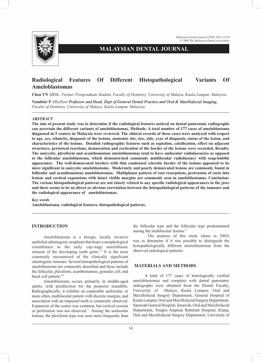
ABSTRACT The aim of present study was to determine if the radiological features noticed on dental panoramic radiographs can ascertain the different variants of ameloblastomas. Methods: A total number of 177 cases of ameloblastoma diagnosed in 5 centers in Malaysia were reviewed. The clinical records of these cases were analyzed with respect to age, sex, ethnicity, diagnosis of the lesions, anatomic site, size, side, year of diagnosis, status of the lesion, and characteristics of the lesions. Detailed radiographic features such as septation, calcification, effect on adjacent structures, periosteal reactions, demarcation and cortication of the border of the lesions were recorded. Results: The unicystic, plexiform and acanthomatous ameloblastomas tend to have unilocular radiolucencies as opposed to the follicular ameloblastoma, which demonstrated commonly multilocular radiolucency with soap-bubble appearance. The well-demarcated borders with thin condensed sclerotic border of the lesions appeared to be more significant in unicystic ameloblastoms. Moderately and poorly demarcated lesions are commonly found in follicular and acanthomatous ameloblastomas. Multiplanar pattern of root resorptions, protrusion of roots into lesions and cortical expansions with intact visible margins are commonly seen in ameloblastomas. Conclusion: The various histopathological patterns are not closely related to any specific radiological appearances in the jaws and there seems to be no direct or obvious correlation between the histopathological patterns of the tumours and the radiological appearance of ameloblastomas.
Key words Ameloblastoma, radiological features, histopathological patterns.
14
Malaysian Dental Journal (2008) 29(1) 14-19© 2008 The Malaysian Dental Association
INTRODuCTION
Ameloblastoma is a benign, locally invasiveepithelialodontogenicneoplasmthatbearsamorphologicalresemblance to the early cap-stage ameloblasticelement of the developing tooth germ.1-3 It is the mostcommonly encountered of the clinically significantodontogenictumours.Severalhistopathologicalpatternsofameloblastomaarecommonlydescribedandtheseincludethefollicular,plexiform,acanthomatous,granularcell,andbasalcellpattern.4-6 Ameloblastoma occurs primarily in middle-agedadults, with predilection for the posterior mandible.Radiographically, it exhibits an expansible unilocular, ormoreoften,multilocularpatternwithdiscretemargins,andassociationwithanimpactedtoothiscommonlyobserved.Expansionofthecortexwascommon,butcorticalerosionor perforationwas not observed.7 Among theunilocularlesions,theplexiformtypewasseenmorefrequentlythan
the follicular type and the follicular type predominatedamongthemultilocularlesions.8 The purpose of this study (done in 2003)was to determine if it was possible to distinguish thehistopathologically different ameloblastomas from theobservedradiologicalpatterns.
MATERIALS AND METHODS
A total of 177 cases of histologically verifiedameloblastomas and complete with dental panoramicradiographs were obtained from the Dental Faculty,University of Malaya, Kuala Lumpur; Oral andMaxillofacial Surgery Department, General Hospital ofKualaLumpur;OralandMaxillofacialSurgeryDepartment,SarawakGeneralHospital,Sarawak;OralandMaxillofacialDepartment, Tengku Ampuan Rahimah Hospital, Klang;OralandMaxillofacialSurgeryDepartment,Universityof
MALAYSIAN DENTAL JOuRNAL
15
Chen / Nambiar
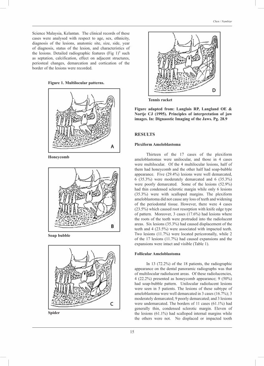
ScienceMalaysia,Kelantan.Theclinicalrecordsofthesecases were analysed with respect to age, sex, ethnicity,diagnosis of the lesions, anatomic site, size, side, yearof diagnosis, status of the lesion, and characteristics ofthe lesions. Detailed radiographic features (Fig 1)9 suchas septation, calcification, effect on adjacent structures,periosteal changes, demarcation and cortication of theborderofthelesionswererecorded.
RESuLTS
Plexiform Ameloblastoma
Thirteen of the 17 cases of the plexiformameloblastomas were unilocular, and those in 4 caseswere multilocular. Of the 4 multilocular lesions, half ofthemhadhoneycomband theotherhalfhadsoap-bubbleappearance. Five(29.4%)lesionswerewelldemarcated,6 (35.3%) were moderately demarcated and 6 (35.3%)were poorly demarcated. Some of the lesions (52.9%)had thincondensedscleroticmarginwhileonly6 lesions(35.3%) were with scalloped margins. The plexiformameloblastomadidnotcauseanylossofteethandwideningof the periodontal tissue. However, there were 4 cases(23.5%)whichcausedrootresorptionwithknifeedgetypeofpattern.Moreover,3cases(17.6%)hadlesionswherethe rootsof the teethwereprotruded into the radiolucentareas.Sixlesions(35.3%)hadcauseddisplacementoftheteethand4(23.5%)wereassociatedwithimpactedteeth.Two lesions (11.7%) were located pericoronally, while 2of the17 lesions (11.7%)hadcausedexpansionsand theexpansionswereintactandvisible(Table1).
Follicular Ameloblastoma
In13 (72.2%)of the18patients, the radiographicappearanceonthedentalpanoramicradiographswasthatofmultilocularradiolucentareas.Oftheseradiolucencies,4 (22.2%) presented as honeycomb appearance; 9 (50%)had soap-bubble pattern. Unilocular radiolucent lesionswere seen in 5 patients. The lesions of these subtype ofameloblastomawerewelldemarcatedin3cases(16.7%);3moderatelydemarcated;9poorlydemarcated;and3lesionswereundemarcated.Thebordersof11cases(61.1%)hadgenerally thin, condensed sclerotic margin. Eleven ofthe lesions (61.1%)had scalloped internalmarginswhilethe others were not. No displaced or impacted tooth
Figure 1. Multilocular patterns.
Honeycomb
Soap bubble
Spider
Tennis racket
Figure adapted from: Langlais RP, Langland OE & Nortje CJ (1995). Principles of interpretation of jaw images. In: Dignaostic Imaging of the Jaws. Pg. 28.9
16
Radiological Features �f Different �istopathological Variants �f Ameloblastomas
wasassociatedwith this typeof ameloblastoma. Fifteen(83.3%) of the lesions had caused missing teeth and therootsofteethwereaffectedin4ofthe18cases(22.2%).Therootswereresorbedinamultiplanarpatterninallthe4cases.Rootsofteethprotrudedintotheradiolucencyin9cases(50.0%).Fivecases(27.8%)wereattheperiapicalregionoftheteethwhile13(72.2%)werenotspecificinrelationtotheneighbourngteeth.Eightcasesofthelesion(44.4%)causedexpansionofthemandiblewhile4ofthebordersof the expanded lesions (50.0%)were intact andvisible on the panoramic radiographs. In 2 cases (25%)theborderswereexpandedbutthemarginswereintactbutinvisibleonradiographs.Intwocases(25.0%)thecorticalmarginswereperforated.
Acanthomatous Ameloblastomas
Of the total 12 cases, 10 lesions (83.3%) appearedas unilocular radiolucencies. Two (16.7%) presentedas multilocular radiolucencies. All the multilocularradiolucencieshadsoap-bubblepatterntypeofappearance,whilemorethanhalfofthelesionshadpoorlydemarcatedborders. There were 2 (16.7%) lesions which appearedwelldemarcated;2moderatelydemarcated(16.7%);and2lesions(16.7%)wereundemarcated. In8cases(66.7%),the margins of certain parts of the lesions were thin,condensed,andsclerotic. Theremaining4(33.3%)werenothavinganyscleroticmarginatall.Twocases(16.7%)oftheacanthomatousameloblastomacauseddestructionoftheperiodontal tissuesand the teethappearedas floatingin the radiographs. The type of root resorption that was
Radiographicfeatures
Plexiform(%)
Follicular(%)
Acanthomatous(%)
MixedPlexiformandFollicular
(%)
Desmoplastic(%)
Unicystic(%)
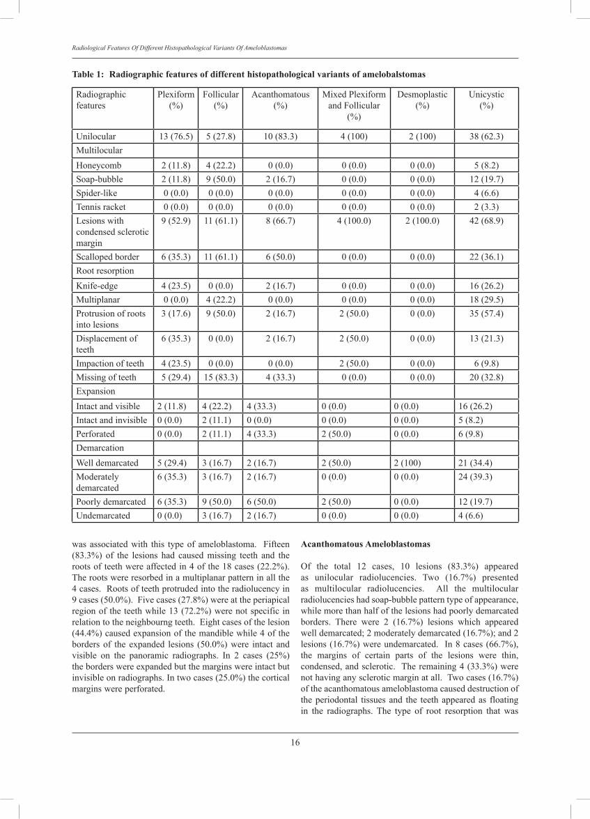
Unilocular 13(76.5) 5(27.8) 10(83.3) 4(100) 2(100) 38(62.3)MultilocularHoneycomb 2(11.8) 4(22.2) 0(0.0) 0(0.0) 0(0.0) 5(8.2)Soap-bubble 2(11.8) 9(50.0) 2(16.7) 0(0.0) 0(0.0) 12(19.7)Spider-like 0(0.0) 0(0.0) 0(0.0) 0(0.0) 0(0.0) 4(6.6)Tennisracket 0(0.0) 0(0.0) 0(0.0) 0(0.0) 0(0.0) 2(3.3)Lesionswithcondensedscleroticmargin
9(52.9) 11(61.1) 8(66.7) 4(100.0) 2(100.0) 42(68.9)
Scallopedborder 6(35.3) 11(61.1) 6(50.0) 0(0.0) 0(0.0) 22(36.1)RootresorptionKnife-edge 4(23.5) 0(0.0) 2(16.7) 0(0.0) 0(0.0) 16(26.2)Multiplanar 0(0.0) 4(22.2) 0(0.0) 0(0.0) 0(0.0) 18(29.5)Protrusionofrootsintolesions
3(17.6) 9(50.0) 2(16.7) 2(50.0) 0(0.0) 35(57.4)
Displacementofteeth
6(35.3) 0(0.0) 2(16.7) 2(50.0) 0(0.0) 13(21.3)
Impactionofteeth 4(23.5) 0(0.0) 0(0.0) 2(50.0) 0(0.0) 6(9.8)Missingofteeth 5(29.4) 15(83.3) 4(33.3) 0(0.0) 0(0.0) 20(32.8)ExpansionIntactandvisible 2(11.8) 4(22.2) 4(33.3) 0(0.0) 0(0.0) 16(26.2)Intactandinvisible 0(0.0) 2(11.1) 0(0.0) 0(0.0) 0(0.0) 5(8.2)Perforated 0(0.0) 2(11.1) 4(33.3) 2(50.0) 0(0.0) 6(9.8)DemarcationWelldemarcated 5(29.4) 3(16.7) 2(16.7) 2(50.0) 2(100) 21(34.4)Moderatelydemarcated
6(35.3) 3(16.7) 2(16.7) 0(0.0) 0(0.0) 24(39.3)
Poorlydemarcated 6(35.3) 9(50.0) 6(50.0) 2(50.0) 0(0.0) 12(19.7)Undemarcated 0(0.0) 3(16.7) 2(16.7) 0(0.0) 0(0.0) 4(6.6)
Table 1: Radiographic features of different histopathological variants of amelobalstomas
17
Chen / Nambiar
causedbythelesionswastheknifeedgepattern.Inanother2casestheteethhadprotrudedintotheradiolucentareas.Thelesionshadalsodisplacedteethin2instances(16.7%)and4(33.3%)caseshadmissingteeth.Inrelationtotheteeth,2cases(16.7%)werelocatedpericoronallyandalso2 (16.7%) were located periapically. For this type ofameloblastoma, 8 lesions (66.7%) had expanded corticalmargins. Among theexpanded lesions,halfof thecaseshadtheborderintactandvisibleandtheotherhalfhadthemarginsperforated.
Desmoplastic Ameloblastomas
Thelesionsappearedasunilocularonthepanoramicradiographs.Theborderswerewelldemarcatedwiththincondensedscleroticmargin.Therewasnorootresorption,wideningoftheperiodontalligamentspace,protrusionofrootsintothelesionsoranyexpansionofthecortex.
Mixed Follicular and Plexiform Ameloblastomas Themixedfollicularandplexiformameloblastomapresented as unilocular radiolucency on the panoramicradiographs. Half of the lesions were well demarcatedand the other half was poorly demarcated. The marginsof 2 lesions were thin, condensed and sclerotic. Rootresorptionswerenoticedin2casesbutitwasnotabletodistinguishwhetheritwasknife-edgeormultiplanartypeofresorption.In2cases,theameloblastomawasassociatedwithdisplacedteethandin2casestheteethwereimpacted.The lesions also caused expansionsof the cortical platesandhalfofthemwereperforated.
unicystic Ameloblastomas
For the unicystic ameloblastoma, 38 (62.3%) panoramicradiographs presented with unilocular radiolucencies and23 (37.7%) with multilocular radiolucencies. Of the 23multilocularlesions,5(21.7%)hadhoneycombappearance,12 (52.2%) had soap-bubble appearance, 2 (8.7%) hadtennis racket appearanceand 4 (17.4%)had spider-likeappearance. Forty-five lesions (73.8%) were well andmoderatelydemarcated.Four(6.6%)wereundemarcatedwhiletheotherswerepoorlydemarcated.Majorityoftheinternal margin of the lesions (68.9%) were of the thincondensedscleroticvarietyand22ofthelesions(36.1%)were scalloped margins. Six cases (9.8%) had floatingteeth appearance in the panoramic radiographs and therewere 2 cases (3.3%) with widened periodontal ligamentspace. Most of the lesions (55.7%) had caused rootresorptions.Sixteen(26.2%)lesionsproducedknifeedgetype of root resorption, whereas 18 (29.5%) cases wereof themultiplanarvariety. Therewere35cases (57.4%)wheretherootsoftheteethwereprotrudedintothelesions.Thirteen lesions (21.3%) caused displacement of teethwhile6cases(9.8%)wereassociatedwithimpactedteeth.Approximatelyonethirdofthecasesdemonstratedmissingteeth.Nine(14.8%)lesionswerelocatedpericoronally,10(16.4%)periapicallyand2(3.3%)wereintheinterradicular
area. Of the61 lesions,27(44.3%)patientshadcorticalexpansion. Sixteen (59.3%)of theexpanded lesionshadmarginsthatwere intact andvisibleonplain radiographs.Five (18.5%) had intact margins but invisible whereas 6(22.2%)hadcorticalperforations.
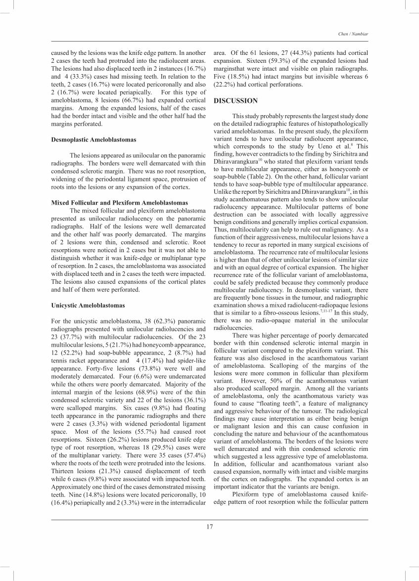
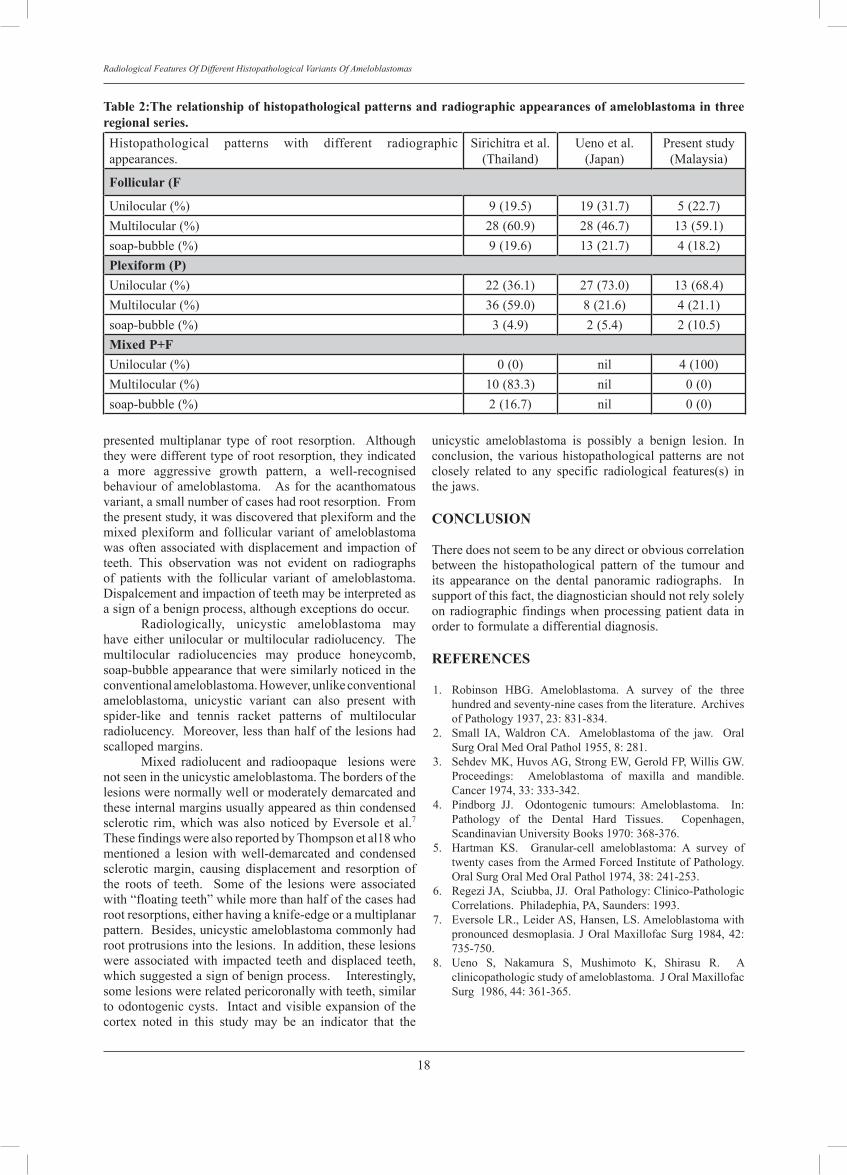
DISCuSSION Thisstudyprobablyrepresentsthelargeststudydoneonthedetailedradiographicfeaturesofhistopathologicallyvariedameloblastomas.Inthepresentstudy,theplexiformvariant tends to have unilocular radiolucent appearance,which corresponds to the study by Ueno et al.8 Thisfinding,howevercontradictstothefindingbySirichitraandDhiravarangkura10whostatedthatplexiformvarianttendstohavemultilocular appearance, either ashoneycomborsoap-bubble(Table2).Ontheotherhand,follicularvarianttendstohavesoap-bubbletypeofmultilocularappearance.UnlikethereportbySirichitraandDhiravarangkura10,inthisstudyacanthomatouspatternalsotendstoshowunilocularradiolucency appearance. Multilocular patterns of bonedestruction can be associated with locally aggressivebenignconditionsandgenerallyimpliescorticalexpansion.Thus,multilocularitycanhelptoruleoutmalignancy.Asafunctionoftheiraggressiveness,multilocularlesionshaveatendencytorecurasreportedinmanysurgicalexcisionsofameloblastoma.Therecurrencerateofmultilocularlesionsishigherthanthatofotherunilocularlesionsofsimilarsizeandwithanequaldegreeofcorticalexpansion.Thehigherrecurrencerateofthefollicularvariantofameloblastoma,couldbesafelypredictedbecausetheycommonlyproducemultilocular radiolucency. In desmoplastic variant, therearefrequentlybonetissuesinthetumour,andradiographicexaminationshowsamixedradiolucent-radiopaquelesionsthatissimilartoafibro-osseouslesions.7,11-17Inthisstudy,there was no radio-opaque material in the unilocularradiolucencies. Therewashigherpercentageofpoorlydemarcatedborder with thin condensed sclerotic internal margin infollicularvariantcomparedtotheplexiformvariant.Thisfeature was also disclosed in the acanthomatous variantof ameloblastoma. Scalloping of the margins of thelesions were more common in follicular than plexiformvariant. However, 50% of the acanthomatous variantalsoproduced scallopedmargin. Amongall thevariantsof ameloblastoma, only the acanthomatous variety wasfound to cause “floating teeth”, a feature of malignancyandaggressivebehaviourof thetumour.Theradiologicalfindings may cause interpretation as either being benignor malignant lesion and this can cause confusion inconcludingthenatureandbehaviouroftheacanthomatousvariantofameloblastoma.Thebordersofthelesionswerewell demarcated and with thin condensed sclerotic rimwhichsuggestedalessaggressivetypeofameloblastoma.In addition, follicular and acanthomatous variant alsocausedexpansion,normallywithintactandvisiblemarginsof thecortexonradiographs. Theexpandedcortex isanimportantindicatorthatthevariantsarebenign. Plexiform type of ameloblastoma caused knife-edgepatternofrootresorptionwhilethefollicularpattern
18
Radiological Features �f Different �istopathological Variants �f Ameloblastomas
presented multiplanar type of root resorption. Althoughtheyweredifferenttypeofrootresorption,theyindicateda more aggressive growth pattern, a well-recognisedbehaviourof ameloblastoma. As for theacanthomatousvariant,asmallnumberofcaseshadrootresorption.Fromthepresentstudy,itwasdiscoveredthatplexiformandthemixed plexiform and follicular variant of ameloblastomawasoftenassociatedwithdisplacementand impactionofteeth. This observation was not evident on radiographsof patients with the follicular variant of ameloblastoma.Dispalcementandimpactionofteethmaybeinterpretedasasignofabenignprocess,althoughexceptionsdooccur. Radiologically, unicystic ameloblastoma mayhave either unilocular or multilocular radiolucency. Themultilocular radiolucencies may produce honeycomb,soap-bubbleappearancethatweresimilarlynoticedintheconventionalameloblastoma.However,unlikeconventionalameloblastoma, unicystic variant can also present withspider-like and tennis racket patterns of multilocularradiolucency.Moreover,lessthanhalfofthelesionshadscallopedmargins. Mixed radiolucent and radioopaque lesions werenotseenintheunicysticameloblastoma.Thebordersofthelesionswerenormallywellormoderatelydemarcatedandtheseinternalmarginsusuallyappearedasthincondensedsclerotic rim, which was also noticed by Eversole et al.7ThesefindingswerealsoreportedbyThompsonetal18whomentioned a lesion with well-demarcated and condensedsclerotic margin, causing displacement and resorption ofthe roots of teeth. Some of the lesions were associatedwith“floatingteeth”whilemorethanhalfofthecaseshadrootresorptions,eitherhavingaknife-edgeoramultiplanarpattern.Besides,unicysticameloblastomacommonlyhadrootprotrusionsintothelesions.Inaddition,theselesionswere associated with impacted teeth and displaced teeth,whichsuggestedasignofbenignprocess.Interestingly,somelesionswererelatedpericoronallywithteeth,similartoodontogeniccysts. Intactandvisibleexpansionofthecortex noted in this study may be an indicator that the
unicystic ameloblastoma is possibly a benign lesion. Inconclusion, thevarioushistopathologicalpatternsarenotclosely related to any specific radiological features(s) inthejaws.
CONCLuSION
Theredoesnotseemtobeanydirectorobviouscorrelationbetween the histopathological pattern of the tumour andits appearance on the dental panoramic radiographs. Insupportofthisfact,thediagnosticianshouldnotrelysolelyon radiographic findingswhenprocessingpatientdata inordertoformulateadifferentialdiagnosis.
REFERENCES
1. Robinson HBG. Ameloblastoma. A survey of the threehundredandseventy-ninecasesfromtheliterature.ArchivesofPathology1937,23:831-834.
2. Small IA, Waldron CA. Ameloblastoma of the jaw. OralSurgOralMedOralPathol1955,8:281.
3. SehdevMK,HuvosAG,StrongEW,GeroldFP,WillisGW.Proceedings: Ameloblastoma of maxilla and mandible.Cancer1974,33:333-342.
4. Pindborg JJ. Odontogenic tumours: Ameloblastoma. In:Pathology of the Dental Hard Tissues. Copenhagen,ScandinavianUniversityBooks1970:368-376.
5. Hartman KS. Granular-cell ameloblastoma: A survey oftwentycasesfromtheArmedForcedInstituteofPathology.OralSurgOralMedOralPathol1974,38:241-253.
6. RegeziJA,Sciubba,JJ.OralPathology:Clinico-PathologicCorrelations.Philadephia,PA,Saunders:1993.
7. EversoleLR.,LeiderAS,Hansen,LS.Ameloblastomawithpronounceddesmoplasia. JOralMaxillofacSurg1984,42:735-750.
8. Ueno S, Nakamura S, Mushimoto K, Shirasu R. Aclinicopathologicstudyofameloblastoma.JOralMaxillofacSurg1986,44:361-365.
Table 2:The relationship of histopathological patterns and radiographic appearances of ameloblastoma in three regional series.Histopathological patterns with different radiographicappearances.
Sirichitraetal.(Thailand)
Uenoetal.(Japan)
Presentstudy(Malaysia)
Follicular (F
Unilocular(%) 9(19.5) 19(31.7) 5(22.7)Multilocular(%) 28(60.9) 28(46.7) 13(59.1)soap-bubble(%) 9(19.6) 13(21.7) 4(18.2)Plexiform (P)Unilocular(%) 22(36.1) 27(73.0) 13(68.4)Multilocular(%) 36(59.0) 8(21.6) 4(21.1)soap-bubble(%) 3(4.9) 2(5.4) 2(10.5)Mixed P+FUnilocular(%) 0(0) nil 4(100)Multilocular(%) 10(83.3) nil 0(0)soap-bubble(%) 2(16.7) nil 0(0)
19
Chen / Nambiar
9. Langlais RP, Langland OE, Nortjé CJ. Principles ofinterpretationof jaw images. In:Diagnostic imagingof thejaws.Williams&Wilkins,Malvern,USA1995:19-41.
10.SirichitraV,DhiravarangkuraP.Intrabonyameloblastomaofthejaws.Ananalysisof147Thaipatients.Int.J.OralSurg1984,13:187-193.
11.Ashman SG, Corio RL, Eisele, DW, and Murphy MT.Desmoplasticameloblastoma.Acasereportandliteraturereview.OralSurgOralMedOralPathol1993,75:479-482.
12.Kaffe I, Buchner A, Taicher S. Radiographic features ofdesmoplastic variant of ameloblastoma. Oral Surg. OralMed.OralPathol1993,76:525-529.
13.OkadaY,SugimuraM,IshidaT:Ameloblastomaaccompaniedbyprominentboneformation.JOralMaxillofacSurg1986,44:555-557.
14.Philipsen HP, Ormiston IW, Reichart PA. The desmo-and osteoplastic ameloblastoma. Histologic variant orclinicopathologicentity?Casereports.IntJOralMaxillofacSurg1992,21:352-357.
15.TanimotoK,TakataT,SueiY,WadaT.Acaseofdesmoplasticvariantofamandibularameloblastoma. J.OralMaxillofacSurg1991,49:94-97.
16.SiarCH,NgKH.Ameloblastoma inMalaysia -A25-yearsreview.AnnalsAcademyofMedicine1993,22:856-860.
17.Waldron CA, El-MoftySK.A histopathologic study of 116ameloblastomas with special reference to the desmoplasticvariant. Oral Surg. Oral Med. Oral Pathol. 1987, 63: 441-451.
18.ThompsonIOC,FerreiraR,VanWykCW.Recurrentunicysticameloblastoma of the maxilla. Br J Oral Maxillofac Surg1993,31:180.
Address for correspondence:
Prof. Dr. Phrabhakaran NambiarDept of �eneral Dental Practice and �ral �� Ma�illofacial �maging, Faculty of Dentistry, University of Malaya, 50603, Kuala Lumpur Malaysia.Tel: 017-3620050Fa�: 03-79674595E-mail: [email protected]
Psychological Impacts Of Dental Fluorosis Among Malaysian School Children Mohd Nor M. BDS(Mal), MSc(Lon), DDPHRCS(Eng) Senior Dental Officer of Port Dickson District, Negeri Sembilan, Ministry Of Health Malaysia.
Sheiham A. BDS, PhD, DHC.Professor and Lecturer, Department of Epidemiology and Public Health, University College London, London,United Kingdom.
Tsakos G. DDS, MSc, PhD. Lecturer, Department of Epidemiology and Public Health, University College London, London,United Kingdom.
ABSTRACT The objectives were to assess the prevalence, severity, the psychological and social impacts of fluorosis among school children and their parents in the Kuala Pilah area, Negeri Sembilan, Malaysia. Methods: A convenience sample of 431 students aged 16-17 years old from 3 secondary schools in Kuala Pilah, Negeri Sembilan were selected. The students were assessed for presence of dental fluorosis using Dean’s Index and to assess impacts. Questionnaires were administered to all the dental fluorosis students and selected matched students with no fluorosis. They constituted a control group for the case control study. Questionnaires were sent to the parents of dental fluorosis and control non fluorosis cases. Results: The prevalence of dental fluorosis was 27.8%; 82% of the fluorosis cases were questionable to mild and 18% moderate to severe fluorosis. 16.1% of students with dental fluorosis and 8.5% of non-fluorosis students had psychological impacts. 12.8% of the parents of children with fluorosis reported that their child had an impact. More girls with fluorosis (35.7%) had psychological impacts compared than boys (25.0%). In the 16.1% of fluorosis cases who had psychological impacts, difficulty smiling and showing teeth, affected 35.7% of girls and 25 % of boys. The percentage of students with dental fluorosis who had psychological impacts on carrying out school work was 2.7%, and 3.6% had impacts related to going out with friends. Conclusions: There were considerable psychological impacts on smiling and showing teeth among Malaysian teenagers with dental fluorosis and some of their parents were concerned about the fluorosis. But the impacts were mild and do not have a major impact on students’ lives. Dental fluorosis is not aesthetically displeasing to most subjects but does have psychological and social impacts on a small percentage with fluorosis.
Key words fluorosis, prevalence, children, psychosocial, impacts, dissatisfaction, tooth colour
20
Malaysian Dental Journal (2008) 29(1) 20-24© 2008 The Malaysian Dental Association
InTRoDucTIon
Seventy percent of Malaysians have access tooptimallyfluoridatedwatersupplies(0.7partpermillion)from224watertreatmentplants.Fluoridationhasbroughtmuch dental health benefits, but as with most beneficialinterventions there are unwanted side-effects, namelyfluorosis.Theextentofthesocialandpsychologicalimpactdependsonhow it isperceivedby thechildrenand theirparents. Astudyonschoolchildreninafluoridatedareafoundthattherewasanincreaseintheprevalenceofdentalfluorosis since the water fluoridation programme began
and45%ofthe12-13year-oldchildrenhadfluorosis.1In1999,a study ina fluoridatedandanon-fluoridatedareain Malaysia reported the prevalence of dental fluorosisin thefluoridatedareawas74.7%and14.2%in thenon-fluoridated area in children aged 16-17 years old. Fromthe study 94 % of the fluorosis cases were questionabletomild. Inthefluoridatedarea25%hadnormalteethcompared than 85% in non-fluoridataed area. There areonlyafewstudiesinSouthEastAsiancountriesthathaveevaluatedthepsychologicalimpactofdentalfluorosis. The perceptions of public about tooth appearanceis important in order to weigh up the benefits and
MALAYSIAn DEnTAL JouRnAL
21
Mohd Nor M / Sheiham / Tsakos
disadvantages of a whole population strategy, such aswater fluoridation.3 The level of fluorosis appears to berelated to the levels of concern of the children and theirparents about tooth aesthetics. In low endemic fluorosisareas with very mild forms of fluorosis, the impact wasnot significant in the children and their parents.4 Wherethere was more severe fluorosis, there was increasedlevels of dissatisfaction, both among the parents and thechildren.5Mostaestheticallyunacceptabledentalfluorosiscancausepsychologicaldistresstotheaffectedindividual;socio-dental measures indicated that the dental fluorosisaffectedtheirqualityoflife.6Becausewaterfluoridationissowidespread inMalaysiaand levelsof fluorosisappearto be increasing, both in prevalence and severity, it wasconsideredimportanttoassesstheimpactsoffluorosisonthequalityoflifeofschoolchildren. Theaimofthisstudywastoassesstheprevalence,severity and psychological impacts of dental fluorosisin children in Kuala Pilah area, Negeri Sembilan. Theobjectivesweretoassesstheprevalenceandseverityandthe psychological and social impacts of fluorosis amongschoolchildreninKualaPilaharea,aswellastheconcernamongtheirparents.
MATERIALS AnD METHoDS
Study Population and Sampling This study included students in a conveniencesample of Forms 4 and 5 children aged 16 to 17 yearsold in 3 of the 20 secondary schools in the Kuala Pilaharea.Selectionof schoolswasbasedonproximity to theKualaPilahDentalClinic.MostofthechildrenhadbeenresidentintheKualaPilahareasincebirth.Exclusioncaseswere orthodontic patients with appliances and childrenwhodidnothaveanteriorteeth,forexamplehypodontia;5 orthodontic patients and 1 hypodontia patient wereexcludedfromthestudy.
Examination method
Childrenwereexaminedseatedinaportabledentalchairinawell-lit roomunderartificial lightfromanoperatinglight, except in one school, Tunku Muhammad Schoolthat had a static clinic and their own dental chair andequipment and light. Teeth were not dried but excessplaquewasremovedwithgauzeforaclearviewforDean’sIndexcoding.Onedentalofficer,1DSA(DentalSurgeonAssistant) and 1 attendant carried out examinations andrecordings. Subjects were screened using Dean’s Index(WHO,1997)usingcolouredphotographsofDean’sIndexgradestodistinguishfluorosisfromothertypesofenamelopacities.7 The score was based on the 2 worst affectedteethinthemouth. A re-examination on a different day of 10% ofsubjectswasdone tocheck for inter-examinervariabilityconsistency between gold standard and examiner. BothpercentagesofagreementandKappascorewerecalculated
for Dean’s index of fluorosis. Kappa score was 0.9 andpercentageofagreementwas90.7%indicatingan‘almostperfectagreement’.Intra-examinervariabilitycouldnotbedoneduetotimeconstraintsbecausethisstudywasdoneinMalaysia and thedata collection shouldbe completedbyscheduledtimeandtheanalyseswillbe continuedinUnitedKingdom. To assess the differences in psychosocial impactsbetween children with and without enamel fluorosis acase-controlstudywascarriedout.Theimpactsrelatingtotoothaestheticsinchildrenwithandwithoutfluorosiswereexamined. First the epidemiological study was done toidentifychildrenwithandwithoutfluorosis.Thenchildrenwithfluorosiswerematched,bysex,ageandschoolwithanon-fluorosiscontrol.Questionnaireswereadministeredto all the fluorosis subjects and selected non-fluorosiscasesafteroralexaminationforfluorosisandeachof thechartingformswascodedforfluorosisandnon-fluorosis.Thepurposeof thecodingwas tomakeiteasierfordatacollection,datasearchinganddataanalysis.Bothfluorosiscasesandselectednon-fluorosiscontrolswereaskedabouttheir problems related to the colour of their teeth andwhethertheyweresatisfiedornotwiththeappearanceoftheir teeth. They were also asked in detail whether theyhad any difficulty or psychological impacts relating tothree typesofproblems,namely:1.smilingandshowingteeth;2.carryingoutschoolworkandlearninginclass;3.goingoutandhavingcontactwithpeople.ThosequestionsarebasedonquestionsfromtheChild-OIDPsocio-dentalindicator.8 All the questionnaires were completed bythe students in the classroom. Those who had fluorosisand selected cases without fluorosis were asked to givequestionnairestotheirparentsandtobringthembackthenextday.Theformswerecollectedon the followingdaybytheclassteacher. Statistical analysis was performed using SPSS(10.1)fortheprevalenceofdentalfluorosisandtoassessthe relationship between students with dental fluorosisand theirparents in termsof thepsychological impactoffluorosis. Statistical analyses were also used to evaluatethefluorosisandnon-fluorosiscase(case-controlstudies)amongthestudents.
RESuLTS
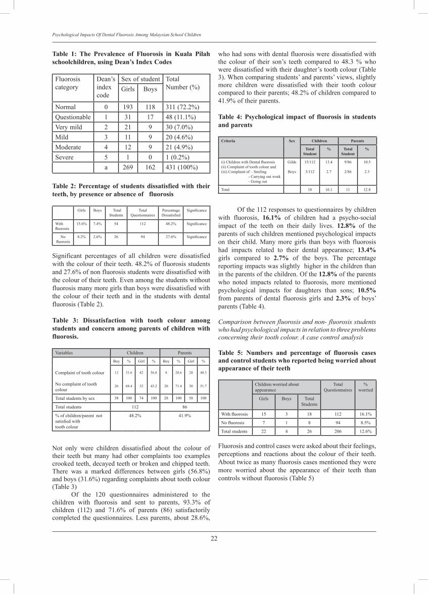
The prevalence of dental fluorosis was 27.8 % inKuala Pilah. There was no difference in the prevalencebetweenboysandgirls;28.2%ofgirlsand27.1%ofboyshad fluorosis. Most of the fluorosis was questionableandverymildbut5.1%hadmoderateorseverefluorosis(Table1).
22
Psychological Impacts Of Dental Fluorosis Among Malaysian School Children
Table 1: The Prevalence of Fluorosis in Kuala Pilah schoolchildren, using Dean’s Index codes
Fluorosiscategory
Dean’sindexcode
Sexofstudent TotalNumber(%)Girls Boys
Normal 0 193 118 311(72.2%)Questionable 1 31 17 48(11.1%)Verymild 2 21 9 30(7.0%)Mild 3 11 9 20(4.6%)Moderate 4 12 9 21(4.9%)Severe 5 1 0 1(0.2%)
a 269 162 431(100%)
Table 2: Percentage of students dissatisfied with their teeth, by presence or absence of fluorosis
Girls Boys TotalStudents
TotalQuestionnaires
PercentageDissatisfied
Significance
Withfluorosis
15.6% 7.4% 54 112 48.2% Significance
Nofluorosis
8.2% 2.6% 26 94 27.6% Significance
Significant percentages of all children were dissatisfiedwiththecolouroftheirteeth.48.2%offluorosisstudentsand27.6%ofnonfluorosisstudentsweredissatisfiedwiththecolouroftheirteeth.Evenamongthestudentswithoutfluorosismanymoregirlsthanboysweredissatisfiedwiththe colour of their teeth and in the students with dentalfluorosis(Table2).
Table 3: Dissatisfaction with tooth colour among students and concern among parents of children with fluorosis.
Variables Children Parents
Complaintoftoothcolour
Nocomplaintoftoothcolour
Boy % Girl % Boy % Girl %
12
26
31.6
68.4
42
32
56.8
43.2
8
20
28.6
71.4
28
30
48.3
51.7
Totalstudentsbysex 38 100 74 100 28 100 58 100
Totalstudents 112 86
%ofchildren/parentnotsatisfied with toothcolour
48.2% 41.9%
Not only were children dissatisfied about the colour oftheir teeth but many had other complaints too examplescrookedteeth,decayedteethorbrokenandchippedteeth.There was a marked differences between girls (56.8%)andboys(31.6%)regardingcomplaintsabouttoothcolour(Table3) Of the 120 questionnaires administered to thechildren with fluorosis and sent to parents, 93.3% ofchildren (112) and 71.6% of parents (86) satisfactorilycompletedthequestionnaires.Lessparents,about28.6%,
whohadsonswithdentalfluorosisweredissatisfiedwiththe colour of their son’s teeth compared to 48.3 % whoweredissatisfiedwiththeirdaughter’stoothcolour(Table3).Whencomparingstudents’andparents’views,slightlymore children were dissatisfied with their tooth colourcomparedtotheirparents;48.2%ofchildrencomparedto41.9%oftheirparents.
Table 4: Psychological impact of fluorosis in students and parents
Criteria Sex Children Parents
TotalStudent
% TotalStudent
%
(i) Children with Dental fluorosis (ii)Complaintoftoothcolourand(iii)Complaintof–Smiling-Carryingoutwork-Goingout
Gilds
Boys
15/112
3/112
13.4
2.7
9/86
2/86
10.5
2.3
Total 18 16.1 11 12.8
Ofthe112responsestoquestionnairesbychildrenwith fluorosis, 16.1% of children had a psycho-socialimpact of the teeth on their daily lives. 12.8% of theparentsofsuchchildrenmentionedpsychologicalimpactson their child.Manymoregirls thanboyswith fluorosishad impacts related to their dental appearance; 13.4%girls compared to 2.7% of the boys. The percentagereportingimpactswasslightlyhigherinthechildrenthanintheparentsofthechildren.Ofthe12.8%oftheparentswho noted impacts related to fluorosis, more mentionedpsychological impacts for daughters than sons; 10.5% from parents of dental fluorosis girls and 2.3% of boys’parents(Table4).
Comparison between fluorosis and non- fluorosis students who had psychological impacts in relation to three problems concerning their tooth colour. A case control analysis
Table 5: numbers and percentage of fluorosis cases and control students who reported being worried about appearance of their teeth
Childrenworriedaboutappearance
TotalQuestionnaires
%worried
Girls Boys TotalStudents
With fluorosis 15 3 18 112 16.1%
No fluorosis 7 1 8 94 8.5%
Totalstudents 22 4 26 206 12.6%
Fluorosisandcontrolcaseswereaskedabouttheirfeelings,perceptions and reactions about the colourof their teeth.Abouttwiceasmanyfluorosiscasesmentionedtheyweremore worried about the appearance of their teeth thancontrolswithoutfluorosis(Table5)
23
Mohd Nor M / Sheiham / Tsakos
Table 6: The distribution of psychological impacts of teeth on three aspects of daily life in students with and without fluorosis, by sex.
Type ofproblems
Fluorosis (54) non Fluorosis (26)
Girls (42) Boys (12) %
Yes
Girls (22) Boys (4) %
YesYes % Yes % Yes % Yes %
Difficultyinsmilling 15 35.7 3 25 16.1 7 31.8 1 25 8.5
Carryoutschoolwork
3 7.1 0 0 2.7 1 4.5 1 25 2.1
Goingoutwithfriends
4 9.5 0 0 3.6 2 9.1 1 25 3.2
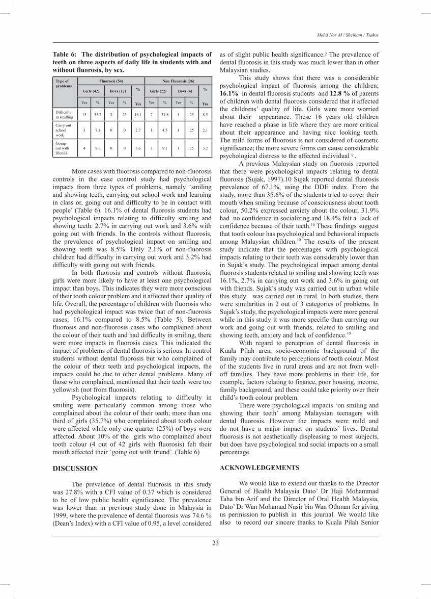
Morecaseswithfluorosiscomparedtonon-fluorosiscontrols in the case control study had psychologicalimpacts from three types of problems, namely ‘smilingandshowingteeth,carryingoutschoolworkandlearninginclassor,goingoutanddifficulty tobe incontactwithpeople’ (Table6).16.1%ofdental fluorosis studentshadpsychological impacts relating to difficulty smiling andshowingteeth.2.7%incarryingoutworkand3.6%withgoing out with friends. In the controls without fluorosis,the prevalence of psychological impact on smiling andshowing teeth was 8.5%. Only 2.1% of non-fluorosischildrenhaddifficultyincarryingoutworkand3.2%haddifficultywithgoingoutwithfriends. In both fluorosis and controls without fluorosis,girlsweremore likely tohaveat leastonepsychologicalimpactthanboys.Thisindicatestheyweremoreconsciousoftheirtoothcolourproblemanditaffectedtheirqualityoflife.Overall,thepercentageofchildrenwithfluorosiswhohadpsychological impactwastwicethatofnon-fluorosiscases; 16.1% compared to 8.5% (Table 5). Betweenfluorosis and non-fluorosis cases who complained aboutthecolouroftheirteethandhaddifficultyinsmiling,therewere more impacts in fluorosis cases.This indicated theimpactofproblemsofdentalfluorosisisserious.Incontrolstudents without dental fluorosis but who complained ofthe colour of their teeth and psychological impacts, theimpacts couldbedue toother dental problems.Manyofthosewhocomplained,mentionedthattheirteethweretooyellowish(notfromfluorosis). Psychological impacts relating to difficulty insmiling were particularly common among those whocomplainedaboutthecolouroftheirteeth;morethanonethirdofgirls(35.7%)whocomplainedabouttoothcolourwereaffectedwhileonlyonequarter(25%)ofboyswereaffected.About10%of the girlswhocomplainedabouttooth colour (4 out of 42 girls with fluorosis) felt theirmouthaffectedtheir‘goingoutwithfriend’.(Table6)
DIScuSSIon The prevalence of dental fluorosis in this studywas27.8%withaCFIvalueof0.37whichisconsideredto be of low public health significance. The prevalencewas lower than in previous study done in Malaysia in1999,wheretheprevalenceofdentalfluorosiswas74.6%(Dean’sIndex)withaCFIvalueof0.95,alevelconsidered
asofslightpublichealthsignificance.2TheprevalenceofdentalfluorosisinthisstudywasmuchlowerthaninotherMalaysianstudies. This study shows that there was a considerablepsychological impact of fluorosis among the children;16.1%indentalfluorosisstudentsand12.8 %ofparentsofchildrenwithdentalfluorosisconsideredthatitaffectedthe childrens’ quality of life. Girls were more worriedabout their appearance. These 16 years old childrenhavereachedaphase in lifewhere theyaremorecriticalabout their appearance and having nice looking teeth.Themildformsoffluorosisisnotconsideredofcosmeticsignificance;themoresevereformscancauseconsiderablepsychologicaldistresstotheaffectedindividual9 . A previous Malaysian study on fluorosis reportedthat there were psychological impacts relating to dentalfluorosis(Sujak,1997).10Sujakreporteddentalfluorosisprevalence of 67.1%, using the DDE index. From thestudy,morethan35.6%ofthestudentstriedtocovertheirmouthwhensmilingbecauseofconsciousnessabouttoothcolour,50.2%expressedanxietyabout thecolour,31.9%hadnoconfidenceinsocializingand18.4%feltalackofconfidencebecauseoftheirteeth.10Thesefindingssuggestthattoothcolourhaspsychologicalandbehavioralimpactsamong Malaysian children.10 The results of the presentstudy indicate that the percentages with psychologicalimpactsrelatingtotheirteethwasconsiderablylowerthaninSujak’sstudy.Thepsychological impactamongdentalfluorosisstudentsrelatedtosmilingandshowingteethwas16.1%,2.7%incarryingoutworkand3.6%ingoingoutwithfriends.Sujak’sstudywascarriedoutinurbanwhilethisstudywascarriedoutinrural.Inbothstudies,thereweresimilarities in2outof3categoriesofproblems. InSujak’sstudy,thepsychologicalimpactsweremoregeneralwhileinthisstudyitwasmorespecificthancarryingourwork and going out with friends, related to smiling andshowingteeth,anxietyandlackofconfidence.10 With regard to perception of dental fluorosis inKuala Pilah area, socio-economic background of thefamilymaycontributetoperceptionsoftoothcolour.Mostof the students live in rural areasandarenot fromwell-off families. They have more problems in their life, forexample,factorsrelatingtofinance,poorhousing,income,familybackground,andthesecouldtakepriorityovertheirchild’stoothcolourproblem. Therewerepsychologicalimpacts‘onsmilingandshowing their teeth’ among Malaysian teenagers withdental fluorosis. However the impacts were mild anddo not have a major impact on students’ lives. Dentalfluorosisisnotaestheticallydispleasingtomostsubjects,butdoeshavepsychologicalandsocialimpactsonasmallpercentage.
ACKNOWLEDGEMENTS WewouldliketoextendourthankstotheDirectorGeneral of Health Malaysia Dato’ Dr Haji MohammadTaha binArif and the Director of Oral Health Malaysia,Dato’DrWanMohamadNasirbinWanOthmanforgivingus permission to publish in this journal.We would likealso to record our sincere thanks to Kuala Pilah Senior
24
Psychological Impacts Of Dental Fluorosis Among Malaysian School Children
Dental Officer and to the staff from the Dental ClinicKuala Pilah, for all their support and assistance, withouttheirco-operation,Iwouldnotbeabletocarryoutthistasksuccessfully.Finally,thankstoalltheparentsandchildrenwhoparticipatedinthestudy.
REFEREncES
1. OralHealthDivision,MinistryofHealthMalaysia (August2001): Oral Health care in Malaysia, MOH/K/GIG/4.2001(BK),Malaysia.
2. Oral Health Division, Ministry of Health Malaysia (2001):FluorideEnamelOpacitiesin16yearsoldSchoolChildren,MOH/GIG/2.2001(RR),Malaysia.
3. Jones,S.,Lennon,M.(1997):Fluoridation,CommunityOralHealth,PineC.Editor,2ndEdition,GreatBritain,TheBathPress.p.222
4. Clarkson,J.J.,O’Mullane,D.M.(1992):Prevalenceofenameldefects/fluorosisinfluoridatedandnon-fluoridatedareasinIreland,Community Dent Oral Epidemiology20,196-9.
5. Melaku,Z.,Ismail,S.(2002):PerceptiononfluoriderelatedhealthproblemsinanareaofendemicfluorosisinEthiopia:An exploratory qualitative study, Ethiopia. Journal Health Dev 16,85-93.
6. Sheiham,A., Spencer, J (2002): Health needs assessment.:CommunityOralHealth:PineC.Editor,2ndEdition,GreatBritain,TheBathPress.p.39.
7. WHO (1997): Oral Health Surveys. Basic Methods, 4thEdition.WorldHealthOrganization.Geneva.
8. Gherunpong,S.,Tsakos,G.,Sheiham,A.(2004):Developingandevaluatinganoralhealth-relatedqualityoflifeindexforchildren;TheCHILD-OIDP.Community Dental Health 21,161-169.
9. Riordan,P.J.(1993):Perceptionofdentalfluorosis,Journal of Dental Research 72,1268-74.
10.Sujak, S.L. (1997): Prevalence and aesthetic perception ofdevelopmentaldefectsamong16yearsoldschoolchildreninPenang.Thesis.MastersinCommunityDentistry,UniversityofMalaya.KualaLumpur.
Address for correspondence:
Dr. Maznah Mohd NorSenior Dental OfficerPort Dickson District HospitalNegeri SembilanMalaysia
Evaluation of Amorphous Calcium Phosphate (ACP) as an alternative liner- An in vivo study Prabhakar A.R. MDS. Professor and Head
Shrirang S. MDS. Post graduate student
Sugandhan S. MDS. Professor
Ameet J. K, MDS. Reader Department of Pedodontics & Preventive Dentistry, Bapuji Dental College and Hospital, Davangere, Karnataka, India.
ABSTRACT The present in vivo study was carried out to evaluate the potential use of amorphous calcium phosphate as an alternative cavity liner in 40 patients in the age group of 6-8 years, having bilateral initial occlusal carious lesions on the mandibular first permanent molars.
The experimental teeth (total 40) were restored with amalgam after the application of amorphous calcium phosphate liner in a conventional Class-I cavity preparation. Sensitivity evaluation was carried out using tactile stimulation (probe), thermal stimulation (air blast, water jet) and osmotic stimulation (glucose solution) and was recorded on a patient discomfort scale. The contra-lateral teeth (total 40) which were restored with amalgam placed over a glass ionomer cement liner (Type III- LC) served as control.
In the control group, the post-operative sensitivity after GIC liner placement, reduced from 65% teeth to 22.5% teeth for probing, from 97.5% to 55% for air blasting. Use of water jet and osmotic stimulation was not done after liner placement for the fear of dilution or chemical reaction with the liner material. No sensitivity was observed in almost all the teeth after amalgam restoration. A follow up of 1-month and 6 months showed almost all the control group teeth (40) were comfortable and vital without any sensitivity.
The data was analyzed using Wilcoxon’s signed rank test (alternative to paired t-test) and Mann-Whitney test (alternative to unpaired t- test).
In the experimental group, the post-operative sensitivity after amorphous calcium phosphate (ACP) liner placement, reduced from 67.5% teeth to 22.5% for probing, from 100% teeth to 47.5% teeth for air blasting. Complete lack of sensitivity was seen in almost the teeth after amalgam restoration. A follow up period of 1-month and 6 months showed that almost all the experimental group teeth (40) were comfortable and vital, without any sensitivity.
It was concluded that amorphous calcium phosphate lining system can be used as a viable alternative to conventional cavity liners below amalgam restorations as the ACP system on clinical evaluation showed equally effective desensitizing and sealing property as compared to a GIC (Type III) liner.
25
Malaysian Dental Journal (2008) 29(1) 25-30© 2008 The Malaysian Dental Association
INTRodUCTIoN
Theaimofconservativetreatmentofvital teethisto maintain them in a healthy and functional state. Theprimary requirement for an ideal restorative material isthat, it should formaperfectunionwith the surroundingtoothstructuretopreventtheingressofsaliva,fluidsanddebris.
Inspiteofmanyadvancesinrestorativematerials,amalgam still remains the choice of material due toits advantages, which include good physical properties,dimensionalstability,easeofmanipulation,nearinsolubilityin the oral cavity and cost effective.1Yet, post-operativesensitivity and susceptibility to fracture still remain itsinherent disadvantages. Post-operative sensitivity mayresultfromtheinabilityoftherestorativematerialtoseal
MALAYSIAN dENTAL JoURNAL
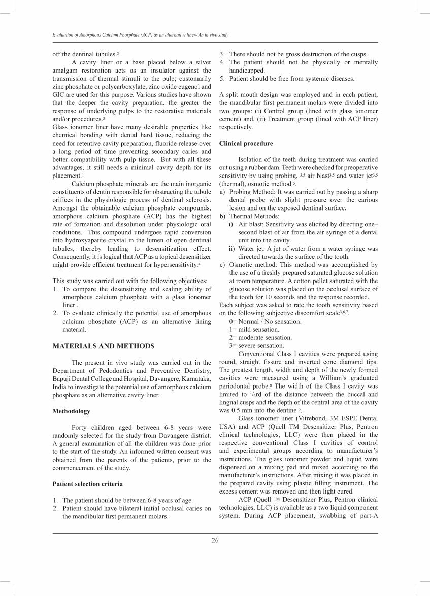
26
Evaluation of Amorphous Calcium Phosphate (ACP) as an alternative liner- An in vivo study
offthedentinaltubules.2 A cavity liner or a base placed below a silveramalgam restoration acts as an insulator against thetransmission of thermal stimuli to the pulp; customarilyzincphosphateorpolycarboxylate,zincoxideeugenolandGICareusedforthispurpose.Variousstudieshaveshownthat the deeper the cavity preparation, the greater theresponse of underlying pulps to the restorative materialsand/orprocedures.3Glass ionomer liner have many desirable properties likechemical bonding with dental hard tissue, reducing theneedforretentivecavitypreparation,fluoridereleaseovera long period of time preventing secondary caries andbetter compatibility with pulp tissue. But with all theseadvantages, it still needs a minimal cavity depth for itsplacement.1 Calciumphosphatemineralsarethemaininorganicconstituentsofdentinresponsibleforobstructingthetubuleorifices in the physiologic process of dentinal sclerosis.Amongst the obtainable calcium phosphate compounds,amorphous calcium phosphate (ACP) has the highestrate of formation and dissolution under physiologic oralconditions. This compound undergoes rapid conversionintohydroxyapatitecrystal in the lumenofopendentinaltubules, thereby leading to desensitization effect.Consequently,itislogicalthatACPasatopicaldesensitizermightprovideefficienttreatmentforhypersensitivity.4
Thisstudywascarriedoutwiththefollowingobjectives:1. To compare the desensitizing and sealing ability of
amorphous calcium phosphate with a glass ionomerliner.
2. Toevaluate clinically thepotentialuseof amorphouscalcium phosphate (ACP) as an alternative liningmaterial.
MATERIALS ANd METHodS
The present in vivo study was carried out in theDepartment of Pedodontics and Preventive Dentistry,BapujiDentalCollegeandHospital,Davangere,Karnataka,Indiatoinvestigatethepotentialuseofamorphouscalciumphosphateasanalternativecavityliner.
Methodology Forty children aged between 6-8 years wererandomly selected for the study fromDavangeredistrict.Ageneralexaminationofall thechildrenwasdonepriortothestartofthestudy.Aninformedwrittenconsentwasobtained from the parents of the patients, prior to thecommencementofthestudy.
Patient selection criteria
1. Thepatientshouldbebetween6-8yearsofage.2. Patientshouldhavebilateral initialocclusalcarieson
themandibularfirstpermanentmolars.
3. Thereshouldnotbegrossdestructionofthecusps.4. The patient should not be physically or mentally
handicapped.5. Patientshouldbefreefromsystemicdiseases.
A split mouth design was employed and in each patient,the mandibular first permanent molars were divided intotwo groups: (i) Control group (lined with glass ionomercement)and, (ii)Treatmentgroup (linedwithACP liner)respectively.
Clinical procedure
Isolationof the teethduring treatmentwascarriedoutusingarubberdam.Teethwerecheckedforpreoperativesensitivitybyusingprobing,3,5airblast3,5andwaterjet3,5(thermal),osmoticmethod5.a)ProbingMethod:Itwascarriedoutbypassingasharp
dental probe with slight pressure over the cariouslesionandontheexposeddentinalsurface.
b)ThermalMethods:i) Airblast:Sensitivitywaselicitedbydirectingone–
secondblastofairfromtheairsyringeofadentalunitintothecavity.
ii)Waterjet:Ajetofwaterfromawatersyringewasdirectedtowardsthesurfaceofthetooth.
c)Osmotic method: This method was accomplished bytheuseofafreshlypreparedsaturatedglucosesolutionatroomtemperature.Acottonpelletsaturatedwiththeglucosesolutionwasplacedontheocclusalsurfaceofthetoothfor10secondsandtheresponserecorded.
Eachsubjectwasaskedtoratethetoothsensitivitybasedonthefollowingsubjectivediscomfortscale3,6,7. 0=Normal/Nosensation. 1=mildsensation. 2=moderatesensation. 3=severesensation. ConventionalClass Icavitieswerepreparedusinground, straight fissure and inverted cone diamond tips.Thegreatestlength,widthanddepthofthenewlyformedcavities were measured using a William’s graduatedperiodontal probe.8 The width of the Class I cavity waslimited to 1/3rd of the distance between the buccal andlingualcuspsandthedepthofthecentralareaofthecavitywas0.5mmintothedentine9. Glass ionomer liner (Vitrebond, 3M ESPE DentalUSA) and ACP (Quell TM Desensitizer Plus, Pentronclinical technologies, LLC) were then placed in therespective conventional Class I cavities of controland experimental groups according to manufacturer’sinstructions. The glass ionomer powder and liquid weredispensed on a mixing pad and mixed according to themanufacturer’sinstructions.Aftermixingitwasplacedinthe prepared cavity using plastic filling instrument. Theexcesscementwasremovedandthenlightcured. ACP (Quell TM Desensitizer Plus, Pentron clinicaltechnologies,LLC)isavailableasatwoliquidcomponentsystem. During ACP placement, swabbing of part-A
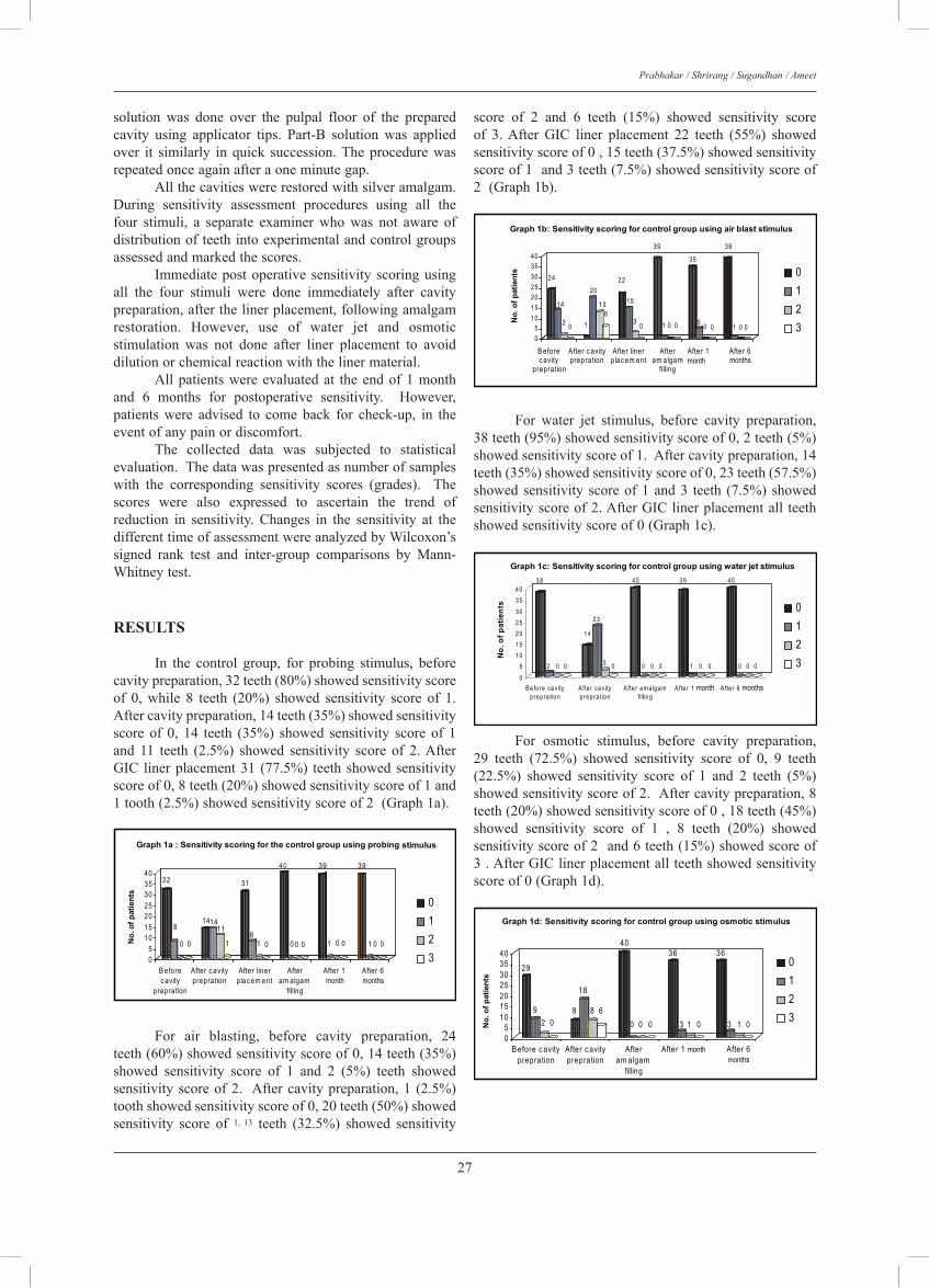
27
Prabhakar / Shrirang / Sugandhan / Ameet
solution was done over the pulpal floor of the preparedcavity using applicator tips. Part-B solution was appliedover it similarly in quick succession.Theprocedurewasrepeatedonceagainafteraoneminutegap. Allthecavitieswererestoredwithsilveramalgam.During sensitivity assessment procedures using all thefour stimuli, a separate examiner who was not aware ofdistributionofteethintoexperimentalandcontrolgroupsassessedandmarkedthescores. Immediatepostoperativesensitivityscoringusingall the four stimuli were done immediately after cavitypreparation,afterthelinerplacement,followingamalgamrestoration. However, use of water jet and osmoticstimulation was not done after liner placement to avoiddilutionorchemicalreactionwiththelinermaterial. Allpatientswereevaluatedat theendof1monthand 6 months for postoperative sensitivity. However,patients were advised to come back for check-up, in theeventofanypainordiscomfort. The collected data was subjected to statisticalevaluation.Thedatawaspresentedasnumberofsampleswith the corresponding sensitivity scores (grades). Thescores were also expressed to ascertain the trend ofreduction in sensitivity. Changes in the sensitivity at thedifferenttimeofassessmentwereanalyzedbyWilcoxon’ssigned rank test and inter-group comparisons by Mann-Whitneytest.
RESULTS
In the control group, for probing stimulus, beforecavitypreparation,32teeth(80%)showedsensitivityscoreof 0, while 8 teeth (20%) showed sensitivity score of 1.Aftercavitypreparation,14teeth(35%)showedsensitivityscoreof 0, 14 teeth (35%) showed sensitivity scoreof 1and 11 teeth (2.5%) showed sensitivity score of 2.AfterGIC linerplacement31 (77.5%) teethshowedsensitivityscoreof0,8teeth(20%)showedsensitivityscoreof1and1tooth(2.5%)showedsensitivityscoreof2(Graph1a).
32
8
0 0
141411
1
31
81 0
40
0 0 0
39
1 0 0
39
1 0 0
05
10152025303540
No.
of p
atie
nts
B eforec avity
prepration
After c avityprepration
After linerplac em ent
Afteram algam
filling
After 1month
After 6months
Graph 1a : Sensitivity scoring for the control group using probing stimulus
0123
24
14
2 0 1
20
136
22
15
3 0
39
1 0 0
35
5 0 0
39
1 0 005
10152025303540
No.
of p
atie
nts
B eforec avity
prepration
After c avityprepration
After linerplac em ent
Afteram algam
filling
After 1month
After 6months
Graph 1b: Sensitivity scoring for control group using air blast stimulus
0123
0123
3 8
2 0 0
1 4
2 3
3 0
4 0
0 0 0
3 9
1 0 0
4 0
0 0 0
05
1 01 52 02 53 03 54 0
No.
of p
atie
nts
Be fo re ca v ityp re p ra tio n
Afte r ca v ityp re p ra tio n
Afte r a ma lg a mfillin g
Afte r 1 month Afte r 6 months
Graph 1c: Sensitivity scoring for control group using water jet stimulus
For air blasting, before cavity preparation, 24teeth(60%)showedsensitivityscoreof0,14teeth(35%)showed sensitivity score of 1 and 2 (5%) teeth showedsensitivityscoreof2. Aftercavitypreparation,1 (2.5%)toothshowedsensitivityscoreof0,20teeth(50%)showedsensitivity score of 1, 13 teeth (32.5%) showed sensitivity
score of 2 and 6 teeth (15%) showed sensitivity scoreof 3.After GIC liner placement 22 teeth (55%) showedsensitivityscoreof0,15teeth(37.5%)showedsensitivityscoreof1and3teeth(7.5%)showedsensitivityscoreof2(Graph1b).
32
8
0 0
141411
1
31
81 0
40
0 0 0
39
1 0 0
39
1 0 0
05
10152025303540
No.
of p
atie
nts
B eforec avity
prepration
After c avityprepration
After linerplac em ent
Afteram algam
filling
After 1month
After 6months
Graph 1a : Sensitivity scoring for the control group using probing stimulus
0123
24
14
2 0 1
20
136
22
15
3 0
39
1 0 0
35
5 0 0
39
1 0 005
10152025303540
No.
of p
atie
nts
B eforec avity
prepration
After c avityprepration
After linerplac em ent
Afteram algam
filling
After 1month
After 6months
Graph 1b: Sensitivity scoring for control group using air blast stimulus
0123
0123
3 8
2 0 0
1 4
2 3
3 0
4 0
0 0 0
3 9
1 0 0
4 0
0 0 0
05
1 01 52 02 53 03 54 0
No.
of p
atie
nts
Be fo re ca v ityp re p ra tio n
Afte r ca v ityp re p ra tio n
Afte r a ma lg a mfillin g
Afte r 1 month Afte r 6 months
Graph 1c: Sensitivity scoring for control group using water jet stimulus
For water jet stimulus, before cavity preparation,38teeth(95%)showedsensitivityscoreof0,2teeth(5%)showedsensitivityscoreof1.Aftercavitypreparation,14teeth(35%)showedsensitivityscoreof0,23teeth(57.5%)showedsensitivityscoreof1and3 teeth (7.5%)showedsensitivityscoreof2.AfterGIClinerplacementall teethshowedsensitivityscoreof0(Graph1c).
32
8
0 0
141411
1
31
81 0
40
0 0 0
39
1 0 0
39
1 0 0
05
10152025303540
No.
of p
atie
nts
B eforec avity
prepration
After c avityprepration
After linerplac em ent
Afteram algam
filling
After 1month
After 6months
Graph 1a : Sensitivity scoring for the control group using probing stimulus
0123
24
14
2 0 1
20
136
22
15
3 0
39
1 0 0
35
5 0 0
39
1 0 005
10152025303540
No.
of p
atie
nts
B eforec avity
prepration
After c avityprepration
After linerplac em ent
Afteram algam
filling
After 1month
After 6months
Graph 1b: Sensitivity scoring for control group using air blast stimulus
0123
0123
3 8
2 0 0
1 4
2 3
3 0
4 0
0 0 0
3 9
1 0 0
4 0
0 0 0
05
1 01 52 02 53 03 54 0
No.
of p
atie
nts
Be fo re ca v ityp re p ra tio n
Afte r ca v ityp re p ra tio n
Afte r a ma lg a mfillin g
Afte r 1 month Afte r 6 months
Graph 1c: Sensitivity scoring for control group using water jet stimulus
For osmotic stimulus, before cavity preparation,29 teeth (72.5%) showed sensitivity score of 0, 9 teeth(22.5%) showed sensitivity score of 1 and 2 teeth (5%)showedsensitivityscoreof2.Aftercavitypreparation,8teeth(20%)showedsensitivityscoreof0,18teeth(45%)showed sensitivity score of 1 , 8 teeth (20%) showedsensitivityscoreof2and6teeth(15%)showedscoreof3.AfterGIClinerplacementallteethshowedsensitivityscoreof0(Graph1d).
0123
0123
0123
29
92 0
8
18
8 6
40
0 0 0
36
3 1 0
36
3 1 005
10152025303540
No.
of p
atie
nts
B efore c avityprepration
After c avityprepration
Afteram algam
filling
After 1 month After 6months
Graph 1d: Sensitivity scoring for control group using osmotic stimulus
34
60 0
1318
72
31
63
0
40
0 0 0
39
1 0 0
39
1 0 0No.
of p
atie
nts
B eforec avity
prepration
After c avityprepration
After linerplac em ent
Afteram algam
filling
After 1month
After 6months
Graph 2a: Sensitivity scoring for experimental group using probing stimulus
05
10152025303540
2117
2 0 0
1817
5
1917
4 0
39
1 0 0
35
50 0
39
1 0 005
10152025303540
No.
of p
atie
nts
B eforec avity
prepration
After c avityprepration
After linerplac em ent
Afteram algam
filling
After 1month
After 6months
Graph 2b: Sensitivity scoring for experimental group using air blast stimulus
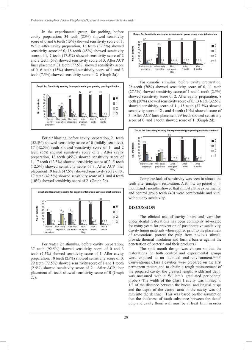
28
Evaluation of Amorphous Calcium Phosphate (ACP) as an alternative liner- An in vivo study
In the experimental group, for probing, beforecavity preparation, 34 teeth (85%) showed sensitivityscoreof0and6teeth(15%)showedsensitivityscoreof1.While after cavity preparation, 13 teeth (32.5%) showedsensitivity score of 0, 18 teeth (45%) showed sensitivityscoreof1,7 teeth(17.5%)showedsensitivityscoreof2and2teeth(5%)showedsensitivityscoreof3.AfterACPlinerplacement31teeth(77.5%)showedsensitivityscoreof 0, 6 teeth (15%) showed sensitivity score of 1 and 3teeth(7.5%)showedsensitivityscoreof2(Graph2a).
0123
0123
0123
29
92 0
8
18
8 6
40
0 0 0
36
3 1 0
36
3 1 005
10152025303540
No.
of p
atie
nts
B efore c avityprepration
After c avityprepration
Afteram algam
filling
After 1 month After 6months
Graph 1d: Sensitivity scoring for control group using osmotic stimulus
34
60 0
1318
72
31
63
0
40
0 0 0
39
1 0 0
39
1 0 0No.
of p
atie
nts
B eforec avity
prepration
After c avityprepration
After linerplac em ent
Afteram algam
filling
After 1month
After 6months
Graph 2a: Sensitivity scoring for experimental group using probing stimulus
05
10152025303540
2117
2 0 0
1817
5
1917
4 0
39
1 0 0
35
50 0
39
1 0 005
10152025303540
No.
of p
atie
nts
B eforec avity
prepration
After c avityprepration
After linerplac em ent
Afteram algam
filling
After 1month
After 6months
Graph 2b: Sensitivity scoring for experimental group using air blast stimulus
Forairblasting,beforecavitypreparation,21teeth(52.5%) showed sensitivity scoreof 0 (mildly sensitive),17 (42.5%) teeth showed sensitivity score of 1 and 2teeth (5%) showed sensitivity score of 2 . After cavitypreparation, 18 teeth (45%) showed sensitivity score of1,17teeth(42.5%)showedsensitivityscoreof2,5teeth(12.5%) showed sensitivity score of 3. After ACP linerplacement19teeth(47.5%)showedsensitivityscoreof0,17teeth(42.5%)showedsensitivityscoreof1and4teeth(10%)showedsensitivityscoreof2(Graph2b).
0123
0123
0123
29
92 0
8
18
8 6
40
0 0 0
36
3 1 0
36
3 1 005
10152025303540
No.
of p
atie
nts
B efore c avityprepration
After c avityprepration
Afteram algam
filling
After 1 month After 6months
Graph 1d: Sensitivity scoring for control group using osmotic stimulus
34
60 0
1318
72
31
63
0
40
0 0 0
39
1 0 0
39
1 0 0No.
of p
atie
nts
B eforec avity
prepration
After c avityprepration
After linerplac em ent
Afteram algam
filling
After 1month
After 6months
Graph 2a: Sensitivity scoring for experimental group using probing stimulus
05
10152025303540
2117
2 0 0
1817
5
1917
4 0
39
1 0 0
35
50 0
39
1 0 005
10152025303540
No.
of p
atie
nts
B eforec avity
prepration
After c avityprepration
After linerplac em ent
Afteram algam
filling
After 1month
After 6months
Graph 2b: Sensitivity scoring for experimental group using air blast stimulus
For water jet stimulus, before cavity preparation,37 teeth (92.5%) showed sensitivity score of 0 and 3teeth (7.5%) showed sensitivity score of 1.After cavitypreparation,10teeth(25%)showedsensitivityscoreof0,29teeth(72.5%)showedsensitivityscoreof1and1tooth(2.5%) showed sensitivity score of 2 . After ACP linerplacement all teeth showed sensitivity scoreof 0 (Graph2c).
0123
0123
37
3 0 0
10
29
1 0
40
0 0 0
38
2 0 0
40
0 0 0No.
of p
atie
nts
B efore c avityprepration
After c avityprepration
Afteram algam
filling
After1 month
After6 months
Graph 2c: Sensitivity scoring for experimental group using water jet stimulus
28
11
1 08
1315
4
39
1 0 0
35
50 0
36
4 0 0No.
of p
atie
nts
B efore c avityprepration
After c avityprepration
Afteram algam
filling
After1 month
After6 months
Graph 2d: Sensitivity scoring for experimental group using osmotic stimulus
05
10152025303540
05
10152025303540
For osmotic stimulus, before cavity preparation,28 teeth (70%) showed sensitivity score of 0, 11 teeth(27.5%)showedsensitivityscoreof1and1tooth(2.5%)showedsensitivityscoreof2.Aftercavitypreparation,8teeth(20%)showedsensitivityscoreof0,13teeth(32.5%)showed sensitivity score of 1 , 15 teeth (37.5%) showedsensitivityscoreof2.and4teeth(10%)showedscoreof3.AfterACPlinerplacement39teethshowedsensitivityscoreof0and1toothshowedscoreof1(Graph2d).
0123
0123
37
3 0 0
10
29
1 0
40
0 0 0
38
2 0 0
40
0 0 0No.
of p
atie
nts
B efore c avityprepration
After c avityprepration
Afteram algam
filling
After1 month
After6 months
Graph 2c: Sensitivity scoring for experimental group using water jet stimulus
28
11
1 08
1315
4
39
1 0 0
35
50 0
36
4 0 0No.
of p
atie
nts
B efore c avityprepration
After c avityprepration
Afteram algam
filling
After1 month
After6 months
Graph 2d: Sensitivity scoring for experimental group using osmotic stimulus
05
10152025303540
05
10152025303540
Completelackofsensitivitywasseeninalmosttheteethafteramalgamrestoration.Afollowupperiodof1-monthand6monthsshowedthatalmostalltheexperimentaland control group teeth (40) were comfortable and vital,withoutanysensitivity.
dISCUSSIoN The clinical use of cavity liners and varnishesunder dental restorations has been commonly advocatedformanyyearsforpreventionofpostoperativesensitivity.Cavityliningmaterialswhenappliedpriortotheplacementof restorations protect the pulp from noxious stimuli,provide thermal insulationand formabarrieragainst thepenetrationofbacteriaandtheirproducts.3 The split mouth design was chosen so that therestorations on both control and experimental groupswere exposed to an identical oral environment.10,11,12Conventional Class I cavities were prepared on the firstpermanentmolarsand toobtaina roughmeasurementofthe prepared cavity, the greatest length, width and depthwas measured with a William’s graduated periodontalprobe.8 The width of the Class I cavity was limited to1/3of thedistancebetween thebuccal and lingual cuspsand the depth of the central area of the cavity was 0.5mminto thedentine. Thiswasbasedon theassumptionthat the thickness of tooth substance between the dentalpulpandcavityfloor/wallmustbeatleast1mminorder
29
Prabhakar / Shrirang / Sugandhan / Ameet
tominimizetheadverseeffectsofcavitypreparationandrestorationsonthedentalpulp.9 The teeth in each patient were divided into 2groups,controlgroup,wherea lightactivatedGIC(TypeIII)wasusedandexperimentalgroup,whereacoatofACPlinerwasappliedinthecavities. GIClinershavemanyadvantagesincludingchemicalbonding to tooth structure and fluoride release. One ofthe main advantages of visible light—activated glassionomerisitsimprovedresistancetoacidsolubility13,14,15.Italsohasgoodthermalinsulation,antibacterialaction,acommandsetandsubsequentlyprovidesimprovedsupportto amalgam restorations 13,14,15. Therefore in the presentstudy,lightactivatedGIClinerwasused. The main component of ACP system namely,calciumphosphateisalsothemaininorganicconstituentof dentine. These mineral rich deposits are known tooccludedentinaltubulessimilartothephysiologicprocessofdentinalsclerosis.Amorphouscalciumphosphateshowsahighcapabilityof rapidconversion intohydroxyapatitecrystal under physiologic oral conditions, which canprecipitate in the lumenof theopendentinal tubules 4,16.Studies have shown that a serial application of calciumchloride followed by potassium phosphate, results indentinal tubule occlusion by ACP formation leading toimmediate relief from dentinal hypersensitivity4. In theexperimentalgroup,thisACPsystemwasusedasacavityliner. All the teeth inboth the experimental andcontrolgroups were restored with silver amalgam. Preoperative,intraoperative and postoperative sensitivity assessmentsweredoneusingprobing(tactile),airblast,water jetandosmoticstimulationmethods.SimilarmethodswereusedinthestudiesbyMinkovetal17,KleinbergI.5,ChawlaH.S3andmostrecentlyPerdigaoetal18. Itwasobservedthatthemostcommonstimuliusedin clinical studies were thermal and tactile stimulation19.Since teeth may often show sensitivity to one but notto another stimulus,more thanonemethodwas requiredfor checking the dentinal sensitivity.5 Therefore in thepresentstudy,allthefourstimuliwereusedforsensitivityassessment. In the current study, the criterion for sensitivityassessmentwassubjectivei.e.,thesubjectwasaskedtoratethetoothsensitivityon‘Thesubjectivediscomfortscale’.AsimilarmethodofsensitivityratingwasusedbyTarbetW.G.etal,7ChawlaH.S.3andGillamD.G.andNewmanH.N.6Greenetal20statedthattheresultsofexperimentationusingonlythepatient’ssubjectiveresponsesshouldbeconstruedashavingminimal significance. On the contraryGillamD.G.andNewmanH.N6 statedthatthissimpledescriptivesensitivity scale offers a precise choice of words, whichrepresentstheexperienceofpainbythepatient.Hencethisstudy successfully evaluated and categorized the patientperceptionof thequalityofreductionofsensitivityusingthevariousmaterialstested. In the control group, the sensitivity scores oncomparisonbefore and after cavitypreparation increasedfrom20% (not sensitive) teeth to65% (mildly sensitive)teeth for probing, from 40% (35% -mildly sensitive and5% moderately sensitive) teeth to 98.5% teeth for air
blasting,from5%(mildlysensitive)teethto65%(57.5%-mildly sensitive and 7.5%-moderately sensitive) teethfor water jet and from 27.5% (22.5% -mildly sensitiveand5%-moderatelysensitive) teethto80%(45%-mildlysensitive, 20%-moderately sensitive and 15%-severelysensitive)teethforosmoticstimulation The reason for increased postoperative sensitivityascitedbyBrannstrom21wasthatthedentaldrillremovesfluid from the dentin, partly by frictional heat causingevaporationandpartly,bymechanicallypressingoutfluidfromthedentinaltubulesleadingtopainorsensitivity. The postoperative sensitivity after GIC linerplacement, reduced from 65% teeth to 22.5% teethfor probing, and from 97.5% to 55% for air blasting.Use of water jet and osmotic stimulation was notdone after liner placement to avoid any dilution orchemical reaction of the water component with theliner material. These results suggest that there was ahighly significant decrease in the sensitivity after theplacement of GIC liner. The results were in conformitywith theresultsobtained inastudybyGordonetalwhoreported that the sensitivitywas reducedwhenGIC linerwasusedunderamalgamrestorations.2 The postoperative sensitivity after ACP linerplacement,reducedfrom67.5%teethto22.5%forprobingandfrom100%teethto47.5%teethforairblasting.TheseresultssuggestthattherewasahighlysignificantdecreaseinthesensitivityafterplacementofACPliner.SincetherewerenodirectstudiesthatevaluatedACPasacavitylinerunder amalgam restoration, this study was compared tothe study conducted by Geiger et al4 who reported thatapplicationACPtotheexposedrootsurfaces,resultedinarapiddecreaseinsensitivitytostimulation. Almostalltheteethinthecontrolandexperimentalgroups showed no sensitivity to any stimulationimmediately, after amalgam restoration, after one monthand6monthsfollowupevaluations. Theseresultspointout that no significant differences existed in both thecontrolandexperimentalgroups. The duration of 6 months of evaluation of thepostoperative sensitivity was in accordance with theguidelines given by Holland et al 22.According to them,the duration should be sufficient to allow the expressionof maximum efficacy of the active agent (experimental),whileminimizingthemagnitudeofanyplacebo(control).They also recommended that the clinical trial designshould recognize the time required to achieve maximumdesensitizing effects that might vary between differentproductsoragents22.
CoNCLUSIoNS The following conclusions were drawn from theresultsofthisstudy:1. Clinical evaluation on ACP system showed equal
desensitizingeffectandsealingpropertycomparedtoaGIC(TypeIII)liner.
2. Amorphous calcium phosphate lining system can beused as a viable alternative to conventional cavitylinersbelowamalgamrestorations.
TheuseofACPasalinercanbeadvantageous,in
30
Evaluation of Amorphous Calcium Phosphate (ACP) as an alternative liner- An in vivo study
that it provides adequate bulk to the restorative materialeven in shallow cavities thus preventing inadvertentsacrificeof sound tooth structure. The sealingabilityofthe material is definitely well proven; nonetheless, theeffectivenessofthematerialinthedeepercavitiesandcosteffectiveness of the material has to be considered beforeweighing it as a replacement to the more conventionalmodalities.Weadvocatefurtherparallelobjectivestudiestoascertaintheefficiencyofthismaterialanditsprospectiveclinicalusefulness.
REFERENCES
1. Chen RS, Liu CC, Cheng MR, Lin CP, BondedAmalgamrestorations.Usingaglassionomerasanadhesiveliner.OperDent2000;25:411-7.
2. Gordan VV, Mjor IA, Moor JE. Amalgam restorations.Postoperativesensitivityasafunctionoflinertreatmentandcavitydepth.OperDent1999;24:377-83.
3. Chawla HS. NaF Iontophoresis as an alternative to cavityliners-An in vivo study. J Ind Soc Pedo Prev Dent 1991;9:17-19.
4. GeigerS,MatalonS.,BiasbalgJ.,TungMS,EichmillerFC.Theclinical effectof amorphouscalciumphosphate (ACP)onrootsurfacehypersensitivity.OperDent2003;28(5):496-500.
5. Kleinberg I, Kaufman HW, Confessor F. Methods ofmeasuringtoothhypersensitivity.DentClinNorthAm1990;34(3):515–29.
6. GillamDGandNewmanHN.Assessmentofpainincervicaldentinalsensitivitystudies.JClinPeriodontol1993;20:383-94.
7. Tarbet WJ, Silverman G, Stoleman JM, Fratarcangelo PA.Anevaluationof2methodsfor thequantitationofdentinalhypersensitivity.JAmDentAssoc1979;90:914-18.
8. Tavares M. Evaluation of a chemomechanical method ofcariesremovalinrootsurfacelesions.QuintessenceInt1988;19(1):29-32.
9. NozakaK,SurugaY,AmariE.Microleakageof compositeresinsincavitiesofupperprimarymolars.IntJPediatrDent1999;9:185-94.
10.Yates R, Owen J, Jackson R, Newcombe RG,Addy M.Asplitmouthplacebocontrolledstudytodeterminetheeffectofamorphouscalciumphosphateinthetreatmentofdentinesensitivity.JClinPeriodontol1998;25:687-92.
11.Donly KJ, Wild T and Jensen ME. Cuspal reinforcementin primary teeth. An in vitro comparison of 3 restorativematerials.PediatrDent1988;10(2):102-4.
12.HubelSandMejareI.Conventionalversusresin-modifiedglass- ionomer cement for Class II restorations in primarymolars.A 3- year clinical study. Int J Pediatr Dent 2003;13:2-8.
13.HiltonTJ.Cavitysealers,linersandbases.Currentphilosophiesandindicationsforuse.OperDent1996;21:134-46.
14.Robbins JW. The placement of bases beneath amalgamrestoration:Areviewof literatureandrecommendationsfortheuse.JAmDentAssoc1986;113:910-2.
15.McCoy RB. Announcement: Bases and cavity varnishesupdate.OperDent1995;20:216.
16.Ishikawa K, Suge T, Yoshiyama M, Kawasaki A, AsaokaK, Ebisu S. Occlusion of dentinal tubules with calciumphosphateusingacidiccalciumphosphatesolutionfollowedbyneutralization.JDentRes1994;73(6):1197-04.
17.Minkov B, Marmari L, Gedalia E, Garfunkel A. TheeffectivenessofsodiumfluoridetreatmentwithandwithoutIontophoresis on the reduction of hypersensitive dentin. JPeriodontol1975;46:246.
18.PerdigaoJ,GraldeliSandHodgesJS.Total-etchversusselfetchedadhesives:Effectonpostoperativesensitivity. JAmDentAssoc2003;134:1621-29.
19.IdeM,WilsonRF,AshleyFP.Thereproducibilityofmethodsof assessment for cervical dentine hypersensitivity. J ClinPeriodontol2001;28:16-22.
20.GreenBL,McfallJ.Calciumhydroxideandpotassiumnitrateas desensitizing agents for hypersensitive root surface. JPeriodontol1977;48:667-2.
21.Brannstrom M. Dentin and pulp in restorative dentistry.Castelnuovo (AT), Italy: Wolfe medical publication Ltd;1982.p.8-43.
22.HollandGR,NorhiMN,AddyM,GangarosaL,OrchardsonR.Guidelinesforthedesignandconductofclinicaltrialsondentinhypersensitivity.JClinPeriodontol1997;24:808-13.
Address for correspondence:
dR. A.R. PRABHAKAR, Professor And HeadDepartment Of Pedodontics & Preventive Dentistry,Bapuji Dental College And Hospital,Davangere – 577004, Karnataka.E-mail:- [email protected] Ph. No:- 91-8192-220575Fax:- 91-8192-220578
The Expert Says….How to diagnose pulpal status? Safura A B DDS (Canada), MClinDent in Endodontology (London), MFDSRCS (Eng), DipCDSc (London) Lecturer, Department of Operative Dentistry, Faculty of Dentistry, Universiti Kebangsaan Malaysia, Kuala Lumpur, Malaysia.
31
Malaysian Dental Journal (2008) 29(1) 31-33© 2008 The Malaysian Dental Association
InTroducTIon The assessment of pulp vitality is a crucialdiagnostic procedure in the practice of endodontics.Dentists should establish their treatment decisions basedon good sound information and in the best interests ofthe patients. Therefore, a definite diagnosis must beestablishedwithcomprehensiveinvestigationandrecordsbefore any treatment is carried out1. Since the dentalpulp is enclosed in an opaque tooth, the assessment oftooth vitality is undertaken indirectly by: looking forclinicalorradiologicalevidenceofpulpnecrosisorapicalperiodontitis;investigatingnerveconduction;orexaminingthebloodflow1,2. Currently, commonly used vitality testers assessthe integrity of the Aδ nerve fibers in the dentine-pulp complex by means of brief application of stimulus tothe outer surface of the tooth. If the Aδ nerve fibers are successfully stimulated, the patient will experiencea brief sharp/tingling sensation from the tooth3,4,5,6.However,thesetestsonlyindicatesthatthenervefibersarefunctioningbutdoesnotgiveanyindicationofbloodflowinthepulp,orwhetheritispartiallydamaged.Pulpvitalityisdeterminedpurelybythefunctionofvasculaturehealth.Thus thismethodof testingmaynotaccuratelydiagnosethe condition of the pulp2, 7, 8. Bhaskar and Rappaport10
concludedintheirstudythattheconventionalvitalitytestsare actually sensitivity tests as they only tests the nerveconductionandhavequestionablepredictivevalueof thevitalityofpulptissue. Accurate information on the status of blood flowin the pulp is essential as the Aδ nerve fibers will cease to functionwhenthereisnooxygensupply2.However,thereareinstancesfollowingtrauma,wherethereisbloodflowin the pulp but the Aδ nerve fibers are not functioning or are injured7, 9, 10. Irritation of the pulp, by whateverstimulus, causes inflammation and with more severeinflammationpulpaltissuemaybepermanentlydamaged.Atpresent,researchersarestilltryingtodevelopadevicethatisabletodeterminethestatusofpulpalcirculationforinstanceusing laserDoppler flowmetry,dualwavelengthspectrophotometry, and pulse oximetry7, 11, 12. Althoughsome of these methods has shown promising results and
able todemonstrate the truevitalityof thepulp, theyarenotyetreadytoreplacethepresentlyavailabletesters.Inthe meantime, these ‘sensitivity tests’ are only availabletoaidwiththepulpaldiagnosis.Despiteitsshortcomings,these tests are cheap, easy to use, useful when used incombinationandareabletoprovidedentistswithsufficientinformationtoestablishadiagnosis. Based on the following clinical and radiographicexaminations,diagnosisofreversiblepulpitis,irreversiblepulpitis, periapical periodontitis and acute apical abscesscanbemadeclearly.
HISTorY
Thefollowingacronymcouldbeusedasaguidetoaskthepatientsaboutsomeimportantinformationonthesigns and symptoms related to pulpal disease. These areusefulasaguidetoclinicalandradiologicalexaminations,andappropriatelyrelatedtotheanswersgivenbypatients.• L - Location of pain – upper arch, lower arch, left or
rightside• o - Origin of pain – tooth/teeth involved• c - Characteristic of the pain – sharp, dull, stabbing,
throbbing,lingeringpain• A - Aggravating factors – hot or cold food/drink,
coldair,spontaneous• T - �i�ing of pain – dayti�e, at night, disturb sleep- �i�ing of pain – dayti�e, at night, disturb sleep �i�ing of pain – dayti�e, at night, disturb sleep• E - Alleviating factors – �edications, avoiding cold/
hotdrink/foodetc.
THErMAL And ELEcTrIc PuLP TESTInG Thermaltestingisbaseduponapplyingheatorcoldtoalocalizedareaofthetoothwithintentionstostimulatethe Aδ nerve fibers within the dental pulp. When the stimulusisappliedtoahealthypulpitresultsinatransientsharp localized pain/tingling sensation, which lasts fora few seconds after removal of the stimulus (positiveresponse). The normal use of thermal tests on teeth hasbeenshowntobesafetohealthypulptissue4.Incontrast,apulpresponse lastingmore thanhalfaminuteafter theremovalofthestimulus(lingeringsensation)isfrequentlyinterpreted as indicating an irreversibly inflamed pulp.
MALAYSIAn dEnTAL JournAL
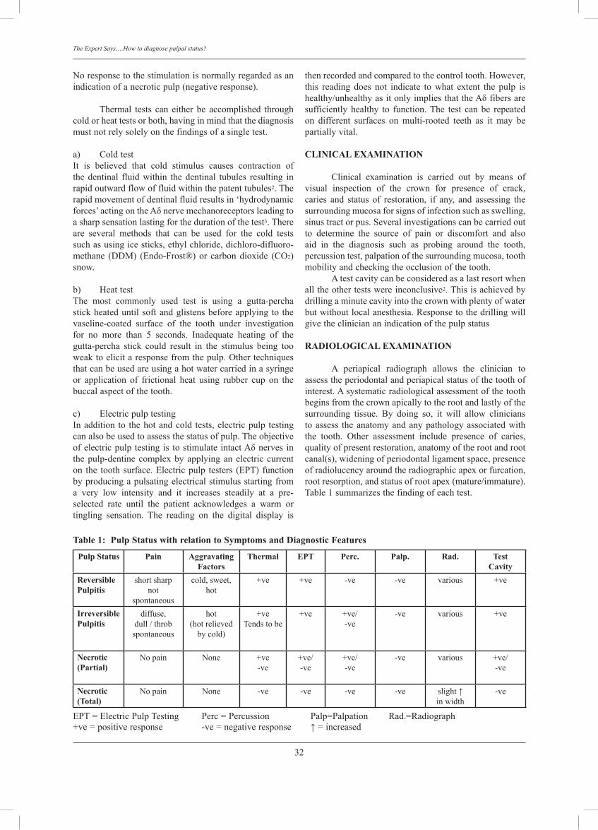
32
The Expert Says….How to diagnose pulpal status?
Noresponsetothestimulationisnormallyregardedasanindicationofanecroticpulp(negativeresponse).
Thermal tests can either be accomplished throughcoldorheattestsorboth,havinginmindthatthediagnosismustnotrelysolelyonthefindingsofasingletest.
a) ColdtestIt is believed that cold stimulus causes contraction ofthedentinal fluidwithin thedentinal tubules resulting inrapidoutwardflowoffluidwithinthepatenttubules2.Therapidmovementofdentinalfluidresultsin‘hydrodynamicforces’ acting on the Aδ nerve �echanoreceptors leading to asharpsensationlastingforthedurationofthetest3.Thereare several methods that can be used for the cold testssuch as using ice sticks, ethyl chloride, dichloro-difluoro-�ethane (DDM) (Endo-Frost®) or carbon dioxide (CO2)snow.
b) Heattest�he �ost co��only used test is using a gutta-percha stickheateduntilsoftandglistensbeforeapplyingto thevaseline-coated surface of the tooth under investigation for no more than 5 seconds. Inadequate heating of thegutta-percha stick could result in the sti�ulus being too weak to elicit a response fro� the pulp. Other techniques thatcanbeusedareusingahotwatercarriedinasyringeor application of frictional heat using rubber cup on thebuccalaspectofthetooth.
c) ElectricpulptestingInaddition to thehotandcold tests,electricpulp testingcanalsobeusedtoassessthestatusofpulp.Theobjectiveof electric pulp testing is to sti�ulate intact Aδ nerves in the pulp-dentine co�plex by applying an electric current on the toothsurface.Electricpulp testers (EPT) functionbyproducingapulsatingelectricalstimulusstartingfroma very low intensity and it increases steadily at a pre-selected rate until the patient acknowledges a warm ortingling sensation. The reading on the digital display is
thenrecordedandcomparedtothecontroltooth.However,this reading does not indicate to what extent the pulp ishealthy/unhealthy as it only i�plies that the Aδ fibers are sufficiently healthy to function.The test can be repeatedon different surfaces on �ulti-rooted teeth as it �ay be partiallyvital.
cLInIcAL EXAMInATIon Clinical examination is carried out by means ofvisual inspection of the crown for presence of crack,caries and status of restoration, if any, and assessing thesurroundingmucosaforsignsofinfectionsuchasswelling,sinustractorpus.Severalinvestigationscanbecarriedoutto determine the source of pain or discomfort and alsoaid in the diagnosis such as probing around the tooth,percussiontest,palpationofthesurroundingmucosa,toothmobilityandcheckingtheocclusionofthetooth. Atestcavitycanbeconsideredasalastresortwhenalltheothertestswereinconclusive2.Thisisachievedbydrillingaminutecavityintothecrownwithplentyofwaterbutwithoutlocalanesthesia.Responsetothedrillingwillgivethecliniciananindicationofthepulpstatus
rAdIoLoGIcAL EXAMInATIon A periapical radiograph allows the clinician toassesstheperiodontalandperiapicalstatusofthetoothofinterest.Asystematicradiologicalassessmentofthetoothbeginsfromthecrownapicallytotherootandlastlyofthesurrounding tissue. By doing so, it will allow clinicianstoassess theanatomyandanypathologyassociatedwiththe tooth. Other assess�ent include presence of caries, qualityofpresentrestoration,anatomyoftherootandrootcanal(s),wideningofperiodontalligamentspace,presenceofradiolucencyaroundtheradiographicapexorfurcation,rootresorption,andstatusofrootapex(mature/immature).Table1summarizesthefindingofeachtest.
Pulp Status Pain AggravatingFactors
Thermal EPT Perc. Palp. rad. Testcavity
reversible Pulpitis
shortsharpnot
spontaneous
cold,sweet,hot
+ve +ve -ve -ve various +ve
Irreversible Pulpitis
diffuse,dull/throbspontaneous
hot(hotrelieved
bycold)
+veTendstobe
+ve +ve/-ve
-ve various +ve
necrotic (Partial)
Nopain None +ve-ve
+ve/-ve
+ve/-ve
-ve various +ve/-ve
necrotic (Total)
Nopain None -ve -ve -ve -ve slight ↑inwidth
-ve
EPT=ElectricPulpTesting Perc=Percussion Palp=PalpationRad.=Radiograph+ve = positive response -ve = negative response ↑ = increased
Table 1: Pulp Status with relation to Symptoms and diagnostic Features
33
Safura
concLuSIonS The determination of pulp vitality requires acomprehensiveinvestigationanditsinterpretationdependsuponacombinationofmanyfactors.Anunderstandingofboththeusefulnessandlimitationsofpulptestingmethodsisessentialforitseffectiveuseinclinicaldentistry.
rEFErEncES
1. Chambers IG.The roleandmethodsofpulp testing inoraldiagnosis: a review. Int Endod J. 1982;15:1-5
2. Rowe A, Pitt Ford �. �he assess�ent of pulp vitality. Int Endod J. 1990;23:77-83
3. Brannstrom M.The hydrodynamic theory of dentinal pain:sensation in preparations, caries and the dentinal cracksyndro�e. J Endod. 1986; 12: 52-57
4. Rickoff B, �rowbridge H, Baker J, Fuss Z, Bender IB. Effects of thermal vitality tests on human dental pulp. J Endod.1988;14:482-485
5. Brannstrom M.The hydrodynamic theory of dentinal pain:sensationinpreparations,cariesanddentinalcracksyndrome.J Endod. 1986;12:453-457
6. �rowbridge HO, Franks M, Korostoff E, E�ling R. Sensory response to thermal stimulation in human teeth. J Endod.1980;6:405-412
7. Gopikrisna V, �inagupta K, Kandaswa�y D. Co�parison of electrical, thermal and pulse oximetry methods forassessingpulpvitalityinrecentlytraumatizedteeth.JEndod.2007;33(5):531-535
8. Petersson K, Soderstro� C, Kiani-Anaraki M, Levy G. Evaluation of the ability of thermal and electrical tests toregister pulp vitality. Endod Dent �rau�atol. 1999;15(3):127-31
9. Bhaskar SN, Rappaport HM. Dental vitality tests and pulpstatus. J A� Dent Assoc. 1973;86:409-411
10.Ehrman EH. Pulp testers and pulp testing with particularreference to the use of dry ice. Aust Dent J. 1977;22:272-279
11. Kahan RS, Gulabivala K, Snook M, Setchell DJ. Evaluation of a pulse oximeter and customized probe for pulp vitalitytesting. J Endod. 1996;22:105-9.
12. Schnettler JM, Wallace JA. Pulse oxi�etry as a diagnostic tool of pulp vitality. J Endod. 1991;17:488-90
Address for correspondence:
SAFurA AnITA BAHArIn DDS (Canada), MClinDent in Endodontology (London), MFDSRCS (Eng), DipCDSc (London)LecturerDepartment of Operative Dentistry,Faculty of Dentistry,Universiti Kebangsaan Malaysia,Jalan Raja Muda Abdul Aziz,Kuala Lumpur MalaysiaTel: 603-92897759E-mail: [email protected]
Effect Of Beverages And Food Source On Wear Resistance Of Composite Resins Seow LL. BDS (Malaya), MSc (London), FDSRCS (England), PhD (Malaya), FICD. Associate Professor, School of Dentistry, International Medical University, Kuala Lumpur, Malaysia
Chong SY, Lau MN, Tiong SG, Yew CC. Final year student 2008. Faculty of Dentistry, University of Malaya, 50603 Kuala Lumpur, Malaysia
ABSTRACT Certain beverages e.g. coffee, tea, soft drinks, fruit juices, alcoholic beverages, may affect the physical properties of composite resins. Objectives: The objectives of this study were to: (1) evaluate the effect of different beverages and chilli sauce on the wear resistance of composite resins, (2) evaluate effect of the duration of immersion in the beverages and chilli sauce on the wear resistance of composite resins. Materials and methods: Disc specimens were fabricated using two different types of composite resins: (i) Filtek Z350 (3M ESPE, USA, nano-filled composite, 40 specimens) and (ii) Solare P (GC Dental Products Corp, Japan, microhybrid composite, 40 specimens). After polymerization, all the specimens were polished using Enhance Polishing System (Dentsply International Inc.,USA). The specimens were air-dried before weighing using Sartorius BP 221S weighing balance (Sartorius AG, Goettingen, Germany). Ten specimens from each type of composite were immersed in distilled water (control group), Coca cola®, orange juice (Peel Fresh®) and chilli sauce (Maggi®) respectively. The duration of immersion was 6 hours and 1 week. A reciprocal compression-sliding system was used to evaluate the wear resistance of the specimens. The specimens were moved back and forth with a loaded counter-body (235g) against sand paper (P1000, 3M ESPE, USA) in running water. The weight of the specimens were measured after 6 hours of immersion and 20,000 wear cycles and also at 1 week of immersion with further 20,000 wear cycles. The wear resistances were tabulated as percentage of weight loss from the specimens. Results were statistically analyzed using one way ANOVA and post-hoc Tukey’s test (p= 0.05). Results: The results showed that Solare P has significantly lower wear resistance compared to Filtek Z350. There was no significant difference in wear resistance for Filtek Z350 when immersed in chili sauce, Coca-cola® and orange juice in comparison with control group for 6 hours and 1 week. Similar findings were observed for Solare P. Conclusion: Within the limitations of this study, it was concluded that Solare P has poorer wear resistance than Filtek Z350. The soaking medium investigated and duration of immersion have no influence on the wear resistance of Solare P and Filtek Z350.
34
Malaysian Dental Journal (2008) 29(1) 34-39© 2008 The Malaysian Dental Association
INTRODUCTION
Owing to aesthetic demand, the advantage ofminimal invasive preparation1 and ability to strengthentherestoredteeth2,compositeresinhasgainedpopularityas a restorative material for posterior teeth. Concern formercury toxicity is not an issue with composite resinin comparison with amalgam. It has therefore becomean important substitute for amalgam. It is an excellentalternative to amalgam for small to medium cavitieswhere minimal invasive preparation techniques1 can beapplied,theabilitytoadheretotoothstructureviaadhesivetechniquehas ruledout theneed tocreateundercut, thuspreservingsoundtoothstructure. Wearofamaterialinvolvesvariousprocessessuchasabrasion,erosionandfatigue;henceitisoneoftheleast
understoodproperty3,4.Preliminaryinvitro investigationsfor the physical properties of a material such as wearcharacteristics, bond strengths, flexural strengths andfatiguebehaviorismandatorytoscreenthematerialpriortoclinicalusage5. Consumptionofcertainbeverages,suchascoffee,tea, softdrinks, fruit juices andalcoholicbeveragesmayaffect theaestheticsandphysicalpropertiesofcompositeresins,henceaffectingtheclinicallifespanofthecompositerestorations6,7,8,9.Sarrettetal.hasshownthatowingtothelowpH,ethanolhas thepotential toproduceerosionandaltersomemechanicalpropertiesofcompositeresins9.Thewear resistance of composite resins decreased with theincrease in thepercentageofethanol in thesolution.Theeffects of beverages of the properties of composite werealsorelatedtothefrequencyandamountofitsintake.
MALAYSIAN DENTAL JOURNAL
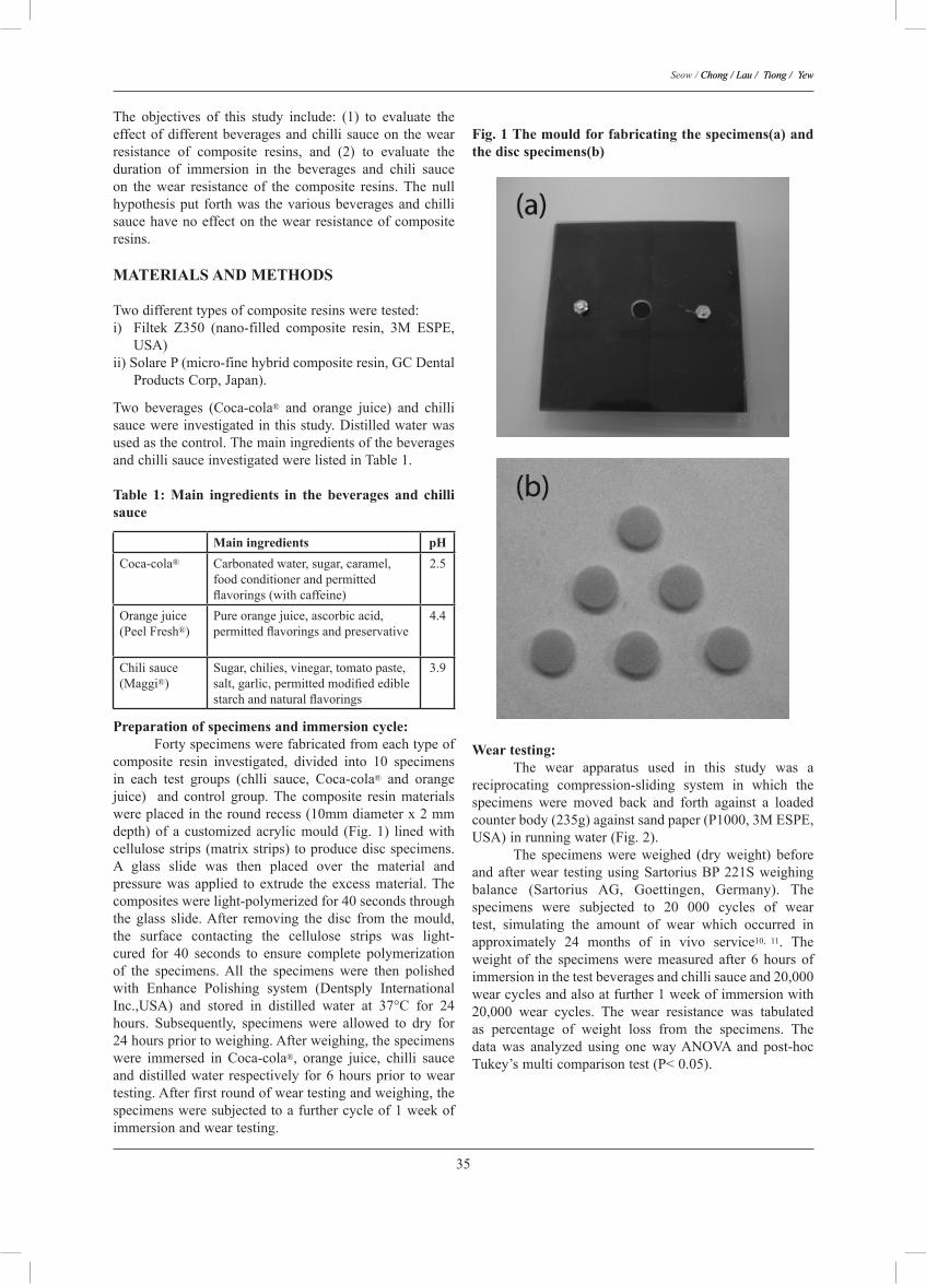
35
Seow / Chong / Lau / Tiong / �ewChong / Lau / Tiong / �ew
The objectives of this study include: (1) to evaluate theeffectofdifferentbeveragesandchillisauceonthewearresistance of composite resins, and (2) to evaluate theduration of immersion in the beverages and chili sauceon the wear resistance of the composite resins. The nullhypothesisput forthwas thevariousbeveragesandchillisaucehavenoeffectonthewearresistanceofcompositeresins.MATERIALS AND METHODS
Twodifferenttypesofcompositeresinsweretested:i) Filtek Z350 (nano-filled composite resin, 3M ESPE,
USA)ii)SolareP(micro-finehybridcompositeresin,GCDental
ProductsCorp,Japan).
Two beverages (Coca-cola® and orange juice) and chillisaucewereinvestigatedinthisstudy.Distilledwaterwasusedasthecontrol.ThemainingredientsofthebeveragesandchillisauceinvestigatedwerelistedinTable1.
Table 1: Main ingredients in the beverages and chilli sauce
Main ingredients pHCoca-cola® Carbonatedwater,sugar,caramel,
foodconditionerandpermittedflavorings (with caffeine)
2.5
Orangejuice(PeelFresh®)
Pureorangejuice,ascorbicacid,permitted flavorings and preservative
4.4
Chilisauce(Maggi®)
Sugar,chilies,vinegar,tomatopaste,salt, garlic, permitted modified edible starch and natural flavorings
3.9
Preparation of specimens and immersion cycle: Fortyspecimenswerefabricatedfromeachtypeofcomposite resin investigated, divided into 10 specimensin each test groups (chlli sauce, Coca-cola® and orangejuice) and control group.The composite resin materialswereplacedintheroundrecess(10mmdiameterx2mmdepth)of a customizedacrylicmould (Fig.1) linedwithcellulosestrips(matrixstrips)toproducediscspecimens.A glass slide was then placed over the material andpressurewas applied to extrude the excessmaterial.Thecompositeswerelight-polymerizedfor40secondsthroughthe glass slide.After removing the disc from the mould,the surface contacting the cellulose strips was light-cured for 40 seconds to ensure complete polymerizationof the specimens.All the specimens were then polishedwith Enhance Polishing system (Dentsply InternationalInc.,USA) and stored in distilled water at 37°C for 24hours. Subsequently, specimens were allowed to dry for24hourspriortoweighing.Afterweighing,thespecimenswere immersed in Coca-cola®, orange juice, chilli sauceanddistilledwater respectively for6hoursprior toweartesting.Afterfirstroundofweartestingandweighing,thespecimensweresubjectedtoafurthercycleof1weekofimmersionandweartesting.
Fig. 1 The mould for fabricating the specimens(a) and the disc specimens(b)
Wear testing: The wear apparatus used in this study was areciprocating compression-sliding system in which thespecimens were moved back and forth against a loadedcounterbody(235g)againstsandpaper(P1000,3MESPE,USA)inrunningwater(Fig.2). The specimens were weighed (dry weight) beforeandafterwear testingusingSartoriusBP221Sweighingbalance (Sartorius AG, Goettingen, Germany). Thespecimens were subjected to 20 000 cycles of weartest, simulating the amount of wear which occurred inapproximately 24 months of in vivo service10, 11. Theweight of the specimens were measured after 6 hours ofimmersioninthetestbeveragesandchillisauceand20,000wearcyclesandalsoatfurther1weekofimmersionwith20,000 wear cycles. The wear resistance was tabulatedas percentage of weight loss from the specimens. Thedata was analyzed using one wayANOVA and post-hocTukey’smulticomparisontest(P<0.05).
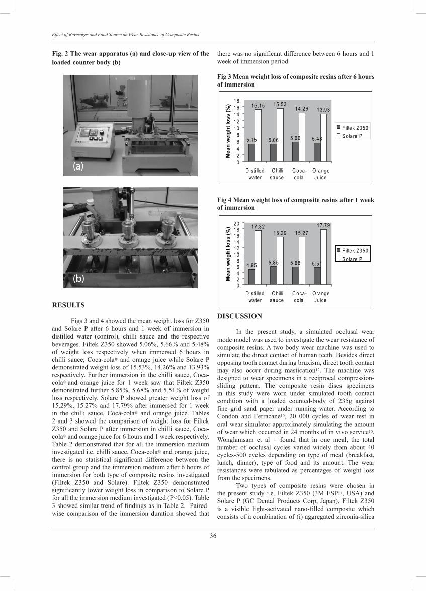
36
Effect of Beverages and Food Source on Wear Resistance of Composite Resins
Fig. 2 The wear apparatus (a) and close-up view of the loaded counter body (b)
RESULTS
Figs3and4showedthemeanweightlossforZ350and Solare P after 6 hours and 1 week of immersion indistilled water (control), chilli sauce and the respectivebeverages.FiltekZ350showed5.06%,5.66%and5.48%of weight loss respectively when immersed 6 hours inchilli sauce, Coca-cola® and orange juice while Solare Pdemonstratedweightlossof15.53%,14.26%and13.93%respectively.Furtherimmersioninthechillisauce,Coca-cola® and orange juice for 1 week saw that Filtek Z350demonstratedfurther5.85%,5.68%and5.51%ofweightloss respectively.SolareP showedgreaterweight lossof15.29%,15.27%and17.79%after immersed for 1weekin the chilli sauce, Coca-cola® and orange juice. Tables2and3showed thecomparisonofweight loss forFiltekZ350andSolarePafter immersioninchillisauce,Coca-cola®andorangejuicefor6hoursand1weekrespectively.Table2demonstrated that for all the immersionmediuminvestigatedi.e.chillisauce,Coca-cola®andorangejuice,there is no statistical significant difference between thecontrolgroupandtheimmersionmediumafter6hoursofimmersionforboth typeofcomposite resins investigated(Filtek Z350 and Solare). Filtek Z350 demonstratedsignificantlylowerweightlossincomparisontoSolarePforalltheimmersionmediuminvestigated(P<0.05).Table3showedsimilartrendoffindingsasinTable2.Paired-wise comparison of the immersion duration showed that
therewasnosignificantdifferencebetween6hoursand1weekofimmersionperiod.
Fig 3 Mean weight loss of composite resins after 6 hours of immersion
5 .15 5 .06 5 .66 5 .48
15 .15 15 .5314 .26 13 .93
02468
1012141618
D isti lledwa te r
C hillisauce
C oca-co la
OrangeJuice
Mea
n w
eigh
t los
s (%
)
F iltek Z350S o la re P
4 .95 5 .85 5 .68 5 .51
17 .3215 .29 15 .27
17 .79
02468
101214161820
D isti lledwa te r
C hillisauce
C oca-co la
OrangeJuice
Mea
n w
eigh
t los
s (%
)
F iltek Z350S o la re P
Fig 4 Mean weight loss of composite resins after 1 week of immersion
5 .15 5 .06 5 .66 5 .48
15 .15 15 .5314 .26 13 .93
02468
1012141618
D isti lledwa te r
C hillisauce
C oca-co la
OrangeJuice
Mea
n w
eigh
t los
s (%
)
F iltek Z350S o la re P
4 .95 5 .85 5 .68 5 .51
17 .3215 .29 15 .27
17 .79
02468
101214161820
D isti lledwa te r
C hillisauce
C oca-co la
OrangeJuice
Mea
n w
eigh
t los
s (%
)
F iltek Z350S o la re P
DISCUSSION In the present study, a simulated occlusal wearmodemodelwasusedtoinvestigatethewearresistanceofcomposite resins.A two-bodywearmachinewasused tosimulatethedirectcontactofhumanteeth.Besidesdirectopposingtoothcontactduringbruxism,directtoothcontactmay also occur during mastication12. The machine wasdesigned towearspecimens ina reciprocalcompression-sliding pattern. The composite resin discs specimensin this study were worn under simulated tooth contactcondition with a loaded counted-body of 235g againstfine grid sand paper under running water. According toCondon and Ferracane10, 20 000 cycles of wear test inoralwearsimulatorapproximatelysimulatingtheamountofwearwhichoccurredin24monthsofinvivoservice10.Wonglamsam et al 11 found that in one meal, the totalnumber of occlusal cycles varied widely from about 40cycles-500 cycles depending on type of meal (breakfast,lunch, dinner), type of food and its amount. The wearresistances were tabulated as percentages of weight lossfromthespecimens. Two types of composite resins were chosen inthe present study i.e. Filtek Z350 (3M ESPE, USA) andSolareP (GCDentalProductsCorp, Japan).FiltekZ350is a visible light-activated nano-filled composite whichconsistsofacombinationof(i)aggregatedzirconia-silica
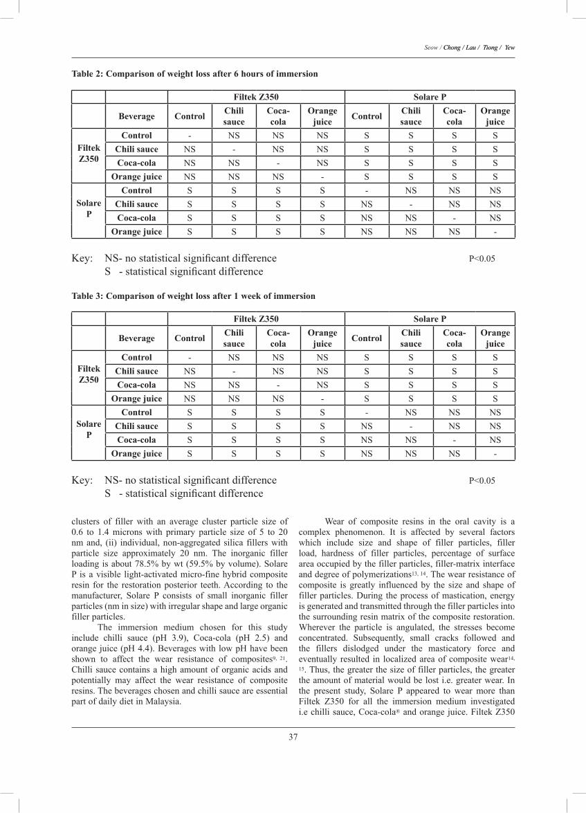
37
Seow / Chong / Lau / Tiong / �ewChong / Lau / Tiong / �ew
clusters of filler with an average cluster particle size of0.6 to 1.4 microns with primary particle size of 5 to 20nmand, (ii) individual,non-aggregated silica fillerswithparticle size approximately 20 nm. The inorganic fillerloadingisabout78.5%bywt(59.5%byvolume).SolareP isavisible light-activatedmicro-finehybridcompositeresin for the restorationposterior teeth.According to themanufacturer, Solare P consists of small inorganic fillerparticles(nminsize)withirregularshapeandlargeorganicfillerparticles. The immersion medium chosen for this studyinclude chilli sauce (pH 3.9), Coca-cola (pH 2.5) andorangejuice(pH4.4).BeverageswithlowpHhavebeenshown to affect the wear resistance of composites9, 21.Chillisaucecontainsahighamountoforganicacidsandpotentially may affect the wear resistance of compositeresins.ThebeverageschosenandchillisauceareessentialpartofdailydietinMalaysia.
Wear of composite resins in the oral cavity is acomplex phenomenon. It is affected by several factorswhich include size and shape of filler particles, fillerload, hardness of filler particles, percentage of surfaceareaoccupiedbythefillerparticles,filler-matrixinterfaceanddegreeofpolymerizations13, 14.Thewearresistanceofcomposite is greatly influenced by the size and shape offillerparticles.During theprocessofmastication,energyisgeneratedandtransmittedthroughthefillerparticlesintothesurroundingresinmatrixofthecompositerestoration.Wherever the particle is angulated, the stresses becomeconcentrated. Subsequently, small cracks followed andthe fillers dislodged under the masticatory force andeventually resulted in localizedareaofcompositewear14,
15.Thus,thegreaterthesizeoffillerparticles,thegreatertheamountofmaterialwouldbelosti.e.greaterwear.Inthe present study, Solare P appeared to wear more thanFiltek Z350 for all the immersion medium investigatedi.echillisauce,Coca-cola®andorangejuice.FiltekZ350
Table 2: Comparison of weight loss after 6 hours of immersion
Filtek Z350 Solare P
Beverage Control Chili sauce
Coca-cola
Orange juice Control Chili
sauceCoca-cola
Orange juice
Filtek Z350
Control - NS NS NS S S S SChili sauce NS - NS NS S S S SCoca-cola NS NS - NS S S S S
Orange juice NS NS NS - S S S S
Solare P
Control S S S S - NS NS NSChili sauce S S S S NS - NS NSCoca-cola S S S S NS NS - NS
Orange juice S S S S NS NS NS -
Key: NS- no statistical significant difference P<0.05 S - statistical significant difference
Table 3: Comparison of weight loss after 1 week of immersion
Filtek Z350 Solare P
Beverage Control Chili sauce
Coca-cola
Orange juice Control Chili
sauceCoca-cola
Orange juice
Filtek Z350
Control - NS NS NS S S S SChili sauce NS - NS NS S S S SCoca-cola NS NS - NS S S S S
Orange juice NS NS NS - S S S S
Solare P
Control S S S S - NS NS NSChili sauce S S S S NS - NS NSCoca-cola S S S S NS NS - NS
Orange juice S S S S NS NS NS -
Key: NS- no statistical significant difference P<0.05 S - statistical significant difference
38
Effect of Beverages and Food Source on Wear Resistance of Composite Resins
is a nano-filled composite with filler particles smallerthanSolareP.The fillerparticles inSolareP is irregularinsize, thereforemoresusceptibletostressconcentrationandcrackpropagation,leadingtothefillersbeingpluckedoutbyocclusalwear.Similarobservationwithregardstothe low wear rate of nano-filled composite in was alsoobservedinotherinvitrostudies16, 17. Surface quality of composite resin may affect thewear resistance of the restorations. A relatively smoothsurfacethatisvoidofsurfaceprotrusionhastheabilitytolowerthefrictionforceandsubsequentlylowerwearloss18.After polishing, composites with larger filler particlestend to produce a polished surface with higher surfaceroughness than those with smaller filler particles19, 20. Ithas been shown that composite resin with nano-fillerswas able to produce superior surface finishing16. Thismayexplain thefindingwhereFiltekZ350,anano-filledcompositeshowssignificantlylowerwearlossthanSolareP,amicrohybridcompositeswherethefillerparticleswerelarger. An earlier study demonstrated that organic acidscausedadecreaseinthehardnessofresincomposite21. Itmay then be speculated that acidic medium would havean effect on the wear resistance of composite resins. Itis, therefore, theaimof thepresent study toevaluate theeffect of acidic beverages and food on composite resins.The present study has shown that chilli sauce, Coca-cola and orange juice did not have significant effect onthe wear resistance of the two types of composite resinsinvestigated. Draughn and Harrison22 have shown thattherewasnodirectcorrelationbetweenabrasivewearandhardness,themechanismsinvolveforthesetwotestmodesisdifferent. One explanation for this finding was that theobservedreduction insurfacehardness isconfined to theouterexposedsuperficialsurfaceofthespecimens.Duringthewear test, thesoftenedsuperficialsurface is removedquickly, and a fresh, relatively hard surface is thenexposed,whichwouldtakealongertimetowearoff.Thiswouldnotadverselyaffecttheoverallwearfactortoagreatnoticeable extent if the weight loss due to the softenedsurface layer was only a small proportion23. Therefore,though the acidic medium affects the surface hardness,oncethesurfaceisbeingwornoff,thewearofinnerhardlayer was not affected to a significant extent. This mayalsoexplainthatthedurationofimmersionof6hoursand1weekhasnosignificanteffectonthewearresistanceofthecompositesinvestigatedinthepresentstudy. Althoughtheeffectofacidicbeveragesandfoodisnotsignificantinthepresentinvitroweartesting,itmayhave effect when a cyclical test model is employed i.eto simulate in the invivoconditionwhere thecompositerestorationswillbeconstantlysubjectedtoviciouscycleofbeingexposedtoacidicbeverages/foodandocclusalwearforces.Once the superficial layerbecomessoftened, it ismoresusceptibletoabrasionanduponitsremoval,afreshsurfacebecomesexposed.Thismay thenbe subjected tothe same softening process in the oral environment.Theoverall effectwouldbemanifest as aprogressive lossofsubstanceoftherestoration.
CONCLUSIONS Within the limitations of the present study, it canbeconcludedthatSolarePwearssignificantlymore thanFiltek Z350. The immersion medium investigated in thepresentstudyi.e.chillisauce,Coca-cola®andorangejuicehasnosignificanteffecton thewear resistanceofSolareP and Filtek Z350. The duration of immersion does notadverselyaffectthewearresistanceofSolarePandFiltekZ350. Posterior restorations are subjected to substantialocclusalloadandocclusalwear,caremustbeexecutedincaseselection,materialselectionandproper technique toensurelongtermclinicalsuccessfortherestorations.
REFERENCES
1. EricsonD,KiddE,McCombD,MjorE,NoackMJ.Minimallyinvasive dentistry – Concepts and techniques in cariology.OralHealthPreventDent2003;1:59-72.
2. Arola D, Galles LA, Sarubin MF. A comparison of themechanical behavior of posterior teeth with amalgam andcompositeMODrestorations.JDent2001;29:63-73.
3. Momoi Y, Hirosaki K, Kohno A, McCabe JF. In vitrotoothbrush-dentifrice abrasion of resin-modified glassionomers.DentMater1997;13:82-88.
4. Kanter J, Koski RE, Martin D. The relationship of weightlosstosurfaceroughnessofcompositeresinsfromsimulatedtoothbrushing.JProsthetDent1982;47:505-513.
5. Roland F, Franklin GG, Ulrich L, Anselm P, Norbert K.Evaluation of resin composite materials. Part I: In vitroinvestigationsAmJofDent2005;18(1):23-27.
6. DietschiK,CampanileG,HolzJ,MeyerJM.Comparisonofthe color stabilityof tennew-generationcomposites:An invitrostudy.DentMater1994;10(6):353-362.
7. Lim BS, Moon HJ, Baek KW, Hahn SH, Kim CW. Colorstabilityofglassionomersandpolyacid-modifiedresin-basedcomposites in various environmental solutions.Am J Dent2001;14(4):241-246.
8. Wiltshire WA, Labuschagne PW. Staining of light-curedaesthetic resin restorative materials by different stainingmedia:Aninvitrostudy.JDentAssocofSouthAfrica1990;45(12):561-565.
9. SarrettDC,ColettiDP,PelusoAR.Theeffectsofalcoholicbeveragesoncompositewear.DentMater2000;16(1):62-67.
10.CondonJR,FerracaneJL.Evaluationofcompositewearwithanewmulti-modeoralwearsimulator.DentMater1996;12:218-223.
11.WonglamsamA,KakutaK,OguraH.Effectsofocclusalandbrushing cycles on wear of composite resins in combinedweartest.DentMaterJ2008;27(2):243-250.
12.AdamSH,ZanderHA.Functionaltoothcontactsinlateralandincentricocclusion.JAmDentAssoc1964;69:465-473.
13. Erica CNT, Jennifer LT, Jeffrey RP, Jeffrey YT. In vitrotoothbrush-dentifriceabrasionoftworestorativecomposites.JEsthetandRestorDent2005;17:172-182.
14.BayneBC,HeymannHO,SwiftEJ.Updateofdentalcompositerestorations.JAmDentAssoc1994;125:687-693.
15. Leinfelder KF, Wilder JAD, Teixeira LC. Wear rates ofposteriorcompositeresins.JAmDentAssoc1986;112:829-835.
39
Seow / Chong / Lau / Tiong / �ewChong / Lau / Tiong / �ew
16.Sumita BM, Dong W, Brian NH. An application ofnanotechnology in advanced dental materials. J Am DentAssoc2003;134:1382-1390.
17. Yap AUJ, Tan CH, Chung SM. Wear behavior of newcompositerestoratives.OperDent2004;29(3):269-274.
18. Yap AUJ. Occlusal contact area(OCA) wear of two newcompositerestoratives.JOralRehab2002;29:194-200.
19.AnthonyHL,DentDR,ClaytonA.Chan.Thepolishabilityofposteriorcomposites.JProsthetDent1989;61(2):138-146.
20.JungM,SehrK,KlimekJ.Surfacetextureoffournanofilledandonehybrid composite after finishing.OperDent2007;32(1):45-52
21. Consani S, Goes MF. Effect of acids on resin composites:Invitrostudyontheeffectoforganicacidsonthehardnessand surface roughness of composites. Revista Gaucha deOdontalgia1998;46(4):201-204.
22.Draughn RA, Harrison A. Relationship between abrasivewearandmicrostructureofcompositeresins.JProsthetDent1978;40:220-224.
23.ChadwickRG.Theeffectofstoragemediauponthesurfacemicrohardnessandabrasionresistanceofthreeecomposites.DentMater1990;6:123-128.
Address for correspondence:
Associate Professor Dr. Seow Liang LinSchool of Dentistry, International Medical University,126, Jalan 19/155B, Bukit Jalil57000, Kuala Lumpur, MalaysiaTel : 603-8656 7228Fax : 603-8656 7229email: [email protected]
Essentials of Clinical Periodontology and Periodontics.Author : Shantipriya ReddyPublisher : Jaypee Brothers Medical Publishers (P) Ltd., New DelhiYear of publication : 2006
Essential of Clinical Periodontology and Periodontics, written by Professor Shantipriya Reddy, gives the reader excellent fundamental knowledge in periodontology. With more than a decade experience in academics, Professor Reddy knew exactly what scholars in this field require from a textbook. The book with 421 pages covered broad topics from the normal anatomy of the periodontium, periodontal diseases and finally to the treatment of periodontal diseases. The book began with a prolog on the history of periodontology in the early civilization until present. Part I of the book introduced the periodontal structures anatomically and physiologically, Part II discussed the classification systems and epidemiology of periodontal diseases. As the content increased, it discussed further into the subject of the aetiopathology (Part III), periodontal pathology (Part IV) and treatment of periodontal disease (Part V). The last part of the book was neatly divided into two sections; Section 1: Diagnosis, Prognosis and Treatment Plan and Section 2: Periodontal Therapy (Non-surgical and Surgical Therapy). The reader will appreciate the way this book was written and organised. Diagrams and tables are included systematically according to the relevant topics. Illustrations were made easy to understand, with labels to help summarized the text. This book contains many clinical photos but perhaps could be more appropriate and enhancing if neatly cropped to focus on certain relevant areas only. Nevertheless, the arrangement of these photos was indeed helpful to show step-by-step procedures. At the end of each chapter, the author cleverly included some review questions for the reader to self-assessed their reading for that particular topics. Bibliography of key references ended each chapter and will assist the reader to seek more information on the topics, if needed. The reviewer congratulates Professor Reddy and the publisher for this excellent book and recommends it to be included in dental practitioners’ booklist. Although this book does not provide exhaustive coverage of current knowledge in periodontology, it can still be recommended for use of dental undergraduates as well as dental nurses for its simplicity.
Special features: an overview• 421 pages of text and photos• 5 essential parts with 50 relevant chapters • a special chapter on Questionnaire for clinical case discussion • Fonts are big, clear and organised to hearten reading• Key Points To Note in some chapters to highlight important issues in the topics• clinical photos and illustrations to enhance further understanding of the text• Index for quick search on particular topics• comes with a photo CDROM
Reviewer:Dr. Shahida Mohd SaidCo-Editor,Malaysian Dental JournalEmail: [email protected]
40
Malaysian Dental Journal (2008) 29(1) 40© 2008 The Malaysian Dental Association
MALAYSIAN DENTAL JOURNAL
Relationship between patients’ perceptions and clinical indications for dental extraction in Mukah Division, Sarawak Ling XF. Dental Officer Oral Surgery Department, Kuala Lumpur General Hospital, Kuala Lumpur, Malaysia.
Chan JA. Assistant Chief Dental Director, Kuching, Sarawak, Malaysia.
ABSTRACT The general aim of this study was to examine the patients’ perception of dental extractions. The specific objectives were to understand the purpose of extractions from the patients’ point of view, to find out the relationship between age and type of teeth extracted and to compare the patients’ perceptions with clinical indications for dental extraction. The survey was done using self administered, structured questionnaires to collect information. From the survey we found that toothache (33%) and the presence of cavities (34%) were the main reasons for extraction from the patients’ point of view. Besides that, the number of patients requiring extractions decreased with age. The main clinical indication for tooth extraction was caries (73%) and of these, 29% of restorable teeth were extracted.
41
Malaysian Dental Journal (2008) 29(1) 41-45© 2008 The Malaysian Dental Association
InTRoDuCTIon
Tooth extraction was one of the most commonprocedures carried out in the two government dentalclinicsinMukahDivision,Sarawak;MukahDentalClinicandDalatDentalClinic.OfthepatientswhovisitedthesedentalclinicsinMay2006,59.4%hadtoothextractions1.InastudyamongMalaysianadultsbyRazakIA,41%ofthesubjectspreferredextractionswhenexperiencingtoothache2.AccordingtotheNationalOralHealthSurveyofAdults(NOHSA)2000,35.8%of theMalaysianpopulationwasassessed as requiring extractions 3.Of the teeth indicatedforextraction,themajority(93.8%)wereduetocaries5.
Thegeneralobjectiveofourstudywastoexaminethepatient’sperceptionondentalextraction.Thespecificobjectives were to understand the reasons for extractionfromthepatient’spointofview,tofindouttherelationshipbetweenageandtypeofteethextracted.Lastly,tocomparethe patients’ perceptions with the clinical indications fordentalextractions.
Patients and methods The survey was done using a self administered,structured questionnaire. The survey was carried outsimultaneously in both the government dental clinics inMay2006.ThequestionnairewastestedinapilotstudyinMukahDentalClinicinOctober2005.ThequestionnairewaswrittenintheMalaylanguage.Atotalof236patients
tookpartinthisstudy.Thesurveywascarriedoutonallpatientswhowere15yearsoldandabovewhorequestedforextractionofpermanentteeth.Demographicdatawererecorded (age, gender, race and education level). Thepatients were asked to answer three questions regardingtheirreasonsfordentalextraction.Thedentalofficerswereaskedtorecordthetoothextracted,whetherthetoothwasrestorableandtheclinicalindicationfordentalextraction.ThesameextractioncriteriawereusedandtheseareshowninTable1.Boththeexaminerswerecalibrated.
Table 1: Inclusion for simple restorative treatment
No Criteria1 • Painofveryshortduration
• Paindoesnotlingerafterthestimulushasbeenremoved
• Painisdifficulttolocalize2 • Toothisnottendertopercussion
• Cariesdoesnotinvolvethepulp3 • Toothisvital
• Nosignofirreversiblepulpitis4 • Noperiapicalpathology5 • Restorativetreatmentinthisstudyexclude
rootcanaltreatment
MALAYSIAn DEnTAL JouRnAL
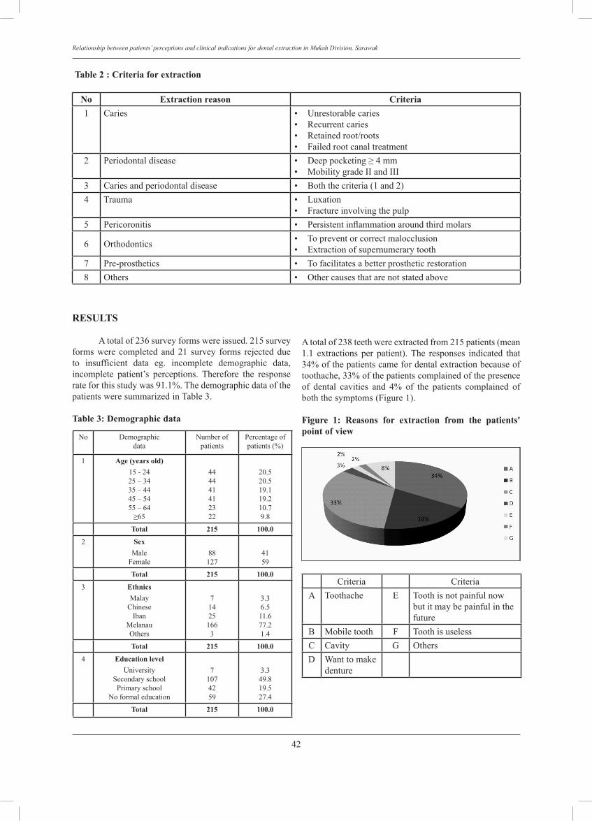
42
Relationship between patients’ perceptions and clinical indications for dental extraction in Mukah Division, Sarawak
Table 2 : Criteria for extraction
no Extraction reason Criteria1 Caries • Unrestorablecaries
• Recurrentcaries• Retainedroot/roots• Failedrootcanaltreatment
2 Periodontaldisease • Deeppocketing≥4mm• MobilitygradeIIandIII
3 Cariesandperiodontaldisease • Boththecriteria(1and2)4 Trauma • Luxation
• Fractureinvolvingthepulp5 Pericoronitis • Persistentinflammationaroundthirdmolars
6 Orthodontics • Topreventorcorrectmalocclusion• Extractionofsupernumerarytooth
7 Pre-prosthetics • Tofacilitatesabetterprostheticrestoration8 Others • Othercausesthatarenotstatedabove
RESuLTS
Atotalof236surveyformswereissued.215surveyformswere completed and21 survey forms rejecteddueto insufficient data eg. incomplete demographic data,incomplete patient’s perceptions. Therefore the responserateforthisstudywas91.1%.ThedemographicdataofthepatientsweresummarizedinTable3.
Table 3: Demographic data
No Demographicdata
Numberofpatients
Percentageofpatients(%)
1 Age (years old)15-2425–3435–4445–5455–64≥65
444441412322
20.520.519.119.210.79.8
Total 215 100.0
2 SexMale
Female88127
4159
Total 215 100.0
3 EthnicsMalay
ChineseIban
MelanauOthers
714251663
3.36.511.677.21.4
Total 215 100.0
4 Education levelUniversity
SecondaryschoolPrimaryschool
Noformaleducation
71074259
3.349.819.527.4
Total 215 100.0
Atotalof238teethwereextractedfrom215patients(mean1.1 extractions per patient).The responses indicated that34%ofthepatientscamefordentalextractionbecauseoftoothache,33%ofthepatientscomplainedofthepresenceof dental cavities and 4% of the patients complained ofboththesymptoms(Figure1).
Figure 1: Reasons for extraction from the patients' point of view
Criteria CriteriaA Toothache E Toothisnotpainfulnow
butitmaybepainfulinthefuture
B Mobiletooth F ToothisuselessC Cavity G OthersD Wanttomake
denture
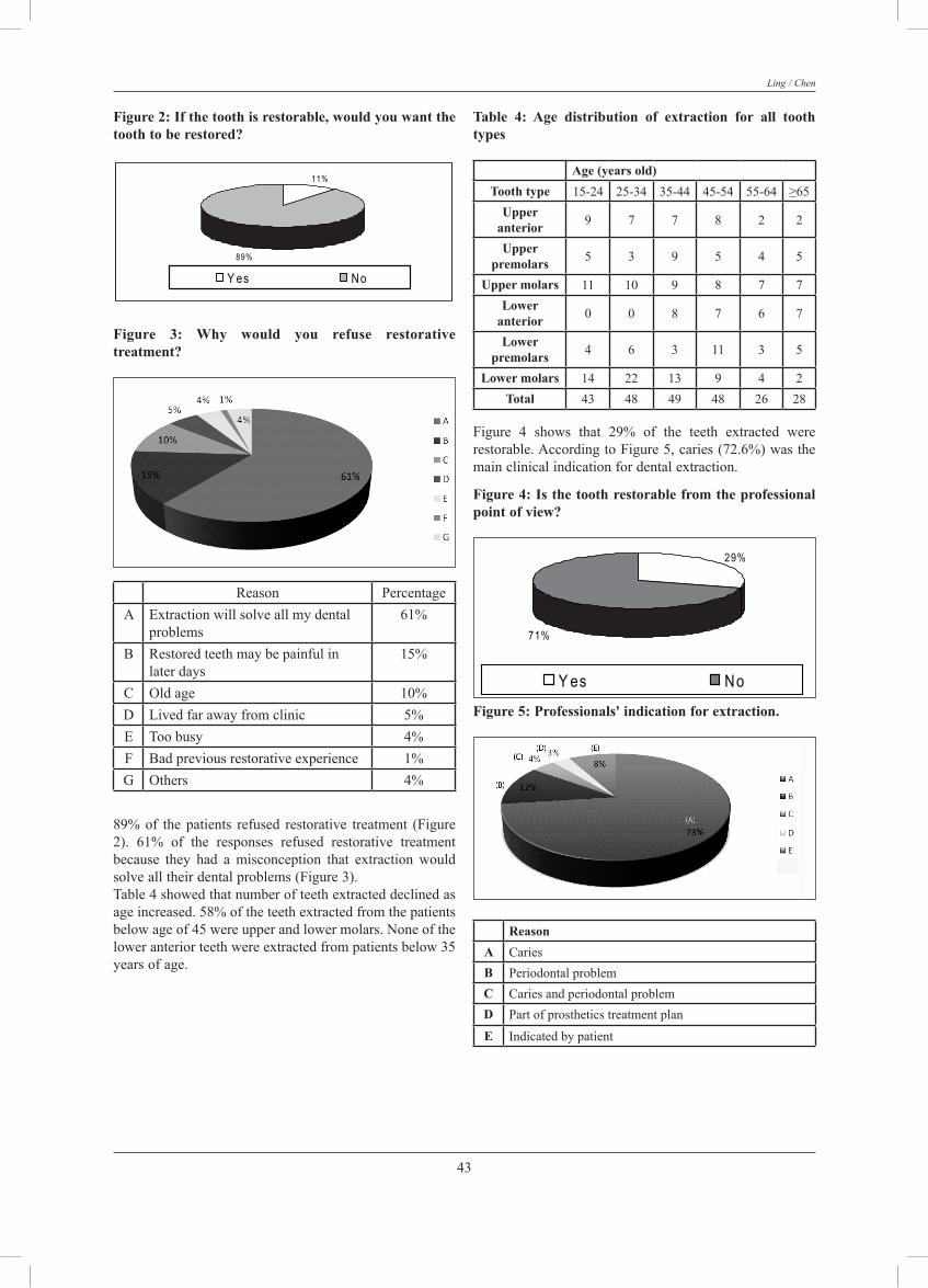
43
Ling / Chen
Figure 2: If the tooth is restorable, would you want the tooth to be restored?
11%
89%
Y es No
Figure 3: Why would you refuse restorative treatment?
Reason PercentageA Extractionwillsolveallmydental
problems61%
B Restoredteethmaybepainfulinlaterdays
15%
C Oldage 10%D Livedfarawayfromclinic 5%E Toobusy 4%F Badpreviousrestorativeexperience 1%G Others 4%
89%of thepatients refused restorative treatment (Figure2). 61% of the responses refused restorative treatmentbecause they had a misconception that extraction wouldsolvealltheirdentalproblems(Figure3).Table4showedthatnumberofteethextracteddeclinedasageincreased.58%oftheteethextractedfromthepatientsbelowageof45wereupperandlowermolars.Noneoftheloweranteriorteethwereextractedfrompatientsbelow35yearsofage.
Table 4: Age distribution of extraction for all tooth types
Age (years old)Tooth type 15-24 25-34 35-44 45-54 55-64 ≥65
upper anterior 9 7 7 8 2 2
upper premolars 5 3 9 5 4 5
upper molars 11 10 9 8 7 7Lower
anterior 0 0 8 7 6 7
Lower premolars 4 6 3 11 3 5
Lower molars 14 22 13 9 4 2Total 43 48 49 48 26 28
Figure 4 shows that 29% of the teeth extracted wererestorable.AccordingtoFigure5,caries(72.6%)wasthemainclinicalindicationfordentalextraction.
Figure 4: Is the tooth restorable from the professional point of view?
29%
71%
Y es No Figure 5: Professionals' indication for extraction.
ReasonA CariesB PeriodontalproblemC CariesandperiodontalproblemD Partofprostheticstreatmentplan
E Indicatedbypatient
44
Relationship between patients’ perceptions and clinical indications for dental extraction in Mukah Division, Sarawak
DISCuSSIon Certain features of study design have been foundto improve response rate and these were included wherepossible4 i.e.aclearandbriefquestionnaire,explanationof the questionnaire at every session and clarification ofevery enquiry.These factors contributed to the relativelyhighresponserate. The number of patients requiring extractionsdecreased with age, ranging from 20.6% in age group15-24 years to 9.8% at age 65 and above. 79.5% ofpatients requiring extractions were 55 years old andbelow.Thismaybedue to the fact thatolderpopulationwas mostly edentulous. According to NOHSA 2000,proportions of population requiring extractions increasedwith age, ranging from 14.8% in age group 15-19 yearsto69.2%among thoseabove75of age 3. Caremustbetaken,however,wheninterpretingtheresults,becausethisstudy only involved patients who attended dental mainclinic where as in NOHSA 2000, it involved the wholepopulation. Majorityofthepatientsinvolvedinthisstudywerefemale(59%).ThisdoesnotcorrespondwiththedatafromJabatanPerangkaanMalaysia,whichstatedthat45.7%ofMukah populations were females.5 This may be due tothe fact that, females were more dental health consciouscomparing tomales.77%of thepatients involved in thisstudywereMelanau.ThiscorrespondswiththedatafromJabatanPerangkaanMalaysia,whichstatedthat82%ofthepopulationconsistsofMelanau.5 From the patients’ perspective, the main reasonsfor tooth extraction were toothache and the presence ofcavities.Mostofthepatientsrefusedrestorativetreatmentbecause they had a misconception that extraction couldsolve all their dental problems. Dental health educationthat promoted teeth for life did not seem to reach theentire population because 27.4% of the patients who didnotundergoformaleducation.Ontheotherhand,despitebeingexposed to regularoralhealthcare throughout theirschoolyears,asizeableproportion(26.7%)of16yearoldsinSibu,reportedthattheywouldchoosetohavetheirteethextractediftheyhaddecayedorpainfulteeth.6 Surprisingly,highpercentagesofmolarteethwereextracted inpatientsbelowageof45yearsold.ReichEconcluded that the third molar was the most frequentlyextracted tooth type7 and Corbet EF, concluded that themost commonly extracted tooth type was the first molarand lower incisiors.8 None of the lower anterior teeth inthis study were extracted from patients below 35 yearsofageperhapsbecause theflushingeffectofsalivafromtheorificeofsubmandiubularductmadetheseteethmoreresistanttocaries. Caries (73%) was the main clinical indication fortooth extraction. In NOHSA 2000, of the teeth indicatedfor extraction, the majority (93.8%) was due to caries.3By comparison to developed nation, the proportion ofextractionsduetocarieswashigherthanthe50%reportedbyKayandBlinkhorninScotland9and48%reportedbyAgerholmandSidiinEnglandandWales10. The high percentage of extractions of restorableteeth(29%)showedthatthepatientswereconvincedthat
extractionswouldsolvealltheirdentalproblems(61.7%)andthepatientsfeltthatrestoredteethmaybepainfulinthe future (14.8%).Other reasons includeoldage (9.7%),living far away from the dental clinic (5.1%) and beingtoo busy for restorative treatment (4.1%). Extraction ofrestorableteethwasjustonefacetofanincreasingproblemof increasing demand for denture.According to NOHSA2000,theIbanshadthehighestprostheticneed(50.1%).11
None of the extractions in this study were dueto orthodontic reasons. This was because orthodonticprocedures were financially and technically demanding.Furthermore, there was no orthodontists in MukahDivision.InScotland,however,Chestnuttfoundthatmoreorthodonticextractionsandfewerextractionsduetocarieswere carried out in 1997 for patients under 21 years ofage12.
ConCLuSIonS
In conclusion, from this study, we found that thenumber of patients requiring extractions decreased withage. Besides that, molar teeth were the most commonlyextractedteethamongpatientsbelowtheageof45yearsold. From the patients’ perspective, the main reasonsfor tooth extraction were toothache and the presence ofcavities.Clinically,caries(73%)wasstillthemainclinicalindicationfortoothextraction,andwefoundthat29%ofrestorableteethwereextracted.
REFEREnCES
1. Sistem Maklumat Pengurusan Kesihatan, Laporan BulananBahagian Mukah Bagi Hasil Kerja Pegawai Pergigian, PG207,May2006.
2. RazakIA,JaafarN,JalalludinRL,EsaR.Patient’spreferenceforexodontiasversuspreventioninMalaysia.
3. National Oral Health Survey of Adults 2000. Oral healthstatus,impactsandtreatmentneedsofMalaysianadults.OralHealthDivisionMinistryofHealthMalaysia:2004;55.
4. TanRT,BurkeFJT.Responseratestoquestionnairesmailedtodentists.Areviewof77publications.InternationalDentalJournal1997:47:349-354.
5. DataextractedfromJabatanPerangkaanMalaysia/JP/SWK/691/2/7Klt5(65),9May2003.
6. ChenJA,EddyA,ChiaJC.Perceptionoforalhealthamong16yearoldschoolchildreninSibu,Sarawak2003;3.
7. Reich E, Hiller KA. Reasons for tooth extraction inwestern states of Germany.Community Dentistry and OralEpidemiology1993;21:379-383.
8. CorbetEF,DaviesWIR.ReasonsgivenfortoothextractioninHongKong.CommunityDentalHealth1991;8:121-130.
9. KayEJ,BlinkhornAS.ThereasonsunderlyingtheextractionofteethinScotland.BrDentJ1986;160:287-290.
10.Agerholm DM, Sidi AD. Reasons given for extraction ofpermanent teeth by general dental practitioners in EnglandandWales.BrDentJ1988;164:345-348.
11.National Oral Health Survey of Adults 2000. Oral healthstatus,impactsandtreatmentneedsofMalaysianadults.OralHealthDivisionMinistryofHealthMalaysia:2004;60.
12.Chestnutt I, Binnie VI, Taylor MM.Reasons for toothextraction in Scotland (short communication).JournalDentistry2000;28:295-297.
45
Ling / Chen
ACKnoWLEGEMEnT
We would like to thank the Director General ofHealthMalaysiaforpermission topublish thispaperandalso Deputy Director of Health (Oral Health) Sarawak,MukahDivisionalDentalOfficer,MrFahamirulHarddy,Dental Surgery Assistants of Mukah Dental Clinic andDalat Dental Clinic and all those who had providedvaluableinputandfeedback.
Address for correspondence:
Dr. Ling Xiao FengDental OfficerOral Surgery DepartmentKuala Lumpur General HospitalJalan Pahang50586 Kuala Lumpur
Case Series Analysis of Oral Cancer and Their Risk Factors Khan AR. MBBS, Grad Dip HSc (Aust), M HSc(Aust) Professor, Department of Community MedicineAsian Institute of Medicine, Science and Technology
Anwar N. BDS (UM) FFDRCS (IRELAND) Head of the Dental Department, Penang Hospital
Manan AHB. MBBS (UM) MPH (UKM), Public Health Specialist and Epidemiology Officer in the District Of Seberang Perai Tengah, Penang
Narayan KA. MBBS, MD Professor, Department of Community Medicine Asian Institute of Medicine, Science and Technology
ABSTRACT Cancer causes approximately 12% of all deaths throughout the world and is the third leading cause of death in developing countries. In Malaysia, Indians have the highest incidence of mouth cancer compared to other races, and females are more affected compared to males. Objective: The main objective of this study was to analyze the cases of oral cancer treated in the dental department of Penang hospital, Malaysia and to determine the risk factors associated with oral cancer. Methodology: We reviewed the medical reports of all the patients with oral cancer treated in the dental department of Penang General Hospital from 1994 to 2004. Results: There were 46 cases of oral cancer treated by the dental department of Penang General Hospital during this time period. 22 were males and 24 females. The mean age of the patients was 61.2 years old. Indians comprised the majority of the cases (n=23; 50%) followed by Malays (n=12; 26.1%) and Chinese (n=11; 23.9%). Of these cases, 54.3% (n=25) had used quid, 39.1% (n=18) smoked cigarettes and 32.6% (n=15) consumed alcohol. Indians made up 76% (n=19) of all quid users (p=<0.05). 56% (n=14) of all quid users used the combination of betel leaves, areca nut and lime (p=<0.05). Females made up 81% (n=17) of the quid users and smokers were solely males (p=<0.05). Chinese were the highest among the races to smoke (n=6; 54.5%) and consume alcohol (n=6; 54.5%). The most common presentation of the tumours was swelling, pain and bleeding (n=16; 34.8%). Oral mucosa was the commonest site of the tumours with 67.4% (n=31) followed by tongue (n=9; 19.6%) and jaw (n=6; 13%). Histopathological examination revealed 91.3% (n=42) of the cases were squamous cell carcinoma. Conclusion: This study though with its limitations, has shown the risk of cancer due to tobacco and betel quid use. There is a need to develop focused promotion programmes such as prevention of betel chewing among Indian women and reduction of smoking among Chinese. Further analytical studies such as case control and qualitative studies are needed to determine other influencing factors.
Key words Prevalence, Oral cancer, Penang General Hospital
46
Malaysian Dental Journal (2008) 29(1) 46-50© 2008 The Malaysian Dental Association
INTROduCTION
Cancer causes approximately 12% of all deathsthroughout the world, and in the developed countriesit is the second leading cause of death whereas in thedevelopingcountriesitisthethirdleadingcauseofdeathmakingup9.8%ofalldeaths.In1996,7.1milliondeathswereattributedtocancerworldwide.1 Cancerofthemouthandpharynxwasthe6thleadingcauseofdeathinmenin1996.Theincidenceofcancerofthemouthandpharynxwas96per1000amongmalesand
32per1000amongfemalesindevelopedcountries.Inthedevelopingcountriesincidenceofthemouthandpharynxwas288per1000amongmalesand160amongfemales.Themortalityforthecancerofthemouthandpharynxfor1996 was 207 per 1000 for males and 117 per 1000 forfemales.InSouthEastAsiathemajorityofcancersareoforalcavityanduterinecervix.1
According to the report from the national cancerregistryofMalaysia,cancerofthegumandmouthisthe6th most common cancer among Indian men and cancerof thetongue,9th mostfrequent.AmongIndianfemales
MALAYSIAN dENTAL JOuRNAL
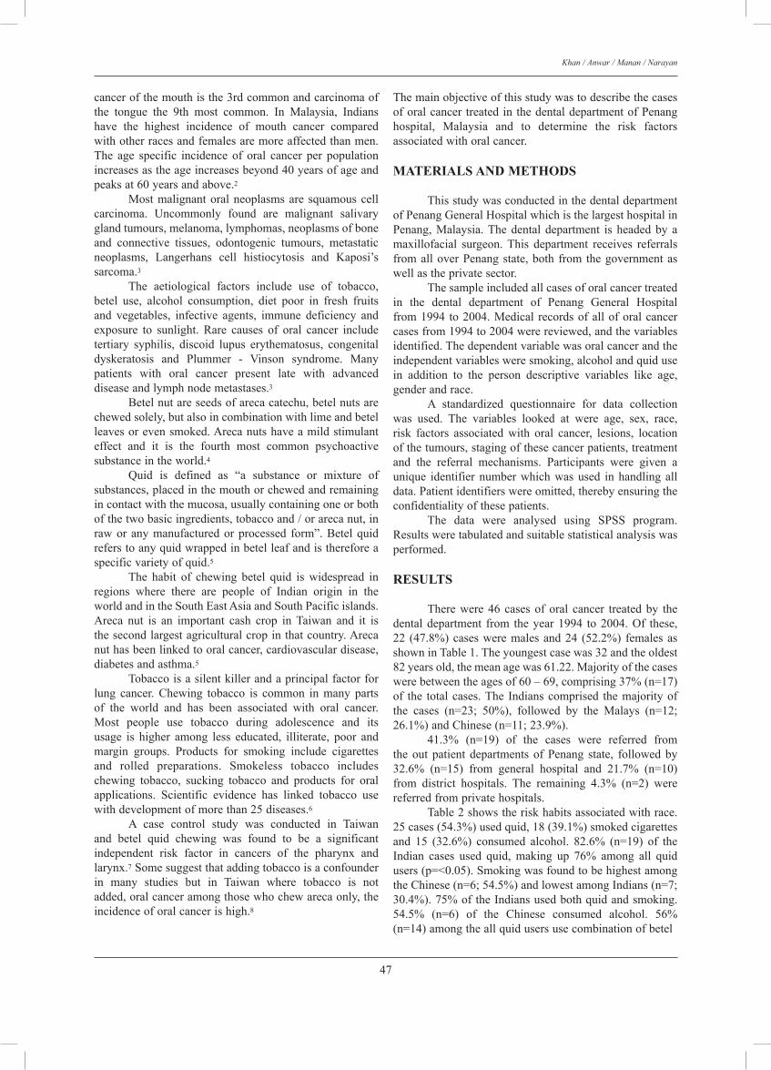
47
Khan / Anwar / Manan / Narayan
cancerofthemouthisthe3rdcommonandcarcinomaofthe tongue the 9th most common. In Malaysia, Indianshave the highest incidence of mouth cancer comparedwithotherracesandfemalesaremoreaffectedthanmen.The age specific incidence of oral cancer per populationincreasesastheageincreasesbeyond40yearsofageandpeaksat60yearsandabove.2 Mostmalignantoralneoplasmsare squamouscellcarcinoma. Uncommonly found are malignant salivaryglandtumours,melanoma,lymphomas,neoplasmsofboneand connective tissues, odontogenic tumours, metastaticneoplasms, Langerhans cell histiocytosis and Kaposi’ssarcoma.3 The aetiological factors include use of tobacco,betel use, alcohol consumption, diet poor in fresh fruitsand vegetables, infective agents, immune deficiency andexposure to sunlight. Rare causes of oral cancer includetertiary syphilis, discoid lupus erythematosus, congenitaldyskeratosis and Plummer - Vinson syndrome. Manypatients with oral cancer present late with advanceddiseaseandlymphnodemetastases.3 Betelnutareseedsofarecacatechu,betelnutsarechewedsolely,butalsoincombinationwithlimeandbetelleavesorevensmoked.Arecanutshaveamildstimulanteffect and it is the fourth most common psychoactivesubstanceintheworld.4 Quid is defined as “a substance or mixture ofsubstances,placedinthemouthorchewedandremainingincontactwiththemucosa,usuallycontainingoneorbothofthetwobasicingredients,tobaccoand/orarecanut,inrawor anymanufacturedorprocessed form”.Betelquidreferstoanyquidwrappedinbetelleafandisthereforeaspecificvarietyofquid.5
The habit of chewing betel quid is widespread inregions where there are people of Indian origin in theworldandintheSouthEastAsiaandSouthPacificislands.Areca nut is an important cash crop in Taiwan and it isthesecondlargestagriculturalcropinthatcountry.Arecanuthasbeenlinkedtooralcancer,cardiovasculardisease,diabetesandasthma.5 Tobaccoisasilentkillerandaprincipalfactorforlung cancer. Chewing tobacco is common in many partsof the world and has been associated with oral cancer.Most people use tobacco during adolescence and itsusage is higher among less educated, illiterate, poor andmargin groups. Products for smoking include cigarettesand rolled preparations. Smokeless tobacco includeschewing tobacco, sucking tobacco and products for oralapplications. Scientific evidence has linked tobacco usewithdevelopmentofmorethan25diseases.6 A case control study was conducted in Taiwanand betel quid chewing was found to be a significantindependent risk factor in cancers of the pharynx andlarynx.7Somesuggestthataddingtobaccoisaconfounderin many studies but in Taiwan where tobacco is notadded,oralcanceramongthosewhochewarecaonly,theincidenceoforalcancerishigh.8
ThemainobjectiveofthisstudywastodescribethecasesoforalcancertreatedinthedentaldepartmentofPenanghospital, Malaysia and to determine the risk factorsassociatedwithoralcancer.
MATERIALS ANd METHOdS
ThisstudywasconductedinthedentaldepartmentofPenangGeneralHospitalwhichisthelargesthospitalinPenang,Malaysia.Thedental department is headedby amaxillofacial surgeon.This department receives referralsfromalloverPenangstate,both fromthegovernmentaswellastheprivatesector. Thesampleincludedallcasesoforalcancertreatedin the dental department of Penang General Hospitalfrom1994to2004.Medical recordsofalloforalcancercasesfrom1994to2004werereviewed,andthevariablesidentified.Thedependentvariablewasoralcancerandtheindependentvariablesweresmoking,alcoholandquidusein addition to the person descriptive variables like age,genderandrace. A standardized questionnaire for data collectionwas used. The variables looked at were age, sex, race,risk factors associated with oral cancer, lesions, locationofthetumours,stagingofthesecancerpatients,treatmentand the referral mechanisms. Participants were given aunique identifiernumberwhichwasused inhandlingalldata.Patientidentifierswereomitted,therebyensuringtheconfidentialityofthesepatients. The data were analysed using SPSS program.Resultsweretabulatedandsuitablestatisticalanalysiswasperformed.
RESuLTS
Therewere46 casesoforal cancer treatedby thedentaldepartment from theyear1994 to2004.Of these,22 (47.8%)casesweremalesand24 (52.2%) femalesasshowninTable1.Theyoungestcasewas32andtheoldest82yearsold,themeanagewas61.22.Majorityofthecaseswerebetweentheagesof60–69,comprising37%(n=17)of the totalcases.The Indianscomprised themajorityofthe cases (n=23; 50%), followed by the Malays (n=12;26.1%)andChinese(n=11;23.9%). 41.3% (n=19) of the cases were referred fromtheoutpatient departmentsofPenang state, followedby32.6% (n=15) from general hospital and 21.7% (n=10)from district hospitals. The remaining 4.3% (n=2) werereferredfromprivatehospitals. Table2showstheriskhabitsassociatedwithrace.25cases(54.3%)usedquid,18(39.1%)smokedcigarettesand 15 (32.6%) consumed alcohol. 82.6% (n=19) of theIndian cases used quid, making up 76% among all quidusers(p=<0.05).SmokingwasfoundtobehighestamongtheChinese(n=6;54.5%)andlowestamongIndians(n=7;30.4%).75%oftheIndiansusedbothquidandsmoking.54.5% (n=6) of the Chinese consumed alcohol. 56%(n=14)amongtheallquidusersusecombinationofbetel
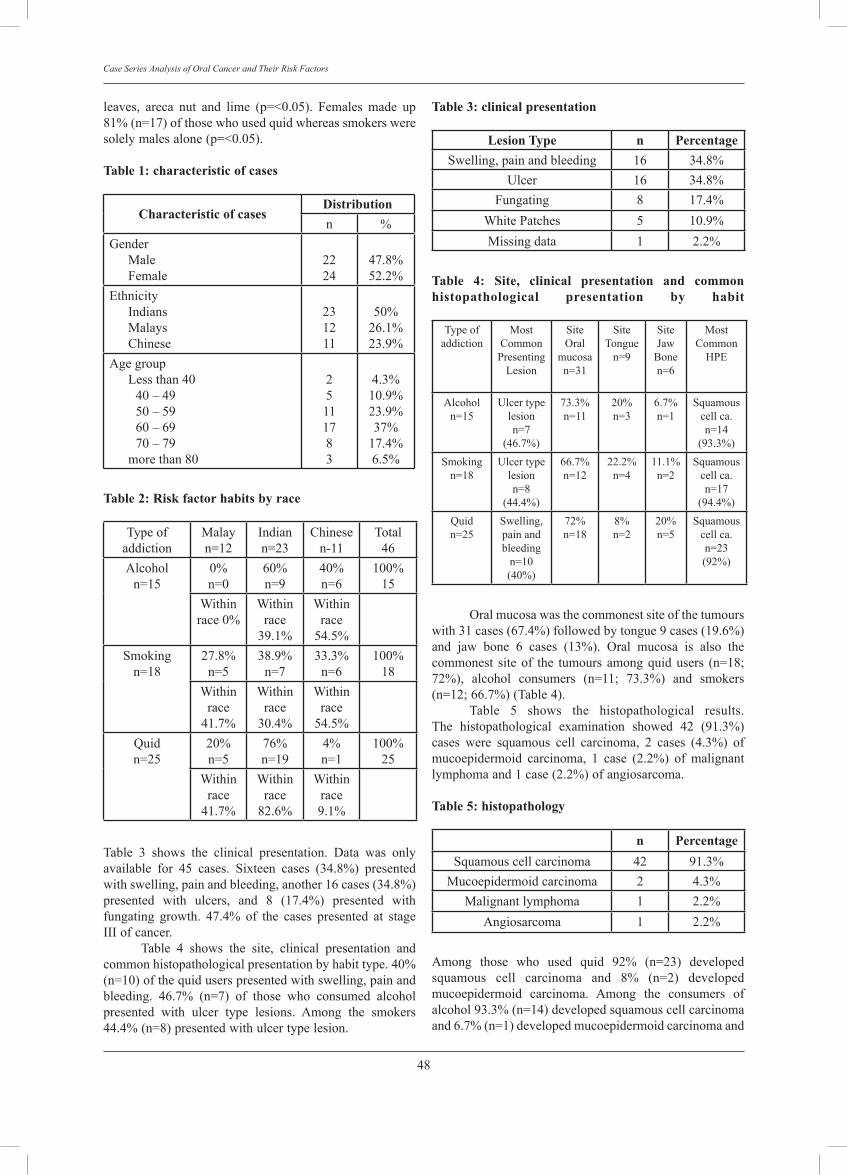
48
Case Series Analysis of Oral Cancer and Their Risk Factors
leaves, areca nut and lime (p=<0.05). Females made up81%(n=17)ofthosewhousedquidwhereassmokersweresolelymalesalone(p=<0.05).Table 1: characteristic of cases
Characteristic of casesdistributionn %
GenderMaleFemale
2224
47.8%52.2%
EthnicityIndiansMalaysChinese
231211
50%26.1%23.9%
AgegroupLessthan40
40–4950–5960–6970–79
morethan80
25111783
4.3%10.9%23.9%37%
17.4%6.5%
Table 2: Risk factor habits by race
Typeofaddiction
Malayn=12
Indiann=23
Chinesen-11
Total46
Alcoholn=15
0%n=0
60%n=9
40%n=6
100%15
Withinrace0%
Withinrace
39.1%
Withinrace
54.5%Smoking
n=1827.8%
n=538.9%
n=733.3%
n=6100%
18Within
race41.7%
Withinrace
30.4%
Withinrace
54.5%Quidn=25
20%n=5
76%n=19
4%n=1
100%25
Withinrace
41.7%
Withinrace
82.6%
Withinrace9.1%
Table 3 shows the clinical presentation. Data was onlyavailable for 45 cases. Sixteen cases (34.8%) presentedwithswelling,painandbleeding,another16cases(34.8%)presented with ulcers, and 8 (17.4%) presented withfungating growth. 47.4% of the cases presented at stageIIIofcancer. Table 4 shows the site, clinical presentation andcommonhistopathologicalpresentationbyhabittype.40%(n=10)ofthequiduserspresentedwithswelling,painandbleeding. 46.7% (n=7) of those who consumed alcoholpresented with ulcer type lesions. Among the smokers44.4%(n=8)presentedwithulcertypelesion.
Table 3: clinical presentation
Lesion Type n PercentageSwelling,painandbleeding 16 34.8%
Ulcer 16 34.8%Fungating 8 17.4%
WhitePatches 5 10.9%Missingdata 1 2.2%
Table 4: Site, clinical presentation and common histopathological presentation by habit
Typeofaddiction
MostCommonPresenting
Lesion
SiteOral
mucosan=31
SiteTongue
n=9
SiteJaw
Bonen=6
MostCommon
HPE
Alcoholn=15
Ulcertypelesionn=7
(46.7%)
73.3%n=11
20%n=3
6.7%n=1
Squamouscellca.n=14
(93.3%)Smoking
n=18Ulcertype
lesionn=8
(44.4%)
66.7%n=12
22.2%n=4
11.1%n=2
Squamouscellca.n=17
(94.4%)Quidn=25
Swelling,painandbleeding
n=10(40%)
72%n=18
8%n=2
20%n=5
Squamouscellca.n=23(92%)
Oralmucosawasthecommonestsiteofthetumourswith31cases(67.4%)followedbytongue9cases(19.6%)and jaw bone 6 cases (13%). Oral mucosa is also thecommonest siteof the tumours amongquidusers (n=18;72%), alcohol consumers (n=11; 73.3%) and smokers(n=12;66.7%)(Table4). Table 5 shows the histopathological results.The histopathological examination showed 42 (91.3%)cases were squamous cell carcinoma, 2 cases (4.3%) ofmucoepidermoid carcinoma, 1 case (2.2%) of malignantlymphomaand1case(2.2%)ofangiosarcoma.Table 5: histopathology
n PercentageSquamouscellcarcinoma 42 91.3%
Mucoepidermoidcarcinoma 2 4.3%Malignantlymphoma 1 2.2%
Angiosarcoma 1 2.2%
Among those who used quid 92% (n=23) developedsquamous cell carcinoma and 8% (n=2) developedmucoepidermoid carcinoma. Among the consumers ofalcohol93.3%(n=14)developedsquamouscellcarcinomaand6.7%(n=1)developedmucoepidermoidcarcinomaand
49
Khan / Anwar / Manan / Narayan
among the smokers 94.4% (n=17) developed squamouscellcarcinoma. Unfortunately most data on the type of treatmentwas missing (n=19). From the data available (n=27), 17hadundergoneradiotherapy,4surgery,3acombinationofsurgery, radiotherapyandchemotherapy,1chemotherapyalone, 1 combination of chemotherapy and radiotherapyand1combinationofsurgeryandradiotherapy.
dISCuSSION A majority of the cases in this series were withinthe age group of between 60-69 years old comprising37% of all cases. This finding is consistent with canceroccurrencewhich takesmanyyearsof repeatedexposureto the risk factor before developing. Indians made themajorityof the cases.These results show similaritywiththefindingsofanepidemiologicalsurveyconductedbyagroupofresearchersfromtheUniversityofMalayawheretheyfoundthattheprevalenceoforalmucosallesionwashighamongtheIndiansandtheleastamongtheChinese.9.Similarly in another survey, findings showed that theprevalence of oral precancer was highest among IndiansandtheindigenouspeopleofSabahandSarawakwhilethelowestamongsttheChinese.10
There are strong indications for an association ofhabit of betel quid chewing with cancer of the mouth,oropharyngeal cavity and upper parts of the digestivetract.11 Incidence for oral cancer in India is among thehighest in world and it has been associated with diet,weightandlifestylefactorsespeciallychewingbetelquid12.Similarlywefoundusersofquidcomprised themajorityof the cases with 54.3% of the patients diagnosed withoralcancers.ThemajorityofthequiduserswereIndianscomprising 82.6%. 81% of these quid users are females.Themostcommoncombinationofquiduserscomprising56% was betel leaves, areca nut and lime. 40% of thesequiduserspresentedwithswelling,painandbleeding. 36.6%ofthecasesconsumedalcohol.Themajorityof alcohol consumers were Chinese (54.5%) while nonewere Malay, probably because alcohol is forbidden inIslamandmostMalaysareMuslims.Mostofthealcoholconsumers presented with ulcer type of lesions. Studieshave linked the occurrence of oral cancer with alcoholusage 7, 13, 14. Some authors have indicated a synergisticincrease of oral cancer among people who consumealcohol,smokeandchewquid.15It is interesting tonotethat themostcommon typeofcancer forall threehabitswas squamous cell carcinoma and the most commonpresentation for alcohol and smoking habits were ulcertype lesions whereas for quid was swelling, pain andbleeding. Smokers comprised 39.1% of all the oral cancercases. Chinese made the majority of the smokers andthe smokers comprised solely of males. Most of thesmokers presented with ulcer type of lesions (44.4%).Mixtures of tobacco products increases the like hoodof developing oral cancer, and duration and frequencyincreases the risk further4,8, 11, 12, 14. Similar studies haveshowedindependentrisksofsmokingtolaryngealcancer7.
This is not surprising as most chewers of quid are alsosmokersandconsumealcoholdrinksasshownbyastudyconductedinTaiwan16. Because of the small number of cases, the site ofthetumourwasgroupedintothreecategoriesoralmucosa,tongueandjawbone.Under thecategoryoforalmucosaincludedpalate,buccalmucosa,retromolar,floorofmouthandlip.Thejawboneincludedthemandibleandmaxilla.Thiscaseseriesanalysisshowedthattheoralmucosawasthemostcommonsiteofthetumourwith67.4%followedby the tongue 19.6%, and jaw bone 13%. Oral mucosawasthemostcommonsiteoftumouramongtheusersofquid,alcoholandsmokers.ThisfindingcontrastswiththefindingsfromScotland,wherethemostcommonsiteofthecanceristhetonguefollowedbythefloorofthemouth13.SimilarlyinanotherstudyinSeychelles,themostcommonsitefororalcancerwasalsothetonguefollowedbyfloorofmouth,pharyngealwall, lipsandpalate17. Whereasina study done in the United States ofAmerica found thatthemostcommonsitefororalcancerwasfloorofmouth,tonguefollowedbythegingival.18 91.3% of all the histopathological examinationreported squamous cell carcinoma. Squamous cellcarcinoma was the most common presentation amongthosewhousedquid,consumedalcoholandsmoked.Thisfinding is in line with the present knowledge that mostmalignantoralneoplasmsaresquamouscellcarcinoma3. Sincethisisaretrospectiveanalysisoftherecords,the information available is limited. No comparativecontrol group could be made hence risk factor analysiscouldnotbedone.
CONCLuSIONS Likemanyother earlier studies, this study thoughwithitslimitations,hasshownthattheriskofcancerdueto tobaccoandbetelquiduse is real. Itdemonstrates thedifferent risky habit pattern among different races. Thisinformationwillbeusefulfordevelopingfocusedpromotionprogrammes such as prevention of betel chewing amongIndian women, reduction of smoking among Chinese.There is need to carry out focused case control andqualitativestudiestodetermineotherinfluencingfactors.
ACKNOWLEdGEMENT We would like to thank Prof Dr Margaret B.Comfort from the Dental Department of Asian InstituteofMedicine,ScienceandTechnologyforallhervaluableadvice.
REFERENCES
1. Park,K.ParksTextbookofPreventiveandSocialMedicine.16thed.2000.M/sBanarsidasBhanot.India.
2. GCCLim.HYahya.SecondCancerReportoftheNationalCancerRegistry.CancerIncidenceinMalaysia2003.NationalCancerRegistry.MinistryOfHealthMalaysia.
3. Crispian,Scully.Porter,Stephen.ABCofOralHealth.OralCancer.BMJ.2000;321:97–100.
4. RoobanT.Elizabeth,Joshua.Anusa,Rooban.Girish,Kumar
50
Case Series Analysis of Oral Cancer and Their Risk Factors
Govind.HealthHazardsofChewingArecanutandProductsContainingArecanut.CalicutMedicalJournal2005;3(2):e3.
5. ZainRB.IkedaN.GuptaPC.WarnakulasuriyaS,VanWykCW.ShresthaP.AxellT.OralMucosalLesionsAssociatedWithBetelQuid,ArecaNut andTobaccoChewingHabits:Consensus from a Workshop Held in Kuala Lumpur,Malaysia,November25-27,1996.JOralPatholMed.1999Jan;28(1):1-4.
6. Mehl,Garet.Stimson,VGary.Riley,Leanne.Ball,Andrew.Youth Tobacco Rapid Assessment and Response Guide.WHO:TobaccoFreeInitiativeandDepartmentofChildandAdolescentHealthandDevelopment.Geneva.
7.KaWoLee.WenReiKuo.ShihMengTsai.Etal.DifferentImpactfromBetelQuid,AlcoholandCigarette:RiskFactorsforPharyngeal andLaryngealCancer. International JournalofCancer.2005;vol.117,Issue5,pp831-836.
8. Warnakulasuriya, Saman. Areca Nut Use: An IndependentRiskFactor forOralCancer.TheHealthProblem isUnderRecognised.BMJ.2002;324(7341):799-800.
9. Taiyeb Ali TB. Razak IA. Raja Latifah RJ. Zain RB. AnEpidemiological Survey of Oral Mucosal Lesions amongElderlyMalayisans.Gerodontology.1995Jul;12(1):37-40.
10.ZainRB.IkedaN.RazakIa.AxellT.MajidZA.GuptaPC.Yaacob M. A National Epidemiological Survey of OralMucosalLesionsinMalaysia.CommunityDentistry&OralEpidemiology.Oct25(5):377-83,1997.
11. ItsuoChiba.PreventionofBetelQuidChewersOralCancerin theAsian PacificArea.Asian Pacific Journal of CancerPrevention,vol2,2001.
12.Sinha,R.Anderson,DE.McDonaldSS.GreenwaldP.CancerRiskandDietinIndia.JPostgradMed2003;49:222-228.
13.MacfarlaneGJ,BoyleP.ScullyC.OralCancerinScotland:ChangingIncidenceandMortality.BMJ1992;305:1121-3.
14.Novella,AntoniaC.TobaccoControl.JAMA,vol270(7),18Aug1993,806.
15. Sylie LousieAvon. Oral Mucosal LesionsAssociatedWithUseofQuid.JournalDeL’AssociationDentaireCanadiene,2004,vol70.244–248
16.GorskyM.EpsteinJB.OakeyC.LeND.HayJ.Stevenson-MooreP.CarcinomaoftheTongue:ACaseSeriesAnalysisofClinicalPresentation,RiskFactors,StagingandOutcome.OralsurgeryOralMedicineOralPathologyOralRadiology&Endodontics.Nov98(5):546-52,2004.
17.Wang,X.C.Thanikachalam,P.M.BrewerR.ClinicopathologicalStudyofOralandOropharyngealCarcinomainSeychelles.SMDJ1997.
18. BaraschA. GofaA. Krutchoff DJ. Eisenberg E. SquamousCellCarcinomaoftheGingiva.ACaseSeriesAnalysis.OralSurgery Oral Medicine Oral Pathology Oral Radiology &Endodontics.Aug80(2):183-7,1995.
Address for correspondence:
Prof. dr. Abdul Rashid Khan, Department of Community MedicineFaculty of Medicine, Health and ScienceSchool Of MedicineAsian Institute of Medicine, Science and Technology2 Persiaran Cempaka. Amanjaya08000 Sungai PetaniKedah, Malaysiaemail: [email protected] [email protected]
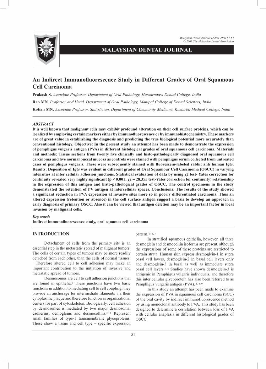
An Indirect Immunofluorescence Study in Different Grades of Oral Squamous Cell Carcinoma Prakash S. Associate Professor, Department of Oral Pathology, Harsarndass Dental College, India
Rao MN. Professor and Head, Department of Oral Pathology, Manipal College of Dental Sciences, India
Kotian MN. Associate Professor, Statistician, Department of Community Medicine, Kasturba Medical College, India
ABSTRACT It is well known that malignant cells may exhibit profound alteration on their cell surface proteins, which can be localized by employing certain markers either by immunofluorescence or by immunohistochemistry. These markers are of great value in establishing the diagnosis and predicting the true biological potential more accurately than conventional histology. Objective: In the present study an attempt has been made to demonstrate the expression of pemphigus vulgaris antigen (PVA) in different histological grades of oral squamous cell carcinoma. Materials and methods: Tissue sections from twenty five clinically and histo-pathologically diagnosed oral squamous cell carcinoma and five normal buccal mucosa as controls were stained with pemphigus serum collected from untreated cases of pemphigus vulgaris. These were subsequently stained with fluorescein-labeled rabbit anti human IgG. Results: Deposition of IgG was evident in different grades of Oral Squamour Cell Cascinoma (OSCC) in varying intensities at inter cellular adhesion junctions. Statistical evaluation of data by using χ2 test- Yates correction for continuity revealed very highly significant (p < 0.001; χ2 = 28.355 test-Yates correction for continuity) relationship in the expression of this antigen and histo-pathological grades of OSCC. The control specimens in the study demonstrated the retention of PV antigen at intercellular spaces. Conclusions: The results of the study showed a significant reduction in PVA expression at invasive sites more so in poorly differentiated carcinoma. Thus an altered expression (retention or absence) in the cell surface antigen suggest a basis to develop an approach in early diagnosis of primary OSCC. Also it can be viewed that antigen deletion may be an important factor in local invasion by malignant cells.
Key wordsIndirect immunofluorescence study, oral squamos cell carcinoma
51
Malaysian Dental Journal (2008) 29(1) 51-54© 2008 The Malaysian Dental Association
INTRODuCTION
Detachment of cells from the primary site is anessentialstepinthemetastaticspreadofmalignanttumors.Thecellsofcertaintypesoftumorsmaybemorereadilydetachedfromeachother,thanthecellsofnormaltissues.1 Therefore altered cell to cell adhesion may make animportant contribution to the initiation of invasive andmetastaticspreadoftumors. Desmosomesarecelltocelladhesionjunctionsthatare found in epithelia.2 These junctions have two basicfunctionsinadditiontomediatingcelltocellcoupling;theyprovideananchoragefor intermediatefilamentsvia theircytoplasmicplaqueandthereforefunctionasorganizationalcentersforpartofcytoskeleton.Biologically,celladhesionby desmosomes is mediated by two major desmosomalcadherins, demogleins and desmocollins.3, 4 Representsmall families of type-1 transmembrane glycoproteins.These show a tissue and cell type – specific expression
pattern.5, 6, 7
Instratifiedsquamousepithelia,however,allthreedesmogleinanddesmocollinisoformsarepresent,althoughtheexpressionsofsomeoftheseproteinsarerestrictedtocertainstrata.Humanskinexpressdesmoglein-1insuprabasal cell layers, desmoglein-2 in basal cell layers onlyand desmoglein-3 in basal as well as immediate suprabasal cell layers.5, 6 Studies have shown desmoglein-3 isantigenicinPemphigusvulgarisindividuals,andthereforethisintercellularglycoproteinhasalsobeenreferredtoasPemphigusvulgarisantigen(PVA).6, 8, 9
InthisstudyanattempthasbeenmadetoexaminetheexpressionofPVAinsquamouscellcarcinoma(SCC)oftheoralcavitybyindirectimmunofluorescencemethodbyusingmonoclonalantibodytoPVA.Thisstudyhasbeendesigned todetermineacorrelationbetween lossofPVAwith cellular anaplasia in different histological grades ofOSCC.
MALAYSIAN DENTAL JOuRNAL
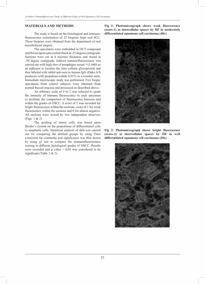
52
An Indirect Immunofluorescence Study in Different Grades of Oral Squamous Cell Carcinoma
MATERIALS AND METHODS
Thestudyisbasedonthehistologicalandimmuno-fluorescence examination of 25 biopsies from oral SCC.Thesebiopsieswereobtainedfromthedepartmentoforalmaxillofacialsurgery. ThespecimenswereembeddedinOCTcompoundandfrozenuponaprecooledchuckat-25degreescentigrade.Sections were cut at 4 microns thickness and stored at-30 degree centigrade, indirect immunofluorescence wascarriedoutwithhightiterofpemphigusserum10 (1:640)asanindicatortolocalizetheintercellularglycoproteinandthenlabeledwithrabbitanti-seratohumanIgG(DakoA/Sproducts)withpropidium-iodide0.01%asacounterstain.Immediatemicroscopicstudywasperformed.Fivebiopsyspecimens from control subjects were obtained fromnormalbuccalmucosaandprocessedasdescribedabove. Anarbitrary scaleof0 to2was selected togradethe intensity of immuno fluorescence in each specimento facilitate the comparison of fluorescence between andwithinthegradesofOSCC.Ascoreof2wasrecordedforbrightfluorescencewithinthesections,scoreof1forweakfluorescencewithinthesectionsand0foralmostnegative.All sections were scored by two independent observers(Figs.1&2) The grading of tumor cells was based uponBroder’ssystemontheproportionsofdifferentiatedcellstoanaplasticcells.Statisticalanalysisofdatawascarriedout for comparing the defined groups by using Yatescorrectionforcontinuityandsignificancewasthendrawnby using χ2 test to compare the immunofluorescence scoring indifferenthistologicalgradesofOSCC.Resultswere recorded and p value < 0.05 was considered to besignificant(Table1&2)
Fig I: Photomicrograph shows weak fluorescence (score-1) at intercellular spaces by IIF in moderately differentiated squamous cell carcinoma (40x)
Fig 2: Photomicrograph shows bright fluorescence (score-2) at intercellular spaces by IIF in well differentiated squamous cell carcinoma (20x)
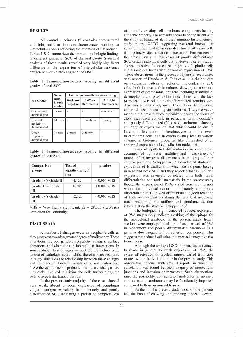
53
Prakash / Rao / Kotian
RESuLTS
All control specimens (5 controls) demonstrateda bright uniform immuno-fluorescence staining atintercellularspacesreflectingtheretentionofPVantigen.Tables1&2summarizestheimmuno-pathologicfindingsin different grades of SCC of the oral cavity. Statisticalanalysis of these results revealed very highly significantdifference in the expression of intercellular substanceantigenbetweendifferentgradesofOSCC.Table 1: Immunofluorescence scoring in different grades of oral SCC
H/P Grades
No. of casesin each grades
Indirect immunoflurescence scoring
0-Almost negative
1-Weak fluorescence
2-Bright fluorescence
Grade-IWelldifferentiated
4cases - - 4uniform
Grade-IImoderatelydifferentiated
14cases - 13uniform 1patchy
Grade-IIIpoorlydifferentiated
7cases 5cases 2patchy -
Table 1: Immunofluorescence scoring in different grades of oral SCC
Comparison groups
Test of significance χ2 test
p value
GradeIv/sGradeII 4.122 <0.001VHSGradeIIv/sGradeIII
6.205 <0.001VHS
GradeIv/sGradeIII
12.128 <0.001VHS
VHS = Very highly significant; χ2 = 28.355 (test-Yatescorrectionforcontinuity)
DISCuSSION A number of changes occur in neoplastic cells astheyprogresstowardsagreaterdegreeofmalignancy.Thesealterations include genetic, epigenetic changes, surfacealterations and alterations in intercellular interactions. Insomeinstancethesechangesarecontributingfactorstothedegreeofpathologynoted,whilsttheothersareresultant,inmanysituationstherelationshipbetweenthesechangesand progression towards neoplasia is not understood.Nevertheless it seems probable that these changes areultimately involved in driving the cells further along thepathtoneoplastictransformation. In thepresent studymajorityof the cases showedvery weak, absent or focal expression of pemphigusvulgaris antigen especially in moderately and poorlydifferentiated SCC indicating a partial or complete loss
of normally existing cell membrane components bearingantigenicproperty.TheseresultsseemstobeconsistentwiththestudyofHirakietal. in their immunohisto-chemicalstudy in oral OSCC, suggesting weekend intercellularadhesionmightleadtoaneasydetachmentoftumorcellsfrom primary site, initiating metastasis.11 Furthermore inthe present study in few cases of poorly differentiatedSCCcertainindividualcellsthatunderwentkeratinisationshowed positive fluorescence, majority of spindle cellsandbizarrecellformsweredevoidofexpressionofPVA.TheseobservationsinthepresentstudyareinaccordancewithreportsofHaradaet al.,Tadaet al.12intheirstudieson expression pattern of adhesion molecules by SCCcells, both in vivo and in culture, showing an abnormalexpressionofdesmosomalantigensincludingdesmoglein,desmoplakin, and plakoglobin in cell lines, and the lackofmoleculewasrelatedtodedifferentiatedkeratinocytes.Also western-blot study on SCC cell lines demonstratedabnormal sizes of desmoglein isoforms. The observationmadeinthepresentstudyprobablysupportstheviewsofafore mentioned authors, in particular with moderatelyand poorly differentiated (20 cases) carcinomas showingan irregular expression of PVA which could be due tolack of differentiation in keratinocytes an initial eventin carcinomacells, and in continummay lead tovariouschanges in biological properties like diminished or anabnormalexpressionofcelladhesionmolecules. Loss of epithelial differentiation in carcinomas,accompanied by higher mobility and invasiveness oftumors often involves disturbances in integrity of intercellular junctions. Schipper et al.13 conducted studies onexpression of E-Cadherin to which desmogleins belonginheadandneckSCCandtheyreportedthatE-Cadherinexpression was inversely correlated with both tumordifferentiation and nodal metastasis. In the present studythough the expression of PVA, varied from area to areawithin the individual tumor in moderately and poorlydifferentiatedSCC,inwelldifferentiated,agoodretentionof PVA was evident justifying the fact that neoplastictransformation is not uniform and simultaneous, thussubstantiatingthestudyofSchipperet al. The biological significance of reduced expressionof PVA may simply indicate masking of the epitope forthe monoclonal antibody. In the present study frozensectionswereemployed,and the reducedor lackofPVAin moderately and poorly differentiated carcinoma is agenuine down-regulation of adhesion component. Thissuggeststhatreducedadhesionintumorcellsmaygiverisetometastasis. AlthoughtheabilityofSCCtometastasizeseemedto relate in general to weak expression of PVA, theextent of retention of labeled antigen varied from areatoareawithinindividual tumorinthepresentstudy.Thisobservation concurs with several reports in which nocorrelation was found between integrity of intercellularjunctions and invasion or metastasis. Such observationsraise the possibility that adhesion molecules in invasiveandmetastaticcarcinomasmaybe functionally impaired,comparedtothoseinnormaltissues. Further in the present study most of the patientshad the habit of chewing and smoking tobacco. Several
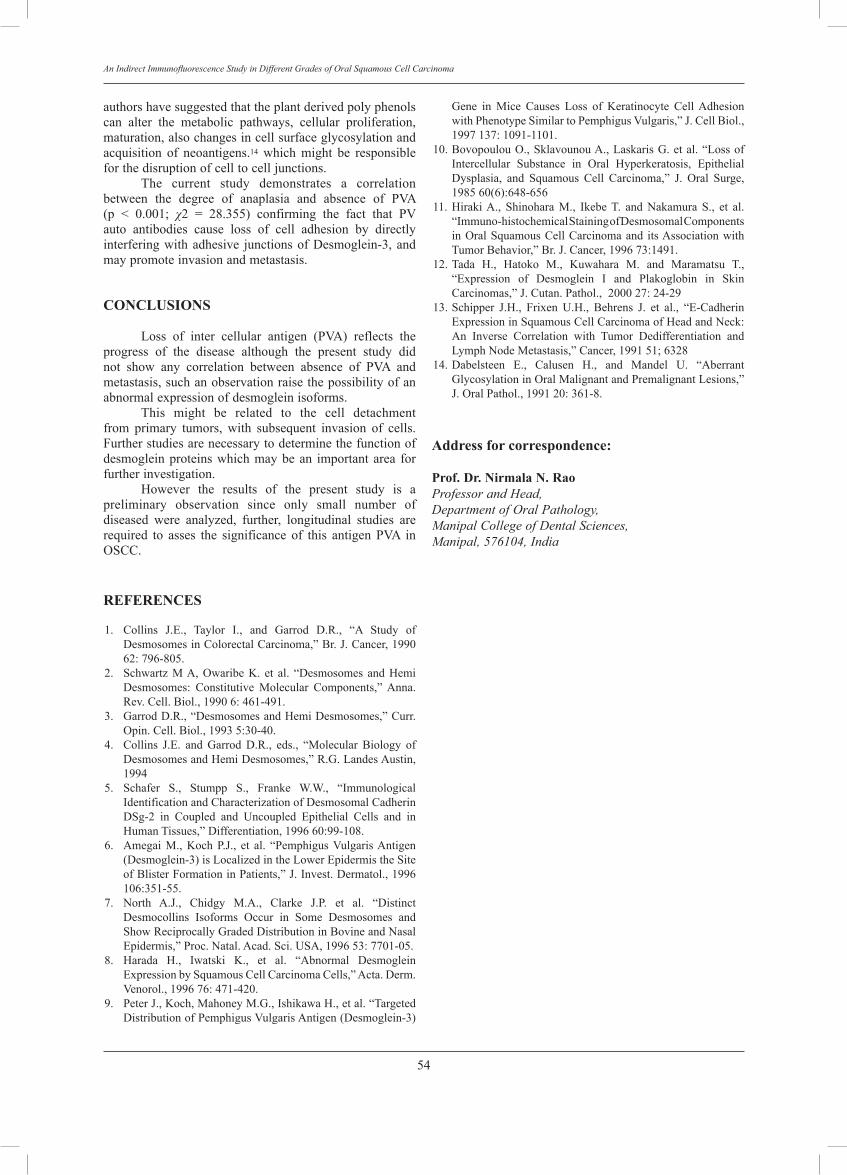
54
An Indirect Immunofluorescence Study in Different Grades of Oral Squamous Cell Carcinoma
authorshavesuggestedthattheplantderivedpolyphenolscan alter the metabolic pathways, cellular proliferation,maturation,alsochangesincellsurfaceglycosylationandacquisition of neoantigens.14 which might be responsibleforthedisruptionofcelltocelljunctions. The current study demonstrates a correlationbetween the degree of anaplasia and absence of PVA(p < 0.001; χ2 = 28.355) confirming the fact that PVauto antibodies cause loss of cell adhesion by directlyinterferingwithadhesive junctionsofDesmoglein-3,andmaypromoteinvasionandmetastasis.
CONCLuSIONS Loss of inter cellular antigen (PVA) reflects theprogress of the disease although the present study didnot show any correlation between absence of PVA andmetastasis,suchanobservationraisethepossibilityofanabnormalexpressionofdesmogleinisoforms. This might be related to the cell detachmentfrom primary tumors, with subsequent invasion of cells.Furtherstudiesarenecessarytodeterminethefunctionofdesmogleinproteinswhichmaybean importantarea forfurtherinvestigation. However the results of the present study is apreliminary observation since only small number ofdiseased were analyzed, further, longitudinal studies arerequired to asses the significance of this antigen PVA inOSCC.
REFERENCES
1. Collins J.E., Taylor I., and Garrod D.R., “A Study ofDesmosomesinColorectalCarcinoma,”Br.J.Cancer,199062:796-805.
2. SchwartzMA,OwaribeK. et al. “Desmosomes andHemiDesmosomes: Constitutive Molecular Components,”Anna.Rev.Cell.Biol.,19906:461-491.
3. GarrodD.R.,“DesmosomesandHemiDesmosomes,”Curr.Opin.Cell.Biol.,19935:30-40.
4. Collins J.E. and Garrod D.R., eds., “Molecular Biology ofDesmosomesandHemiDesmosomes,”R.G.LandesAustin,1994
5. Schafer S., Stumpp S., Franke W.W., “ImmunologicalIdentificationandCharacterizationofDesmosomalCadherinDSg-2 in Coupled and Uncoupled Epithelial Cells and inHumanTissues,”Differentiation,199660:99-108.
6. Amegai M., Koch P.J., et al. “PemphigusVulgarisAntigen(Desmoglein-3)isLocalizedintheLowerEpidermistheSiteofBlisterFormationinPatients,”J.Invest.Dermatol.,1996106:351-55.
7. North A.J., Chidgy M.A., Clarke J.P. et al. “DistinctDesmocollins Isoforms Occur in Some Desmosomes andShowReciprocallyGradedDistributioninBovineandNasalEpidermis,”Proc.Natal.Acad.Sci.USA,199653:7701-05.
8. Harada H., Iwatski K., et al. “Abnormal DesmogleinExpressionbySquamousCellCarcinomaCells,”Acta.Derm.Venorol.,199676:471-420.
9. PeterJ.,Koch,MahoneyM.G.,IshikawaH.,etal.“TargetedDistributionofPemphigusVulgarisAntigen(Desmoglein-3)
Gene in Mice Causes Loss of Keratinocyte CellAdhesionwithPhenotypeSimilartoPemphigusVulgaris,”J.CellBiol.,1997137:1091-1101.
10.BovopoulouO.,SklavounouA.,LaskarisG.etal.“LossofIntercellular Substance in Oral Hyperkeratosis, EpithelialDysplasia, and Squamous Cell Carcinoma,” J. Oral Surge,198560(6):648-656
11.HirakiA., Shinohara M., IkebeT. and Nakamura S., et al.“Immuno-histochemicalStainingofDesmosomalComponentsinOralSquamousCellCarcinomaand itsAssociationwithTumorBehavior,”Br.J.Cancer,199673:1491.
12.Tada H., Hatoko M., Kuwahara M. and Maramatsu T.,“Expression of Desmoglein I and Plakoglobin in SkinCarcinomas,”J.Cutan.Pathol.,200027:24-29
13.Schipper J.H., Frixen U.H., Behrens J. et al., “E-CadherinExpressioninSquamousCellCarcinomaofHeadandNeck:An Inverse Correlation with Tumor Dedifferentiation andLymphNodeMetastasis,”Cancer,199151;6328
14.Dabelsteen E., Calusen H., and Mandel U. “AberrantGlycosylationinOralMalignantandPremalignantLesions,”J.OralPathol.,199120:361-8.
Address for correspondence:
Prof. Dr. Nirmala N. Rao Professor and Head,Department of Oral Pathology,Manipal College of Dental Sciences,Manipal, 576104, India
Collaborative Learning: A Peer Group Teaching In Oral Hygiene Instructions Activity Yahya NA. Department of Dental Public Health, Faculty of Dentistry, Universiti Kebangsaan Malaysia (UKM)
ABSTRACT Introduction: The objectives of this survey were to investigate whether an oral health education activity through collaborative learning and peer group teaching by students would give a positive impact and to evaluate its effectiveness, sufficiency and relevancy of the activity content. Methods: Three third year dental students in each group were assigned to demonstrate to their group mates the myriad of oral hygiene device for plaque removal and how they are used. Fifteen students were appointed each as a ‘toothbrushing specialist’, ‘flossing specialist’ and interdental toothbrush specialist’ respectively. The others acted as participants in the group. The students then teach each other in the group on how to use these oral hygiene measures. A self-administered questionnaire assessing the activity outcome was given to fifty-two third year dental students who attended the demonstration. The data were entered in the computer using the SPSS version 12.0 for analysis. Results: Overall response rate was 100%. Slightly more than half of the students felt confident to teach their patients effective toothbrushing and flossing and able to indicate the usage of interdental toothbrush. Most of the students preferred a small group teaching rather than a larger one. The result shows that student’s knowledge, attitudes and practices on effective oral hygiene has gained. Conclusion: Collaborative learning in the clinical environment appears to give positive impact on the dental student’s ability to impart oral health education to their patients.
Key words Collaborative learning, dental students, oral hygiene, small group, peer group teaching.
55
Malaysian Dental Journal (2008) 29(1) 55-58© 2008 The Malaysian Dental Association
INTROduCTION
Inacollaborativelearningenvironment,participantsarebrought together tosimultaneouslyworkona task tolearn from this task work and teamwork. 1 Collaborativelearning is an umbrella term for a variety of educationalapproaches involving joint intellectualeffortbystudents,orstudentsandteacherstogether.4Collaborativelearningcenter on student’s exploration or application of thecourse material, not simply the teacher’s presentation ofit.4 Collaboration is defined as the process of buildingand maintaining a shared conception of a problem ortask, distributing responsibility across members of thegroup, sharing expertise, and mutually constructing andnegotiatingcognition.2 Theoralhygieneinstructionsdemonstrationactivityforthe3rdyeardentalstudentsintheFacultyofDentistry,National University of Malaysia (UKM) previously wasconducted in a large group with total teaching was doneby the lecturer.Unfortunately, therearemanydrawbackswhen students learn in a large group environment. It istoo easy for students to becomepassiveobservers rather
than active participants. Groups larger than six mightlessen their opportunity to speak frequently and lessentheir responsibility towards their duty in the learningprocess.3 Collaborative learning represents a significantshift away from the typical teacher-centred or lecture-centredmilieuinclassrooms.4
The objectives of this survey were to investigatewhether an oral health education activity throughcollaborativelearningandpeergroupteachingbystudentswouldgiveapositiveimpact.Therationaleofthissurveyistoacknowledgethatcollaborativelearningandpeergroupteaching can be applied successfully in any academicsubjectespeciallyindentistry.
MATERIALS AND METHODS
The population of this study consisted of thirdyear dental undergraduate students of the Faculty ofDentistry, National University of Malaysia (UKM)attending a demonstration on oral hygiene instructionsfor their Introduction to Clinical Dentistry Module.
MALAYSIAN DENTAL JOURNAL
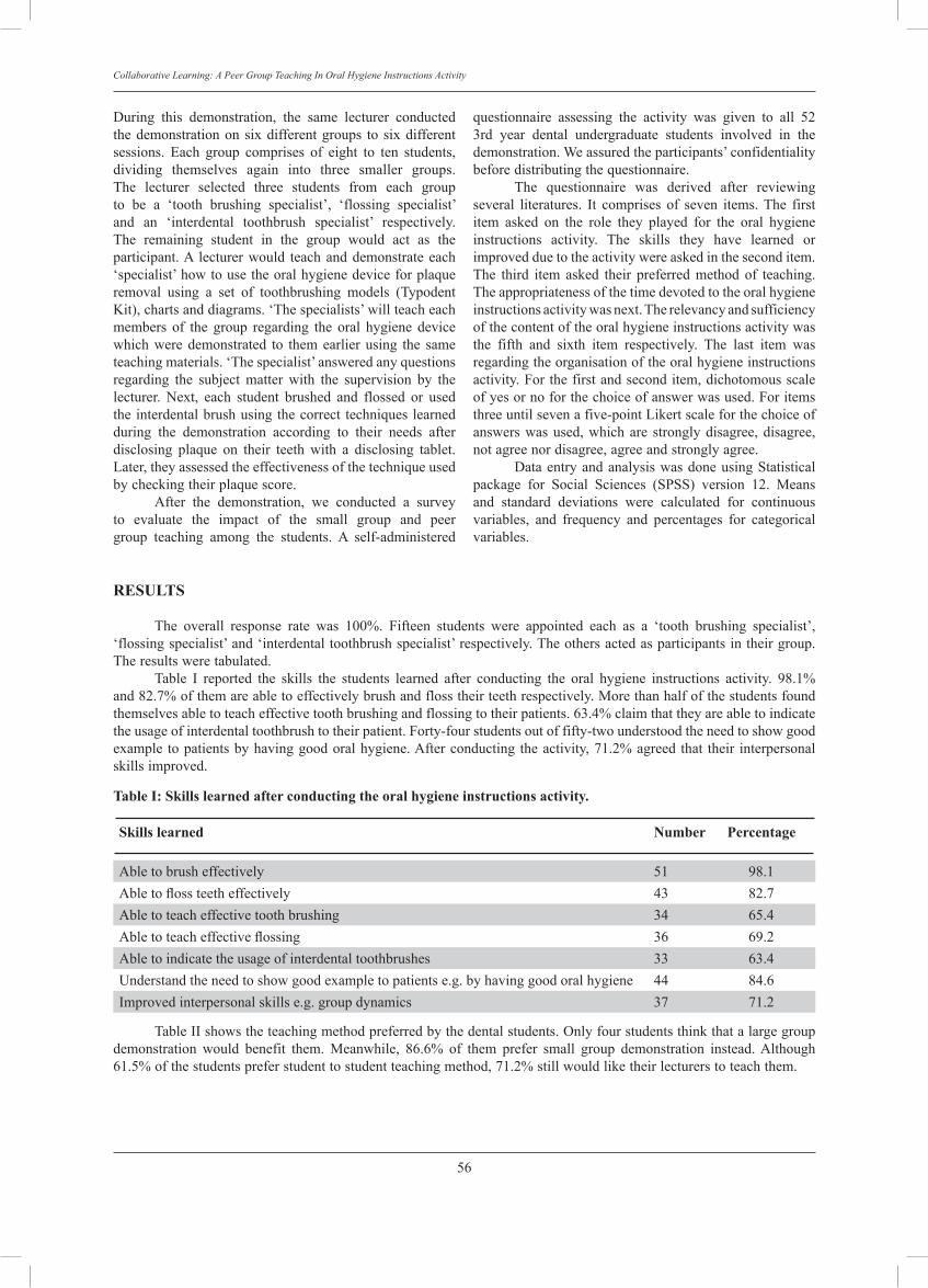
56
Collaborative Learning: A Peer Group Teaching In Oral Hygiene Instructions Activity
During this demonstration, the same lecturer conductedthedemonstrationonsixdifferentgroups to sixdifferentsessions. Each group comprises of eight to ten students,dividing themselves again into three smaller groups.The lecturer selected three students from each groupto be a ‘tooth brushing specialist’, ‘flossing specialist’and an ‘interdental toothbrush specialist’ respectively.The remaining student in the group would act as theparticipant.A lecturerwould teachanddemonstrateeach‘specialist’howtousetheoralhygienedeviceforplaqueremoval using a set of toothbrushing models (TypodentKit),chartsanddiagrams.‘Thespecialists’willteacheachmembers of the group regarding the oral hygiene devicewhichweredemonstrated to themearlierusing the sameteachingmaterials.‘Thespecialist’answeredanyquestionsregarding the subject matter with the supervision by thelecturer. Next, each student brushed and flossed or usedtheinterdentalbrushusingthecorrect techniqueslearnedduring the demonstration according to their needs afterdisclosing plaque on their teeth with a disclosing tablet.Later,theyassessedtheeffectivenessofthetechniqueusedbycheckingtheirplaquescore. After the demonstration, we conducted a surveyto evaluate the impact of the small group and peergroup teaching among the students. A self-administered
questionnaire assessing the activity was given to all 523rd year dental undergraduate students involved in thedemonstration.Weassuredtheparticipants’confidentialitybeforedistributingthequestionnaire. The questionnaire was derived after reviewingseveral literatures. It comprises of seven items.The firstitem asked on the role they played for the oral hygieneinstructions activity. The skills they have learned orimprovedduetotheactivitywereaskedintheseconditem.The third item asked their preferred method of teaching.Theappropriatenessofthetimedevotedtotheoralhygieneinstructionsactivitywasnext.Therelevancyandsufficiencyofthecontentoftheoralhygieneinstructionsactivitywasthe fifth and sixth item respectively. The last item wasregardingtheorganisationoftheoralhygieneinstructionsactivity.Forthefirstandseconditem,dichotomousscaleofyesornoforthechoiceofanswerwasused.Foritemsthreeuntilsevenafive-pointLikertscaleforthechoiceofanswerswasused,whichare stronglydisagree,disagree,notagreenordisagree,agreeandstronglyagree. DataentryandanalysiswasdoneusingStatisticalpackage for Social Sciences (SPSS) version 12. Meansand standard deviations were calculated for continuousvariables, and frequency and percentages for categoricalvariables.
RESULTS
The overall response rate was 100%. Fifteen students were appointed each as a ‘tooth brushing specialist’,‘flossingspecialist’and‘interdentaltoothbrushspecialist’respectively.Theothersactedasparticipantsintheirgroup.Theresultsweretabulated. Table I reported the skills the students learned after conducting the oral hygiene instructions activity. 98.1%and82.7%ofthemareabletoeffectivelybrushandflosstheirteethrespectively.Morethanhalfofthestudentsfoundthemselvesabletoteacheffectivetoothbrushingandflossingtotheirpatients.63.4%claimthattheyareabletoindicatetheusageofinterdentaltoothbrushtotheirpatient.Forty-fourstudentsoutoffifty-twounderstoodtheneedtoshowgoodexample topatientsbyhavinggoodoralhygiene.Afterconductingtheactivity,71.2%agreedthat their interpersonalskillsimproved.
Table I: Skills learned after conducting the oral hygiene instructions activity.
Skills learned Number Percentage
Abletobrusheffectively 51 98.1Able to floss teeth effectively 43 82.7Abletoteacheffectivetoothbrushing 34 65.4Able to teach effective flossing 36 69.2Abletoindicatetheusageofinterdentaltoothbrushes 33 63.4Understandtheneedtoshowgoodexampletopatientse.g.byhavinggoodoralhygiene 44 84.6Improvedinterpersonalskillse.g.groupdynamics 37 71.2
TableIIshowstheteachingmethodpreferredbythedentalstudents.Onlyfourstudentsthinkthatalargegroupdemonstration would benefit them. Meanwhile, 86.6% of them prefer small group demonstration instead.Although61.5%ofthestudentspreferstudenttostudentteachingmethod,71.2%stillwouldliketheirlecturerstoteachthem.
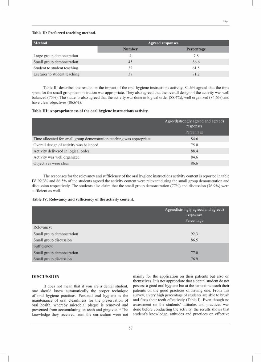
57
Yahya
Table II: Preferred teaching method.
Method Agreed responsesNumber Percentage
Largegroupdemonstration 4 7.8Smallgroupdemonstration 45 86.6Studenttostudentteaching 32 61.5Lecturertostudentteaching 37 71.2
TableIIIdescribestheresultsontheimpactoftheoralhygieneinstructionsactivity.84.6%agreedthatthetimespentforthesmallgroupdemonstrationwasappropriate.Theyalsoagreedthattheoveralldesignoftheactivitywaswellbalanced(75%).Thestudentsalsoagreedthattheactivitywasdoneinlogicalorder(88.4%),wellorganized(84.6%)andhaveclearobjectives(86.6%).
Table III: Appropriateness of the oral hygiene instructions activity.
Agreed(stronglyagreedandagreed)responsesPercentage
Timeallocatedforsmallgroupdemonstrationteachingwasappropriate 84.6Overalldesignofactivitywasbalanced 75.0Activitydeliveredinlogicalorder 88.4Activitywaswellorganized 84.6Objectiveswereclear 86.6
TheresponsesfortherelevancyandsufficiencyoftheoralhygieneinstructionsactivitycontentisreportedintableIV.92.3%and86.5%ofthestudentsagreedtheactivitycontentwererelevantduringthesmallgroupdemonstrationanddiscussionrespectively.Thestudentsalsoclaimthatthesmallgroupdemonstration(77%)anddiscussion(76.9%)weresufficientaswell.
Table IV: Relevancy and sufficiency of the activity content.
Agreed(stronglyagreedandagreed)responsesPercentage
Relevancy:Smallgroupdemonstration 92.3Smallgroupdiscussion 86.5Sufficiency:Smallgroupdemonstration 77.0Smallgroupdiscussion 76.9
dISCuSSION It does not mean that if you are a dental student,one should know automatically the proper techniqueof oral hygiene practices. Personal oral hygiene is themaintenance of oral cleanliness for the preservation oforal health, whereby microbial plaque is removed andpreventedfromaccumulatingonteethandgingivae.4Theknowledge they received from the curriculum were not
mainly for the application on their patients but also onthemselves.Itisnotappropriatethatadentalstudentdonotpossessagoodoralhygienebutatthesametimeteachtheirpatients on the good practices of having one. From thissurvey,averyhighpercentageofstudentsareabletobrushandflosstheirteetheffectively(TableI).Eventhoughnoassessment on the students’ attitudes and practices wasdonebeforeconductingtheactivity,theresultsshowsthatstudent’s knowledge, attitudes and practices on effective
58
Collaborative Learning: A Peer Group Teaching In Oral Hygiene Instructions Activity
oralhygienehasgained.ThisiscomparablewiththestudybyRongWSetal,wheretherewasasignificantincreasein the number of year five dental students who used amorethoroughbrushingtechniqueandhavemorepositiveattitudetooralhealth.5 DuringtheconductingoftheIntroductiontoClinicalDentistrymodule,thestudentswerenotincontactwithanypatients yet. This is the stage of preparing the studentson the clinical environment and revising some clinicalproceduresthattheyhavelearnedtheoreticallyinthepasttwoyears.Thus,thisexplainedthatonlyslightlymorethanhalf of the students felt confident to teach their patientseffective toothbrushing and flossing and able to indicatetheusageofinterdentaltoothbrush(TableI). A high percentage of students claimed that theyhaveactuallyimprovedtheir interpersonalskillsafter theactivity(TableI).Thissuggeststhat,theinformalsettingofcollaborativelearningfacilitateddiscussionandinteraction.This group interaction helped the students to learn fromeachother’s,skillsandexperiences.6Collaborationdoesnotjusthappenbecauseindividualsco-present;individualsmustmakeaconscious,continuedefforttocoordinatetheirlanguage and activity with respect to shared knowledge.Studentslearnbestwhentheyareactivelyinvolvedintheprocess.7Fromthissurvey,mostofthestudentspreferredasmallgroupteachingratherthanalargerone(TableII).Moreover,mostofthemalsoagreedthattheoralhygieneinstructions activity was a well organized, delivered inlogical order, balanced design and have clear objectives(TableIII).Thisisconcurrentwithreportsfromresearchersthat,regardlessofthesubjectmatter,studentsworkinginsmallgroupstendtolearnmoreofwhatistaughtandretainitlongerthanwhenthesamecontentispresentedinotherinstructionalformats.3 Collaborative learning in a classroom has itschallenges and dilemmas. Shown from the survey werea high percentage of students preferring their lecturerto teach rather than their peers (Table II). Challenges atthe classroom level are compounded by the traditionalstructures andcultureof theacademy,whichcontinue toperpetuatetheteacher-centred,transmission-of-informationmodelofteachingandlearning.4Teachingincollaborativesettings puts front and centre the tension between theprocess of student learning and content coverage. It isdifficulttoensurestudentsarelearningandmasteringkeyskills in the activity while at the same time addressingallthematerialofthecourse.However,fromthissurvey,the students reported that the activity content during thesmall group demonstration teaching and the small groupdiscussionwasrelevantandsufficient(TableIV).
CONCLuSIONS Dentalstudentswereabletopracticeeffectiveoralhygiene behaviours and were confident enough to teachtheirpatientsinpracticinggoodoralhygiene.Therationaleofthissurveyistoacknowledgethatcollaborativelearningand peer group teaching can be applied successfully inanyacademicsubjectespeciallyindentistry.Thestudentsunderstoodwell that they shouldbegood rolemodels to
theirpatients.Thisreflectsthatcollaborativelearningandpeergroupteachingmotivatethestudentsbygettingthemmore actively engaged and succeeded in their learningabilitiestogainknowledgeandskills.
RECOMMENDATIONS From this survey, many students still prefer theirteachers/ lecturers to do the teaching rather than theirpeers.Thiswasprobablyduetothelackoftrust towardstheir peers in teaching them. In addition, peer groupteaching might not be their norms in most of the dentalcurricular activity. Therefore, collaborative learningshouldbeintroducedandusedmoreoftenintothedentalcurricular to enhance a better achievement in knowledgeand skills amongdental students specifically, in thenextmillennium.
REFERENCES
1. Van den Bossche et al. Social and Cognitive DrivingTeamwork in Collaborative Learning Environments: TeamLearning Beliefs and Behaviours. Small Group Research2006;37:490.
2. Roschelle, J. Learning by collaborating: Convergentconceptual change. Journal of the Learning Sciences 1992,2:235-76.
3. Davis BG.Tools for teaching. Jossey-Bass Publishers: SanFrancisco,1993.
4. Goodsell A, Maher M, Tinto V,Smith BL, MacGregor JT.CollaborativeLearning:ASourcebookforHigherEducation.National Center on Postsecondary Teaching, Learning andAssessment:PennsylvaniaStateUniversity,1992.
5. ChooA,DelacDM,MesserLB.Oralhygienemeasuresandpromotion: Review and considerations. Australian DentalJournal2001;46:166-73.
6. RongWS,WangWJ,YipHK.Attitudesofdentalandmedicalstudentsintheirfirstandfinalyearsofundergraduatestudytooralhealthbehaviour.EuropeanJournalofDentalEducation2006;10:178-84.
7. Gokhale AA. Collaborative learning enhances criticalthinking.2002.
Address for correspondence:
dr. Nurul Asyikin Yahya Faculty of Dentistry, Universiti Kebangsaan Malaysia (UKM), Jalan Raja Muda Abdul Aziz, 50300 Kuala Lumpur, Malaysia.Telephone no: 603 40405854 Fax no: 603 26982944Email address: [email protected]
59
ABSTRACTS OF SCIENTIFIC PAPERS PRESENTED AT THE 15TH FDI/MDA SCIENTIFIC CONVENTION AND TRADE EXHIBITION, 25th - 27th JANUARY 2008
A 6 YEARS CLINICAL REVIEW OF TRAUMA TO YOUNG PERMANENT TEETH (2000-2006) IN DEPARTMENT OF PAEDIATRIC DENTISTRY HOSPITAL SULTANAH BAHIYAH, ALOR STAR, KEDAH.1Dewi Mayang Sari Kamarozaman, 2Azillah Mohd Ali
1Dental Officer, Department of Paediatric Dentistry of Hospital Sultanah Bahiyah.2Consultant and HOD Paediatric Dentistry of Hospital Sultanah Bahiyah, Alor Setar, Kedah.
Objectives: To evaluate the prevalence, treatment modalities and clinical outcomes of dental trauma in young permanent teeth in the Department of Paediatric Dentistry, Hospital Alor Star from the year 2000-2006.
Materials and Methods: A retrospective review was conducted of all dental record of children registered in the dental paediatric clinic under category of trauma cases (labeled green). During the period of Jan 2000-November 2006; a total of 82 patients presented with 200 traumatized permanent teeth. The data regarding demographic characteristic, prevalence off dental trauma, treatment modalities and clinical outcome were analyzed.
Results: The study revealed a total of 82 patients with 200 traumatized permanent teeth presented during the 6 years interval. The age of patients ranged from 6 to 16 years old. Male experienced more dental trauma in 3:2 ratio. The majority ethnic affected was Malay (84 %) followed by Chinese and Indian. The major cause of dental trauma was fall (55%) followed by MVA and alleged hit. Maxillary incisors were the most affected (84%). The most common dental injury was avulsion (24%) and the least was extrusion (8%). 149 out of 200 teeth were successfully managed with various approaches depending on types and severity of trauma. There were 51 cases of defaulted treatment. 11 cases of complications were recorded.
Conclusion: 149 out of 200 traumatized teeth were successfully managed with various approaches. Immediate proper treatment followed by subsequent review visit ensures a good long-term prognosis of treatment provided. Unfortunately there was incidence of patient or parent defaulted treatment. Hence, preventive, educational program and periodic check up should be instituted among the public and schoolchildren.
Keywords: young permanent teeth, trauma, children,
HOW TO REPAIR A FRACTURED FISSURE SEALANT? G. Qadri*, S.N.F. Mohd Noor and D. Mohamad.
School of Dental Sciences, Universiti Sains Malaysia, Kelantan
Summary: The aim of this study was to investigate whether differences in the technique of repair, influence a seal of a repaired nano-filled resin based sealant, placed on occlusal surfaces of permanent molar teeth in-vitro.
Sample and methods: 112 human extracted molars were randomly allocated to one of four groups, of 28 teeth each. A nano filled resin sealant (Filtek Z350) was placed on their occlusal surface following cleaning by prophylaxis and acid etching. Duplication of sealant failure was carried out. The teeth were then subjected to four different methods of repair: Group 1: With a slow speed prophylaxis brush followed by acid etching; Group 2: Prophylaxis brush, acid etching and application of bonding agent; Group 3: Light curing for 5s; and Group 4: Light curing for 20s. Then, they were stored for one week in artificial saliva, painted with two layers of varnish: their apices were sealed with wax, and the teeth were immersed in 1% methylene blue. The teeth were then sectioned to achieve three cuts. A total
Malaysian Dental Journal (2008) 29(1) 59-73© 2008 The Malaysian Dental Association
MALAYSIAN DENTAL JOURNAL
60
of 648 sections from 112 teeth were scored for microleakage.
Results: Statistical analysis using chi-squared did not demonstrate any one single method of repair to be superior to the control method for reapplication of the sealant.
Conclusion: Group no. 1, seems to be the most appropriate, as this method is likely to be agreeable to most children because of its time saving nature.
Keywords: Repair, sealant, resin based sealant
MAXIMIZING THE VALUE OF MICROSOFT ACCESS DATABASE IN MANAGING SURGICAL WAITING LISTJabar MN*, Ramli R, Rahman RA
OS Department, UKM.
We presents the use of Microsoft Access as a standard Microsoft Office program as a simple, stand-alone database in managing a waiting list for our third molar surgical patients. Readily available in all machines running on Windows platform, the database can be built to collect, analyze and monitor all patients scheduled for surgical removal. The novelty of this record keeping system not only lies in its simplicity to build and use, but to its cost effectiveness, security and can be used to collect standardized demographic and clinical data compared to conventional paper or book waiting list. The data stored can be quickly viewed or even shared over the network if configured. Appointment booking and monitoring can be done effectively avoiding patient drop-outs. Although not utilizes the web, it allow back-up and copying to the off-the-shelf thumbdrive by doctors that they can have access to the list at any time.
Keywords: Waiting list, Microsoft Access, Microsoft Office
DEVELOPMENT OF ELECTROPALATOGRAPH (EPG) ARTIFICIAL PALATE FOR SPEECH THERAPY OF CLEFT LIP AND PALATE CHILDREN. Siti Noor Fazliah Mohd Noor*, Rosmawati Aman, Abdul Rani Samsudin, Abdul Hakim Abdul Basir.
Department of Community Dentistry, USM
Objective: To determine the speech characteristics focusing on the parameters of articulation between normal and cleft lip and/or palate subjects.
Materials and methods: Normal and cleft lip and/or palate subjects were selected according to the criteria. Impression of the upper arch was taken and EPG was constructed for each subjects. The EPG artificial palate was validated and speech assessment was done. EPG data was collected from spontaneous response after naming the pictures on selected target sounds. Data was analyzed for the error pattern of the cleft-type characteristics (CTC) and also the idea of tongue-palate contact during the error production of sounds was achieved.
Results: There is a difference in the tongue palate contact between the normal and cleft lip and/or palate subjects.
Conclusions: EPG can be used as a tool to improve the error pattern of the cleft-type characteristics (CTCs) among cleft lip and/or palate patients and suggested to be used by other subjects with articulation problems.
The study was supported by eScience Fund Grant from MOSTI (305/PPSG/6113101)
Keywords: speech, cleft-type characteristics, speech assessment
SOFT TISSUE RECONSTRUCTION OF RIDGE DEFORMITIES: MODIFIED ONLAY-INTERPOSITIONAL CONNECTIVE TISSUE GRAFT COMBINED WITH LABIALLY POSITIONED FLAP AND TEMPORARY REMOVAL PARTIAL DENTURES. A CASE SERIES. R.Ayob1 & M.Jaya2
1. Periodontal specialist, Peringgit Dental Clinic Malacca, Ministry of Health1
2. Clinical Dental Officer, Central Malacca Dental Clinic, Ministry of Health2
Aims: The purpose of this paper is to describe the new modified onlay-interpositional connective tissue graft (CTG) technique combined with labially positioned flap, to demonstrate whether the removal partial dentures (RPD) is able to support the healing stage and to evaluate the clinical results post surgery.
61
Materials and methods: Four stabilized chronic and generalised aggressive periodontitis patients from Periodontal Clinic with severe aesthetical prosthesis problem and ridge deformities of Siebert class III morphology on the anterior maxilla were selected. A removable partial denture (RPD) was specially designed prior surgery. The new modified technique is described in detail. The bridge-works constructed at six to eight weeks post surgery. The clinical assessment in terms of degree of the ridge deformities, texture, colour, scar tissue, papilla classification and gingival line were recorded at baseline, six weeks and 6 months.
Results: All patients showed a similar pattern of uncomplicated healing, clinically using the RPD. At six weeks post surgery, all bucco-lingual and apico-coronal ridge deformities were corrected. At six months, the ridge deformities corrected were excellently maintained; inter-dental papilla reconstruction was appreciable, aesthetic quality of texture and color of tissues without scarring noted. The gingival line was symmetrical.
Conclusion: The specially designed RPD managed to assist the healing of the new modified technique of the CTG inserted in the labially positioned flap. Clinical results showed that the ridge deformities have been corrected with improvement in papilla level and gingival line.
Keywords: soft tissue reconstruction, connective tissue graft, partial denture
THE CLINICAL REVIEW ON DENTAL AVULSION AMONGS CHILDREN IN HOSPITAL SULTANAH BAHIYAH 2001-2006.Hazwani Hassan1, Azillah Mohd Ali2
1Dental Officer, Department of Paediatric Dentistry, Hospital Alor Setar2Consultant and HOD Paediatric Dentistry of Hospital Sultanah Bahiyah, Alor Setar
Introduction: Dental trauma occurs in childhood and adolescence with consequences in time and cost for both patient and family. For some individuals, dental trauma will result in long, time consuming and costly treatments in childhood which will continue into adulthood. Tooth avulsion is defined as total displacement of the tooth out of its alveolar socket. It accounts for 0.5 % to 16% of traumatic injuries in the permanent dentition. Avulsion of permanent teeth occurs most often in children 7 to 9 years old, an age when the relatively resilient alveolar bone provides only minimal resistance to extrusive forces.
Objectives: 1)To identify the causes of tooth avulsion 2)To determine the age range, gender, and race of the patient with tooth avulsion 3)To determine the period elapsed between avulsion and time seeking dental care 4)To identify sources of referral 5)To identify the treatment provided to the patient.
Methodology: Retrospective study of the cases treated in Department of Paediatric Dentistry, Hospital Sultanah Bahiyah from year 2001-2006.
Results: From the data collected, 30 avulsions identified involving either 1 tooth or more than one tooth. Causes of tooth avulsion identified are alleged Motor vehicle accident (57%), alleged fall (40%) and alleged hit by object (3%). Out of 30 avulsion identified, 70% were male while 30% were female From the data collected, 80 % of avulsion cases involving Malays while 20% of cases involving Chinese, none of other races. From 30 cases of avulsion, 87% of cases referred by Casualty Hospital Alor Setar, 3% of cases referred from other dental clinic and 10% are from other resources. Other resources include Periodontic Clinic, Casualty Hospital Sik and Surgical Department Hospital Alor Setar.
Conclusion: The result of this study reveals that parents/patient are not aware that the avulsed teeth can be replanted. In addition there is a delay in seeking treatment more than 24 hours. The percentage of patients attended for treatment immediately was very low when compared with other studies. Preventive Educational Program should be instituted in state of Kedah, which should be directed to parents, teachers and other health care personal. Successful management of the avulsed tooth begins at the site of accident.
Keywords: dental avulsion, children, accident
62
THE RELATIONSHIP BETWEEN GENDER, AGE, JOB RANK, MARITAL STATUS AND LEVEL OF MENTAL HEALTH AMONGST GOVERNMENT DENTISTS. Mason L 1*, AbdulKadir R2, Noriah Y1, Zuraida NZ3
1Department of Community Dentistry, Faculty of Dentistry, University Malaya2Department of Dental Public Health, Faculty of Dentistry, National University of Malaysia 3Department of Psychological Medicine, Faculty of Medicine, University Malaya.
Objective: The aim of this study was to determine whether gender, age, job rank and marital status were associated with the level of mental health of Malaysian’s government dentists.
Method: A self-administered questionnaire survey was conducted on all dentists with at least one year working experience in government agencies. In all, 682 survey forms from a total of 864 forms distributed were returned (81% response rate). The study explored eight mental health domains namely; vitality, somatic experience, behavior, cognitive symptoms, emotions, sense of coherence, interpersonal relationship and sense of accomplishment experienced by respondents measured by structured statements on a five point Likert Scale.
Results: Although none reported extreme negative conditions, 18.9% and 75.9% respondents were reported to have poor or moderate level of mental health respectively. Only 5.2 % reported excellent status. Gender, age and job rank were found to be significantly associated with level of mental health at p <0.05 but marital status was not (p >0.05). Increasing age and higher job rank were significantly related to better mental health experience (p <0.05) regardless of location of practice and practice types. The most important finding of the study was that most dentists (95%) experience mental stress and that about one in every five (18.9%) was serious.
Clinical Implications: Persistent mental stress is harmful to the dentists’ overall mental health and may raise issues of work quality and productivity. Work policy, training opportunities and supportive environment should be looked into to help dentists cope with work and life more productively.
Conclusion: The survey concluded that dentists are prone to mental stress. The selected variables studied contribute significantly to a better understanding of how their level of mental health was affected by them.
Keywords: Government dentist, age, marital status, job rank, mental health.
ORAL AND PERIORAL TRAUMA IN CHILDREN ATTENDING THE PAEDIATRIC DENTAL DEPARTMENT IN MELAKA GENERAL HOSPITAL.Dr Sa’adah Atan. Paediatric Dental Specialist, Hospital Melaka.
The Paediatric Dental Department in Hospital Melaka started its operation in January 2007 and is the referral centre for paediatric oral management for the state of Melaka and adjacent areas of Johor and Negeri Sembilan.
The response has been very encouraging: until November 2007 the attendance of patients had reached 2172 with 736 new cases referred. In this first year alone, 137 patients were seen for management of traumatic oral and perioral injuries to child patients, up to the age of 16. The data on the types of traumatic events, types of dental and bony injury sustained by the patients has been analysed. A large number (61%) of injuries were sustained due to falls, followed by injuries due to motor vehicle accidents (30%).
Though peak incidence are said to occur at 2-4 years and 8-10 years of age (Welbury, 1997), our findings showed no such predilection to any age groups. The main injuries sustained were found to be soft tissue injuries followed by luxative dental injuries. Some children also presented with bony fractures.
This study serves to show the pattern and types of trauma cases seen at this department.
Keywords: oral trauma, children, perioral trauma, referral
63
THE CORONAL SEALING ABILITY OF A NOVEL NANO HYDROXYAPATITE - FILLED ENDODODNTIC SEALER. S.M.A. Alomari, Z. Ab Ghani*, A. Husein, S.M. Masudi
School of Dental Sciences, Universiti Sains Malaysia (USM)
Objectives: To evaluate the sealing ability of a novel nano hydroxyapatite (HA)-containing endodontic sealer in preventing coronal leakage before and after post preparation and to compare it with the commercial AH26 sealant.
Materials and Methods: A total of 152 extracted human single-rooted teeth were instrumented using NiTi files and the crowns were then amputated. After cleaning and shaping procedure were completed using step back technique, samples were randomly divided into two groups. The two groups were obturated with either gutta percha and AH 26 sealer or gutta percha with the nano HA-containing sealer. All teeth were then stored at 37° C for 7 days to allow the sealer to set. Each group was then further sub divided into two groups where one of them were prepared for post using para-post drill and the other group was left intact. The resulting 4 study groups contained 38 samples per group (n = 38). The teeth were then thermal cycled at 5°C and 55°C in water baths at dwell time of 30 seconds for a total of 500 cycles. External surfaces of the roots were coated with two layers of nail varnish that did not cover the coronal opening. Specimens were then submerged in 2% methylene blue dye for 24 hours. Each root was sectioned vertically into two halves, and microleakage was measured under microscope (x36) by taking the maximum linear dye penetration corono-apically. Micro leakage readings were analyzed by the independent t test (α=0.05).
Results: The result showed that there was no statistically significant difference in the coronal sealing ability between the two sealers, before and after preparation for post. The experimental nanoHA-containing and AH26 sealers with post space preparation showed significantly more leakage compared to sealers with no post space preparation.
Conclusions: Preparation for post caused a significant decrease in the coronal sealing ability of both sealers. The preparation for post did not create any difference between the performances of the two sealers. The novel nanoHA-containing sealer tested had a comparable coronal sealing ability with the commercial AH26 sealer.
Keywords: coronal seal, sealer, post preparation
TRADITIONAL MALAYSIAN AND ORIENTAL HERB EXTRACTS AS ANTI-PLAQUE AGENT: IN VITRO STUDY. Fouad Houssain AL-Bayaty*, Mahmood Amen Abdulla
Department of Oral Pathology, Oral Medicine and Periodontology, Faculty of Dentistry, University of Malaya
The effectiveness of antimicrobial agents in mouthwashes to control bacterial plaque both in vitro and in vivo has been proven by several considerable amount of research. Synthetic drugs produce many side effects compared to the majority of natural remedies that are devoid of such serious side effects even as they provide therapeutic benefit.
Objective: To evaluate the antibacterial activity of aqueous and ethanol extracts of Malaysian herbs: 1-Cinnamomum zeylanicum, 2-Syzygium aromaticum, 3-Illicium verum, 4-Syzygium aromaticum in combination with Illicium verum, 5-Mangifera indica, and Oriental herbs: 1-salvadora persica (miswak), 2-mytrus communis , 3-cappris spinosa, 4-Qureucus infectoria on dental plaque and compeer it with chlorhexidine mouth wash which used as positive control and sterilized distell water as negative control.
Methods: A pool samples of dental plaque was collected and incubated aerobically and anaerobically, disc diffusion test was used to assess the antibacterial effects of all the extracts.
Results: Demonstrate promising antibacterial effect of all the herbal extracts; ethanol extracts represent a higher antibacterial effect than aqueous extracts but lower than chlorhexidine mouth wash.
Keywords: antimicrobial agents, mouthwash, traditional, herb extracts
64
SKINPLAST FOR MOUTH ULCERS AND WOUNDS. Hashim Yaacob1*, Sri Nurestri Abd Malek2 Kim Kah Hwi3and Norhanom Abd Wahab4 1Department of Oral Pathology, Oral Medicine and Periodontology, Faculty of Dentistry, University of Malaya.2Institute of Biological Sciences, Faculty of Science, University of Malaya 3Department of Physiology, Faculty of Medicine, University of Malaya
Some Malaysian villagers have been using for ages the bark of ZX plant to treat oral recurrent aphthous ulcerations (RAU) and skin wounds. RAU affects about 25% of the world population. RAU causes pain, difficulty in eating, swallowing, speaking and in performing other oral functions. It’s serious form, called Bechet’s Syndrome may cause blindness, genital ulceration, and neurological, dermal, joint and gut disorders. There is no satisfactory treatment for RAU at the moment. Many drugs that are in current use (e.g. steroids) to treat RAU are not satisfactory in relieving pains from RAU and skin wounds, and killing it for a reasonably long period. They are not easy to use and often cause devastating side-effects. A herbal product called SKINPLAST have been developed to treat RAU. The active ingredient of SKINPLAST is an extract from the bark of the plant ZX. It is the first topical anaesthetic agent to be produced in the form of a film. Clinical trials have shown that SKINPLAST is beneficial for the treatment of RAU and for external skin wounds. It relieved pain within three minutes of application, produced long pain-free period (4-5 hours), promoted healing, shortened the number of ulcer days (from the usual 14 days, reduced to 8 days), caused no side-effects, cheap and easy to use. Investigations showed that the plant extract ZX possessed anti-inflammatory, anti-ulcerogenic and anaesthetic properties. It also hastened healing-time and did not promote tumour development. The ED50 for wound healing is 350mg/kg while LD50 is 1198mg/kg.
Keywords: skinplast, ulcer, wound healing
SATISFACTION AND DENTURE USAGE AMONG DENTURE WEARERS. Abrizah A
Principal Assistant Director, Dental Division , Negeri Sembilan
A study to explore full denture usage and satisfaction was conducted among patients of age 60 and above in the District of Rembau, Negeri Sembilan. Subjects were interviewed via telephone using questionnaires which were developed and pretested by the researcher.
Findings: The study revealed that 56 % of respondents reported using dentures that were issued in the year 2004. Dissatisfaction was reported among all the 14 patients who did not use dentures and 2 who were still using the dentures made by dental technicians in 3 dental clinics in the District of Rembau.. The factors cited for dissatisfaction were related to function, aesthetics, speech and others with function being the highest dissatisfaction factor. Analysis using Fisher Exact Test to investigate the difference between dissatisfaction and denture usage revealed that there exist a significant difference between the two variables (p = 0.001; 95% C.I). Significant differences also exist between dissatisfaction and factors such as function, aesthetics and speech (p= 0.000; 95% C.I). It was also found that 50 % of respondents who faced problems with dentures, had difficulty in going back to the dental clinics to seek corrective actions. Among factors cited were problems with transportation, time and location of clinic on the upper floor.
Conclusion: The key to a successful denture construction lies in good communication with patients which encompasses understanding patients’ needs, managing expectations and overseeing challenges whilst maximizing available resources. .Patients should be encouraged to come for follow-up treatment after issue of dentures so that problems faced could be resolved. Furthermore, to strengthen the services new technologies should be incorporated in the government dental services and operators should be updated so as to ensure that the dentures produced are of high quality and fulfill patients’ satisfactions.
Keywords: satisfaction, denture usage, function, aesthetic, speech
65
EFFECT OF WHITENING TOOTHPASTES ON STAIN REMOVAL AND SURFACE ROUGHNESS OF COMPOSITE RESINS. Chong Sum Ying, Lim Thai Boon, Seow Liang Lin*.
Faculty of Dentistry, University of Malaya, Kuala Lumpur
Objectives: To assess i) the staining susceptibility of composite resins. ii) the ability of whitening toothpastes in removing stains from composite resins. iii) the effect of whitening toothpastes on the surface roughness of composite resins.
Materials and Methods: Thirty specimens from each composite resins: Filtek Z350 (3M ESPE), Filtek Z250 (3M ESPE) and Beautifil (Shofu Inc.) were fabricated. After polishing, specimens were immersed in coffee for 3 days. Specimens were then brushed twice a day for 1 week using Colgate Total (Colgate-Palmolive, control group), Colgate Advanced Whitening (Colgate-Palmolive, test group) and Darlie All Shiny White (Hawley & Hazel Chemical Co., test group). Colour changes (∆E*) were measured using Spectrophotometer at baseline, after coffee immersion and after brushing. The surface roughness before and after brushing was evaluated using Profilometer. Results were statistically analyzed using one way ANOVA and Tukey’s test.
Results: There was significant difference in terms of colour changes for Filtek Z350, Filtek Z250 and Beautifil after coffee immersion (P<0.05). There was no significant difference in the ability to remove stains amongst the toothpastes investigated (P>0.05). The Darlie All Shiny White group exhibited significantly higher surface roughness compared to the control group (P<0.05).
Conclusions: Filtek Z350 was able to resist staining by coffee better than Filtek Z250 and Beautifil. The whitening toothpaste does not offer added advantage in terms of ability to remove stains compared to ordinary toothpaste. Darlie All Shiny White is more abrasive than Colgate Total.
Keywords: whitening toothpaste, stain, removal, surface, roughness
VARIATION IN PERIODONTAL MANAGEMENT AMONG GENERAL DENTAL PRACTITIONERS (GDPs) IN KUALA LUMPUR. Badiah Baharin1*, Noor Aswani Abu Bakar2, Normazian Mohd Noor2, Nor Azura Bahagia2
1 Periodontology Department, Universiti Kebangsaan Malaysia2 General Dental Practitioner.
Objective: To investigate the trends on the management of periodontal patients by general dental practitioners in Kuala Lumpur.
Methodology: A questionnaire was sent to all general dental practitioners in Kuala Lumpur. This questionnaire investigated the management of periodontal disease in general dental service which also included diagnosis and provision of periodontal treatment.
Results: Out of 420 questionnaires posted, 240 usable returns were achieved which gave a response rate 57 %. Majority of the respondents used visualisation (81.1%) as method of diagnosing periodontal disease and basic periodontal examination (BPE) was only performed by 68.5% of the respondents. 85.5% of the GDPs surveyed, were confident in managing periodontal cases which was mostly in the form of non-surgical treatment (95.5% scaling and polishing and 71.5 % root debridement). Surgical treatment was also performed by the GDPs (45.6 %). Patient refusal to treatment (55.2.%) was seen as the major barrier to disease management. 81.7% of the GDPs referred cases to mostly private specialist clinic (68.8%) and only 60% of GDPs maintained their periodontal patients after completion of treatment.
Conclusion: Variation in periodontal management exists among GDPs in Kuala Lumpur where visualisation and non-surgical treatment are the most common diagnosing method and treatment provided respectively. Routine usage of (BPE) should be emphasized in identifying the disease and assessing complexity of cases for better management of patients in general practices.
Keywords: periodontal, management, dental practitioners
66
PRIORITY POINTS AND WAITING PERIOD IN LOWER THIRD MOLAR SURGERY. Jabar MN*, Ramli R, Rahman RA
OS Department, UKM.
The objective of this study was twofold, namely to evaluate the adherence to clinical practice guideline on lower third molar management to the (i) referral indications and (ii) knowledge of a dentist to a specific factors that have priority in the waiting list. The study included 120 consecutive patient referred for surgical removal of lower third molar under local anesthesia. Patients were categorized at acceptance of cases based on the indication for surgical removal. Priority scoring system based on demographic, seriousness of indication and disease progression were used to see the whether this has effect to the dentist decision on surgical waiting time. Waiting period for surgery was calculated and compared to the indication and priority point of each case.
Keywords: Priority points, waiting period, lower third molar
CLINICAL PARAMETERS OF PERIODONTAL ABSCESS: A CASE SERIES OF 14 ABSCESSES. Chan Yoong Kian.
Outpatient Dental Clinic, Johor Bahru.
According to the Consensus report (1999), periodontal abscess could be defined as a localised purulent infection within the tissue adjacent to the periodontal pocket that may lead to the destruction of periodontal ligaments and alveolar bone. The prevalence of periodontal abscesses in emergency dental clinics was found to be between 8%-14%.
Methods: The purpose of this study was to study the clinical features of periodontal abscesses seen in a specialist periodontal unit. The period of the study was from November 2006 to November 2007.
Results: There were 14 patients with equal distribution of gender. The mean age was 39.6 years. Twelve upper teeth (85.7%) were found to be involved as compared to two lower teeth (14.3%). There were more posterior teeth involved, a total of nine teeth (64.3%) as compared to five anterior teeth (35.7%). The mean pocket depth associated with the abscesses was found to be 7.4mm. There were ten buccal sites (71.4%) as compared to four palatal sites (28.6%). Average temperature of patients was 36.9° C. Only one patient was found to have cervical lymphadenopathy (7%). The teeth involved were found to be mostly mobile with mobility of grade I to III (71%). The mean Plaque Index was found to be 1.0 and the Gingival Index was found to be 0.9.
Conclusion: Posterior, upper teeth and buccal sites were found to be more affected by periodontal abscesses. The abscesses were found to be associated with deep pockets of more than 6 mm. Most of the patient were found to be afebrile and without any cervical lymphadenopathy.
Keywords: clinical, parameters, periodontal abcess
PREVALENCE OF HYPODONTIA IN REPAIRED CLEFT LIP AND PALATE PATIENTS IN KELANTAN. Zreaqat M.H* , Shurrab H Y
School of Dental Sciences, Universiti Sains Malaysia, Kelantan
Purpose of the study: To determine the prevalence of hypodontia in repaired cleft lip and palate patients in Kelantan in the late mixed and early permanent dentition.
Materials and Methods: Forty-eight orthopanthomograms (OPG) of repaired cleft lip and palate patients at the age range of 7-14 and 48 OPGs of control group at the same age range were recruited from the Kelantan Combined Cleft Lip and Palate and Craniofacial Deformities Clinic (KCCCDC), and Hospital Universiti Malaysia (HUSM). All subjects of the study were children who had their lip and palate repaired. Syndromic cases of cleft lip and palate were excluded. Eruption pattern and dentition status of the involved subjects were assessed to determine the
67
incidence of hypodontia in both groups and set significant differences among them. Third molars were excluded from the assessment.
Results: Prevalence of congenitally absent teeth in the cleft group was found in 23 cases (47.9%), while in control group was found only in 2 cases (4.2%). The difference between both groups was statistically significant (P<0.001). Within the cleft group, the congenital absence of 2 teeth was found in 26.1% of cases, 3 teeth in 13% of cases, 4 teeth in 8.7% of cases, 5 teeth or more in 4.3% of cases.
Conclusion: Higher prevalence of congenitally absent teeth in the cleft group compared to the control group. Lack of tissues continuity is the major factor in hypodontia in cleft lip and palate patients.
Keywords: hypodontia, cleft lip and palate, prevalence
TOOTH WEAR: THE INFLUENCE OF DIETARY INTAKE AMONG 16-YEAR-OLD SCHOOL CHILDRENBibi Saerah NAK1, NM Ismail2, Naing L2, AR Ismail2
1 Pegawai Pergigian Daerah, Daerah Hulu Perak2 School of Dental Sciences, Health Campus Universiti Sains Malaysia 16150 Kubang Kerian, Kelantan
Introduction: Tooth wear describes the non-carious pathological loss of tooth tissue, which results from attrition, erosion and abrasion that occurs singly or in combination.
Objectives: To investigate dietary intake patterns in relation to tooth wear and to determine the relationship between tooth wear and dietary intake.
Methods: This case-control study involved 576 randomly selected 16-year-old school children from a secondary school in Kota Bharu town. The Smith and Knight tooth wear index (1984) was used. Data were analyzed using a simplified software program (Naing, 2004) based on the index to quantify pathological tooth wear. Controls were subjects with no pathological tooth wear indicated by zero scores on all tooth surfaces. Cases were subjects with pathological tooth wear having at least one surface scoring 1 for tooth wear. Data on the rate and frequency of consumption of drinks, foods and fruits were obtained from food frequency questions in the self-administered questionnaires.
Results: Over 95% of the children consumed carbonated drinks. Less than 5% of the children consumed it twice per day. The method of drinking was not significantly related to tooth wear but the duration of intake of carbonated drinks, orange juices, consumption of certain sport and carbonated drinks and intake of dairy products were significantly associated with tooth wear (simple logistic regression analysis with p value< 0.05).
Conclusion: Most children consumed carbonated drinks daily but at low frequency. There were significant relationship between particular drinks and fruits with the amount tooth wear. Further investigation of the erosive potential of these drinks and fruit is required.
Keywords: tooth wear, dietary patterns, schoold children, Smith and Knight index
EXPLOITATION ON THE USE OF POLYVINYL SILOXANE IMPRESSION MATERIAL: AN INVESTIGATION AMONG DENTAL STUDENTS Razak, NR Mohd, MMHamirudin*, M Ahmad, NA Tarib
Department of Prosthodontics, Faculty of Dentistry, UKM
Aim of The Study: To asses the manipulation of polyvinyl siloxane impression material among dental student that may affect the result of the impression.
Materials and Methods: This study evaluated randomly selected dental student during the manipulation of polyvinyl siloxane impression material. Students were observed using self-evaluation assessment form that was designed to
68
evaluate the manipulation of PVS impression material among UKM dental students. Data collected were presented with SPSS analysis.
Results: A total of 86 students were evaluated during manipulation polyvinyl siloxane impression materials. 11 (12.79%) were taken by year 4, 55(63.95 %) by year 5 and 20(23.26%) by semester 3 student. From out of 86 impression surfaces that had been assessed, no perfect impression surface were found. 36 (41.86%) were unacceptable and have to be repeated. 32 (37.21%) were acceptable with minor error at non critical area. 14 (16.28%) were acceptable with minor error at critical area and 4 (4.65%) had major error at non critical area.
Conclusion: Overall, we had achieved the aim of the study to obtain better understanding of the reason that can lead to failure of the impression. Failure of the impression can happened in any of these stages starting from tray construction, mouth preparation, tray loading and removal of the polyvinyl siloxane impression material.
Keywords: polyvinyl siloxane impression material, exploitation, dental students, assessment
DENTAL SCREENING OF UNDERGRADUATES OF USIMWan Nor Syariza*, Wan Mohamad Nasir
Islamic Science University of Malaysia (USIM)
Introduction: Dental screening is proven effective in diagnosing and reducing dental caries. A dental screening was organised in conjunction with the 5th Convocation Festival of USIM on 28th till 30th July 2007.
Objective: The aim of this screening programme was to determine the prevalence, severity and dental treatment needs for dental caries. Methods: A total of 118 undergraduate students of USIM (73 female and 45 male) aged between 18-24 years old were examined using disposable mouth mirrors and probes under portable dental light. Dental caries status was evaluated using the DMFX(T) Index.
Results: The prevalence of dental caries among the subjects was 79.5%. The mean DMFX(T) Index for this group of subjects was 3.3 with a mean of 1.7 decayed untreated teeth (DT),1.25 filled teeth (FT) and a mean of 0.3 missing and teeth indicated for extraction (M+X)T.
Conclusion: The data indicated that the prevalence of dental caries was relatively high. The study provides valuable information on the need for establishment of the student dental clinic at USIM.
Keywords: dental screening, undergraduate, dental caries, prevalence
THE SEM VIEW OF THE EROSION BY ACIDIC SOLUTIONS ON TOOTH-COLOURED DENTAL MATERIALSW.Z. Wan Bakar*, J. McIntyre
Universiti Sains Malaysia, Kelantan, Malaysia
Objectives: Not many study been done to assess the effect of erosion on dental materials. This in vitro study was to assess the surface damage of tooth-coloured dental restorative materials by erosive acids.
Methods: Sixty eight anterior tooth crowns were restored with four types of restoration materials which are conventional GIC (Fuji IX and Ketac Fil +) , RMGIC (Fuji II LC) and composite resin (Z100). They were painted with nail varnish to leave a 2 mm border around the restorations before exposed to 0.113% HCl, 5.0% citric acid, 0.02% orthophosphoric acids or DDW for 2, 4, 8 and 16 hours. Half of the surfaces of sixteen porcelain sample (Ducera, Germany) were painted before similar acids exposure, weekly up to a month. The surface effect of erosion was analysed using Scanning Electron Microscope (SEM).
Results: The conventional GICs sustained severe erosion by the HCl and citric acid solutions. Minor effect seen on Fuji II and Z100 showed very stable. They are less affected by the phosphoric acids at the concentration tested. No effect seen on dental porcelain even after one month.
69
Conclusions: The conventional GIC materials could sustain severe damage if tooth exposed to high acidic environment or erosion. Composite resin and porcelain could survive better in this sort of environment. This study could help dentist to choose material for patient with erosion problems.
Keywords: SEM view, erosion, acidic solutions, tooth-coloured restorative materials
THE FLEXURAL STRENGHTS OF FIVE COMMERCIALLY AVAILABLE TOOTH-COLOURED RESTORATIVE MATERIALSE. Sulaiman*, Y.M. Yeo, Y.T. Chong
Department of Conservative Dentistry, Faculty of Dentistry, Universiti Malaya
Objective: To evaluate the flexural strengths of five commercially available tooth-coloured restorative materials : Alpha-Dent (composite resin, Dental Technologies Inc.), Solare Anterior (composite resin, GC), F2000 (polyacid-modified composite resin, 3M), Beautifil (giomer, Shofu) and Fuji II LC (resin-modified glass ionomer cement, GC] using the ISO 4049 specifications.
Materials and Method: Ten specimens of (25±0.2)mm x (2±0.1)mm x (2±0.1)mm from each material were prepared using a customized metal mould. After light polymerization, the specimens were stored in distilled water at 37ºC for 24 hours. The specimens were then subjected to flexural testing using an Instron Universal Testing Machine with a crosshead speed of 0.5mm min¯¹. The flexural strengths were calculated from the maximum load exerted on the specimens. Data were analysed using one-way ANOVA and scheffe’s post-hoc multiple comparison tests at a significance level of 0.05.
Results: The results showed that the mean flexural strengths of Beautifil, Solare Anterior and Alpha-Dent were above 80 MPa and those of F2000 and Fuji II LC were below 80 MPa. The results of one-way ANOVA and Scheffe’s post-host tests demonstrated that Beautifil had significantly higher mean flexural strength compared to Fuji II LC, F2000 and Alpha-Dent (P<0.05). Both Solare Anterior and Alpha-Dent showed significantly higher mean flexural strengths than Fuji II LC and F2000 (P<0.05).
Conclusions: Under the experimental conditions, Beautifil (BF) showed significantly higher mean flexural strength compared to Fuji II LC (FL), F2000 (F2) and Alpha-Dent (AD) (BF>FL, F2, AD). The mean flexural strengths of Beautifil, Solare Anterior and Alpha-Dent were above the minimum requirement of ISO 4049 for occlusal fillings (80 MPa), therefore can be used in stress-bearing areas.
Keywords: flexural strengths, tooth-coloured restorative materials, stress-bearing areas
TOBACCO CESSATION PROGRAM: A BETTER OUTCOME IN PERIODONTAL THERAPYNurul Asyikin Yahya
Department of Dental Public Health, Faculty of Dentistry, UKM
Objective: To critically review the significant of tobacco use cessation as part of initial treatment in treating periodontal disease or placing implants in patients who use tobacco.
Methods: Searches for eligible literature written in English or translated in English were performed via electronic databases such as Medline through HUKM library website. Keywords or phrases used were tobacco cessation, periodontal disease.
Results: There is a significant need also to address the issue of tobacco use cessation in the management of periodontitis. Approximately half of periodontitis cases have been attributed to either current or former smoking. Both cigar and cigarette smokers have significantly greater loss of bone height than nonsmokers, and there is a trend for pipe smokers to have more bone loss than nonsmokers. Refractory periodontitis has been shown to occur almost exclusively among current smokers and tobacco use cessation is recommended prior to periodontal treatment. Smoking may alter the quality of the flora. The oxygen tension in the periodontal pocket is lower in smokers, which may favor anaerobic species. Smokers were 3.1 times more likely to exhibit Actinobacillus actinomycetemcomitans

70
infection and 2.3 times more likely to be infected with Bacteroides forsythus than former or never smokers. There is strong evidence that smoking affects the innate and immune host response. Smoking impairs gingival blood flow, revascularisation of bone and soft tissues, which could have a major impact on wound healing, particularly as it relates to regenerative and periodontal and implants therapies.
Conclusion: Data from epidemiological, cross-sectional and case-control studies strongly suggest that tobacco use cessation is beneficial to patients following periodontal treatments for a better outcome.
Keywords: tobacco cessation, periodontal disease, implants
SELF-REPORTED KNOWLEDGE AND AWARENESS OF PERIODONTAL HEALTH AMONG PREGNANT WOMENN S Abbas, N A Zakaria, N A Yahya*
Department of Dental Public Health, Faculty of Dentistry, UKM
Introduction: Certain condition may have an effect of gingival status and may aggravate pre-existing disease especially in person with poor oral hygiene. Pregnancy is one of these conditions.
Objective: To assess the knowledge of pregnant women with regard to periodontal disease and its effect on pregnancy.
Materials and Method: A cross-sectional study was done. A self administered close ended questionnaire with an introductory letter of the study was distributed to 45 pregnant women at Obstetrics and Gynaecology Clinic, Hospital Kuala Lumpur. Data were entered and analyze using the SPSS version 12.0. Frequency and percentages were calculated for categorical variables. Results: Only 26.7% (n=12) know what plaque is, while only 40% (n=18) know the effect of plaque. Only 31.1% (n=14) know the causes of periodontal disease among pregnant women.
Conclusions: Majority of the pregnant women have limited knowledge and awareness about periodontal disease.
Keywords: periodontal health, pregnant, knowledge, awareness
JOB STRESS AMONG DENTAL SURGERY ASSISTANT IN FACULTY OF DENTISTRY, UNIVERSITI KEBANGSAAN MALAYSIANN Noor Zamry, S Ahmad, NA Yahya*
Department of Dental Public Health,Faculty of Dentistry, UKM
Introduction: Job stress is one of the public health problems and can have an impact on the quality of life. Objective: To study the prevalence of job stress among dental surgery assistants in Faculty of Dentistry, UKM and to determine the contributing risk factors.
Materials and Method: A cross-sectional study in sixty-three dental surgery assistants was conducted between May and June 2007. A self-administered Malay version of the validated Karasek’s Job Content questionnaire with an introductory letter of the study was distributed. Data entry and analysis was done using the SPSS version 12.0. Means and standard deviations were calculated for continuous variables, frequency and percentages for categorical variables. Independent t-test was used to compare mean differences and chi-square test for categorical data between two groups (high strain and non-high strain) with the level of significance set at 0.05.
Results: A response rate of 79.4 percent consented for the study. The prevalence of high job strain in dental surgery assistant in Universiti Kebangsaan Malaysia was 26 percent (n=13). Active group was 30 percent (n=15), passive group was 8 percent (n=4) and low strain 34 percent (n=17).
Conclusions: A high proportion of dental surgery assistants in faculty of dentistry, Universiti Kebangsaan Malaysia experienced low job strain and job insecurity in the workplace posed significant risks of job strain in these workers.
Keywords: job stress, dental surgery assistant, quality of life
71
SUBMARINER’S EXPECTATION OF THE MALAYSIA ARMED FORCES DENTAL SERVICENor Azman AR*, Saub R, Raja Latifah RJ
University of Malaya, Kuala Lumpur, Malaysia
Objective: To investigate the submariner’s expectation of Malaysia Armed Forces Dental Service (MAFDS) in supporting their oral health.
Method: Eighty six Royal Malaysia Navy submariners training in France, who had undergone underwater training, were selected to participate in a qualitative study. Eight ‘officers’ and eight ‘other rank’ were then conveniently selected from those who had undergone two cycles (24 days) of underwater training to form an ‘officer’ and an ‘other rank’ group. Group discussions using the nominal group technique (NGT) were conducted to collect qualitative data to achieve the aim of the study.
Results: The officers prioritized “provision of quality services” while the ‘other rank’ felt that the “number of dental officers and other staff in the dental clinic” was most important. Both groups frequently mentioned that “regular check-ups are important” and “it is important to have a dental check-up before going underwater”.
Conclusion: It is concluded that submariners have high expectations of the MAFDS in supporting their oral health. This is important when planning oral health services for submariners as it reflects a positive attitude towards the service.
Keywords: submariner, dental service, oral health, nominal group technique
WHAT DENTAL STUDENTS THINK ABOUT FIXED PROSTHODONTICS e-LEARNINGMarlynda A1*, Natasya AT1, Salleh MAM2
1Department of Prosthodontics, Faculty of Dentistry, UKM2School of Media and Communication Studies, Faculty of Social Sciences and Humanities, UKM
Introduction: Nowadays, there is a worldwide trend in universities to utilize the benefits of the e-learning as a mechanism to facilitate improvements with respect to the quality of learning. This technology is becoming more stable and mature. However, there is still debate about the effectiveness of usage of e-learning technology in the universities.
Objective: The aim of this study is to evaluate the relative effectiveness of Fixed Prosthodontics e-Learning or FPeL. Materials and Methods: This was a retrospective analysis of questionnaire data, collected from fourth and fifth year dental undergraduates of Faculty of Dentistry UKM. For fixed prosthodontics (FP), all students had received a series of traditional classroom lectures, seminars, video demonstration, preclinical and Fixed Prosthodontics e-Learning or FPeL in semester 1 and 2 during 4th year. The questionnaire of 4 sections is administered after students completed fixed prosthodontics course.
Results: Questionnaires were returned by 136 students (80.9% response rate). Unfortunately, the response rate dropped 12.4% when only 115 students accessed and used the FPeL (68.5%). Majority of FPeL users felt PFeL beneficial to their learning process of FP. PFeL users recommend printable version with detailed explanations, illustrated with more photos and with addition of references.
Conclusion: PFeL assisted users in learning process of fixed prosthodontics.
Keywords: e-learning, fixed prosthodontics, quality of learning
72
ZINC OXIDE EUGENOL IMPRESSION MATERIAL AND ITS EXPLOITATIONShahril MF, Nordin EAB, Marlynda A*, Natasya AT, Hamiruddin MM
Department of Prosthodontics, Faculty of Dentistry, UKM
Introduction: Impression making is an area of restorative dentistry where much material abuse occurs. Many dental prostheses are constructed in the laboratory using casts produced from dental impressions. The fit of the prostheses depend on how well the cast replicates the oral tissues. While the accuracy of the dental cast depends on the accuracy of the impression in which it was cast.
Objective: Our purpose is to study the manipulation of zinc oxide eugenol impression material among UKM dental undergraduates. Methods: The participants comprised of UKM dental undergraduates (Year 3, 4 and 5) of 2007/2008 academic year. The selection of impression tray, variation of the mixing proportion of zinc oxide eugenol impression pastes and the manipulation during impression making using this material were evaluated. A standardized clinical evaluation guideline was prepared to ease the clinical observation.
Results: Only 37 students out of 239 students participated in this study resulting in a low response rate of 15.58%. Out of 37 students being evaluated, 24 (65%) students were not satisfied with their final impression. Most of the impressions were unacceptable [25 (68%)]. However, 12 (32%) students were able to get an acceptable final impression during the first attempt even though minor errors occurred in critical areas and non critical areas.
Conclusion: In UKM dental polyclinics, dental students experience a repeated impression making for each patient daily which is maybe due to lack of knowledge and skills resulting to incorrect clinical manipulation.
Keywords: zinc oxide eugenol, exploitation, impression making, dental undergraduates
UNDERSTANDING THE USAGE OF INDEX OF ORTHODONTIC TREATMENT NEED (IOTN) AMONG GENERAL DENTAL PRACTITIONERS (GDPS)Alizae Marny Mohamed*, Joan Wong, Mary Elota
Faculty of Dentistry, Universiti Kebangsaan Malaysia
Objectives: To assess the understanding and usage of Index of Orthodontic Treatment Need (IOTN) among general dental practitioners (GDPs) in Kuala Lumpur.
Methods: Sixty (N=60) GDPs from the government sector (41.7%, n=25), private sector (40%; n=24) and universities (18.3%; n=11) around Kuala Lumpur participated in this study. The study involved completing questionnaires survey form and analysis of four pairs of casts (W, X, Y, Z). The participants analysed the casts and recorded the IOTN grades for the dental health component (grade 1-5). Following that, they indicated whether the case should be referred for specialist care.
Result: Majority of the GDPs (80%; n=48) use IOTN to make treatment decision, in which 53.3% (n=32) of them indicated time constrain, difficulty in remembering the criteria of indices and/or application of the usage in clinical practice as the barriers to the usage in their practices. The percentage of agreement for the casts and IOTN index were W: IOTN 2 (63.3%; n= 38), X: IOTN 2 (48.3%; n=29), Y: IOTN 4 (65%; n=39) and Z: IOTN 5 (60%; n=36). However in nine instances, the GDPs indicated for specialist care when it is unnecessarily to.
Conclusions: Majority of the GDPs surveyed understood the usage of IOTN but there was variation in the application of usage in clinical practice.
Keywords: IOTN, GDP, survey
73
HANDLING AND MANIPULATION OF ALGINATE IMPRESSION MATERIALBaharom MN, Awang AS, Natasya AT*, Marlynda A, Hamirudin MM
Department of Prosthodontics, Faculty of Dentistry, UKMObjective: To compare student’s manipulation of alginate impression material with the gold standard based from literature review as retrieved from the literature review.
Materials and Methods: Handling and manipulation of alginate impression material during impression making by dental undergraduates (3rd, 4th and 5th year) was assessed and examined. Each step and stage of the impression making was recorded based on an evaluation form designed. Exclusion and inclusion criteria were formulated. Written consent was obtained from all the students prior to clinical evaluation.
Results: 73 alginate impression making were evaluated. Alginate adhesive was not used in 39 of the samples, while 47% of the impressions were made with exposed alginate powder. All 5th year student mixed powder to water, in contrast with all 3rd year students. Majority of the mixture outcome was creamy and grainy and in pink colour. Only some of the impressions were made based on the manufacturer’s instructions. Most of the impressions were acceptable in 3rd and 5th year, while most impressions made by 4th year students were unacceptable.
Conclusions: Most of the alginate impressions were acceptable even if they did not follow the gold standard in manipulation of the alginate or manufacturer’s instruction. Dental students have knowledge of the properties of the impression materials before their clinical sessions. This knowledge will help in the handling and manipulation of the material and subsequently, with practice leads to development of good clinical skills.
Keywords: alginate, impression making, handling, manipulation
CONDYLAR FRACTURES IN KOTA BHARU, KELANTAN: PATTERNS AND TREATMENT OUTCOMESAhmad Y 1*, Mokhtar SM 2
1Klinik Pergigian Pediatrik, Hospital Sultanah Nur Zahirah, Kuala Terengganu, Terengganu2Klinik Pergigian Pediatrik, Hospital Raja Perempuan Zainab II, Kota Bharu, Kelantan
Objectives: To evaluate the pattern of condylar fractures of the mandible, the management and factors that affects the treatment outcome in Kota Bharu, Kelantan.
Methods: This is a retrospective analysis of the records of 39 patients involved with condylar fracture of the mandible registered at Raja Perempuan Zainab II Hospital in Kelantan from year 2004 to 2006. We analyzed the age group, sex type and the aetiology of the fracture. We also investigate the characteristics of the condylar fracture using patients’ Orthopantomography (laterality, location, types of fracture and associated fractures). The evaluations were mouth opening, occlusion and jaw deviation, which was 6 weeks after reduction.
Results: Condylar fracture of the mandible commonly involved the young males (67% in group age 11-20 and males comprised of 77%). The most common aetiology was motor vehicle accident (87%) followed by falls (10%). The pattern of condylar fractures showed that most patient had unilateral fracture (85%), 42% had fracture at subcondylar level followed by at neck level (34%). 85% of the patients had displacement / deviation/ dislocation and 5% showed no displacement at all. The popular management carried out was closed reduction with intermaxillary fixation (72%) followed by conservative treatment (26%). After 6 weeks review, there is no significant difference (p>0.05) in the treatment outcome between patients with good mandibular functions (51%) and patients with some mandibular dysfunctions (49%). Mandibular dysfunctions was frequently observed in female and patients with unilateral condylar fracture (p<0.05). The age, location, type of fracture, presence of associated fracture and treatment method did not affect the outcome.
Conclusions: Condylar fractures commonly involved the young males and the aetiology was mostly motor-vehicle-accidents. Most patients showed unilateral involvement, displacement fracture and at subcondylar level. Preferred management was closed reduction with intermaxillary fixation. At 6 weeks post treatment, there is no significant difference in the percentage of patients with good mandibular function and those with some mandibular dysfunctions. However, gender and laterality were important factors that affect the treatment outcomes.
Keywods: condylar fractures, pattern, management, treatment outcome
74
Malaysian Dental Journal (2008) 29(1) 74-75© 2008 The Malaysian Dental Association
MALAYSIAN DENTAL JOURNAL
CONTINUING PROFESSIONAL DEVELOPMENT QUIZ (CPD POINTS= 2)
Dear Colleagues,
In this issue of the MDJ, we continue with column of Continuing Professional Development Quiz whereby you will get two (2) CPD points by just trying out the quizzes. This is a self-administered test and is designed to help colleagues accumulate CPD points. Your feedback is greatly appreciated. These quiz questions were kindly provided by Prof AR Prabhakar, Prof Prabhakaran Nambiar, Dr. Maznah Mohd Nor, Prof Abdul Rashid Khan and Assoc. Prof. Seow Liang Lin.Thank you.
Assoc. Prof. Seow Liang Lin,Editor, Malaysian Dental Journal.
1. The composition of saliva secreted depends on:
A. Duration and nature of stimulus B. Neurological control C. Differential gland contribution D. All of the above 2. Saliva saturated with calcium and phosphate inhibits
caries by:
A. Inhibiting carcinogenic bacteria B. In such environment remineralisation overrides
demineralization C. Increasing fluoride content of saliva D. Decreasing plaque and calculus formation.
3. Alpha amylase catabolizes:
A. Starch and glucose B. Sugars and proteins C. Starch and glycogen D. Glycogen and glucose
4. Calcium content is more in:
A. Parotid saliva B. Submandibular saliva C. Mixed saliva D. Serous saliva
5. A multilocular lesion is a:
A. Lesion containing tooth-like material B. Radiolucent lesion containing septa (Answer) C. Radiolucent lesion without septa D. Lesion without a sclerotic margin
6. Multilocular pattern of destruction can be associated with the following, except:
A. Definitely a malignant lesion (Answer)
B. Locally aggressive benign lesion C. May cause cortical expansion D. May present as soap bubble, honeycomb or
tennis racket pattern
7. Main composition of ACP contains:
A. Calcium chloride and Potassium phosphate. B. Potassium chloride and Calcium phosphate. C. Calcium carbinate and Potassium phosphate. D. Calcium hydroxide and Calcium phosphate.
8. Mechanism of action of ACP for desensitization effect is,
A. Surface layer formation.
B. Nerve desensitization. C. Dentinal tubule occlusion. D. Iontophoresis.
9. In the study on the evaluation of amorphous calcium phosphate (ACP) as an alternative liner, the depth of the cavity was extended to,
A. 0.5 mm into the dentin.
B. 1 mm into the dentin. C. 1 mm into the enamel. D. Dentinoenamel junction.
75
10. In the article on the psychological impacts of dental fluorosis, the students were asked about psychological impacts such as
A. Anxiety
B. Lack of confidence C. Their quality of life D. The difficulty of smiling and showing out their
teeth 11. Which one from these statements is correct
A. In both fluorosis and controls without fluorosis , about equal percentage of girls and boys dissatisfied with their teeth colour
B. 16.1% of dental fluorosis children had psychological impact compared than 12.8% of non fluorosis students
C. 16.1% of dental fluorosis students compared than 8.5% of their parents
D. About twice as many fluorosis cases mentioned they were more worried about the appearance of their teeth than controls without fluorosis
12. The following factor contributes to the invasion and metastasis of carcinoma:
A. changes in the characteristics of tumour cells
B. extra-cellular stimulation C. altered cell to cell adhesion D. nature of carcinogens
13. In the case series analysis of oral cancer, what is the sequence of risk factors ranging from the highest to lowest:
A. Cigarette smoking, alcohol consumption, betel quid chewing
B. Cigarette smoking, betel quid chewing, alcohol consumption
C. Betel quid chewing, cigarette smoking, alcohol consumption
D. Alcohol consumption, betel quid chewing, cigarette smoking
14. Which pairing of type of composite is accurate?
A. Filtek Z350- micro-fine hybrid composite resin, Solare P- micro-fine hybrid composite resin
B. Filtek Z350- nano-filled composite resin, Solare P- micro-fine hybrid composite resin
C. Filtek Z350- nano-filled composite resin, Solare P- nano-filled composite resin
D. Filtek Z350- micro-fine hybrid composite resin, Solare P- nano-filled composite resin
15. The desirable physical properties of composite resins as posterior restorative materials include:
(i) high flexural strength (ii) high luster (iii) high wear resistance (iv) high surface roughness
A. i & ii B. i & iii C. i & iv D. i, ii & iii
1. D 2. B 3. C 4. B 5. B 6. A 7. A 8. C
9. A 10. D 11. D 12. C 13. C 14. B 15. B
ANSWERS:
76
Malaysian Dental Journal (2007) 28(2) 76-77© 2007 The Malaysian Dental Association
MALAYSIAN DENTAL JOURNAL
Aim And ScopeThe Malaysian Dental Journal covers all aspects of work in Dentistry and supporting aspects of Medicine. Interaction with other disciplines is encouraged. The contents of the journal will include invited editorials, original scientific articles, case reports, technical innovations. A section on back to the basics which will contain articles covering basic sciences, book reviews, product review from time to time, letter to the editors and calendar of events. The mission is to promote and elevate the quality of patient care and to promote the advancement of practice, education and scientific research in Malaysia.
PublicationThe Malaysian Dental Journal is an official publication of the Malaysian Dental Association and is published half yearly (KDN PP4069/12/98)
Instructions to contributorsOriginal articles, editorial, correspondence and suggestion for review articles should be sent to:
Editor,Malaysian Dental Journal
Malaysian Dental Association54-2, Medan Setia 2, Plaza Damansara
50490 Kuala Lumpur, Malaysia
Authors are requested to submit three copies of their typescript and illustration and also copy of their work in a file on a CD. The editor cannot accept responsibility for any damage or loss of typescripts. The editorial currently is unable to support electronic submissions; apart from the use of e-mailing for correspondence.
A paper is accepted for publication on the understanding that it has not been submitted simultaneously to another journal in the English Language. Rejected papers will be returned to authors. The editor reserves the right to make editorial and literary corrections. Any opinion expressed or policies advocated do not necessarily reflect the opinion or policies of the editors.
CopyrightAuthors submitting a paper do so on the understanding the work has not been published before. The submission of the manuscript by the authors means that the authors automatically agree to sign exclusive copyright to the Editor and the publication committee if and when the publication is accepted for publication. The copyright transfer agreement can be downloaded at the MDA webpage (www.mda.org.my). A copy of the agreement must be signed by the principal author before any paper can be published. We assure that no limitation will be put on your freedom to use material contained in the paper without requesting permission provide acknowledgement is made to the journal as the original source of publication. Presentation of manuscriptManuscript should be submitted in journal style. Spelling preferably be either British or American. Articles typed in double spacing throughout on good, white A4 paper with a margin of at least 3 cm all around. Type only on one side of the paper. Three copies of the typescript and illustration should be submitted and the authors retain a copy for reference. In addition to the typed manuscript we also require every article be submitted as a file on a newly formatted 3.5 inch floppy disk. The manuscripts and the file on the disk must be identical and the disk should not contain other files. The disk must be clearly labeled with the title of the article, the names of the author(s) and the computer operating system (eg DOS, Macintosh) and WP version (word 2.0, WordPerfect 5.0) used. Files should not be converted to ASCII format.
77
Full papersPapers should be set out as follows with each beginning in a separate sheet: title page, summary, text, acknowledgements, references, tables, caption to illustrations. Title page. The title page should give the following information: 1) title of the article; 2) initials, name and address of each author, with higher academic qualifications and positions held; 3) name, address, telephone, fax and e-mail address. Text. Normally only two categories of heading should be used: major ones should be typed in capital in the centre of the page and underlined; minor ones should be typed in lower case (with an initial capital letter) at the left hand margin and underlined.Do not use he or she if the sex of the person is unknown; e.g. 'the patient'. References. The accuracy of the references is the responsibility of the author. References should be entered consecutively by Arabic numerals in superscript in the text. The reference list should be in numerical order on a separate sheet in double spacing. Reference to journals should include the author's name and initials (list all authors when six or fewer; when seven or more list only the first three and add ‘et al.’), the title of paper, Journal name abbreviated, using index medicus abbreviations, year of publication, volume number, first and last page numbers (ie. Vancouver style). For example:
Ellis A, Moos K, El-Attar. An analysis of 2067 cases of zygomatico-orbital fractures. J Oral Maxillofac Surg 1985;43:413-417.
Reference to books should be sent out as follows: Scully C, Cawson RA, Medical Problems in Dentistry 3rd edn. Wright 1993:175.
Tables. These should be double spaced on separate sheets and contain only horizontal rules. Do not submit tables as photographs. A short descriptive title should appear above each table and any footnotes, suitably identified below. Care must be taken to ensure that all units are included. Ensure that each table is cited in the text.
IllustrationsLine illustration. All line illustrations should present a crisp black image on an even white background (127 mm x 173 mm or 5 x 7 inches) or no larger than 203 mm x 254 mm or 8 x l0 inches.Photographic illustrations and radiographs. These should be submitted as clear lightly contrasted black and white prints (unmounted) sizes as above. Photomicrographs should have the magnification and details of the staining technique shown. Radiographs should be submitted as photographic prints carefully made to bring out the details to be illustrated, with an overlay indicating the area of importance.All illustration should be carefully marked (by label pasted on the back or by a soft crayon) with figure number and authors name and the top of the figure should be indicated by an arrow. Never use ink of any kind. Do not use paper clips as these can scratch or mark illustrations. Caption should be typed, double spaced on separate sheets from the manuscript.Patient confidentiality. Where illustrations must include recognizable individuals living or dead and of whatever age, great care must be taken to ensure that consent for publication has been given. Otherwise, the patient’s eyes or any indentifiable anatomy should be covered.Permission to reproduce, borrowed illustration or table or identifiable clinical photographs. Written permission to reproduce, borrowed material (illustrations and tables) must be obtained form the original publisher and authors and submitted with the typescript. Borrowed material should be acknowledged in the caption in this style. 'Reproduced by the kind permission of...... (publishers) from /....(reference)'.
Page ProofsPage proofs are sent to the author for checking. The proofs with any minor corrections must be returned by fax or post to the editor within 48 hours of receipt.
Proprietary namesProprietary names of drugs, instruments etc. should be indicated by the use of initial capital letters.
Abbreviations and unitsAvoid abbreviations in the title and abstract. All unusual abbreviations should be fully explained at their first occurrence in the text. All measurements should be expressed in SI units. Imperial units are also acceptable.
OffprintsTen free offprints are supplied to the author. An offprint order form will be sent to the author with the page proof.