evaluation of microbial water quality of … of... · i hereby declare that this thesis entitled...
TRANSCRIPT
EVALUATION OF MICROBIAL WATER QUALITY OF SELECTED SWIMMING POOLS IN
KUCHING, SARAWAK
Wong Sin Siong
TD
W872 2013
Bachelor of Science with Honours (Resource Biotechnology)
2013
380
Pusat Khidmat Maklumat Akademik UNIVERSlll MALAYSIA SARAWAK
Evaluation of Microbial Water Quality of Selected Swimming Pools in Kuching, Sarawak
P.KHIDMAT MAKLUMAT AKADEMIK
111111111 fli'~Aillllllllll 1000246632
Wong Sin Siong
(28661)
Department of Molecular Biology
Faculty of Research Science and Technology
UNIVERSITY MALAYSIA SARA W AK
2013
I

Acknowledgement
First of all, I would like to express my deepest gratitude to my supervisor, Mdm Fazia Mohd
Sinang to have faith in me for letting me to learn valuable knowledge and experience from a
specific project under her. Her advices, supervision, guidance, encouragement, kindness and
readiness to guide me to ensure my project to be accomplished by helping me to face every
problem I faced during the project progress. Her generous heart also gave me a greatest
comfort to completing my project in within the expected time. Moreover, I would also like to
thank my co-supervisor, Dr Samuel Lihan for his willingness to support me when I facing the
problem of methodology. Next, I would like to express my greatest appreciation to Dr.
Awang Ahmad Salleh bin Awang Husaini, Dr Lesley Maurice Bilung, Dr. Cirilo Nolasco
Hipolito and Dr. Hairul Azman Roslan, for their kindness of letting me to use their apparatus,
materials or reagents and facilities in their labs so that I can proceed my proposed lab work
within the estimated time.
I would like to thank master students of Virology Laboratory, Miss Kathleen, Miss Felicia,
Miss Joyce and Miss Leong for their generosity in assistance and guidance throughout the
project. I would also like to thank master student from Microbiology laboratory, Kak Vel for
her generosity of giving some materials and advices which assisting my project progress.
Moreover, I would also like to thank maste: students from Molecular Genetic Laboratory for
providing the materials needed throughout the project. Last but not least, I would also like to
thank my friends and family for their advices, supports and encouragement when I am facing
frustration during my project.
II
DECLARATION
I hereby declare that this thesis entitled "Evaluation of Microbial Water Quality of Swimming
Pool in Kuching Area" is the result of my own effort and work. It has not been submitted
anywhere for any award. No portion of the work referred to in this dissertation has been
submitted in support of an application for another degree of qualification of this or any other
university or institution ofhigher learning.
Signature:
Name: WONG SIN SIONG
Programme of Resource Biotechnology
Department of Molecular Biology
Faculty ofResource Science and Technology
University Malaysia Sarawak
III
Pusat Khidmat MakJumat Akadcmik UNlVERSITJ MALAYSIA SARAWAK
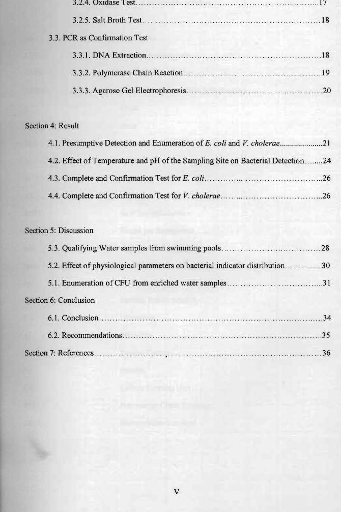
Table of Content
Title & front page...................... ... ..... . . .................. . ... ....... ............................ .. I
Acknowledgement. ........ ...... ... . .... ... .... . ................. . ........ ...... ....... . ....... .. .........11
Declaration .......................... .... ................................. . .. .... ..... . ........... ... ....... III
Table of content. ................. ............ ... ... .......... ..... ... ...... ... ...... . ....... .. ....... ...... V
List of abbreviation............ . ........ ............... ... . . .... ............. . ....... .. . ................. VII
List of table and figure ... ... ....... ............... ............................ . ........................VIII
Abstract. ...... . ........ . . ... .......... .......... .................................... . . . .... .. ................ 1
Section I: Introduction ................. .... .. ...... ................... .. ...... ..... . . . ....... .... ... ......2
Section 2: Literature Review
2.1. Pathogens in Swimming Pool .. . ... . ...... .. ... ... .. .... ................... ..... ... ........4
2.1.1. Factors Associated with Bacterial Contamination in Swimming Pool. ... 5
2.1.2. Transmission of Pathogenic Microorganism ........... . . ... ... . ..............5
2.2. Evidence of Microbiological Contaminations Related to Swimming Pool . . .. .. . ... 6
2.3. Guidelines of Water Quality Monitoring for Swimming Pools ..... ......... ..........9
2.4. Spread Plate Technique .................. . .. ... . . .... . ..... . . ..... .. ... ... . .......... .. .. .. 11
2.5. Polymerase Chain Reaction..... . ... . . . ... ....................................... ... . .... .12
Section 3: Materials and Methods
3.1. Sample Preparations
3.1.1. Sample Collection ...... ....... . .... . . ............................... ..... . ...... 13
3.1.2. Enumeration by Spread Plate Method ........................ ... ............ 13
3.2. Biochemical Detection
3.2.1. Isolation Purification ........... ... ............ ......................... ... .. .... 15
3.2.2. Sulfide, Indole and Motility Test .... . .. ....................... .. ...... ....... 16
3.2.3. Voges-Proskauer Test .............. . ..... .... ...... .... ........ ..... .... ....... 16
IV
3.2.4. Oxidase Test .......... . ...... ................................. . ..... . ... .... . .... 17
3.2.5. Salt Broth Test . ... .. ... .. ...................... . . . .......... . . ... ... . .. ... . ..... IS
3.3. PCR as Confirmation Test
3.3.1. DNA Extraction .... ... ... .... .... . . . ... .. . . .... . .. . . . .......... . ......... ....... IS
3.3.2. Polymerase Chain Reaction ..... ... .. ... . .... ... ... .. ... .. .. ... .. . ... ......... 19
3.3.3. Agarose Gel Electrophoresis . . . . . .... .. ................ .... . . ..................20
Section 4: Result
4.1. Presumptive Detection and Enumeration of E. coli and V cholerae .. ...................21
4.2. Effect of Temperature and pH of the Sampling Site on Bacterial Detection .. ......24
4.3. Complete and Confirmation Test for E. coli .......... ....... ...... . ... ... . ...... . ....... 26
4.4. Complete and Confirmation Test for V cholerae ...... ...... ... . .. ... ... ... . ...........26
Section 5: Discussion
5.3. Qualifying Water samples from swimming pools ....... .. ... . .. ......... . ............ 2S
5.2. Effect of physiological parameters on bacterial indicator distribution ........... . ..30
5.1. Enumeration of CFU from enriched water samples .......... .. ..... . ... . . . ........... .31
Section 6: Conclusion
6.1. Conclusion ................................. . .. . .. . .. . . ........ .. ... ........... . ......... ..... 34
6.2. Recommendations .... .. .... . ...... .. .. ...... . . . ... .. ............. ... . . ............... . .... . 35
Section 7: References .... .. ................... . , . ........ . . . ... . .............. ... ........................ 36
v
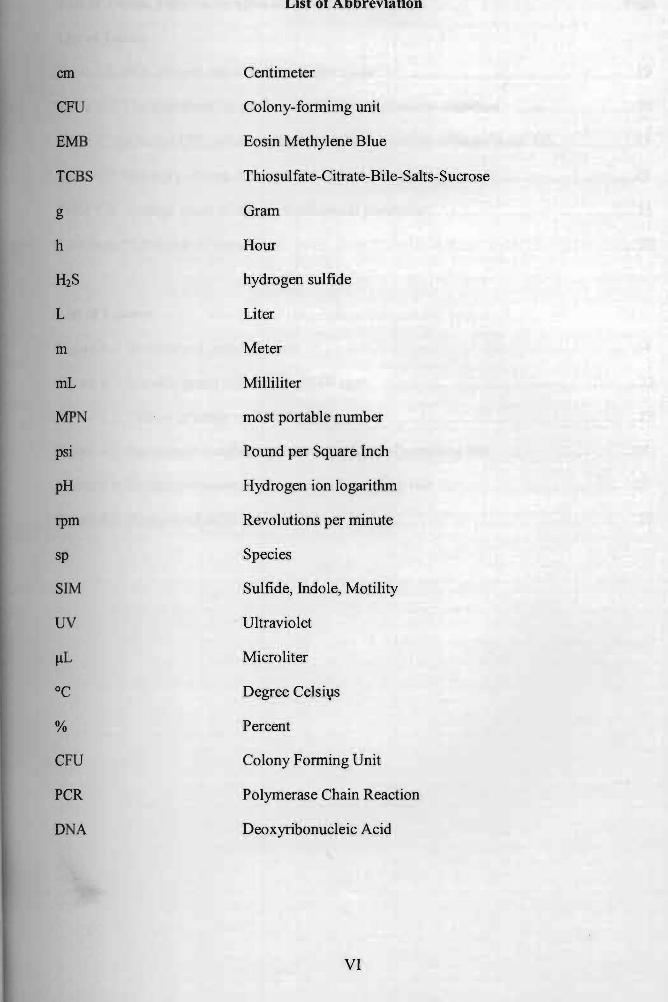
List of Abbreviation
em Centimeter
CFU Colony-formimg unit
EMB Eosin Methylene Blue
TeBS Thiosulfate-Citrate-Bile-Salts-Sucrose
g Gram
h Hour
H2S hydrogen sulfide
L Liter
m Meter
mL Milliliter
MPN most portable number
psi Pound per Square Inch
pH Hydrogen ion logarithm
rpm Revolutions per minute
sp Species
SIM Sulfide, Indole, Motility
UV Ultraviolet
ilL Microliter
°C Degree Celsil}s
% Percent
CFU Colony Forming Unit
PCR Polymerase Chain Reaction
DNA Deoxyribonucleic Acid
VI
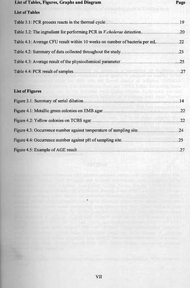
List of Tables, Figures, Graphs and Diagram Page
List of Tables
Table 3.1 : PCR process reacts in the thennal cycle .... .... .. . ...... . .. . . . ... ..... .. . .. ..... . ... .. .. 19
Table 3.2: The ingredient for perfonning PCR in Vcholerae detection.... . . ... ...... ... ...... .. 20
TabJe 4.1: Average CFU result within 10 weeks on number ofbacteria per mL .... , . ... ..... .. 22
Table 4.2: Summary of data collected throughout the study .. ... . . ...... . .... . .. .. ..... ' " . . ......23
Table 4.3: Average result of the physiochemical parameter. ..... . ......... .. . ... ... . . .... .. ....... 25
Table 4.4: PCR result of samples . .... , ... .. , ... .. , . ..... . .. . ..... .... . .. . . .. . . .. ... ... .. .. .. ... . . . ... .. 27
List of Figures
Figure 3.1: Summary of serial dilution . .. .. . ... .. .. .. .. ... ..... .. .. . . ..... . . . . ... . . ..... .. . . ... .... .... 14
Figure 4.1: Metallic green colonies on EMB agar .. .. ..... . .. . .... ... ... . .... . . ... ... .. .... .... ..... .22
Figure 4.2: Yellow colonies on TCBS agar .... .... . ... .. .. .. ..... .... . ... ... .. .... .. . .... . ... .... .... 22
Figure 4.3: Occurrence number against temperature of sampling site .... . . ... ..... ..... . .. .... . . 24
Figure 4.4: Occurrence number against pH of sampling site ... .. ... ... .. . ...... .. ..... ... ........ . 25
Figure 4.5: Example of AGE result .. .... ... ...... . . ... .... .... ... .. .. .... .. .. . ..... ..... ... ... ... .... .27
VII
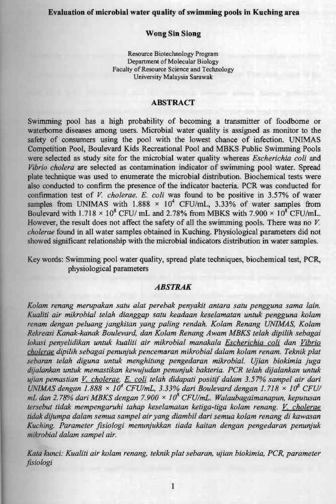
Evaluation of microbial water quality of swimming pools in Kuching area
Wong Sin Siong
Resource Biotechnology Program Department of Molecular Biology
Faculty of Resource Science and Technology University Malaysia Sarawak
ABSTRACT
Swimming pool has a high probability of becoming a transmitter of foodborne or waterborne diseases among users. Microbial water quality is assigned as monitor to the safety of consumers using the pool with the lowest chance of infection. UNIMAS Competition Pool, Boulevard Kids Recreational Pool and MBKS Public Swimming Pools were selected as study site for the microbial water quality whereas Escherichia coli and Vibrio cholera are selected as contamination indicator of swimming pool water. Spread plate technique was used to enumerate the microbial distribution. Biochemical tests were also conducted to confirm the presence of the indicator bacteria. PCR was conducted for confirmation test of V. cholerae. E. coli was found to be positive in 3.57% of water
104 samples from UNIMAS with 1.888 x CFU/mL, 3.33% of water samples from Boulevard with 1.718 x 10⁸ CFUI mL and 2.78% from MBKS with 7.900 x 10⁸ CFU/mL. However, the result does not affect the safety of all the swimming pools. There was no V. cholerae found in all water samples obtained in Kuching. Physiological parameters did not showed significant relationship with the microbial indicators distribution in water samples.
Key words: Swimming pool water quality, spread plate techniques, biochemical test, PCR, physiological parameters
ABSTRAK
Kolam renang merupakan satu alat perebak penyakit antara satu pengguna sama lain. Kualiti air mikrobial telah dianggap satu keadaan keselamatan untuk pengguna kolam renam dengan peluang jangkitan yang paling rendah. Kolam Renang UN/MAS, Kolam Rekreasi Kanak-kanak Boulevard, dan Kolam Renang Awam MBKS telah dipilih sebagai tokasi penyelidikan untuk kualiti air mikrobial manakala Escherichia coli dan Vibrio cholerae dipilih sebagai penunjuk pencemaran mikrobial dalam kolam renam. Teknik plat sebaran telah diguna untuk menghitung pengedaran mikrobial. Ujian biokimia juga dijalankan untuk memastikan kewujudan penunjuk bakteria. PCR telah dijalankan untuk ujian pemastian V. cholerae. E. coli telah didapati positif dalam 3.57% sampel air dari UN/MAS dengan 1.888 x 10⁴ CFUlmL, 3.33% dari Boulevard dengan 1.718 x ](1 CFUI mL dan 2.78% dari MBKS dengan 7.900 x 1rI CFUlmL. Walaubagaimanapun, keputusan tersebut tidak mempengaruhi tahap keselamatan ketiga-tiga kolam renang. V. cholerae tidak dijumpa dalam semua sam pel air yang diambil dari semua kolam renang di kawasan Kuching. Parameter fisiologi menunjukkan tiada kaitan dengan pengedaran penunjuk mikrobial dalam sampel air.
Kata kunci: Kualiti air kolam renang, teknik plat sebaran, ujian biokimia, PCR, parameter lzsiologi
1
Section 1: Introduction
Swimming pools is usually opened for public usage where it can accommodate more than
hundreds people used a pool for water based-recreational activities. Therefore, water in the
pool is a good transmission tool for infectious diseases among human population
throughout the world (Atallah et aI, 2008). Contamination of water can lead to spreading of
diseases from those infected to thousands of healthy individual. Disinfectant such as
chlorine powder or chlorine gas is added into the pool to keep the water sanitary in
standard condition. However, there are standards to monitor the halogen-based disinfectant
in the water regularly to avoid the development of evasive ability of genns against
disinfectant (WHO, 2006). Cost saving procedure is the most important fact of water
quality maintenance of the public pools in developing countries (Issam and Samir, 2004).
Although monitoring is done in schedule, there is still leakage of the monitor procedure
which could easily ignore the possibility of improper sanitization of the water and lead to
contamination such as Cryptosporidium and Giardia contamination of swimming pools as
reported by Schets et al (2004) in Netherlands. Another case reported in Spain that
adenovirus type 4 outbreak which was detected to be related to swimming pool (Artieda et
aI, 2009). The situation consequences increased the transmission rate and waterborne
diseases outbreak in swimming pool.
Therefore, it is necessary to have monitors the level of water quality in swimming
pool to determine microbiological quality as it is the most important parameter for the
safety of swimming pool water. It is also reported some parameter have the probability to
affect the microbial population in water sample, for example pH and temperature (Erdinger
et I, 1997). Water quality monitoring criteria included total coliform count, presence or
absence detection and membrane filtration (CDC, 2010). Spread plate technique is a
2
conventional bacterial enumeration technique which taking advantage of rapid and cost
saving than other techniques (Munsch-Alatossava et ai, 2007). Therefore, spread plate
qualtification test was used as water quality monitoring procedure in this study.
The main objective of this study:
1. To enumerate the microbial indicator of water quality of 3 selected swimming
pools in Kuching area.
2. To detect the presence of Vibrio cholera and Escherichia coli as fecal indicators in
water samples from 3 swimming pools in Kuching area.
3. To determine the physiochemical condition of selected sWImmmg pools and
associates that with V cholerae and E. coli distribution.
3
Section 2: Literature Review
2.1. Pathogens in Swimming Pool
According to Kathy Pond (2005a), there are several pathogens had included as indicator of
water safety for monitoring the water quality. Campylobacter jejuni, Escherichia coli
0157:H7, Helicobacter pulori, Legionella sp., Mycobacterium avium complex, Shigella sp.,
and Vibrio sp. are common fresh water bacterial pathogens that can be found in most water
recreation and some are lethal to human (Barrell et ai, 2000). The pathogenic protozoa
such as Cryptosporidium, Giardia, Microsporidia, and Naegeriafowleri can be commonly
found in swimming pool (Kathy, 2005a). Some of these protozoa are resistance to chlorine
disinfection such as Microsporidia and Cryptosporidium. Schistosoma sp. is the pathogenic
nematode which can survive under chlorine disinfectant in the swimming pool (Franca et
ai, 2002). Viruses such as adenovirus, coxsackievirus, echovirus, Hepatitis A virus and
Hepatitis B virus should also be considered as one of the disease transmission factor in the
public swimming pool. These organisms are highly contagious in little dose through their
point of entry on human body (Kathy, 2005b). Kathy (2005b) also stated that consumer can
be infected with waterborne diseases in .contact of pathogen-containing water pools,
however, the infection is depending on the dose and physical condition of the consumer at
the time of exposure.
4
Pusat jdma R tal Akad 11k UI\1VERSm MALAYSIA SARAWAK
2.1.1. Factors Associated with Bacterial Contamination in Swimming Pool
Microorganisms which are continuously introduced into a well-maintained swimming pool
should be kept lower than threshold level which compliance with standard regulation
(Atallah, 2008). The nitrogen-based nutrient such as urea and phosphate-based nutrient is
excreted from the human body and the water of the swimming pool washed them away
from skin due to the movement of the body in the water.
Apart from that, fecal contamination from enteric system can lead to distribution of
carbon sources into the pool water. Some heavy metals or dust from atmosphere can also
contaminate the water of the public outdoor swimming pools (Erdinger et at, 1997). In
addition the heat energy from sunlight warms up the water surface, causing the rise of
water temperature which provide the optimum temperature for the microbes to grow
actively. Phytoplankton in the water is able also absorb sunlight and provides carbon
sources to the bacteria in the water.
2.1.2. Transmission of pathogenic microorganism
Pathogenic microorganisms are transmitted to a human host through route of entry which
may invade the human host to continue their survival (Gerald et at, 2007). Transmission of
pathogens from swimming pool water to human host is commonly through respiratory tract,
skin and gastrointestinal tract. Air-transmitted microorganism can transmit to host through
the osol which released from swimmers to the inhaler. Gastrointestinal tract is also a
favourite route for microorganism such as E.coU as fecal coliform to infect a host in
5
swimming pool water (lssam and Samir, 2003). Fecal coliform are able to survive in the
hwnan bacteriostatic barrier in gastrointestinal tract such as acidic HCI and bile salt
(Gerald et ai, 2007).
2.2. Evidence of Microbiological Contaminations Related to Swimming Pool
According to Crone (1974), from a total of 227 total of water samples collected from 20
swimming pools in London, Staphylococcus aureus was present as 67% in water samples
with concentration of which less than 100 CFU/ 100 mL (43 %) and more than 100 CFU/
100 mL (53 %). The prevalence of S. aureus indicated the ability to survive and grow in
swimming pool and cause staphylococcus related outbreak.
From the annual report concluded by Michael from CDC in 1985, two issues which
related to swimming pool had been highlighted in the report. Giardiasis outbreak was cause
by the lack of pool water filtering system to filter out giardia cysts from the water (Michael,
1988). Apart from that, Pseudomonas folicultis outbreak which first reported in 1975 was
majorly related to swimming pool due to inadequate pH level with free residue chlorine
level.
Issam and Samir (2003) had investigated the swimming pool water samples which
collected from at West Bank of Palestine and found that most of swimming pool water
samples were not complied with Palestinian and WHO standards. There were about 91.3%
of swimming pools which were contaminated with Salmonella sp. and about 40% of the
sampJ from Total Bacterial Count test did not compliance to the standard. These results
6
had concluded that the developing countries have poor swimming pool water quality
maintaining system.
In 2004, Vermont Department of Health (VDH) announced the outbreak of
Norovirus gastroenteritis due to swimming club in Vermont. The malfunctioned chlorine
feeding system had leaded to the outbreak due to decreasing of free residue chlorine level
which to have decreased disinfection effect against the pathogen in the swimming pool
(Vermont Department ofHealth , 2004).
In the same year, Cryptosporidium and Giardia present was reported to be found on
7 swimming pool filters in Netherlands (Schets et aI, 2004). In the report, 11 .8% of
swimming pool water found to be positive which 4.6 % was Cryptosporidium, 5.9 % was
Giardia and 1.3 % of both Cryptosporidium and Giardia. Cryptosporidium oocysts and
Giardia cysts were detected from one toddler pool and a learner pool water samples
collected. The risk assessment of infection was indicated as one infection per 10,000
persons per year (Schets et aI, 2004).
In 2006, the report of E. coli strain 0157 outbreak had been summarized. The
Outbreak of E. coli 0157 was reported related with swimming pool (Verma et al., 2006).
The infected persons are frequently been found at the group of age less than 5 years old.
There are 106 cases reported regarding to t~e outbreak in England and Wales in between
1995 and 2000. However, as the technology becomes advanced, the maintained free
residue chlorine at the adequate level had showed the decreasing of susceptible cases of
hemorrhagic E. coli strain outbreak in United Kingdom.
There was microbiological contamination investigated at Tirana and Durres region of
Alb i in 2008. According to the investigation done by Mirela et al (2008), 6 pools were
selected and monitored twice a month from May to September in 2008. A total of 260
7
water samples were collected from the pools and analyzed. From the result obtained, most
samples had standard Total Coliform Count. However, the fecal indicators exceeded the
referenced value. Apart from laboratory investigation, 826 patients who suffered the
waterborne illness symptoms were interviewed. From the total, 72 cases were positive with
both Chlamydia trachomatis and Candida albicans infection, 72 cases were positive of
only C. trachomatis infection and 448 cases were positive of only C. albicans infection.
The rest of 234 cases were negative from the infection. Not only that, from 122
staphylococcus infection cases, 39% were infected with Staphylococcus aureus and 5%
were Staphylococcus epidermis positive. The rest of the cases were negative from both
species .infection.
Lastly, pharyngoconjunctival fever outbreak was reported in July 2008 at Spain.
From the report published by Artieda (2009), 59 children were affected by the outbreak
resulted from in contacted with swimming pool water. Approximately 5 out of 6
pharyngeal swab samples Adenovirus type 4 were found to be positive which obtained
from the affected children. From the interview, 73% of primary cases were confirmed
contacted with municipal swimming pool. 25% of secondary cases were confirmed
contacted with primary cases.
From the case study above, it became a necessary reason to conduct the
investigation of microbial water quality of swimming pool in Kuching. The sanitary
condition of public swimming pool water should be studied to ensure the safety of pool
user and also to reduce the risk of waterborne-illness which related to swimming pool.
8
2.3. Guidelines of Water Quality Monitoring for Swimming Pools
A well-organized pool had monitoring procedure guideline to maintain the water quality of
the pool. The following is the guideline from World Health Organization (2006) used to
standardize the local swimming pool water quality.
For a well-maintained pool, the tum over period should be between three to four
hours for competitive pools which is 50 m long as shown in Table 1 in Section Appendix
and the disinfectant dosing and filter operation should be 24 hours continuously operated
(WHO, 2004). For the microbiologica] criteria, there had been separated into few type of
microorganisms: heterotropic colony count, thermotolerant (fecal) coliform or Escherichia
coli, Pseudomonas auruginosa, staphylococcus aureus, and giardia and cryptosporidium.
Heterotropic colony count (HPC) is the enumeration test which is culture-based
intended to grow microorganism found in water samples (WHO, 2003). The test should be
conducted at 35°C-37°C, and the colony counted should be less than 200CFU/mL of water
samples (WHO, 2006). As long as no coliform or E.coli present in the water samples
collected and condition of pool maintenance is within satisfactory level, the incidentally
higher count is still acceptable (Queensland, 2004). However, the sudden rise of the count
should raise a concern immediately. If the 'high count is still persisting, the result can be
concluded that the pool maintenance operating system is unsatisfied and immediate
investigation of the system is required (Queensland, 2004).
Another criterion is the thermotolerant coliform or E.coli. In laboratory method,
col'forms are defined as lactose fermenter and gas production group of microorganism
including Escherichia coli (CDC, 2010). The concentration of thermotolerant coliform or
E. coli should be less than I II 00 mL within operational level (WHO, 2006). It is acceptable
9
of the count of coliform is less than 10 per 100 mL of water sample. However, if it persist
to be more than 10 counts per 100mL of water samples, investigation is necessary be
performed (Barrell et ai, 2000). The presence of the coliform or E.coli indicates that
possibility of fecal contamination (Queensland, 2004). It can also indicated that the failure
of treatment process to maintain sanitary of the pool at the time of sampling. Any count in
100 mL required concerns and repetition of test shall be conducted (Queensland, 2004).
Apart from that, Pseudomonas auruginosa is also an indicator of microbiological
criteria. P. auruginosa is a waterborne pathogen which causes gastrointestinal infection to
the consumer (WHO, 2003). It shall not be present in a well-maintained pool. The presence
of P. auruginosa indicates that the possible of other environmental pathogens such as
legwnella sp. present in the water (Queensland, 2004). Therefore, if P.auruginosa count
more than 100/1 OOmL is detected in water samples, it is recommended to repeat the test
after turbidity, disinfectant residual and pH is re-measured and one turnover is passed
(WHO, 2006).
Staphylococcus aureus is another criterion of microbiological monitoring. However,
it is seldom to have a routine test for s'aureus which is not always necessary to be tested
(Queensland, 2004). According to CDU of Queensland government (2004), when there is
any link between disease outbreak and pool suspected, the test for presence of S.aureus
should be included as a part of water investigation as monitoring water quality. Well
maintained pools and proper sample collection normally does not result in detectable of
s.aureus in 100mL of water samples (Queensland, 2004). However, if s'aureus is detected,
the count should be less than 100 mL (WHO, 2006).
Giardia and Cryptosporidium are pathogenic microorganism which can be
transmitted through contaminated water such as swimming pools. It is necessary not to
10
detect on the presence of Giardia and Crytosporidium in water samples (Queensland,
2004). CDU of Queensland (2004) stated that if the adult or cyst of either Giardia or
Cryptosporidium or both detected in the water samples, however, it can be indicated that
there are particular problem present in the operating and maintaining system of the pools.
Lastly, there is also possibility of unsatisfactory microbiological results as a criterion
of microbiological monitoring. Any unsatisfactory results obtained from the laboratory
investigation, resampling for microbiological testing should be conducted to increase the
effectiveness of the corrective actions (Queensland, 2004). However, if the problem is
persisted, failure of pool maintaining and operating system may be indicated.
1.4. Spread Plate Technique
Spread plate technique is developed since the technique used by Robert Koch to fix the
microbiological organism on the microscopic slides in 1877 which utilized the gelatin and
microscopic slide in order to visualize the disease causing microorganism (Brock, 1988).
Afterwards, spread plate technique is toen widely used recently for the bacterial
quantification steps. Spread plate technique is a technique which spreads the bacterial
suspension evenly over the agar plate surface (Wise, 2006). By using a small amount of
bacterial suspension, the bacterial is spread by sterile bent glass rod so that the growth of
colony can be enumerated subsequently during incubation. This technique is mostly used
for e aerobic culture of microorganism (Jaisai, 2010). The single colony formed on the
agar represents one colony forming unit which might cause by the single cell
11
multiplication or growth. The acceptable colony count is within the range of 30 and 300
colonies. The colony count less than 30 CFU is considered as too few to count because it
will be statistically unreliable. The colony count more than 300 CFU is considered as too
numerous to count because it may cause counting errors. The advantage of spread plate
techniques is that spread plate method can have a low colony count (CFU/mL) which
disinfectant treated swimming pool water may have extremely low amount of bacteria
compared to MPN method which gives higher colony count (Hunsinger et aI, 2005.).
2.5. Polymerase Chain Reaction
Presumptive test alone is insignificant to prove that there is any specific speCIes of
microorganism in the swimming pool which should be reported back to the pool manager.
Because of most the biochemical metabolism of microorganism shared among species, it
makes us difficult to make a significant result as detection of some pathogenic
microorganism which could harm the pool user. Molecular identification is necessary for
confinning the existence of the specific species. Polymerized Chain Reaction (PCR) is one
of the methods to molecularly detect the specific gene which only present in the specific
species, which can used to differentiate the species from whole cloud of the microbial
population (Sharbatkhori et aI, 2009). The process amplifies the specific gene using primer
to tag the location of gene where it can be amplified into thousands of copies so that we
can easily detect the presence of the gene using gel electrophoresis. OmpW is the gene
'ch presence in Vibrio cholerae. OmpW encodes the outer membrane protein of the
V.c ae which makes it specific nutrient utilization and metabolism processes in
environmental change adaptation (Nandi et aI, 2005).
12
Section 3: Materials and Methods
3.1. Sample Preparation
3.1.1. Sample Collection
Sample collection was perfonned with reference of WHO sampling techniques (WHO,
1997). Falcon tube and I % sodium thiosulfate were pre-sterilized through autoclaving
before use. Then swimming pool water was collected at 20 cm from water surface. Then
450 J.l.1 of I % sodium thiosulfate solution was added to the water samples to deactivate the
residue chlorine in the water samples to prevent further disinfectant effects on
microbiological population. Then the water samples collected were preserved in ice. The
water samples were processed within 2 hours after sample collected. The temperature and
pH of the water samples were measured and recorded.
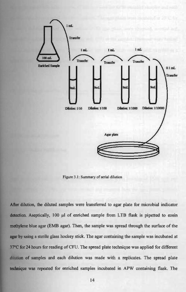
3.1.2 Enumeration by Spread Plate Method
About 10 ml ofwater samples were transferred to conical flasks containing 90 ml of lauryl
tryptose broth, LTB (ratio I: I 0) and incubated in shaking incubator at 37°C for 16-24
hours. The steps were repeated for the enrichment for Vibrio cholera in water sample by
using alkaline peptone water, (pH pre-adjusted to 8.6). After incubation, the enriched
les were proceeded for serial dilution of 10-1, 10-2 and until 10-7
• Figure 3.1 showed
the summary of the serial dilution on enumerating bacterial concentration in water samples:
13
~ I mLI mL ImL
~~ Transfer Transfer
Dilution: 1,'10 Dilution: n 00 DiIuboo: I '1000
Agar plate
Figure 3.1: Summary of serial dilution
After dilution, the diluted samples were transferred to agar plate for microbial indicator
detection. Aseptically, 100 ~l of enriched sample from LTB flask is pipetted to eosin
methylene blue agar (EMB agar). Then, the sample was spread through the surface of the
agar by using a sterile glass hockey stick. The agar containing the sample was incubated at
37°C for 24 hours for reading of CFU. The spread plate technique was applied for different
. n of samples and each dilution was made with a replicates. The spread plate
technique was repeated for enriched samples incubated in APW containing flask. The
14
thiosulfate-citrate-bile-salts-sucrose, TCBS agar used for APW enriched samples and each
dilution was also proceeded for a two replicate. The agar plates were incubated at 29°C for
24 hours. Colonies fonned on the surface of the agar plates were observed, counted and
recorded to calculate the colony forming unit, CFU in the samples. However, only desired
colony and number of colony formed on the agar plates were counted and recorded as a
measurement. The following is the CFU per mL calculation formula:
1 1 CFU/mL = No.of colonies x dilution factor X volume used in mL
3.2. Biochemical detection
3.2.1 Isolate Purification
Once the desired colony on agar plates such as metallic green colonies on EMB agar and
yellow colonies on TCBS agar were counted and recorded from the agar plates, positive
colony on the agar plates was isolated usi,ng streak plate technique for obtaining the pure
culture from the massive mixture of the bacterial population on the agar plate. By using a
sterile inoculating loop, a metallic green colony grew on the EMB agar was streaked into a
new sterile EMB agar through streak plate method. Then the agar plate was incubated at
37°e for 24 hours for the further test. For colony grew on the TCBS agar, the yellow
ny fonned on the TCBS agar was inoculated using sterile inoculating loop to a new
sterile TeSS agar plate through streak plate technique aseptically. Then the agar plate was
15
_:ubllted at 29°C for 24 hours. After the pure culture on the plates was isolated, the
• ·ve colony fonned on EMB agar and TCBS agar was further isolated in Nutrient agar,
for further test.
3.2.2 Sulfide, Indole and Motility Test
SIM test is used complete the detection of the presence of Escherichia coli found on the
EMB agar. Using a sterile inoculating stab, a metallic green colony isolated on EMB agar
as swabbed and then stabbed into a tube containing 10 ml of SIM medium. Then the tube
was incubated in incubator at 37°C for 24 hours. After incubation, the medium was added
• few drops of Kovach' s reagent for indole test (MacWilliams, 2009). Indole positive
appears to change the colourless reagent to pink. Motility positive shows increased in
turbidity of the medium whereas hydrogen sulfide production positive turns the medium
hlack. A positive result for E.coli in SIM medium supposed to be indole and motility
positive but sulfide negative.
3.2.3 Voges-Proskauer Test
oges-Proskauer, VP test was used to complete the detection of E.coli and V.cholerae
the isolates of different agar plates. Before the test ran, the testing culture was first
Incubated in MR-VP broth. One colony from NA culture isolated from previous agar plate
16