daftar pustaka 7689
TRANSCRIPT
8/10/2019 daftar pustaka 7689
http://slidepdf.com/reader/full/daftar-pustaka-7689 1/20
American Thoracic Societv
Diagnostic Standards and Classification of Tuberculosis in Adults and ChildrenT HIS O FFICIAL S TATEMENT OF THE A MERICAN T OR X S OCIETY AND THE C ENTERS FOR D ISEASE C ONTROL AND P REVENTION
WAS ADOWED BY THE ATS B OARD OF DIRE~ORS J ULY 1999. T HIS S TATEMENT WAS ENDORSED BY THE C OUNCIL OF TH E
INFEWOUS D ISEASE S OCIETY OF A MERICA , SEPTEM ER 1999
CONTENTS
IntroductionI. EpidemiologyII. Transmission of M ycobacterium t uberculosis III. Pathogenesis of TuberculosisIV. Clinical Manifestations of Tuberculosis
A. Systemic Effects of TuberculosisB. Pulmonary TuberculosisC. Extrapulmonary Tuberculosis
V. Diagnostic MicrobiologyA. Laboratory Services for Mycobacterial DiseasesB. Collection of Specimens for Demonstration of
Tubercle BacilliC. Transport of Specimens to the LaboratoryD. Digestion and Decontamination of SpecimensE. Staining and Microscopic ExaminationF. Identification of Mycobacteria Directly from
Clinical SpecimensG. Cultivation of MycobacteriaH. Identification of Mycobacteria from CultureI. Drug Susceptibility TestingJ. Genotyping of M ycobacterium t uberculosis
K. Assessment of Laboratory PerformanceVI. Tuberculin Skin Test
A. TuberculinB. Immunologic Basis for the Tuberculin ReactionC. Administration and Reading of TestsD. Interpretation of Skin Test ReactionsE. Boosted Reactions and Serial Tuberculin TestingF. Previous Vaccination with BCGG. Definition of Skin Test ConversionsH. Anergy Testing in Individuals Infected with HIV
VII. Classification of Persons Exposed to and/or Infected withM ycobacterium t uberculosis
VIII. Reporting of TuberculosisReferences
INTRODUCTION
The “Diagnostic Standards and Classification of Tuberculosisin Adults and Children” is a joint statement prepared by theAmerican Thoracic Society and the Centers for Disease Con-trol and endorsed by the Infectious Disease Society of America.The Diagnostic Standards are intended to provide a framework for and understanding of the diagnostic approaches to tuber-
m rit Care Me d Vol 161. pp 1376-l 395 200OInternet address: www atsjournals org
culosis infection/disease and to present a classification schemethat facilitates management of all persons to whom diagnostictests have been applied.
The specific objectives of this revision of the DiagnosticStandards are as follows.
1.
2.
To define diagnostic strategies for high- and low-risk pa-tient populations based on current knowledge of tuberculo-sis epidemiology and information on newer technologies.To provide a classification scheme for tuberculosis that is based on pathogenesis. Definitions of tuberculosis diseaseand latent infection have been selected that (a) aid in anaccurate diagnosis; (b) coincide with the appropriate re-sponse of the health care team, whether it be no response,treatment of latent infection, or treatment of disease; (c)
provide the most useful information that correlates withthe prognosis; (d) provide the necessary information for appropriate public health action; and (e) provide a uni-form, functional, and practical means of reporting. Becausetuberculosis, even after it has been treated adequately, re-mains a pertinent and lifelong part of a person’s medicalhistory, previous as well as current disease is included inthe classification.
This edition of the Diagnostic Standards has been preparedas a practical guide and statement of principles for all personsinvolved in the care of patients with tuberculosis. Referenceshave been included to guide the reader to texts and journal ar-ticles for more detailed information on each topic.
I. EPIDEMIOLOGY
Tuberculosis remains one of the deadliest diseases in the world.The World Health Organization (WHO) estimates that eachyear more than 8 million new cases of tuberculosis occur andapproximately 3 million persons die from the disease (1). Ninety-five percent of tuberculosis cases occur in developing coun-tries, where few resources are available to ensure proper treatment and where human immunodeficiency virus (HIV)infection may be common. It is estimated that between 19 and43% of the world’s population is infected with M ycobacterium tuberculosis, the bacterium that causes tuberculosis infectionand disease (2).
In the United States, an estimated 15 million people are in-fected with M . tuberculosis (3). Although the tuberculosis caserate in the United States has declined during the past fewyears, there remains a huge reservoir of individuals who areinfected with M . tu berculo sis. Without application of effectivetreatment for latent infection, new cases of tuberculosis can beexpected to develop from within this group.
Tuberculosis is a social disease with medical implications. Ithas always occurred disproportionately among disadvantaged
populations such as the homeless, malnourished, and over-
8/10/2019 daftar pustaka 7689
http://slidepdf.com/reader/full/daftar-pustaka-7689 2/20
American Thoracic Society 13 77
crowded. Within the past decade it also has become clear thatthe spread of HIV infection and the immigration of personsfrom areas of high incidence have resulted in increased num- bers of tuberculosis cases.
II. TRANSMISSION OF Mycobacterium tuberculosis
Tuberculosis is spread from person to person through the air by droplet nuclei, particles 1 to 5 pm in diameter that containM. tuberculosis complex (4). Droplet nuclei are producedwhen persons with pulmonary or laryngeal tuberculosis cough,sneeze, speak, or sing. They also may be produced by aerosoltreatments, sputum induction, aerosolization during bron-choscopy, and through manipulation of lesions or processingof tissue or secretions in the hospital or laboratory. Dropletnuclei, containing two to three M . t uberculosis organisms (5)are so small that air currents normally present in any indoor space can keep them airborne for long periods of time (6).Droplet nuclei are small enough to reach the alveoli within thelungs, where the organisms replicate. Although patients withtuberculosis also generate larger particles containing numer-ous bacilli, these particles do not serve as effective vehicles for transmission of infection because they do not remain airborne,
and if inhaled, do not reach alveoli. Organisms deposited onintact mucosa or skin do not invade tissue. When large parti-cles are inhaled, they impact on the wall of the upper airways,where they are trapped in the mucous blanket, carried to theoropharynx, and swallowed or expectorated (7).
Four factors determine the likelihood of transmission of M .tuberculosis: I ) the number of organisms being expelled intothe air, (2) the concentration of organisms in the air deter-mined by the volume of the space and its ventilation, (3) thelength of time an exposed person breathes the contaminatedair, and (4) presumably the immune status of the exposed indi-vidual. HIV-infected persons and others with impaired cell-mediated immunity are thought to be more likely to becomeinfected with M . t uberculosis after exposure than persons withnormal immunity; also, HIV-infected persons and others withimpaired cell-mediated immunity are much more likely to de-velop disease if they are infected. However, they are no morelikely to transmit M . tuberculo sis (8).
Techniques that reduce the number of droplet nuclei in agiven space are effective in limiting the airborne transmission of tuberculosis. Ventilation with fresh air is especially important,
particularly in health care settings, where six or more room-air changes an hour is desirable (9). The number of viable airbornetubercle bacilli can be reduced by ultraviolet irradiation of air inthe upper part of the room (5). The most important means toreduce the number of bacilli released into the air is by treatingthe patient with effective antituberculosis chemotherapy (10). If masks are to be used on coughing patients with infectious tu-
berculosis, they should be fabricated to filter droplet nuclei and
molded to fit tightly around the nose and mouth. Measures suchas disposing of such personal items as clothes and bedding, ster-ilizing fomites, using caps and gowns and gauze or paper masks,
boiling dishes, and washing walls are unnecessary because theyhave no bearing on airborne transmission.
There are five closely related mycobacteria grouped in theM . tuberculosis complex: M . tuberculosis, M . bovis, M . a f r i -canum, M. mi croti , and M . canett i (11, 12). M ycobacterium t u- berculosis is transmitted through the airborne route and thereare no known animal reservoirs. M ycobacter ium bovis may
penetrate the gastrointestinal mucosa or invade the lymphatictissue of the oropharynx when ingested in milk containinglarge numbers of organisms. Human infection with M . bo v i s has decreased significantly in developed countries as a result
of the pasteurization of milk and effective tuberculosis control programs for cattle (13). Airborne transmission of both M . bo -vis and M . africanum can also occur (14-16). M ycobacter ium bovis BCG is a live-attenuated strain of M . bovis and is widelyused as a vaccine for tuberculosis. It may also be used as anagent to enhance immunity against transitional-cell carcinomaof the bladder. When used in this manner, adverse reactionssuch as dissemination may be encountered, and in such casesM . bovis BCG may be cultured from nonurinary tract systemspecimens, i.e., blood, sputum, bone marrow, etc. (17).
III. P THOG N SIS OF TUBERCULOSIS
After inhalation, the droplet nucleus is carried down the bron-chial tree and implants in a respiratory bronchiole or alveolus.Whether or not an inhaled tubercle bacillus establishes an in-fection in the lung depends on both the bacterial virulence andthe inherent microbicidal ability of the alveolar macrophagethat ingests it 4,18). If the bacillus is able to survive initial de-fenses, it can multiply within the alveolar macrophage. The tu-
bercle bacillus grows slowly, dividing approximately every 25to 32 h within the macrophage. M ycobacterium t uberculosis has no known endotoxins or exotoxins; therefore, there is no
immediate host response to infection. The organisms grow for 2 to 12 wk, until they reach lo3 to lo4 in number, which is suffi-cient to elicit a cellular immune response (19, 20) that can bedetected by a reaction to the tuberculin skin test.
Before the development of cellular immunity, tubercle ba-cilli spread via the lymphatics to the hilar lymph nodes andthence through the bloodstream to more distant sites. Certainorgans and tissues are notably resistant to subsequent multi-
plication of these bacilli. The bone marrow, liver, and spleenare almost always seeded with mycobacteria, but uncontrolledmultiplication of the bacteria in these sites is exceptional. Or-ganisms deposited in the upper lung zones, kidneys, bones,and brain may find environments that favor their growth, andnumerous bacterial divisions may occur before specific cellu-lar immunity develops and limits multiplication.
In persons with intact cell-mediated immunity, collectionsof activated T cells and macrophages form granulomas thatlimit multiplication and spread of the organism. Antibodiesagainst M . t uberculosi s are formed but do not appear to be
protective (21). The organisms tend to be localized in the cen-ter of the granuloma, which is often necrotic (22). For themajority of individuals with normal immune function, prolifer-ation of M . tuberculosis is arrested once cell-mediated immu-nity develops, even though small numbers of viable bacilli mayremain within the granuloma. Although a primary complexcan sometimes be seen on chest radiograph, the majority of pulmonary tuberculosis infections are clinically and radio-graphically inapparent (18). Most commonly, a positive tuber-culin skin test result is the only indication that infection with
M . tuberculosis has taken place. Individuals with latent tuber-culosis infection but not active disease are not infectious andthus cannot transmit the organism. It is estimated that approx-imately 10% of individuals who acquire tuberculosis infectionand are not given preventive therapy will develop active tu-
berculosis. The risk is highest in the first 2 yr after infection,when half the cases will occur (23). The ability of the host torespond to the organism may be reduced by certain diseasessuch as silicosis, diabetes mellitus, and diseases associated withimmunosuppression, e.g., HIV infection, as well as by corti-costeroids and other immunosuppressive drugs. In these cir-cumstances, the likelihood of developing tuberculosisdiseaseis greater. The risk of developing tuberculosis also appears to be greater during the first 2-yr of life.
8/10/2019 daftar pustaka 7689
http://slidepdf.com/reader/full/daftar-pustaka-7689 3/20
1378 AMERICAN JOURNAL OF RESPIRATORY AND CRITICAL CARE MEDICINE VO’ 161 2000
HIV-infected persons, especially those with low CD4’ cellcounts, develop tuberculosis disease rapidly after becoming in-fected with M . tu berculo sis; up to 50% of such persons may doso in the first 2 yr after infection with M . tuberculosi s (24). Con-versely, an individual who has a prior latent infection with M .tuberculosis (not treated) and then acquires HIV infection willdevelop tuberculosis disease at an approximate rate of 510 per year (2526).
In a person with intact cell-mediated immunity, the re-sponse to infection with the tubercle bacillus provides protec-tion against reinfection. The likelihood of reinfection is a func-tion of the risk of reexposure, the intensity of such exposure,and the integrity of the host’s immune system. In the UnitedStates the risk of reexposure to an infectious case is low. Fur-thermore, in an otherwise healthy, previously infected person,any organisms that arc deposited in the alveoli are likely to bekilled by the cell-mediated immune response. Exceptions mayoccur, but in immunocompetent individuals, clinical and labo-ratory evidence indicates that disease produced by the inha-lation of a second infecting strain is uncommon. However,reinfection has been documented to occur both in persons withoutrecognized immune compromise and in persons with advancedHIV infection (27-29).
IV. CLINICAL MANIFESTATIONS OF TUBERCULOSIS
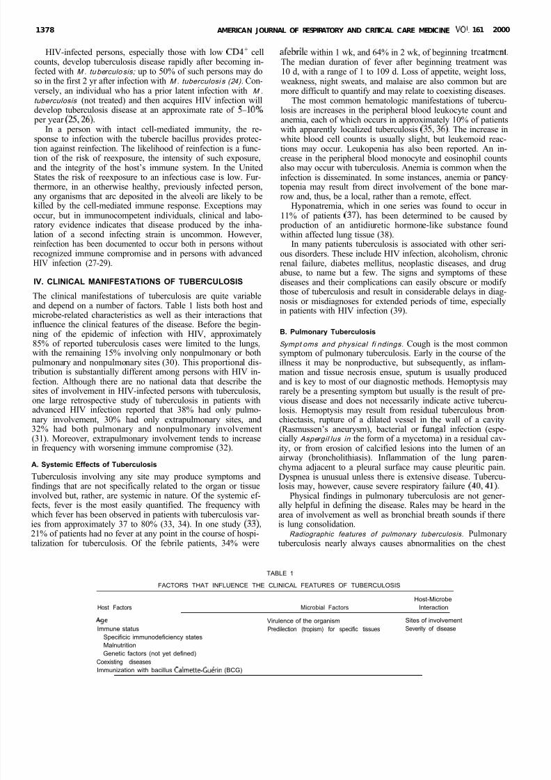
The clinical manifestations of tuberculosis are quite variableand depend on a number of factors. Table 1 lists both host andmicrobe-related characteristics as well as their interactions thatinfluence the clinical features of the disease. Before the begin-ning of the epidemic of infection with HIV, approximately85% of reported tuberculosis cases were limited to the lungs,with the remaining 15% involving only nonpulmonary or both
pulmonary and nonpulmonary sites (30). This proportional dis-tribution is substantially different among persons with HIV in-fection. Although there are no national data that describe thesites of involvement in HIV-infected persons with tuberculosis,one large retrospective study of tuberculosis in patients withadvanced HIV infection reported that 38% had only pulmo-nary involvement, 30% had only extrapulmonary sites, and32% had both pulmonary and nonpulmonary involvement(31). Moreover, extrapulmonary involvement tends to increasein frequency with worsening immune compromise (32).
A. Systemic Effects of Tuberculosis
Tuberculosis involving any site may produce symptoms andfindings that are not specifically related to the organ or tissueinvolved but, rather, are systemic in nature. Of the systemic ef-fects, fever is the most easily quantified. The frequency withwhich fever has been observed in patients with tuberculosis var-ies from approximately 37 to 80% (33, 34). In one study 33)21% of patients had no fever at any point in the course of hospi-
talization for tuberculosis. Of the febrile patients, 34% were
afebrile within 1 wk, and 64% in 2 wk, of beginning treatment.The median duration of fever after beginning treatment was10 d, with a range of 1 to 109 d. Loss of appetite, weight loss,weakness, night sweats, and malaise are also common but aremore difficult to quantify and may relate to coexisting diseases.
The most common hematologic manifestations of tubercu-losis are increases in the peripheral blood leukocyte count andanemia, each of which occurs in approximately 10% of patientswith apparently localized tuberculosis (35,36). The increase inwhite blood cell counts is usually slight, but leukemoid reac-tions may occur. Leukopenia has also been reported. An in-crease in the peripheral blood monocyte and eosinophil countsalso may occur with tuberculosis. Anemia is common when theinfection is disseminated. In some instances, anemia or pancy-topenia may result from direct involvement of the bone mar-row and, thus, be a local, rather than a remote, effect.
Hyponatremia, which in one series was found to occur in11% of patients (37), has been determined to be caused by
production of an antidiuretic hormone-like substance foundwithin affected lung tissue (38).
In many patients tuberculosis is associated with other seri-ous disorders. These include HIV infection, alcoholism, chronicrenal failure, diabetes mellitus, neoplastic diseases, and drug
abuse, to name but a few. The signs and symptoms of thesediseases and their complications can easily obscure or modifythose of tuberculosis and result in considerable delays in diag-nosis or misdiagnoses for extended periods of time, especiallyin patients with HIV infection (39).
B. Pulmonary Tuberculosis
Sympt oms and physical fi ndings. Cough is the most commonsymptom of pulmonary tuberculosis. Early in the course of theillness it may be nonproductive, but subsequently, as inflam-mation and tissue necrosis ensue, sputum is usually producedand is key to most of our diagnostic methods. Hemoptysis mayrarely be a presenting symptom but usually is the result of pre-vious disease and does not necessarily indicate active tubercu-losis. Hemoptysis may result from residual tuberculous bron-chiectasis, rupture of a dilated vessel in the wall of a cavity(Rasmussen’s aneurysm), bacterial or fungal infection (espe-cially Aspergil lus in the form of a mycetoma) in a residual cav-ity, or from erosion of calcified lesions into the lumen of anairway (broncholithiasis). Inflammation of the lung paren-chyma adjacent to a pleural surface may cause pleuritic pain.Dyspnea is unusual unless there is extensive disease. Tubercu-losis may, however, cause severe respiratory failure 40,41).
Physical findings in pulmonary tuberculosis are not gener-ally helpful in defining the disease. Rales may be heard in thearea of involvement as well as bronchial breath sounds if thereis lung consolidation.
Radiographic features of pulmonary tuberculosis. Pulmonary
tuberculosis nearly always causes abnormalities on the chest
TABLE 1
FACTORS THAT INFLUENCE THE CLINICAL FEATURES OF TUBERCULOSIS
Host Factors Microbial FactorsHost-Microbe
Interaction
AgeImmune status
Specificic immunodeficiency statesMalnutritionGenetic factors (not yet defined)
Coexisting diseases
Virulence of the organismPredilection (tropism) for specific tissues
Sites of involvementSeverity of disease
Immunization with bacillus Calmette Cuerin (BCG)
8/10/2019 daftar pustaka 7689
http://slidepdf.com/reader/full/daftar-pustaka-7689 4/20
Am er ic an Thor ac ic Soci ety 1 3 7 9
film, although an endobronchial lesion may not be associatedwith a radiographic finding. In addition, in patients with pulmo-nary tuberculosis disease and HIV infection, a normal chest filmis more common than in persons with tuberculosis disease with-out immune suppression. In primary tuberculosis occurring as aresult of recent infection, the process is generally seen as a mid-dle or lower lung zone infiltrate, often associated with ipsilat-era1 hilar adenopathy. Atelectasis may result from compressionof airways by enlarged lymph nodes. This manifestation is morecommon in children. If the primary process persists beyond thetime when specific cell-mediated immunity develops, cavitationmay occur (so-called “progressive primary” tuberculosis) (42).
Tuberculosis that develops as a result of endogenous reacti-vation of latent infection usually causes abnormalities in the up-
per lobes of one or both lungs. Cavitation is common in thisform of tuberculosis. The most frequent sites are the apical and
posterior segments of the right upper lobe and the apical-poste-rior segment of the left upper lobe. Healing of the tuberculouslesions usually results in development of a scar with loss of lung
parenchymal volume and, often, calcification. In the immuno-competent adult with tuberculosis, intrathoracic adenopathy isuncommon but may occur, especially with primary infection. Incontrast, intrathoracic or extrathoracic lymphatic involvement is
quite common in children. As tuberculosis progresses, infectedmaterial may be spread via the airways into other parts of thelungs, causing a patchy bronchopneumonia. Erosion of a paren-chymal focus of tuberculosis into a blood or lymph vessel maylead to dissemination of the organism and a “miliary” (evenlydistributed small nodules) pattern on the chest film. Dissemi-nated tuberculosis can occur in primary disease and may be anearly complication of tuberculosis in children (both immuno-competent and immunocompromised). When it occurs in chil-dren, it is most common in infants and the very young (< 5 yr).
Old, healed tuberculosis presents a different radiologic ap- pearance from active tuberculosis. Dense pulmonary nodules,with or without visible calcification, may be seen in the hilar areaor upper lobes. Smaller nodules, with or without fibrotic scars,are often seen in the upper lobes, and upper-lobe volume loss of-ten accompanies these scars. Nodules and fibrotic lesions of oldhealed tuberculosis have well-demarcated, sharp margins and areoften described as “hard.” Bronchiectasis of the upper lobes is anonspecific finding that sometimes occurs from previous puhno-nary tuberculosis. Pleural scarring may be caused by old tubercu-losis but is more commonly caused by trauma or other infections.
Nodules and fibrotic scars may contain slowly multiplying tuber-cle bacilli with significant potential for future progression to ac-tive tuberculosis. Persons who have nodular or fibrotic lesionsconsistent with findings of old tuberculosis on chest radiographand a positive tuberculin skin test reaction should be consideredhigh-priority candidates for treatment of latent infection regard-less of age. Conversely, calcified nodular lesions (calcified granu-loma) or apical pleural thickening poses a much lower risk for fu-ture progression to active tuberculosis (42,43).
In patients with HIV infection, the nature of the radio-graphic findings depends to a certain extent on the degree of immunocompromise produced by the HIV infection. Tuber-culosis that occurs relatively early in the course of HIV infec-tion tends to have the typical radiographic findings describedabove (44, 45). With more advanced HIV disease the radio-graphic findings become more “atypical”: cavitation is uncom-mon, and lower lung zone or diffuse infiltrates and intratho-racic adenopathy are frequent.
C. Extrapulmonary TuberculosisExtrapulmonary tuberculosis usually presents more of a diag-nostic problem than pulmonary tuberculosis. In part this re-
lates to its being less common and, therefore, less familiar tomost clinicians 46,47). In addition, extrapulmonary tubercu-losis involves relatively inaccessible sites and, because of thenature of the sites involved, fewer bacilli can cause muchgreater damage. The combination of small numbers of bacilliand inaccessible sites causes bacteriologic confirmation of adiagnosis to be more difficult, and invasive procedures are fre-quently required to establish a diagnosis.
Extrapul monary t uberculosis in HI V-i nfected pati ents. Pre-sumably, the basis for the high frequency of extrapulmonarytuberculosis among patients with HIV infection is the failureof the immune response to contain M . t uberculosi s, therebyenabling hematogenous dissemination and subsequent in-volvement of single or multiple nonpulmonary sites. Becauseof the frequency of extrapulmonary tuberculosis among HIV-infected patients, diagnostic specimens from any suspectedsite of disease should be examined for mycobacteria. More-over, cultures of blood and bone marrow may reveal M . tuber- culosis in patients who do not have an obvious localized site of disease but who are being evaluated because of fever.
Disseminated tuberculosis. Disseminated tuberculosis oc-curs because of the inadequacy of host defenses in containingtuberculous infection. This failure of containment may occur
in either latent or recently acquired tuberculous infection. Be-cause of HIV or other causes of immunosuppression, the or-ganism proliferates and disseminates throughout the body.Multiorgan involvement is probably much more common thanis recognized because, generally, once M . t uberculosis is iden-tified in any specimen, other sites are not evaluated. The term“miliary” is derived from the visual similarity of some dissemi-nated lesions to millet seeds. Grossly, these lesions are l- to2-mm yellowish nodules that, histologically, are granulomas.Thus disseminated tuberculosis is sometimes called “miliary”tuberculosis. When these small nodules occur in the lung, theresulting radiographic pattern is also termed “miliary.”
Because of the multisystem involvement in disseminated tu- berculosis, the clinical manifestations are protean. The present-ing symptoms and signs are generally nonspecific and are dom-inated by systemic effects, particularly fever, weight loss, nightsweats, anorexia, and weakness (48-52). Other symptoms de- pend on the relative severity of disease in the organs involved.A productive cough is common because most patients with dis-seminated disease also have pulmonary involvement. Head-ache and mental status changes are less frequent and are usu-ally associated with meningeal involvement (49). Physicalfindings likewise are variable. Fever, wasting, hepatomegaly,
pulmonary findings, lymphadenopathy, and splenomegaly oc-cur in descending order of frequency. A finding that is stronglysuggestive of disseminated tuberculosis is the choroidal tuber-cle, a granuloma located in the choroid of the retina (53).
The chest film is abnormal in most but not all patients withdisseminated tuberculosis. In the series reported by Griecoand Chmel 48) only 14 of 28 patients (50%) had a miliary
pattern on chest film, whereas 90% of 69 patients reported byMunt (49) had a miliary pattern. Overall, it appears that at thetime of diagnosis approximately 85% of patients have thecharacteristic radiographic findings of miliary tuberculosis.Other radiographic abnormalities may be present as well.These include upper lobe infiltrates with or without cavitation, pleural effusion, and pericardial effusion. In patients with HIVinfection the radiographic pattern is usually one of diffuse in-filtration rather than discrete nodules.
Ly mph node tuberculosis. Tuberculous lymphadenitis usually presents as painless swelling of one or more lymph nodes. Thenodes involved most commonly are those of the posterior or an-terior cervical chain or those in the supraclavicular fossa. Fre-
8/10/2019 daftar pustaka 7689
http://slidepdf.com/reader/full/daftar-pustaka-7689 5/20
1380 AMERICAN JOURNAL OF RESPIRATORY AND CRITICAL C RE MEDICINE VOL 161 2000
quently the process is bilateral and other noncontiguous groupsof nodes can be involved (54). At least initially the nodes are dis-crete and the overlying skin is normal. With continuing diseasethe nodes may become matted and the overlying skin inflamed.Rupture of the node can result in formation of a sinus tract,which may be slow to heal. Intrathoracic adenopathy may com-
press bronchi, causing atelectasis leading to lung infection and perhaps bronchiectasis. This manifestation is particularly com-mon in children. Needle biopsy or surgical resection of the nodemay be needed to obtain diagnostic material if the chest radio-graph is normal and the sputum smear and culture are negative.
In persons not infected with HIV but with tuberculous lym- phadenitis, systemic symptoms are not common unless there isconcomitant tuberculosis elsewhere. The frequency of pulmo-nary involvement in reported series of patients with tubercu-lous lymphadenitis is quite variable, ranging from approxi-mately 5 to 70%. In HIV-infected persons lymphadenitis iscommonly associated with multiple organ involvement.
Pleural tuberculosis. There are two mechanisms by whichthe pleural space becomes involved in tuberculosis. The differ-ence in pathogenesis results in different clinical presentations,approaches to diagnosis, treatment, and sequelae. Early in thecourse of a tuberculous infection a few organisms may gain ac-
cess to the pleural space and, in the presence of cell-mediatedimmunity, cause a hypersensitivity response (55, 56). Com-monly, this form of tuberculous pleuritis goes unnoticed, andthe process resolves spontaneously. In some patients, how-ever, tuberculous involvement of the pleura is manifested asan acute illness with fever and pleuritic pain. If the effusion islarge enough, dyspnea may occur, although the effusions gen-erally are small and rarely are bilateral. In approximately 30%of patients there is no radiographic evidence of involvementof the lung parenchyma; however, parenchymal disease isnearly always present, as evidenced by findings of lung dissec-tions (57).
The second variety of tuberculous involvement of the pleura is empyema. This is much less common than tubercu-
lous pleurisy with effusion and results from a large number of organisms spilling into the pleural space, usually from ruptureof a cavity or an adjacent parenchymal focus via a bronchop-leural fistula (58). A tuberculous empyema is usually associ-ated with evident pulmonary parenchymal disease on chestfilms and air may be seen in the pleural space. In the absenceof concurrent pulmonary tuberculosis, diagnosis of pleural tu-
berculosis requires thoracentesis and, usually, pleural biopsy.Genitourinary tuberculosis. In patients with genitourinary
tuberculosis, local symptoms predominate and systemic symp-toms are less common (59, 60). Dysuria, hematuria, and fre-quent urination are common, and flank pain may also benoted. However, the symptoms may be subtle, and, often,there is advanced destruction of the kidneys by the time a di-agnosis is established (61). In women genital involvement ismore common without renal tuberculosis than in men andmay cause pelvic pain, menstrual irregularities, and infertilityas presenting complaints (60). In men a painless or onlyslightly painful scrotal mass is probably the most common pre-senting symptom of genital involvement, but symptoms of
prostatitis, orchitis, or epididymitis may also occur (59). Asubstantial number of patients with any form of genitourinarytuberculosis are asymptomatic and are detected because of anevaluation for an abnormal routine urinalysis. In patients withrenal or genital tuberculosis, urinalyses are abnormal in morethan 90 , the main finding being pyuria, and/or hematuria.The finding of pyuria in an acid urine with no routine bacterialorganisms isolated from a urine culture should prompt anevaluation for tuberculosis by culturing the urine for myco-
bacteria. Acid-fast bacillus (AFB) smears of the urine should be done, but the yield is low. The suspicion of genitourinarytuberculosis should be heightened by the presence of abnor-malities on the chest film. In most series, approximately 40 to75% of patients with genitourinary tuberculosis have chest ra-diographic abnormalities, although in many these may be theresult of previous, not current, tuberculosis 59,60).
Skeletal tuberculosis. The usual presenting symptom of skeletal tuberculosis is pain (62). Swelling of the involved jointmay be noted, as may limitation of motion and, occasionally,sinus tracts. Systemic symptoms of infection are not common.Since the epiphyseal region of bones is highly vascularized ininfants and young children, bone involvement with tuberculo-sis is much more common in children than adults. Approxi-mately 1% of young children with tuberculosis disease will de-velop a bony focus (63). Because of the subtle nature of thesymptoms, diagnostic evaluations often are not undertakenuntil the process is advanced. Delay in diagnosis can be espe-cially catastrophic in vertebral tuberculosis, where compres-sion of the spinal cord may cause severe and irreversible neu-rologic sequelae, including paraplegia.
Fortunately, such neurologic sequelae represent the more se-vere end of the spectrum. Early in the process the only abnor-mality noted may be soft tissue swelling. Subsequently, subchon-dral osteoporosis, cystic changes, and sclerosis may be noted
before the joint space is actually narrowed. The early changes of spinal tuberculosis may be particularly difficult to detect by stan-dard films of the spine. Computed tomographic scans and mag-netic resonance imaging of the spine are considerably more sen-sitive than routine films and should be obtained when there is ahigh index of suspicion of tuberculosis. Bone biopsy may beneeded to obtain diagnostic material if the chest radiograph isnormal and the sputum smear and culture are negative.
Central nervous system tuberculosis. Tuberculous meningitisis a particularly devastating disease. Meningitis can result fromdirect meningeal seeding and proliferation during a tubercu-lous bacillemia either at the time of initial infection or at the
time of breakdown of an old pulmonary focus, or can resultfrom breakdown of an old parameningeal focus with ruptureinto the subarachnoid space. The consequences of subarach-noid space contamination can be diffuse meningitis or localizedarteritis. In tuberculous meningitis the process is located pri-marily at the base of the brain (64). Symptoms, therefore, in-clude those related to cranial nerve involvement as well asheadache, decreased level of consciousness, and neck stiffness.The duration of illness before diagnosis is quite variable andrelates in part to the presence or absence of other sites of in-volvement. In most series more than 50% of patients with men-ingitis have abnormalities on chest film, consistent with an oldor current tuberculous process, often miliary tuberculosis.
Physical findings and screening laboratory studies are not particularly helpful in establishing a diagnosis. In the presenceof meningeal signs on physical examination, lumbar punctureis usually the next step in the diagnostic sequence. If there arefocal findings on physical examination or if there are sugges-tions of increased intracranial pressure, a computerized tomo-graphic scan of the head, if it can be obtained expeditiously,should be performed before the lumbar puncture. With men-ingitis, the scan may be normal but can also show diffuseedema or obstructive hydrocephalus. Tuberculomas are gen-erally seen as ring-enhancing mass lesions.
The other major central nervous system form of tuberculo-sis, the tuberculoma, presents a more subtle clinical picturethan tuberculous meningitis (65). The usual presentation isthat of a slowly growing focal lesion, although a few patientshave increased intracranial pressure and no focal findings. The
8/10/2019 daftar pustaka 7689
http://slidepdf.com/reader/full/daftar-pustaka-7689 6/20
American Thoracic Society 1381
cerebrospinal fluid is usually normal, and the diagnosis is es-tablished by computed tomographic or magnetic resonancescanning and subsequent resection, biopsy, or aspiration of any ring-enhancing lesion.
Abdomi nal tuberculosis. Tuberculosis can involve any in-traabdominal organ as well as the peritoneum, and the clinicalmanifestations depend on the areas of involvement. In the gutitself tuberculosis may occur in any location from the mouth tothe anus, although lesions proximal to the terminal ileum areunusual. The most common sites of involvement are the termi-nal ileum and cccum, with other portions of the colon and therectum involved less frequently (66). In the terminal ileum or cecum the most common manifestations are pain, which may
be misdiagnosed as appendicitis, and intestinal obstruction. A palpable mass may be noted that, together with the appearanceof the abnormality on barium enema or small bowel films, caneasily be mistaken for a carcinoma. Rectal lesions usually
present as anal fissures, fistulae, or perirectal abscesses. Be-cause of the concern with carcinoma, the diagnosis often ismade at surgery. However, laparoscopy or colonoscopy with biopsy may be sufficient to obtain diagnostic material.
Tuberculous peritonitis frequently causes pain as its pre-senting manifestation, often accompanied by abdominal swell-ing (66-69). Fever, weight loss, and anorexia are also common.Active pulmonary tuberculosis is uncommon in patients withtuberculous peritonitis. Because the process frequently coex-ists with other disorders, especially hepatic cirrhosis with as-cites, the symptoms of tuberculosis may be obscured. Thecombination of fever and abdominal tenderness in a personwith ascites should always prompt an evaluation for intraab-dominal infection, and a paracentesis should be performed.However, this is often not diagnostic, and laparoscopy with bi-opsy is recommended if tuberculosis is suspected.
Peri cardial t uberculosi s. The symptoms, physical findings,and laboratory abnormalities associated with tuberculous peri-carditis may be the result of either the infectious process itself or the pericardial inflammation causing pain, effusion, and eventu-
ally hemodynamic effects. The systemic symptoms produced bythe infection are quite nonspecific. Fever, weight loss, and nightsweats are common in reported series (70-72). Symptoms of car-diopulmonary origin tend to occur later and include cough, dys-
pnea, orthopnea, ankle swelling, and chest pain. The chest painmay occasionally mimic angina but usually is described as beingdull, aching, and often affected by position and by inspiration.
Apart from fever, the most common physical findings arethose caused by the pericardial fluid or fibrosis+ardiac tampon-ade or constriction. Varying proportions of patients in reportedseries have signs of full-blown cardiac constriction when firstevaluated. It is assumed that in these patients the acute phase of the process was unnoticed. In the absence of concurrent extracar-disc tuberculosis, diagnosis of pericardial tuberculosis requiresaspiration of pericardial fluid or, usually, pericardial biopsy.
V. DIAGNOSTIC MICROBIOLOGY
The contribution of the microbiology laboratory to the diag-nosis and management of tuberculosis involves the detectionand isolation of mycobacteria, the identification of the myco-
bacterial species or complex isolated, and the determinationof susceptibilities of the organisms to antimycobacterial drugs.Only laboratories having a sufficient volume of work and as-sured competence should provide clinical mycobacteriologyservices. Such procedures are time-consuming and employ re-agents and special techniques not used routinely in the studyof bacteria in other genera. Furthermore, handling of myco-
bacterial specimens requires special safety precautions and
suitable isolation areas that may place a burden on some labo-ratories.
A. Laboratory Services for Mycobacterial Diseases
With the closing of most tuberculosis sanatoria in the 1970streatment of patients with tuberculosis moved to general hos-
pitals and outpatient clinics. The supporting mycobacteriologyservices were spread diffusely through more and more labora-tories, each processing fewer and fewer specimens. Recently,managed care plans that centralize laboratory processing haveincreased the number of specimens that are sent to regionalreference laboratories for processing, further decreasing thenumbers of mycobacteriology specimens processed locally.Maintenance of laboratory proficiency requires continuingand frequent performance of the required tests. When testsare performed so infrequently that it is impractical to maintainthe materials and expertise required for proficiency, a decisionmust be made concerning referral to another laboratory for testing. In addition, because tuberculosis can be transmitted tolaboratory personnel who handle clinical specimens, adequatetraining in proper techniques and the availability of specialcontainment areas are required for the safe manipulation of clinical specimens. Protection of laboratory personnel and en-
vironment can be achieved by observing standard laboratory practices and techniques, using appropriate safety equipment properly, and designing a safe laboratory layout that includes proper air handling (73,74).
The laboratory and the clinicians requesting service must beconfident of the results the laboratory provides. However, be-cause of patients with multidrug-resistant tuberculosis and in-creased numbers of immunodeficient patients, results from di-agnostic studies must also be timely. Waiting for well-grownsubcultures of mycobacteria to send to reference laboratoriesmay cause significant delays. Laboratories should use efficient
procedures, refer specimens to specialized laboratories as earlyas possible, and ideally be staffed 7 d/wk to provide the mostrapid results possible. A laboratory may choose to develop or
maintain the skills for only some of the procedures required,depending on the frequency with which specimens are receivedfor isolation of mycobacteria, the nature of the clinical commu-nity being served, and the availability of a specialized referralservice. All laboratories doing clinical mycobacteriology must
participate in recognized proficiency testing programs (ClinicalLaboratory Improvement Amendments [CLIA 42 CFR-4931)and levels of service should be established and limited by thequality of performance demonstrated in these examinations.Laboratories with a low volume of work should refer speci-mens/cultures to laboratories that have chosen to maintain ca-
pabilities in mycobacteriology. This will save the time, effort,and expense of setting up and maintaining quality control stan-dards for tests that are performed only rarely. The full spec-trum of bacteriologic support should be concentrated in thelaboratories in a given community or region where profes-sional expertise and complete and safe facilities are available.Physicians and laboratories must cooperate to achieve thehighest quality clinical mycobacteriology service (75).
B. Collection of Specimens for Demonstrationof Tubercle Bacilli
Because the identification of organisms is so critical in diag-nosing tuberculosis, it is of utmost importance that careful at-tention be given to the collection and handling of specimens.Success in isolating mycobacteria from clinical materials de-
pends on the manner in which specimens are handled after collection. For optimal results, specimens should be collectedin clean, sterile containers and held under conditions that in-
8/10/2019 daftar pustaka 7689
http://slidepdf.com/reader/full/daftar-pustaka-7689 7/20
1382 AMERICAN JOURNAL OF RESPIRATORY AND CRITICAL CARE MEDICINE VOL 161 2000
hibit growth of contaminating organisms, since most speci-mens will contain bacteria other than mycobacteria.
Because mycobacterial disease may occur in almost any sitein the body, a variety of clinical materials may be submittedto the laboratory for examination. In addition to the com-mon specimens, such as sputum (natural or induced) and gas-tric aspirate, others include urine, cerebrospinal fluid, pleuralfluid, bronchial washings, material from abscesses, endome-trial scrapings, bone marrow, and other biopsy specimens or resected tissue. The methods for collecting specimens are
briefly outlined below. All specimen collection proceduresthat produce aerosols that potentially contain M . tuberculosis (e.g., sputum, bronchoalveolar lavage, etc.) should be per-formed in properly ventilated areas or safety cabinets by per-sonnel using adequate respiratory protection (76, 77). If the
patient is ill, therapy should not be delayed as the diagnosis is being pursued.
Sputum. Patients need to be instructed as to the proper method of sputum collection. It is important that the patient
be informed that nasopharyngeal discharge and saliva are notsputum; rather, the material brought up from the lungs after a
productive cough constitutes the material desired. Whenever possible, attending personnel should observe the sputum col-
lection. A series of at least three single specimens (but usuallynot more than six) should be collected initially (preferably ondifferent days) from sputum-producing patients. For optimalresults, sputum should be collected and processed in the samecontainer. Commercially available sputum collection devicesusing a 50-r-m plastic, single-use, disposable centrifuge tube isrecommended (78). Alternatively, a sterile, wide-mouth speci-men container with a tightly fitting screwtop lid is adequate.Specimens should be clearly labeled with patient-identifyinginformation and the date of collection. Every effort should bemade to prevent the exterior of the container from becom-ing contaminated during the collection. The container should
be placed in a disposable watertight plastic bag before beingtransported to the laboratory.
I nduced sputum . For patients who have difficulty produc-ing sputum, there are several methods of obtaining a speci-men. Inhalation of an aerosol of sterile hypertonic saline (3-15%), usually produced by an ultrasonic nebulizer, can beused to stimulate the production of sputum (79). Even thoughaerosol-induced specimens may appear thin and watery, theyshould be processed. The specimen should be clearly labeledas “induced sputum’ko it will not be discarded by the labora-tory as an inadequate specimen. Because the cough induced
by this method may be violent and uncontrolled, patientsshould be in areas with adequate environmental controls suchas a hood or booth fitted with a high-efficiency particulate air (HEPA) filter to prevent transmission. They should be at-tended by qualified personnel using appropriate respiratory
protection (9).Gastri c aspir ati on. Gastric aspiration may be necessary for
those patients, particularly children, who cannot produce spu-tum even with aerosol inhalation. About 50 ml of gastric con-tents should be aspirated early in the morning, after the pa-tient has fasted for at least 8 to 10 h, and preferably while the
patient is still in bed. Gastric aspirates should be neutralizedimmediately on collection. For these reasons, gastric aspira-tion is best performed with hospitalized patients, according toa standard protocol (SO). In children, M . tuberculosis can berecovered from gastric aspirates in about 40% of those withradiographic evidence of significant pulmonary disease (81,82).
Bronchial w ashings, bronchoalv eolar l avage, tr ansbronchial biopsy. For patients in whom a diagnosis of tuberculosis hasnot been established from sputum, fiberoptic bronchoscopy
performed with appropriate infection control precautions may be needed with bronchoalveolar lavage, and/or transbronchial biopsy (83). Even in the presence of significant pulmonary dis-ease, the smears of bronchoalveolar lavage fluid may be nega-tive. The topical agents used to anesthetize the airway mucosamay be lethal to M . tuberculosi s, so these agents should beused judiciously. Patients should be placed in a room with ap-
propriate infection controls during and after the procedure.Patient’s sputum produced after bronchoscopy (during the re-covery phase and the next morning) should also be collectedand examined. The procedure may cause the patient to con-tinue producing sputum for several days. These later speci-mens should also be collected and examined. Physical cleaningof the bronchoscope followed by chemical sterilization is abso-lutely essential since documented transmission through a con-taminated bronchoscope has been reported (84-86).
Urine. The first morning-voided midstream specimen is preferred. Multiple specimens are advised to demonstrate the presence of mycobacteria. Smears of urine are usually nega-tive and therefore may not be cost-effective to perform. It is
preferable that the patient not be receiving broad-spectrumantibiotics at the time of collection because the antibioticsmay inhibit growth of mycobacteria from urine.
Blood. Blood for mycobacterial culture should be anticoagu-lated with heparin and processed with a commercially availablelysis centrifugation system or inoculated into commercially avail-able broth media designed for mycobacterial blood cultures.Blood collected in ethylenediaminetetraacetic acid EDTA/pur-
ple-topped tube) is not suited for mycobacterial culture.Cerebrospinalfluid. Cerebrospinal fluid should be analyzed
for protein and glucose (compared with simultaneous serumtotal protein and glucose). Total white blood cell and differen-tial counts should also be obtained. A high protein (> 50% of the serum protein concentration), lymphocytosis, and low glu-cose are typical of tuberculous meningitis. A minimum of 5 mlshould be submitted to the laboratory in a sterile container for mycobacterial culture. The AFB smear of cerebrospinal fluid
is usually negative; however, the culture may be positive. If the laboratory concentrates the fluid before smear and cul-ture, a greater volume (> 10 ml) can lead to increased yield,
but may also increase complications of the procedure.Ti ssue and oth er body f l ui ds. Under a variety of circum-
stances, when noninvasive techniques have not provided a di-agnosis, tissue or other body fluids should be obtained for his-tologic evaluation and culture (for both mycobacteria andfungi). Expeditious and appropriate handling of the specimenmust be assured before the physician performs an invasive
procedure to obtain the specimen. Especially important israpid transportation to the laboratory in an appropriate con-tainer, either without preservative, or in the correct mediumfor the culture, according to the laboratory’s instructions. The
portion of the specimen put in formalin for histologic exami-nation cannot be used for culture.
Pleural, peritoneal, and pericardial fluids may be analyzedfor protein and glucose (compared with simultaneous serumtotal protein and glucose). Cell and differential counts should
be obtained. A high protein (> 50% of the serum protein con-centration), lymphocytosis, and a low glucose are usuallyfound in tuberculous infections, but neither their presence nor their absence is diagnostic. Adenosine deaminase (ADA), a
purine-degrading enzyme that is necessary for the maturationand differentiation of lymphoid cells, has been reported in anumber of studies to be elevated in these fluids when tubercu-losis involves these sites (87, 88). However, the utility of theroutine measurement of ADA has not been determined andthis test is not generally available. The numbers of organisms

8/10/2019 daftar pustaka 7689
http://slidepdf.com/reader/full/daftar-pustaka-7689 8/20
American Thoracic Society 13 83
in the pleural fluid from most cases of tuberculous pleuritis isrelatively low, with positive cultures found in less than 25% of cases. Pleural biopsy shows granulomatous inflammation inapproximately 60% of patients. However, when culture of three biopsy specimens is combined with microscopic exami-nation, the diagnosis can be made in up to 90% of cases (89).Pleuroscopy-guided biopsies increase the yield in pleural sam-
pling. Peritoneal biopsies are best obtained via laparoscopy.Tissue biopsy. Invasive procedures to obtain specimens
from the lung, pericardium, lymph nodes, bones and joints, bowel, salpinges, and epididymis should be considered whennoninvasive techniques do not provide a diagnosis. Many of these areas are amenable to closed techniques such as percuta-neous needle biopsy or aspiration, transbronchial biopsy, or
brushing, precluding a need for formal surgical procedures. In patients with hematogenous or disseminated disease, bonemarrow biopsy, lung biopsy, and liver biopsy for histologic ex-amination and culture should be considered. Appropriatemeasures must be taken when collecting these specimens tominimize aerosolation of M. tuberculosis organisms and pre-vent transmission of infection to personnel.
C. Transport of Specimens to the Laboratory
Clinical specimens, which must be labeled clearly and accu-rately, should be transported or mailed to the laboratory and processed as soon as possible after collection. To minimize tran-sit time, the use of overnight delivery should be considered. If adelay is anticipated, the specimen should be refrigerated until
prompt delivery can be assured. The Interstate Shipment of tiologic Agents (42 CFR, Part 72) provides specific instructionsfor packing and labeling of infectious agents. Authorized pack-aging and components include the following: (1) an inner pack-age that contains a watertight primary receptacle, watertightsecondary packaging, and absorbent material between the pri-mary and secondary receptacle; and (2) outer packaging that isof adequate strength for its capacity, mass, and intended use.Clear labeling of packages specifying the contents is necessary.Standardized biohazard labels for this purpose are available.Additional information can be obtained from the Centers for Disease Control and Prevention, Office of Health and Safety(1600 Clifton Road, Atlanta, GA 30333; website: www.cdc.gov/ad/ohs/). If the specimen cannot be shipped promptly to the lab-oratory, it should be refrigerated until shipped. No fixative or
preservation agents should be used.
D Digestion and Decontamination of Specimens
Most clinical specimens contain an abundance of nonmyco- bacterial organisms. Unless an attempt is made to inhibit theseusually fast-growing contaminants, they can quickly overgrowthe generally more slowly reproducing (18- to 24-h generationtime in culture) mycobacteria on the culture medium. It is also
necessary to liquefy the organic debris (tissue, serum, andother proteinaceous material) surrounding the organisms inthe specimen so that decontaminating agents may kill undesir-able microbes, and surviving mycobacteria may gain access tothe nutrients of the medium onto which they are subsequentlyinoculated. Because mycobacteria are more refractory toharsh chemicals than are most other microorganisms, chemi-cal decontamination procedures have been successfully ap-
plied to ensure the recovery of acid-fast bacteria from clinicalmaterials.
On arrival in the laboratory, most specimens are homoge-nized with a mucolytic agent (such as N-acetyl-r_-cysteine) anddecontaminant (such as a l-2 sodium hydroxide solution) torender the bacterial contaminants nonviable. The mildest de-
contamination procedure that provides sufficient control of thecontaminants without killing the mycobacteria is likely to yieldthe best results (78). However, even under optimal conditions,these procedures kill all but 10 to 20% of the mycobacteria inthe specimen (90,91). As a general rule for Lowenstein-Jensenmedium, there should be approximately 2-5% of sputum spec-imens that are contaminated. If fewer than 2% of specimensare contaminated, the process may be killing many of the my-cobacteria as well as contaminants. If more than 5% of the cul-tures are contaminated, the decontamination process is inade-quate (92). Tissues may be ground in homogenizers anddecontaminated. Specimens collected from normally sterilesites may be placed directly into the culture medium.
E. Staining and Microscopic Examination
The detection of acid-fast bacilli (AFB) in stained smears ex-amined microscopically is the first bacteriologic evidence of the presence of mycobacteria in a clinical specimen. It is theeasiest and quickest procedure that can be performed, and it provides the physician with a preliminary confirmation of thediagnosis. Also, because it gives a quantitative estimation of the number of bacilli being excreted, the smear is of vital clini-cal and epidemiologic importance in assessing the patient’s in-
fectiousness.Smears may be prepared directly from clinical specimens or from concentrated preparations. The acid-fast staining proce-dure depends on the ability of mycobacteria to retain dyewhen treated with mineral acid or an acid-alcohol solution.Two procedures are commonly used for acid-fast staining: thecarbolfuchsin methods, which include the Ziehl-Neelsen andKinyoun methods, and a fluorochrome procedure using au-ramine-0 or auramine-rhodamine dyes. Several quantitativestudies have shown that there must be 5,000 to 10,000 bacilli
per milliliter of specimen to allow the detection of bacteria instained smears (93). In contrast, 10 to 100 organisms are neededfor a positive culture (94). Concentration procedures in whicha liquefied specimen is centrifuged and the sediment is usedfor staining increases the sensitivity of the test; thus, smears of concentrated material are preferred. Negative smears, how-ever, do not preclude tuberculosis disease. Various studieshave indicated that 50 to 80% of patients with pulmonary tu-
berculosis will have positive sputum smears. Factors influenc-ing the sensitivity of smears include staining technique, cen-trifugation speed, reader experience, and the prevalence of tuberculosis disease in the population being tested.
In reading smears, the microscopist should provide the cli-nician with a rough estimate of the number of AFB detected.Table 2 shows a frequently used scheme to quantify organismsseen on AFB smear.
Acid-fast bacteria seen on smear may represent either M.tuberculosis or nontuberculous mycobacteria. However, be-cause of the infectious potential of M. tuberculosis, acid-fast
TABLE 2
QUANTITATION SCALE FOR ACID-FAST BACILLUSSMEARS ACCORDING TO STAIN USED
Carbolfuchsin X 1,000)
No AFB/300 fieldsl-2 AFB/300 fieldsl-9 FB l 00 fieldsl-9 AFB/lO fieldsl-9 AFB/field> 9 AFB/field
Fluorochrome X 250)
No AFB/30 fieldsl-2 AFB/30 fieldsl-9 FB l 0 fields1-9 AFB/field1 O-90 AFB/field 90 AFB/field
Quantity Reported
No AFB seenDoubtful, repeat testRare (1Few 2+Moderate 3+Numerous 4+
8/10/2019 daftar pustaka 7689
http://slidepdf.com/reader/full/daftar-pustaka-7689 9/20
1384 AMERICAN JOURNAL OF RESPIRATORY AND CRITICAL CARE MEDICINE VOL 161 2000
stains should be performed within 24 h of receipt in the labo-ratory and results should be reported to the physician immedi-ately (95). Hospitalized patients should be placed in respira-tory isolation until the absence of M . t uberculosis is definitelyknown or the patient has been treated with medications and isno longer thought to be infectious. There is no need to hospi-talize a person solely because they are infectious. Outpatientsshould be instructed to remain at home, without visitors, untilthey are no longer thought to be infectious. Also, individualswho are particularly susceptible to developing tuberculosisdisease if they become infected (small children, immunocom-
promised individuals) should not visit or live with the patientwhile they can transmit infection.
The percentage of specimens shown positive by smear butnegative in culture should be less than 1% (96). Most smear-
positive, culture-negative specimens are seen in patients whoare taking antimycobacterial therapy. Laboratory errors, pro-longed specimen decontamination, shortened incubation timesof culture, cross-contamination of smears, and water or stainscontaminated with acid-fast organisms can result in smear-
positive, culture-negative specimens (78).
F. Identification of Mycobacteria Directly fromClinical Specimens
A dramatic improvement in the direct detection and identifi-cation of M . tuberculosis has resulted from methods using nu-cleic acid amplification techniques. These technologies allowfor the amplification of specific target sequences of nucleic ac-ids that can then be detected through the use of a nucleic acid probe. Both RNA and DNA amplification systems 97,98) arecommercially available. The uses of nucleic acid amplificationfor the diagnosis of tuberculosis are rapidly evolving and thediscussion below represents a cautious view that should bemodified as additional information becomes available.
Nucleic acid amplification methods can be applied to clini-cal specimens within hours. In the research laboratory, these procedures can produce a positive result from specimens con-taining as few as 10 bacilli (79); however, in clinical laborato-ries, the sensitivity is somewhat less. When evaluating the util-ity of these tests, it is important to keep in mind that initialstudies were performed from the perspective of the labora-tory, not from the clinical perspective. In clinical respiratoryspecimens that are AFB smear positive, the sensitivity of theamplification methods is approximately 95 , with a specific-ity of 98%. In specimens that contain fewer organisms and areAFB smear negative, the nucleic acid amplification test is pos-itive in 48-53 of patients with culture-positive tuberculosisand the specificity remains approximately 95% (99). Thus, theCDC included a positive nucleic acid amplification test in thesetting of a positive smear as confirmation of the diagnosis of tuberculosis (100). An “enhanced” nucleic acid amplificationtest has been approved by a Food and Drug Administration
(FDA) advisory panel for use on both smear-positive andsmear-negative respiratory specimens from patients who areclinically suspected of having tuberculosis. This recommenda-tion was based on a clinical trial in which the “suspicion” of tu-
berculosis disease was quantified (101). In patients (AFBsmear positive and negative) where the clinician had an inter-mediate or high suspicion of tuberculosis disease, the sensitiv-ity of the enhanced nucleic acid amplification test was 7588and the specificity was 100%. The clinical use of nucleic acidamplification in this setting needs to be confirmed and effortsto clarify appropriate uses are underway (100,102). On the ba-sis of available information, decisions about when and how touse nucleic amplification tests should be individualized. Thesetests may enhance diagnostic certainty, particularly in patients
where prompt treatment is imperative (e.g., immunocompro-mised) 103) but should be interpreted in a clinical contextand on the basis of local laboratory performance (100). Newer methods for nucleic acid amplification are being developed toincrease the sensitivity and specificity of the test.
Nucleic acid amplification methods do not replace the needfor routine AFB smear and culture, especially when drug sus-ceptibility tests are to be performed. However, these tests cangreatly increase confidence in the clinical diagnosis pendingculture results. As with other laboratory techniques, resultsfrom nucleic acid amplification methods must be interpretedin the context of the patient’s signs and symptoms, and the
prevalence of tuberculosis within the community. Laboratorycontamination, and technician and sampling errors, can causefalse-positive results. Also, nucleic acid amplification proce-dures can detect nucleic acids from dead as well as live M . tu- berculosis and, therefore, can remain positive for long periodsin patients who have completed tuberculosis therapy. Thus,this method should be used only for initial diagnosis and notfollow-up evaluations of patients who are receiving antimyco-
bacterial drugs.
C. Cultivation of Mycobacteria
All clinical specimens suspected of containing mycobacteriashould be inoculated (after appropriate digestion and decon-tamination, if required) onto culture media for four reasons:(1) culture is much more sensitive than microscopy, being ableto detect as few as 10 bacteria/ml of material (94); (2) growthof the organisms is necessary for precise species identification;(3) drug susceptibility testing requires culture of the organ-isms; and (4) genotyping of cultured organisms may be usefulto identify epidemiological links between patients or to de-tect laboratory cross-contamination. In general, the sensitivityof culture is 8085 with a specificity of approximately 98%(104,105).
Although the diagnosis of tuberculosis disease is mainly bacteriologic in adults, it is usually epidemiologic and, thus, in-direct in children (106). For example, from 1985 to 1988 in theUnited States, 90% of tuberculosis cases in adults were bacte-riologically confirmed, compared with 28% in children (107).In HIV-infected pediatric tuberculosis cases, the bacteriologicconfirmation of disease appears to be much higher, possibly
because of the dissemination of the organism or higher bacte-rial burdens.
Three different types of traditional culture media are avail-able: egg based Lowenstein-Jensen), agar based Middle-
brook 7HlO or 7Hll medium), and liquid (Middlebrook 7H12and other commercially available broths), and each can bemade into selective media by adding antibiotics. Of the solidmedia, growth of mycobacteria tends to be slightly better onthe egg-based medium but more rapid on the agar medium.Growth in liquid media is faster than growth on solid media.
However, liquid media can be used for primary isolation of mycobacteria from nonsterile sites only if supplemented withan antibiotic cocktail.
A major improvement in mycobacteriology has been thedevelopment of commercial broth systems for mycobacterialgrowth detection. Automated culture systems such as BACTEC460 Becton Dickinson Microbiology Systems, Sparks, MD),mycobacterial growth indicator tube (MGIT) systems, ESP(Extra Sensing Power) Myco-ESPculture System II (Trek Di-agnostic Systems, Inc., Westlake, OH), and BacT/ALERT MBSusceptibility Kit Organon Teknika, Durham, NC) use Mid-dlebrook 7H12 media with added material for detection of mycobacteria (radiometric or calorimetric systems). Liquidsystems allow for rapid growth [detection of mycobacterial
8/10/2019 daftar pustaka 7689
http://slidepdf.com/reader/full/daftar-pustaka-7689 10/20
American Thoracic Society 1385
growth within l-3 wk compared with solid media, wheregrowth takes 3-8 wk (104)], whereas agar media provide anopportunity to examine colony morphology and detect mixedcultures. At least one container of solid medium should be in-oculated and used in conjunction with broth culture systems.Egg-based media such as Liiwenstein-Jensen slants are an im-
portant backup for rare M . t uberculosi s strains that may notgrow on the other media. Automated liquid systems should bechecked at least every 2-3 d for growth while solid mediashould be checked once or twice a week.
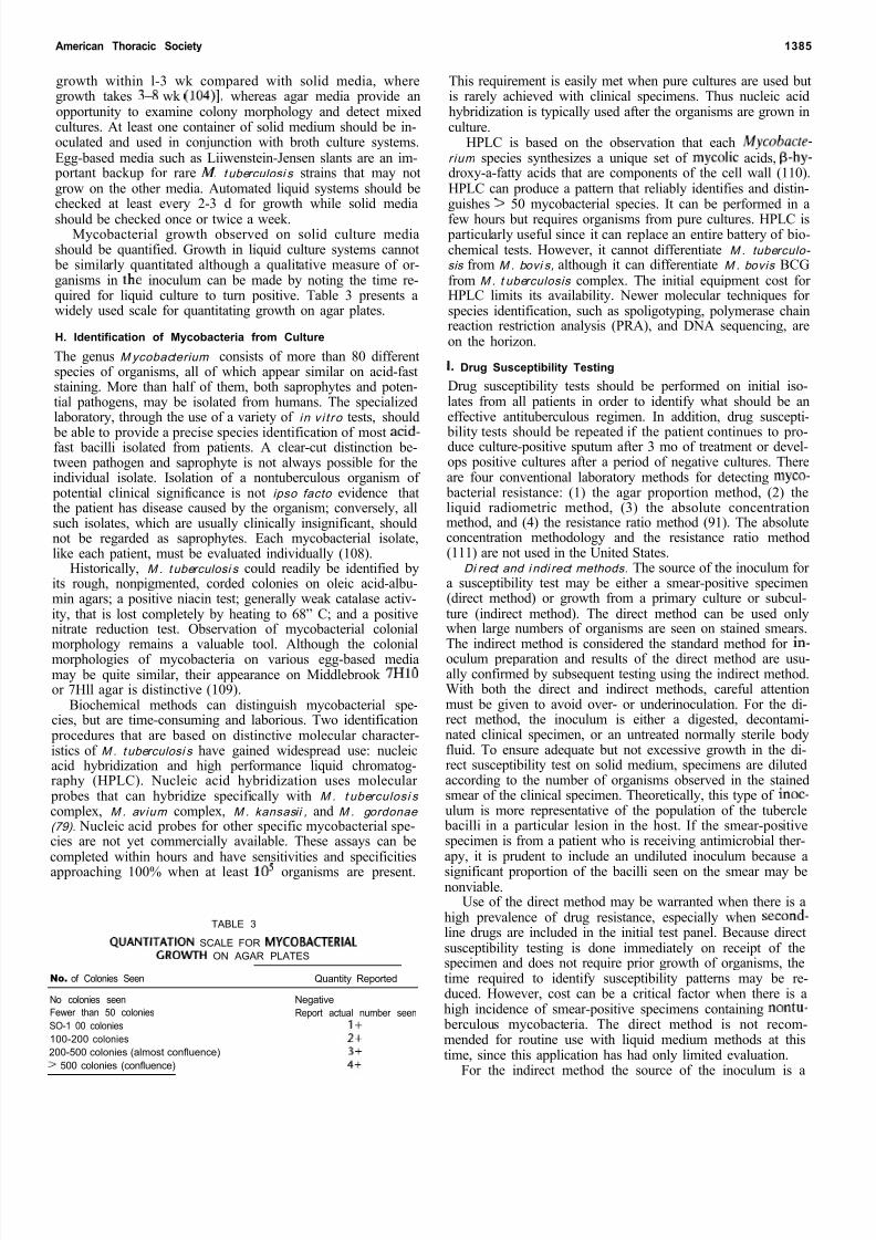
Mycobacterial growth observed on solid culture mediashould be quantified. Growth in liquid culture systems cannot
be similarly quantitated although a qualitative measure of or-ganisms in the inoculum can be made by noting the time re-quired for liquid culture to turn positive. Table 3 presents awidely used scale for quantitating growth on agar plates.
H. Identification of Mycobacteria from Culture
The genus M ycobacterium consists of more than 80 differentspecies of organisms, all of which appear similar on acid-faststaining. More than half of them, both saprophytes and poten-tial pathogens, may be isolated from humans. The specializedlaboratory, through the use of a variety of i n v i t ro tests, should
be able to provide a precise species identification of most acid-fast bacilli isolated from patients. A clear-cut distinction be-tween pathogen and saprophyte is not always possible for theindividual isolate. Isolation of a nontuberculous organism of
potential clinical significance is not ipso facto evidence thatthe patient has disease caused by the organism; conversely, allsuch isolates, which are usually clinically insignificant, shouldnot be regarded as saprophytes. Each mycobacterial isolate,like each patient, must be evaluated individually (108).
Historically, M . t uberculosi s could readily be identified byits rough, nonpigmented, corded colonies on oleic acid-albu-min agars; a positive niacin test; generally weak catalase activ-ity, that is lost completely by heating to 68” C; and a positivenitrate reduction test. Observation of mycobacterial colonialmorphology remains a valuable tool. Although the colonialmorphologies of mycobacteria on various egg-based mediamay be quite similar, their appearance on Middlebrook 7HlOor 7Hll agar is distinctive (109).
Biochemical methods can distinguish mycobacterial spe-cies, but are time-consuming and laborious. Two identification
procedures that are based on distinctive molecular character-istics of M . t uberculosi s have gained widespread use: nucleicacid hybridization and high performance liquid chromatog-raphy (HPLC). Nucleic acid hybridization uses molecular
probes that can hybridize specifically with M . t uberculosi s complex, M . av ium complex, M . kansasii , and M . gordonae (79). Nucleic acid probes for other specific mycobacterial spe-cies are not yet commercially available. These assays can becompleted within hours and have sensitivities and specificitiesapproaching 100% when at least IO 5 organisms are present.
TABLE 3
QUANTITATION SCALE FOR MYCOBACTERIALGRO\NTH ON AGAR PLATES
No. of Colonies Seen Quantity Reported
No colonies seen NegativeFewer than 50 colonies Report actual number seenSO-1 00 colonies 1+100-200 colonies 2+200-500 colonies (almost confluence) 3+> 500 colonies (confluence) 4+
This requirement is easily met when pure cultures are used butis rarely achieved with clinical specimens. Thus nucleic acidhybridization is typically used after the organisms are grown inculture.
HPLC is based on the observation that each M ycobacte-r ium species synthesizes a unique set of mycolic acids, B-hy-droxy-a-fatty acids that are components of the cell wall (110).HPLC can produce a pattern that reliably identifies and distin-guishes > 50 mycobacterial species. It can be performed in afew hours but requires organisms from pure cultures. HPLC is
particularly useful since it can replace an entire battery of bio-chemical tests. However, it cannot differentiate M . tuberculo- sis from M . bovi s, although it can differentiate M . bovis BCGfrom M . t uberculosis complex. The initial equipment cost for HPLC limits its availability. Newer molecular techniques for species identification, such as spoligotyping, polymerase chainreaction restriction analysis (PRA), and DNA sequencing, areon the horizon.
I Drug Susceptibility Testing
Drug susceptibility tests should be performed on initial iso-lates from all patients in order to identify what should be aneffective antituberculous regimen. In addition, drug suscepti-
bility tests should be repeated if the patient continues to pro-duce culture-positive sputum after 3 mo of treatment or devel-ops positive cultures after a period of negative cultures. Thereare four conventional laboratory methods for detecting myco-
bacterial resistance: (1) the agar proportion method, (2) theliquid radiometric method, (3) the absolute concentrationmethod, and (4) the resistance ratio method (91). The absoluteconcentration methodology and the resistance ratio method(111) are not used in the United States.
Di rect and i ndi rect methods. The source of the inoculum for a susceptibility test may be either a smear-positive specimen(direct method) or growth from a primary culture or subcul-ture (indirect method). The direct method can be used onlywhen large numbers of organisms are seen on stained smears.The indirect method is considered the standard method for in-oculum preparation and results of the direct method are usu-ally confirmed by subsequent testing using the indirect method.With both the direct and indirect methods, careful attentionmust be given to avoid over- or underinoculation. For the di-rect method, the inoculum is either a digested, decontami-nated clinical specimen, or an untreated normally sterile bodyfluid. To ensure adequate but not excessive growth in the di-rect susceptibility test on solid medium, specimens are dilutedaccording to the number of organisms observed in the stainedsmear of the clinical specimen. Theoretically, this type of inoc-ulum is more representative of the population of the tubercle
bacilli in a particular lesion in the host. If the smear-positivespecimen is from a patient who is receiving antimicrobial ther-apy, it is prudent to include an undiluted inoculum because asignificant proportion of the bacilli seen on the smear may benonviable.
Use of the direct method may be warranted when there is ahigh prevalence of drug resistance, especially when second-line drugs are included in the initial test panel. Because directsusceptibility testing is done immediately on receipt of thespecimen and does not require prior growth of organisms, thetime required to identify susceptibility patterns may be re-duced. However, cost can be a critical factor when there is ahigh incidence of smear-positive specimens containing nontu- berculous mycobacteria. The direct method is not recom-mended for routine use with liquid medium methods at thistime, since this application has had only limited evaluation.
For the indirect method the source of the inoculum is a
8/10/2019 daftar pustaka 7689
http://slidepdf.com/reader/full/daftar-pustaka-7689 11/20
8/10/2019 daftar pustaka 7689
http://slidepdf.com/reader/full/daftar-pustaka-7689 12/20
American Thoracic Society 1387
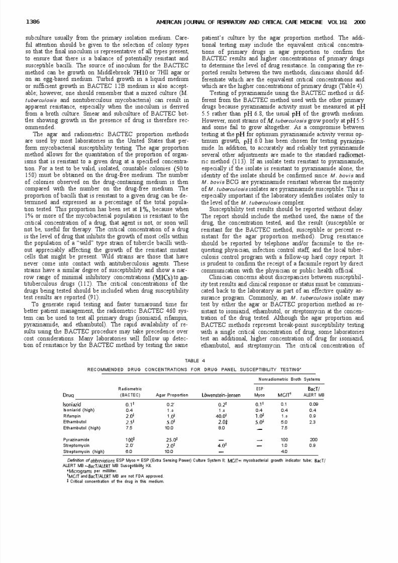
drug determines whether the isolate is considered resistant.The additional higher concentration of drugs, however, can
provide the physician with information about the level of drugresistance in deciding whether to continue therapy with a drugeither at the recommended dose or at an increased dose. Whendrug resistance is noted, it is important that the clinician withless experience seek assistance from experts in the field or from the local tuberculosis control program and the strainshould be retested and confirmed as to its resistance pattern.
Investigations into mechanisms of antimicrobial drug ac-tion and of resistance in M . tubercu losis have benefited fromstudies in other bacterial species, especially Escherichia ol i
(114). A particular focus of such studies has been rifampin re-sistance because of the pivotal role of rifampin in the treat-ment of tuberculosis, the conserved nature of the genetic basisfor resistance (> 96% of rifampin resistance in M . tubercul osis correlates with mutations in an 81-bp segment of the rpo
gene), and the use of rifampin resistance as a marker of multi-drug-resistant M . tubercul osis. The methods employed to de-tect rpo mutations include PCR amplification of the targetsequence and detection by DNA sequencing, line probe assay,single-strand conformation polymorphism, and other molecu-lar procedures. Before these techniques become widespread,
technical simplification or automation, as well as outcomeanalysis in order to justify the anticipated increased costs com- pared with conventional approaches, will be required.
J Cenotyping of Mycobacterium tuberculosic
Genotyping or DNA fingerprinting of tuberculosis has re- placed phage typing as a method for determining the clonalityof bacterial cultures. The Southern blotting method is used,whereby cultured organisms are heat killed and their DNA isisolated, cut with specific restriction enzymes, separated in anagarose gel by electrophoresis, transferred to a membrane,and probed for specific genetic sequences. A standardized
protocol has been developed to permit comparison of geno-types from different laboratories around the world (115).
Genotyping is useful in confirming laboratory cross-contarn-nation (116), investigating outbreaks of tuberculosis (117), eval-uating contact investigations (118), and determining whether new episodes of tuberculosis are due to reinfection or reacti-vation (26). In addition, genotyping is useful for elucidatingsites and patterns of M . tubercul osis transmission within com-munities (119,120).
In response to several nosocomial outbreaks and a dra-matic increase in tuberculosis among HIV-infected patientsin the early 1990s the Centers for Disease Control and Pre-vention established a National Tuberculosis Genotyping andSurveillance Network. The merger of modem molecular pro-tocols for strain identification at the DNA level and conven-tional epidemiological methodologies has given birth to an en-
hanced collaborative strategy to impact tuberculosis controlefforts. Regional tuberculosis genotyping laboratories can becontacted through the state public health laboratories or tu-
berculosis control programs.
K. Assessment of Laboratory Performance
There has been a growing body of new and exciting methodsin mycobacteriology, but there is still no single test that is di-agnostic in all situations. Complementary techniques should
be used to generate complete and rapid information. The lab-oratory director needs to decide which tests will be best per-formed in-house, taking into consideration the need for rapidresults, particularly AFB smear results. Possible options for small laboratories would include splitting a specimen, with
one portion processed locally for AFB smear and the other portion sent for culture to a reference laboratory; or obtain-ing two specimens, with one processed locally so that smear results can be obtained within 24 h and the other specimenshipped to a reference laboratory for further processing. Deci-sions as to which specimens should be sent to a reference labo-ratory should be based on the community served and the re-sources available, as well as in consultation with infectiousdisease, pulmonary, or other affected physicians. With this
partnership, physicians will then share the responsibility for the quality and timeliness of laboratory results.
VI. TUBERCULIN SKIN TEST
The tuberculin skin test is currently the only widely usedmethod for identifying infection with M . tubercu losis in per-sons who do not have tuberculosis disease. Although currentlythe tuberculin skin test antigens that are available are substan-tially less than 100% sensitive and specific for detection of in-fection with M . tubercul osis, no better diagnostic method iswidely available. Proper use of the tuberculin skin test re-quires a knowledge of the antigen used (tuberculin), the im-munologic basis for the reaction to this antigen, the tech-
nique(s) of administering and reading the test, and the resultsof epidemiologic and clinical experience with the test.
A. Tuberculin
The tuberculin test is based on the fact that infection with M .tuberculosis produces a delayed-type hypersensitivity reactionto certain antigenic components of the organism that are con-tained in extracts of culture filtrates called “tuberculins.” Twocompanies manufacture tuberculin purified protein derivative(PPD) in the United States: Parke-Davis Pharmaceuticals(Aplisol) and Pasteur Merieux-Connaught Laboratories Tu-
bersol). Tuberculin PPD is isolated from a culture filtrate of tubercle bacilli by protein precipitation. The most commonlyemployed methods of precipitation use either ammonium sul-fate (AS) or trichloroacetic acid (TCA). Aplisol is isolated bythe AS method; Tubersol is isolated by the TCA method.Standardization should guarantee equivalent potency be-tween PPD preparations isolated by different precipitationmethods and between successive lots isolated by a singlemethod. However, the difference in the chemical compositionof the PPDs produced by the different precipitation methodsimplies that the actual antigens in the two preparations do dif-fer. Most of the constituents of PPD are small proteins withmolecular masses of approximately 10,000 Da, but there arealso polysaccharides and some lipids present (121). The rela-tively small size of the protein constituents in PPD is the rea-son that PPD does not sensitize individuals who have not beenexposed to mycobacteria (121).
A batch of PPD (lot 49608) called PPD-S, which was pro-duced by Seibert and Glenn in 1939, has continued to serve asthe international standard as well as the standard referencematerial in the United States (122). All PPD lots must be bio-assayed to demonstrate equal potency to PPD-S (123). “TU-
berculins” and “PPDs” have been prepared from other myco- bacterial species, but these materials are less sensitive andspecific for diagnosis of nontuberculous mycobacterial infec-tions than is PPD for M . tubercul osis infections. These prepara-tions are occasionally used for epidemiologic purposes 124) but have little clinical utility.
The standard 5-tuberculin unit (TU) dose of PPD-S is de-fined as the delayed skin test activity contained in a PPD-S doseof 0.1 mg/O.l ml. The standard test dose of a commercial PPD
preparation is defined as the dose of the product that is biologi-
8/10/2019 daftar pustaka 7689
http://slidepdf.com/reader/full/daftar-pustaka-7689 13/20
1388 AMERICAN J OURN I OF RESPIRATORY AND CRITICAL CARE MEDICINE VOL 161 2000
tally equivalent to that contained in 5 TU of PPD-S (i.e., it elic-its reactions of equivalent size 20%). Other doses of tubercu-lin, such as 1 and 250 TU, are remnants of the old graduatedsystem of administration (125) and represent the smallest andlargest doses of tuberculin that were administered. Thesestrengths of tuberculin are not commonly available and, even if available, are not standardized by bioassay. The clinical utilityof these concentrations of tuberculin is minimal.
PPD, when diluted in a buffered diluent, is adsorbed invarying amounts by glass and plastics. A small amount of thedetergent Tween 80 is added by the manufacturer to the dilu-ent for PPD to reduce adsorption (126). To minimize reduc-tion in potency by adsorption, tuberculin should never betransferred from one container to another, and skin testsshould be given as soon after the syringe has been filled as
possible. Following these procedures will also help avoid con-tamination: test doses should always be removed from the vialunder strictly aseptic conditions, and the remaining solutionshould be kept refrigerated (not frozen). Tuberculin should bestored in the dark as much as possible and exposure to stronglight should be avoided.
8. Immunologic Basis for the Tuberculin Reaction
The reaction to intracutaneously injected tuberculin is theclassic example of a delayed (cellular) hypersensitivity reac-tion. T cells sensitized by prior infection are recruited to theskin site where they release lymphokines (127). These lym-
phokines induce induration through local vasodilatation,edema, fibrin deposition, and recruitment of other inflamma-tory cells to the area (128). Features of the reaction include (I)its delayed course, reaching a peak more than 24 h after in-
jection of the antigen; (2) its indurated character; and (3) itsoccasional vesiculation and necrosis. Reactivity of the PPD
provides a general measure of a person’s cellular immune re-sponsiveness (121).
Typically, the reaction to tuberculin begins 5 to 6 h after in- jection, causes maximal induration at 48 to 72 h, and subsidesover a period of days. In a few individuals (the elderly andthose who are being tested for the first time), the reaction maynot peak until after 72 h (129). Such delayed reactions do notalter the intrepretation of the test. Immediate hypersensitivityreactions to tuberculin or constituents of the diluent can alsooccur. These reactions disappear by 24 h, and should not beconfused with delayed hypersensitivity reactions. However, if the immediate reaction is severe, it may be prudent not to re-test. Symptom screening is suggested if these individuals are
part of a regular tuberculin skin testing program (e.g., health-care workers).
C. Administration and Reading of Tests
The tuberculin test, like all medical tests, is subject to variabil-ity, but many of the inherent variations in administration andreading of tests can be avoided by careful attention to details.The test is administered by injecting 0.1 ml of 5-TU PPD intra-dermally (Mantoux method) into the volar or dorsal surface of the forearm. Other areas may be used, but the forearm is pre-ferred. The use of a skin area free of lesions and away fromveins is recommended. The injection is made using a one-quarter- to one-half-inch, 27-gauge needle and a tuberculin sy-ringe. The tuberculin should be injected just beneath the sur-face of the skin, with the needle bevel upward or downward(130). A discrete, pale elevation of the skin (a wheal) 6 to 10mm in diameter should be produced when the injection isdone correctly. If it is recognized that the first test was im-
properly administered, another test dose can be given at once,selecting a site several centimeters away from the original in-
jection. A note in the record should indicate the site chosenfor the second test.
Tests should be read between 48 and 72 h after injection,when the induration is maximum. Tests read after 72 h tend tounderestimate the true size of induration. Reading should be
performed in a good light, with the forearm slightly flexed atthe elbow. The basis of reading is the presence or absence of induration, which may be determined by inspection (from aside view against the light as well as by direct light) and by pal-
pation. For standardization, the diameter of induration should be measured transversely to the long axis of the forearm andrecorded in millimeters (130, 131). The absence of indurationshould be recorded as “0 mm,” not “negative.” Interobserver variability may be decreased by using the ball-point penmethod of Sokal to measure induration (132,133).
The multiple puncture test introduces tuberculin into theskin either by puncture with an applicator with points coatedwith dried tuberculin or by puncturing through a film of liquidtuberculin. All multiple puncture tests presently available useconcentrated tuberculin. The quantity of tuberculin intro-duced into the skin by the multiple puncture test cannot be
precisely controlled. For these reasons, multiple-puncturetests are not as reliable as the Mantoux method of skin testing
(123) and should not be used as a diagnostic test. If a multiple puncture test has been placed, the test should be repeated us-ing the Mantoux method, regardless of the result of the multi-
ple puncture test unless there is vesiculation. In this case, thetest should be considered positive and no Mantoux skin test
placed. For each tuberculin test, a record should be made of the technique of administration (Mantoux or multiple-punc-ture), the kind and dose of tuberculin, and the size of reactionin millimeters of induration.
D. Interpretation of Skin Test Reactions
To interpret the tuberculin skin test appropriately, one mustunderstand the sensitivity and specificity of the test as well asthe positive and negative predictive value of the test. The sen-sitivity of a test is the percentage of people with the conditionwho have a positive test. If false-negative results are uncom-mon, the sensitivity is high. The PPD skin test has a reportedfalse-negative rate of 25% during the initial evaluation of per-sons with active tuberculosis (134). This high false-negativerate appears to be due to poor nutrition and general health,overwhelming acute illness, or immunosuppression.
Immunosuppression can be either specific, which may beseen early during disease, or nonspecific, which can be the re-sult of medications, malignancy, or HIV infection (135). Be-cause of the low sensitivity of the test, especially in acutely ill
patients and those who are infected with HIV, the tuberculintest cannot be used to eliminate the possibility of active tuber-culosis (136). Other factors that may result in a false-negativetest are shown in Table 5.
Vaccination with live-attenuated virus can cause suppres-sion of the PPD response in patients known to be infectedwith M . t uberculosi s. Live-attenuated vaccines that may causefalse-negative PPD results are measles, mumps, rubella, oral
polio, varicella, yellow fever, BCG, and oral typhoid TY2la).This suppression does not appear within the first 48 h after measles vaccination, so the Advisory Committee on Immuni-zation Practices recommends that tuberculin testing be doneeither on the same day as vaccination with live virus or 4-6 wk later (137-140).
The specificity of the test is the percentage of people with-out the condition who have a negative test. False-positive re-sults decrease the specificity of a test. False-positive tuberculintests occur in individuals who have been infected with other
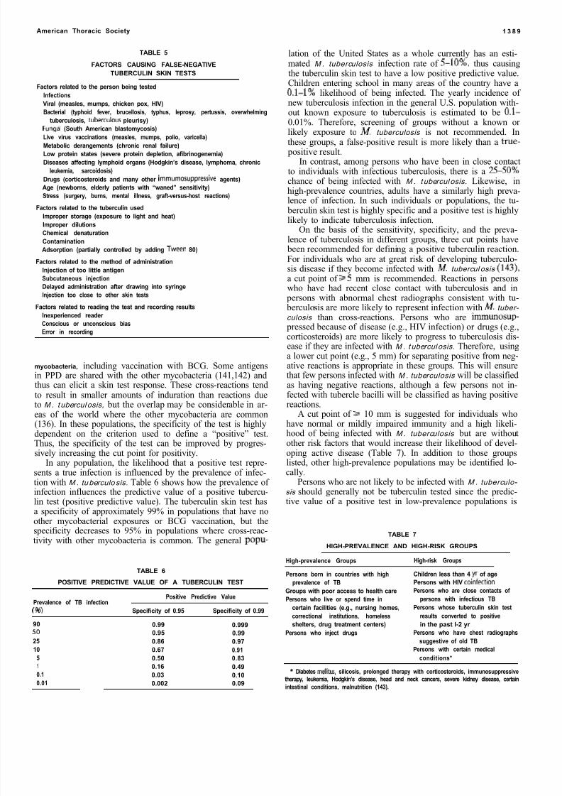
8/10/2019 daftar pustaka 7689
http://slidepdf.com/reader/full/daftar-pustaka-7689 14/20
American Thoracic Society 1 3 8 9
TABLE 5
FACTORS CAUSING FALSE-NEGATIVETUBERCULIN SKIN TESTS
Factors related to the person being testedInfectionsViral (measles, mumps, chicken pox, HIV)Bacterial (typhoid fever, brucellosis, typhus, leprosy, pertussis, overwhelming
tuberculosis, tuberculous pleurisy)Fungal (South American blastomycosis)
Live virus vaccinations (measles, mumps, polio, varicella)Metabolic derangements (chronic renal failure)Low protein states (severe protein depletion, afibrinogenemia)Diseases affecting lymphoid organs (Hodgkin’s disease, lymphoma, chronic
leukemia, sarcoidosis)Drugs (corticosteroids and many other immumosuppressive agents)Age (newborns, elderly patients with “waned” sensitivity)Stress (surgery, burns, mental illness, graft-versus-host reactions)
Factors related to the tuberculin usedImproper storage (exposure to light and heat)Improper dilutionsChemical denaturationContaminationAdsorption (partially controlled by adding Tween 80)
Factors related to the method of administrationInjection of too little antigen
Subcutaneous injectionDelayed administration after drawing into syringeInjection too close to other skin tests
Factors related to reading the test and recording resultsInexperienced reader Conscious or unconscious biasError in recording
mycobacteria, including vaccination with BCG. Some antigensin PPD are shared with the other mycobacteria (141,142) andthus can elicit a skin test response. These cross-reactions tendto result in smaller amounts of induration than reactions dueto M . t ubercul osis, but the overlap may be considerable in ar-
eas of the world where the other mycobacteria are common(136). In these populations, the specificity of the test is highlydependent on the criterion used to define a “positive” test.Thus, the specificity of the test can be improved by progres-sively increasing the cut point for positivity.
In any population, the likelihood that a positive test repre-sents a true infection is influenced by the prevalence of infec-tion with M . tu berculo sis. Table 6 shows how the prevalence of infection influences the predictive value of a positive tubercu-lin test (positive predictive value). The tuberculin skin test hasa specificity of approximately 99% in populations that have noother mycobacterial exposures or BCG vaccination, but thespecificity decreases to 95% in populations where cross-reac-tivity with other mycobacteria is common. The general popu-
TABLE 6
POSITIVE PREDICTIVE VALUE OF A TUBERCULIN TEST
Prevalence of TB infection )
Positive Predictive Value
Specificity of 0.95 Specificity of 0.99
90 0.99 0.99950 0.95 0.9925 0.86 0.9710 0.67 0.91
5 0.50 0.831 0.16 0.490.1 0.03 0.100.01 0.002 0.09
lation of the United States as a whole currently has an esti-mated M . tuberculosis infection rate of S-10 , thus causingthe tuberculin skin test to have a low positive predictive value.Children entering school in many areas of the country have a0.1-l likelihood of being infected. The yearly incidence of new tuberculosis infection in the general U.S. population with-out known exposure to tuberculosis is estimated to be O.l-0.01%. Therefore, screening of groups without a known or likely exposure to M. tuberculosis is not recommended. Inthese groups, a false-positive result is more likely than a true-
positive result.In contrast, among persons who have been in close contact
to individuals with infectious tuberculosis, there is a 2550chance of being infected with M . t ubercul osis. Likewise, inhigh-prevalence countries, adults have a similarly high preva-lence of infection. In such individuals or populations, the tu-
berculin skin test is highly specific and a positive test is highlylikely to indicate tuberculosis infection.
On the basis of the sensitivity, specificity, and the preva-lence of tuberculosis in different groups, three cut points have
been recommended for defining a positive tuberculin reaction.For individuals who are at great risk of developing tuberculo-sis disease if they become infected with M. tubercul osis 143),
a cut point of 2 5 mm is recommended. Reactions in personswho have had recent close contact with tuberculosis and in
persons with abnormal chest radiographs consistent with tu- berculosis are more likely to represent infection with M . tuber- culosis than cross-reactions. Persons who are immunosup-
pressed because of disease (e.g., HIV infection) or drugs (e.g.,corticosteroids) are more likely to progress to tuberculosis dis-ease if they are infected with M . t ubercul osis. Therefore, usinga lower cut point (e.g., 5 mm) for separating positive from neg-ative reactions is appropriate in these groups. This will ensurethat few persons infected with M . tuberculosis will be classifiedas having negative reactions, although a few persons not in-fected with tubercle bacilli will be classified as having positivereactions.
A cut point of 3 10 mm is suggested for individuals whohave normal or mildly impaired immunity and a high likeli-hood of being infected with M . tuberculosis but are withoutother risk factors that would increase their likelihood of devel-oping active disease (Table 7). In addition to those groupslisted, other high-prevalence populations may be identified lo-cally.
Persons who are not likely to be infected with M . tuberculo- sis should generally not be tuberculin tested since the predic-tive value of a positive test in low-prevalence populations is
TABLE 7
HIGH-PREVALENCE AND HIGH-RISK GROUPS
High-prevalence Groups High-risk Groups
Persons born in countries with highprevalence of TB
Groups with poor access to health carePersons who live or spend time in
certain facilities (e.g., nursing homes,correctional institutions, homelessshelters, drug treatment centers)
Persons who inject drugs
Children less than 4 yr of agePersons with HIV coinfectionPersons who are close contacts of
persons with infectious TBPersons whose tuberculin skin test
results converted to positivein the past l-2 yr
Persons who have chest radiographssuggestive of old TB
Persons with certain medicalconditions*
Diabetes mellitus silicosis, prolonged therapy with corticosteroids, immunosuppressivetherapy, leukemia, Hodgkin’s disease, head and neck cancers, severe kidney disease, certainintestinal conditions, malnutrition (143).
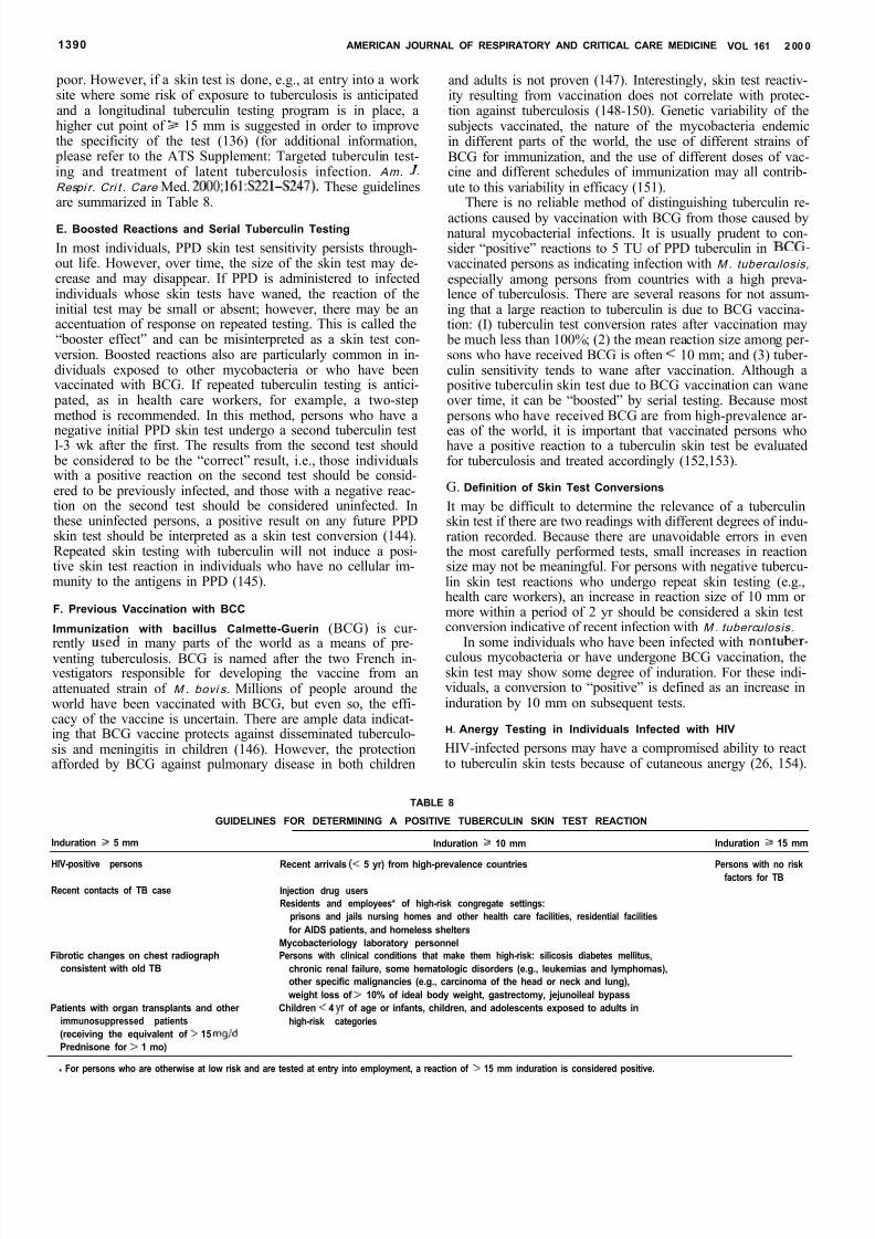
8/10/2019 daftar pustaka 7689
http://slidepdf.com/reader/full/daftar-pustaka-7689 15/20
1390 AMERICAN JOURNAL OF RESPIRATORY AND CRITICAL CARE MEDICINE VOL 161 2 00 0
poor. However, if a skin test is done, e.g., at entry into a work site where some risk of exposure to tuberculosis is anticipatedand a longitudinal tuberculin testing program is in place, ahigher cut point of 2 15 mm is suggested in order to improvethe specificity of the test (136) (for additional information,
please refer to the ATS Supplement: Targeted tuberculin test-ing and treatment of latent tuberculosis infection. Am. J.Respi r. Cri t . Care Med. 2000;161:S221-S247). These guidelinesare summarized in Table 8.
E. Boosted Reactions and Serial Tuberculin Testing
In most individuals, PPD skin test sensitivity persists through-out life. However, over time, the size of the skin test may de-crease and may disappear. If PPD is administered to infectedindividuals whose skin tests have waned, the reaction of theinitial test may be small or absent; however, there may be anaccentuation of response on repeated testing. This is called the“booster effect” and can be misinterpreted as a skin test con-version. Boosted reactions also are particularly common in in-dividuals exposed to other mycobacteria or who have beenvaccinated with BCG. If repeated tuberculin testing is antici-
pated, as in health care workers, for example, a two-stepmethod is recommended. In this method, persons who have a
negative initial PPD skin test undergo a second tuberculin testl-3 wk after the first. The results from the second test should be considered to be the “correct” result, i.e., those individualswith a positive reaction on the second test should be consid-ered to be previously infected, and those with a negative reac-tion on the second test should be considered uninfected. Inthese uninfected persons, a positive result on any future PPDskin test should be interpreted as a skin test conversion (144).Repeated skin testing with tuberculin will not induce a posi-tive skin test reaction in individuals who have no cellular im-munity to the antigens in PPD (145).
F. Previous Vaccination with BCC
Immunization with bacillus Calmette-Guerin (BCG) is cur-rently used in many parts of the world as a means of pre-venting tuberculosis. BCG is named after the two French in-vestigators responsible for developing the vaccine from anattenuated strain of M . bovi s. Millions of people around theworld have been vaccinated with BCG, but even so, the effi-cacy of the vaccine is uncertain. There are ample data indicat-ing that BCG vaccine protects against disseminated tuberculo-sis and meningitis in children (146). However, the protectionafforded by BCG against pulmonary disease in both children
and adults is not proven (147). Interestingly, skin test reactiv-ity resulting from vaccination does not correlate with protec-tion against tuberculosis (148-150). Genetic variability of thesubjects vaccinated, the nature of the mycobacteria endemicin different parts of the world, the use of different strains of BCG for immunization, and the use of different doses of vac-cine and different schedules of immunization may all contrib-ute to this variability in efficacy (151).
There is no reliable method of distinguishing tuberculin re-actions caused by vaccination with BCG from those caused bynatural mycobacterial infections. It is usually prudent to con-sider “positive” reactions to 5 TU of PPD tuberculin in BCG-vaccinated persons as indicating infection with M . tuberculosis,especially among persons from countries with a high preva-lence of tuberculosis. There are several reasons for not assum-ing that a large reaction to tuberculin is due to BCG vaccina-tion: (I) tuberculin test conversion rates after vaccination may
be much less than 100%; (2) the mean reaction size among per-sons who have received BCG is often < 10 mm; and (3) tuber-culin sensitivity tends to wane after vaccination. Although a
positive tuberculin skin test due to BCG vaccination can waneover time, it can be “boosted” by serial testing. Because most
persons who have received BCG are from high-prevalence ar-
eas of the world, it is important that vaccinated persons whohave a positive reaction to a tuberculin skin test be evaluatedfor tuberculosis and treated accordingly (152,153).
G. Definition of Skin Test Conversions
It may be difficult to determine the relevance of a tuberculinskin test if there are two readings with different degrees of indu-ration recorded. Because there are unavoidable errors in eventhe most carefully performed tests, small increases in reactionsize may not be meaningful. For persons with negative tubercu-lin skin test reactions who undergo repeat skin testing (e.g.,health care workers), an increase in reaction size of 10 mm or more within a period of 2 yr should be considered a skin testconversion indicative of recent infection with M . tuberculosis.
In some individuals who have been infected with nontuber-culous mycobacteria or have undergone BCG vaccination, theskin test may show some degree of induration. For these indi-viduals, a conversion to “positive” is defined as an increase ininduration by 10 mm on subsequent tests.
H. Anergy Testing in Individuals Infected with HIV
HIV-infected persons may have a compromised ability to reactto tuberculin skin tests because of cutaneous anergy (26, 154).
TABLE 8
GUIDELINES FOR DETERMINING A POSITIVE TUBERCULIN SKIN TEST REACTION
Induration 5 mm Induration 10 mm Induration 15 mm
HIV-positive persons Recent arrivals < 5 yr) from high-prevalence countries Persons with no riskfactors for TBRecent contacts of TB case
Fibrotic changes on chest radiographconsistent with old TB
Patients with organ transplants and other immunosuppressed patients(receiving the equivalent of > 15 mgldPrednisone for > 1 mo)
Injection drug usersResidents and employees* of high-risk congregate settings:
prisons and jails nursing homes and other health care facilities, residential facilitiesfor AIDS patients, and homeless shelters
Mycobacteriology laboratory personnelPersons with clinical conditions that make them high-risk: silicosis diabetes mellitus,
chronic renal failure, some hematologic disorders (e.g., leukemias and lymphomas),other specific malignancies (e.g., carcinoma of the head or neck and lung),weight loss of > 10% of ideal body weight, gastrectomy, jejunoileal bypass
Children < 4 yr of age or infants, children, and adolescents exposed to adults inhigh-risk categories
l For persons who are otherwise at low risk and are tested at entry into employment, a reaction of > 15 mm induration is considered positive.
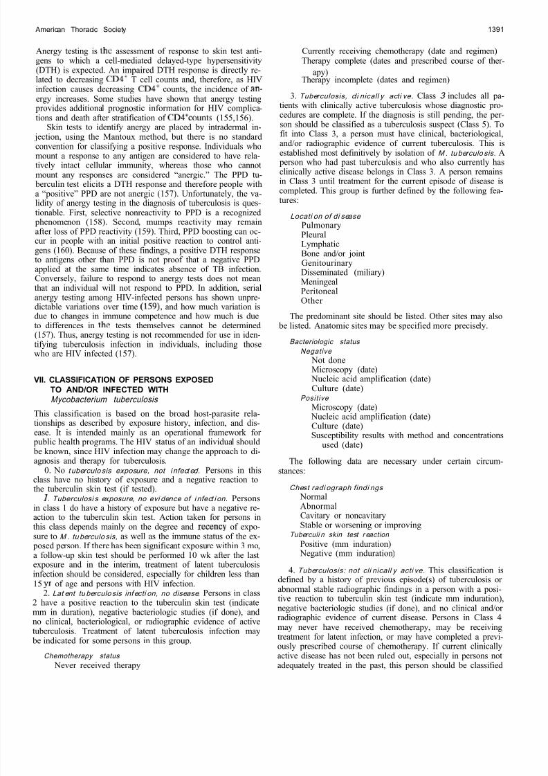
8/10/2019 daftar pustaka 7689
http://slidepdf.com/reader/full/daftar-pustaka-7689 16/20
American Thoracic Society 1391
Anergy testing is the assessment of response to skin test anti-gens to which a cell-mediated delayed-type hypersensitivity(DTH) is expected. An impaired DTH response is directly re-lated to decreasing CD4+ T cell counts and, therefore, as HIVinfection causes decreasing CD4+ counts, the incidence of an-ergy increases. Some studies have shown that anergy testing
provides additional prognostic information for HIV complica-tions and death after stratification of CD4+counts (155,156).
Skin tests to identify anergy are placed by intradermal in- jection, using the Mantoux method, but there is no standardconvention for classifying a positive response. Individuals whomount a response to any antigen are considered to have rela-tively intact cellular immunity, whereas those who cannotmount any responses are considered “anergic.” The PPD tu-
berculin test elicits a DTH response and therefore people witha “positive” PPD are not anergic (157). Unfortunately, the va-lidity of anergy testing in the diagnosis of tuberculosis is ques-tionable. First, selective nonreactivity to PPD is a recognized
phenomenon (158). Second, mumps reactivity may remainafter loss of PPD reactivity (159). Third, PPD boosting can oc-cur in people with an initial positive reaction to control anti-gens (160). Because of these findings, a positive DTH responseto antigens other than PPD is not proof that a negative PPD
applied at the same time indicates absence of TB infection.Conversely, failure to respond to anergy tests does not meanthat an individual will not respond to PPD. In addition, serialanergy testing among HIV-infected persons has shown unpre-dictable variations over time 159) and how much variation isdue to changes in immune competence and how much is dueto differences in the tests themselves cannot be determined(157). Thus, anergy testing is not recommended for use in iden-tifying tuberculosis infection in individuals, including thosewho are HIV infected (157).
VII. CLASSIFICATION OF PERSONS EXPOSEDTO AND/OR INFECTED WITHMycobacterium tuberculosis
This classification is based on the broad host-parasite rela-tionships as described by exposure history, infection, and dis-ease. It is intended mainly as an operational framework for
public health programs. The HIV status of an individual should be known, since HIV infection may change the approach to di-agnosis and therapy for tuberculosis.
0. No t uberculo sis exposure, not i nfect ed. Persons in thisclass have no history of exposure and a negative reaction tothe tuberculin skin test (if tested).
I. Tuberculosi s exposure, no evi dence of i nfect i on. Personsin class 1 do have a history of exposure but have a negative re-action to the tuberculin skin test. Action taken for persons inthis class depends mainly on the degree and recency of expo-sure to M . tu berculo sis, as well as the immune status of the ex-
posed person. If there has been significant exposure within 3 mo,a follow-up skin test should be performed 10 wk after the lastexposure and in the interim, treatment of latent tuberculosisinfection should be considered, especially for children less than15 yr of age and persons with HIV infection.
2. Lat ent tu berculo sis inf ecti on, no disease. Persons in class2 have a positive reaction to the tuberculin skin test (indicatemm in duration), negative bacteriologic studies (if done), andno clinical, bacteriological, or radiographic evidence of activetuberculosis. Treatment of latent tuberculosis infection may
be indicated for some persons in this group.
Chemotherapy status Never received therapy
Currently receiving chemotherapy (date and regimen)Therapy complete (dates and prescribed course of ther-
apy)Therapy incomplete (dates and regimen)
3. Tuberculosis, cli nicall y acti ve. Class 3 includes all pa-tients with clinically active tuberculosis whose diagnostic pro-cedures are complete. If the diagnosis is still pending, the per-son should be classified as a tuberculosis suspect (Class 5). To
fit into Class 3, a person must have clinical, bacteriological,and/or radiographic evidence of current tuberculosis. This isestablished most definitively by isolation of M . tu berculo sis. A
person who had past tuberculosis and who also currently hasclinically active disease belongs in Class 3. A person remainsin Class 3 until treatment for the current episode of disease iscompleted. This group is further defined by the following fea-tures:
Locati on of di sease PulmonaryPleuralLymphaticBone and/or jointGenitourinaryDisseminated (miliary)MeningealPeritonealOther
The predominant site should be listed. Other sites may also be listed. Anatomic sites may be specified more precisely.
Bacteriologic status Negative
Not doneMicroscopy (date)
Nucleic acid amplification (date)Culture (date)
Posi t ive
Microscopy (date) Nucleic acid amplification (date)Culture (date)Susceptibility results with method and concentrations
used (date)
The following data are necessary under certain circum-stances:
Chest radi ograph findi ngs NormalAbnormalCavitary or noncavitaryStable or worsening or improving
Tuberculi n skin test r eaction
Positive (mm induration) Negative (mm induration)
4. Tuberculosis: not cli nicall y acti ve. This classification isdefined by a history of previous episode(s) of tuberculosis or abnormal stable radiographic findings in a person with a posi-tive reaction to tuberculin skin test (indicate mm induration),negative bacteriologic studies (if done), and no clinical and/or radiographic evidence of current disease. Persons in Class 4may never have received chemotherapy, may be receivingtreatment for latent infection, or may have completed a previ-ously prescribed course of chemotherapy. If current clinicallyactive disease has not been ruled out, especially in persons notadequately treated in the past, this person should be classified
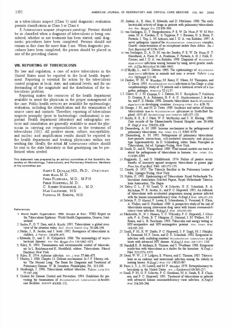
8/10/2019 daftar pustaka 7689
http://slidepdf.com/reader/full/daftar-pustaka-7689 17/20
8/10/2019 daftar pustaka 7689
http://slidepdf.com/reader/full/daftar-pustaka-7689 18/20

8/10/2019 daftar pustaka 7689
http://slidepdf.com/reader/full/daftar-pustaka-7689 19/20
8/10/2019 daftar pustaka 7689
http://slidepdf.com/reader/full/daftar-pustaka-7689 20/20