assessing neuroplasticity using magnetoencephalography...
TRANSCRIPT
63
Jurnal Sains Kesihatan Malaysia 16(1) 2018: 63-69DOI : http://dx.doi.org./10.17576/JSKM-2018-1601-08
Artikel Asli/Original Article
Assessing Neuroplasticity Using Magnetoencephalography (MEG) in Patient with Left-Temporo-Parietal Pilocytic Astrocytomas Treated with Endoscopic Surgery
(Menilai Keplastikan-neuro Menggunakan Magnetoensefalografi (MEG) bagi Penghidap Tumor Pilocytic Astrocytomas pada Bahagian Parietal-Temporo-Kiri yang Dirawat dengan Pembedahan Endoskopi)
HANANI ABDUL MANAN, ZAMZURI IDRIS, MOHAMMED FARUQUE REZA, HAZIM OMAR & JAFRI MALIN ABDULLAH
ABSTRACT
Neuroplasticity has been subjected to a great deal of research in the last century. Recently, significant emphasis has been placed on the global effect of localized plastic changes throughout the central nervous system, and on how these changes integrate in a pathological context. The present study aimed to demonstrate the functional cortical reorganization before and after surgery using magnetoencephalography (MEG) in a participant with brain tumor. Results of Visual Evoked Magnetic Field (VEF) based on functional MEG study revealed significantly different of MEG N100 waveforms before and after surgery. Larger and additional new locations for visual activation areas after the surgery were found suggesting neuroplasticity. The present study highlight a physiological plasticity in a teenage brain and the alterations regarding neural plasticity and network remodeling described in pathological contexts in higher-order visual association areas.
Keywords: Magnetoencephalography; pilocyticastrocytoma; neuroplasticity; endoscopy; visual evoked field
ABSTRAK
Proses keplastikan neural adalah berdasarkan kepada bukti penyelidikan dalam abad yang lalu. Baru-baru ini, penekanan telah diberikan kepada kesan global perubahan keplastikan setempat di seluruh sistem saraf pusat, dan bagaimana perubahan ini diintegrasikan dalam konteks patologi. Kajian ini bertujuan untuk menunjukkan penyusunan semula kefungsian kortikal dalam otak pesakit tumor otak, sebelum dan selepas pembedahan menggunakan magnetoencephalography (MEG). Keputusan Visual Medan Magnet (VEF) dengan menggunakan gelombang MEG N100 menunjukkan perbezaan yang ketara di antara sebelum dan selepas pembedahan. Keputusan kajian ini juga menunjukkan terdapatnya lokasi baru dan pengaktifan yang lebih besar bagi kawasan pengaktifan visual selepas pembedahan. Lokasi-lokasi baru dan kawasan pengaktifan yang lebih besar dicadangkan adalah disebabkan oleh proses keplastikan neuro. Kajian ini juga menunjukkan keplastikan fisiologi dalam otak remaja dan pembentukan semula rangkaian diterangkan dalam konteks patologi yang berkaitan dengan kawasan yang melibatkan pemprosesan visual.
Kata kunci: Magnetoencephalography; pilocytic astrocytoma; keplastikan-neuro; endoskopi; visualevoked field
INTRODUCTION
The role of neuroimaging techniques such as Magnetoencephalography (MEG) in evaluating brain activities and functions. The role of neuroimaging techniques such as Magnetoencephalography (MEG) in evaluating brain activities and function has been subject to numerous report (Majchrzak et al. 2012). There are many research reports describing the role and clinical applications of MEG in assessing auditory, motor and visual processing (Fruchter & Marantz 2015; Liu, Harris & Kanwisher 2002; Hund et al. 1997; Hara et al. 2007; Kikuchi et al. 2015). Moreover, MEG technique is also used for mapping onto patients with neurological disorders or brain damage to further understand the effect of morbidity and mechanism of neuroplasticity to the brain function (Idris et al. 2014; Kakisaka et al. 2013).
This study aims to highlight functional reorganization and neuroplasticity of the brain after surgery involving visual-specific cortex. Recent understanding proposes that the brain is ‘plastic’ and changes its own wiring and networks continuously in an environmentally dependent manner (Rosler et al. 2014; Strey, Baertsch & Baker-Herman 2013). Previous study has also demonstrated that connections between neurons become more extensive, more or less active, or even extend into new areas of the brain (Baertsch & Baker-Herman 2013). Neuroplasticity and cortical reorganization or any enduring changes in the cortical properties both functional and morphological are of major interest as they may play a role in functional recovery and learning after brain injury.
Chap 8.indd 63 22/02/2018 11:41:03
64
Understanding the principles behind the self-organization of biological nervous systems is the key to understanding cognition. Brain lesions such as brain tumors, may disrupt a particular step of an activity that occurs in a specific part of the brain. This disruption may alter the brain’s normal functioning and may interfere with the way the brain normally works. The early descriptions of clinical consequences of brain impairment or abnormality have documented a large variety of behavioral disturbances, and these behavioral disturbances are depending on the area of the lesion (Abraham & Bear 1996). However, the mechanism of the neural changes, reorganization and plasticity postoperatively is not always clear.
In order to achieve the aim, a better fundametal understanding about the brain changes and plasticity in postoperatively need to be fully understood. The present study investigates the neural changes and plasticity of a participant with pilocytic astrocytoma (PA). A non-invasive neuroimaging techniques MEG, using pattern-reversal visual evoked field (VEF) will be used to measure the neural activity in the brain, before and after surgery. The correlation between brain activation and the changes observed was investigated.
MATERIALS AND METHODS
PARTICIPANT
A 10-year old girl diagnosed with a Pilocytic Astrocytoma (PA) at the left temporal-parietal lobe. Participant had
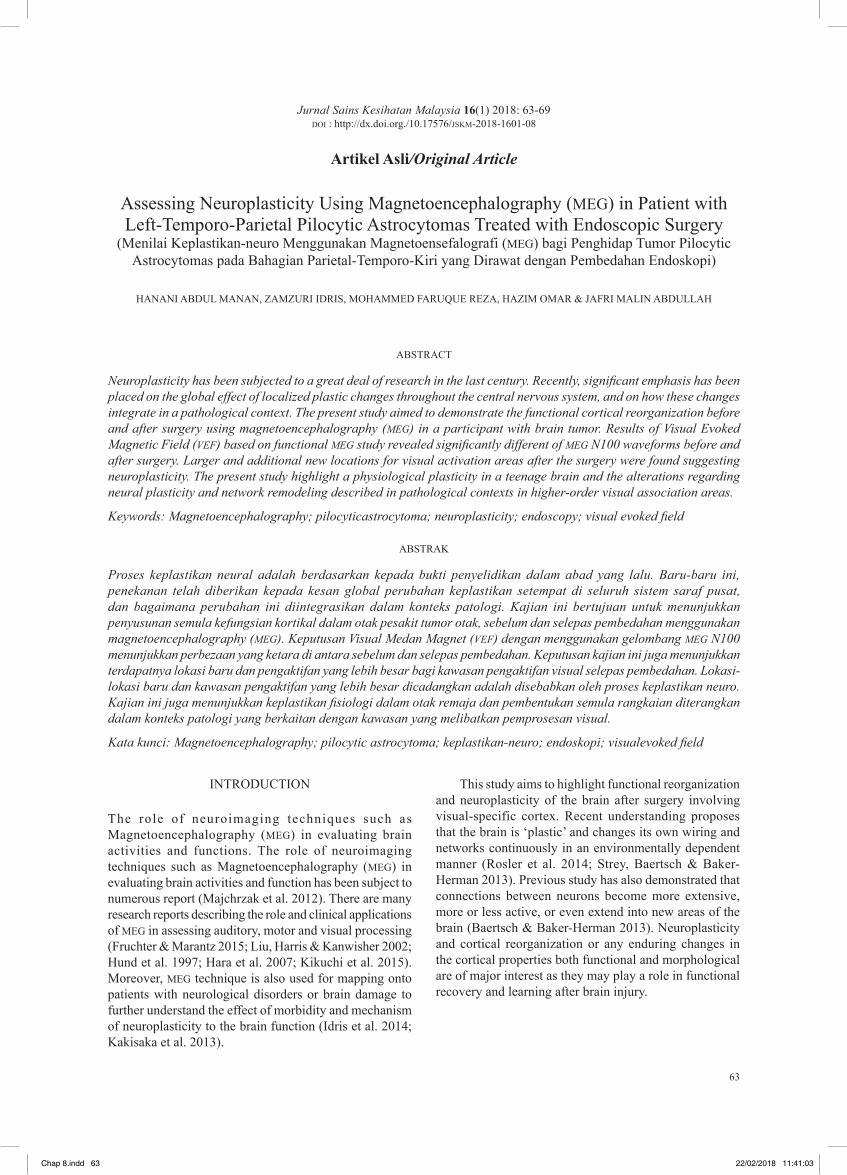
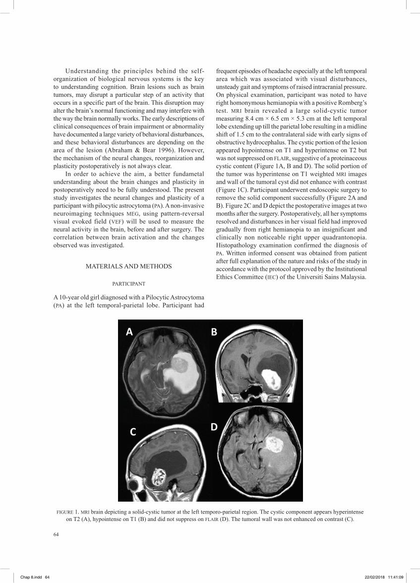
frequent episodes of headache especially at the left temporal area which was associated with visual disturbances, unsteady gait and symptoms of raised intracranial pressure. On physical examination, participant was noted to have right homonymous hemianopia with a positive Romberg’s test. MRI brain revealed a large solid-cystic tumor measuring 8.4 cm × 6.5 cm × 5.3 cm at the left temporal lobe extending up till the parietal lobe resulting in a midline shift of 1.5 cm to the contralateral side with early signs of obstructive hydrocephalus. The cystic portion of the lesion appeared hypointense on T1 and hyperintense on T2 but was not suppressed on FLAIR, suggestive of a proteinaceous cystic content (Figure 1A, B and D). The solid portion of the tumor was hyperintense on T1 weighted MRI images and wall of the tumoral cyst did not enhance with contrast (Figure 1C). Participant underwent endoscopic surgery to remove the solid component successfully (Figure 2A and B). Figure 2C and D depict the postoperative images at two months after the surgery. Postoperatively, all her symptoms resolved and disturbances in her visual field had improved gradually from right hemianopia to an insignificant and clinically non noticeable right upper quadrantonopia. Histopathology examination confirmed the diagnosis of PA. Written informed consent was obtained from patient after full explanation of the nature and risks of the study in accordance with the protocol approved by the Institutional Ethics Committee (IEC) of the Universiti Sains Malaysia.
FIGURE 1. MRI brain depicting a solid-cystic tumor at the left temporo-parietal region. The cystic component appears hyperintense on T2 (A), hypointense on T1 (B) and did not suppress on FLAIR (D). The tumoral wall was not enhanced on contrast (C).
Chap 8.indd 64 22/02/2018 11:41:09
65
FIGURE 2. Intraoperative endoscopic surgery to remove the tumor. A: the tumor was shrinked by the laser. B: The solid component was removed endoscopically. Postoperative images show absence of solid component (C) and a lot smaller cystic component
(D) of the pilocystic astrcoytomas.
TASK DESIGN OF MAGNETOENCEPHALOGRAPHY (MEG) STUDY
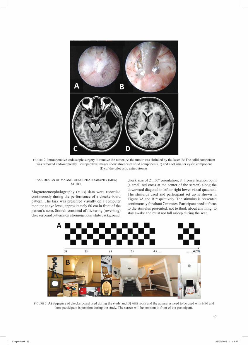
Magnetoencephalography (MEG) data were recorded continuously during the performance of a checkerboard pattern. The task was presented visually on a computer monitor at eye level, approximately 60 cm in front of the patient’s nose. Stimuli consisted of flickering (reversing) checkerboard patterns on a homogenous white background:
check size of 2°, 50° orientation, 8° from a fixation point (a small red cross at the center of the screen) along the downward diagonal in left or right lower visual quadrant. The stimulus used and participant set up is shown in Figure 3A and B respectively. The stimulus is presented continuously for about 7 minutes. Participant need to focus to the stimulus presented, not to think about anything, to stay awake and must not fall asleep during the scan.
FIGURE 3. A) Sequence of checkerboard used during the study and B) MEG room and the apparatus need to be used with MEG and how participant is position during the study. The screen will be position in front of the participant.
Chap 8.indd 65 22/02/2018 11:41:22
66
MEG AND DATA PROCESSING
MEG data were acquired using a 306-channel Vector view system (Elekta Neuromag, Helsinki) while participant engaged in the task, sitting upright in a light Elekta-Neuromag magnetically-shielded room. A magnetometer and two orthogonal planar gradiometers were located at each of the 102 positions. Vertical and horizontal eye movements were recorded using paired electrooculography electrodes. Head position stability was measured before and after acquisition run using four Head-Position Indicator (HPI) coils. HPI were used to monitor the head position. A
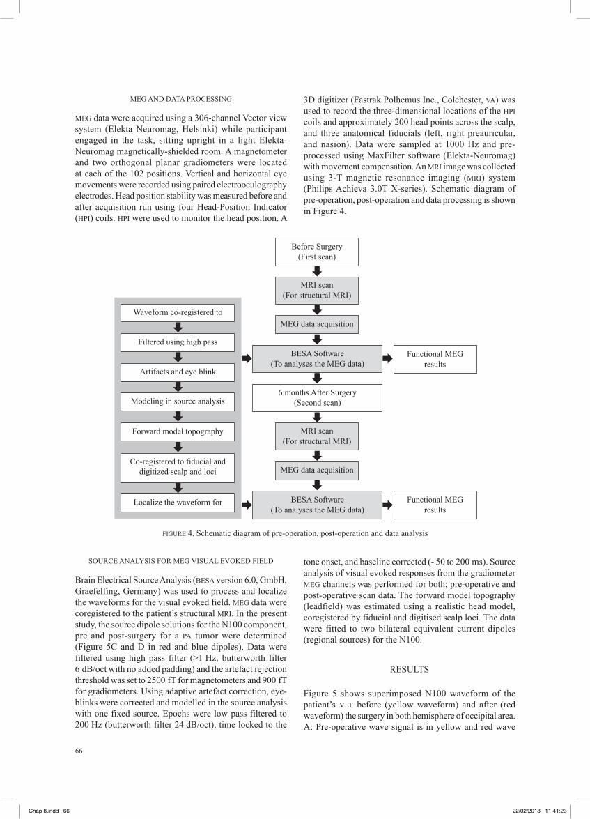
3D digitizer (Fastrak Polhemus Inc., Colchester, VA) was used to record the three-dimensional locations of the HPI coils and approximately 200 head points across the scalp, and three anatomical fiducials (left, right preauricular, and nasion). Data were sampled at 1000 Hz and pre-processed using MaxFilter software (Elekta-Neuromag) with movement compensation. An MRI image was collected using 3-T magnetic resonance imaging (MRI) system (Philips Achieva 3.0T X-series). Schematic diagram of pre-operation, post-operation and data processing is shown in Figure 4.
FIGURE 4. Schematic diagram of pre-operation, post-operation and data analysis
Before Surgery(First scan)
MRI scan(For structural MRI)
Functional MEGresults
Functional MEGresults
MRI scan(For structural MRI)
BESA Software(To analyses the MEG data)
BESA Software(To analyses the MEG data)
6 months After Surgery(Second scan)
MEG data acquisition
MEG data acquisition
Waveform co-registered to
Artifacts and eye blink
Filtered using high pass
Modeling in source analysis
Forward model topography
Localize the waveform for
Co-registered to fiducial and digitized scalp and loci
SOURCE ANALYSIS FOR MEG VISUAL EVOKED FIELD
Brain Electrical Source Analysis (BESA version 6.0, GmbH, Graefelfing, Germany) was used to process and localize the waveforms for the visual evoked field. MEG data were coregistered to the patient’s structural MRI. In the present study, the source dipole solutions for the N100 component, pre and post-surgery for a PA tumor were determined (Figure 5C and D in red and blue dipoles). Data were filtered using high pass filter (>1 Hz, butterworth filter 6 dB/oct with no added padding) and the artefact rejection threshold was set to 2500 fT for magnetometers and 900 fT for gradiometers. Using adaptive artefact correction, eye-blinks were corrected and modelled in the source analysis with one fixed source. Epochs were low pass filtered to 200 Hz (butterworth filter 24 dB/oct), time locked to the
tone onset, and baseline corrected (- 50 to 200 ms). Source analysis of visual evoked responses from the gradiometer MEG channels was performed for both; pre-operative and post-operative scan data. The forward model topography (leadfield) was estimated using a realistic head model, coregistered by fiducial and digitised scalp loci. The data were fitted to two bilateral equivalent current dipoles (regional sources) for the N100.
RESULTS
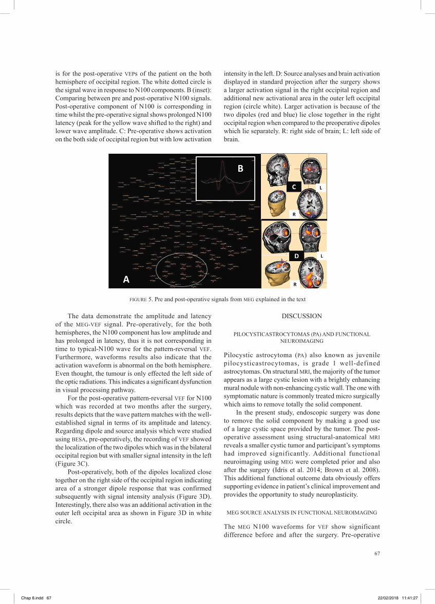
Figure 5 shows superimposed N100 waveform of the patient’s VEF before (yellow waveform) and after (red waveform) the surgery in both hemisphere of occipital area. A: Pre-operative wave signal is in yellow and red wave
Chap 8.indd 66 22/02/2018 11:41:23
67
is for the post-operative VEPs of the patient on the both hemisphere of occipital region. The white dotted circle is the signal wave in response to N100 components. B (inset): Comparing between pre and post-operative N100 signals. Post-operative component of N100 is corresponding in time whilst the pre-operative signal shows prolonged N100 latency (peak for the yellow wave shifted to the right) and lower wave amplitude. C: Pre-operative shows activation on the both side of occipital region but with low activation
intensity in the left. D: Source analyses and brain activation displayed in standard projection after the surgery shows a larger activation signal in the right occipital region and additional new activational area in the outer left occipital region (circle white). Larger activation is because of the two dipoles (red and blue) lie close together in the right occipital region when compared to the preoperative dipoles which lie separately. R: right side of brain; L: left side of brain.
FIGURE 5. Pre and post-operative signals from MEG explained in the text
The data demonstrate the amplitude and latency of the MEG-VEF signal. Pre-operatively, for the both hemispheres, the N100 component has low amplitude and has prolonged in latency, thus it is not corresponding in time to typical-N100 wave for the pattern-reversal VEF. Furthermore, waveforms results also indicate that the activation waveform is abnormal on the both hemisphere. Even thought, the tumour is only effected the left side of the optic radiations. This indicates a significant dysfunction in visual processing pathway.
For the post-operative pattern-reversal VEF for N100 which was recorded at two months after the surgery, results depicts that the wave pattern matches with the well-established signal in terms of its amplitude and latency. Regarding dipole and source analysis which were studied using BESA, pre-operatively, the recording of VEF showed the localization of the two dipoles which was in the bilateral occipital region but with smaller signal intensity in the left (Figure 3C).
Post-operatively, both of the dipoles localized close together on the right side of the occipital region indicating area of a stronger dipole response that was confirmed subsequently with signal intensity analysis (Figure 3D). Interestingly, there also was an additional activation in the outer left occipital area as shown in Figure 3D in white circle.
DISCUSSION
PILOCYSTICASTROCYTOMAS (PA) AND FUNCTIONAL NEUROIMAGING
Pilocystic astrocytoma (PA) also known as juvenile pilocysticastrocytomas, is grade 1 well-defined astrocytomas. On structural MRI, the majority of the tumor appears as a large cystic lesion with a brightly enhancing mural nodule with non-enhancing cystic wall. The one with symptomatic nature is commonly treated micro surgically which aims to remove totally the solid component.
In the present study, endoscopic surgery was done to remove the solid component by making a good use of a large cystic space provided by the tumor. The post-operative assessment using structural-anatomical MRI reveals a smaller cystic tumor and participant’s symptoms had improved significantly. Additional functional neuroimaging using MEG were completed prior and also after the surgery (Idris et al. 2014; Brown et al. 2008). This additional functional outcome data obviously offers supporting evidence in patient’s clinical improvement and provides the opportunity to study neuroplasticity.
MEG SOURCE ANALYSIS IN FUNCTIONAL NEUROIMAGING
The MEG N100 waveforms for VEF show significant difference before and after the surgery. Pre-operative
Chap 8.indd 67 22/02/2018 11:41:27

68
N100 waveform has lower in amplitude (deformed wave) and longer in its latency (Figure 5). Concerning the dipole localization for VEF, the pre-operative dipoles are spread equally into the right and left occipital areas, whilst for the post-operative dipoles, they are localized only in the right occipital area. These brainwave data provide some functional evidence for visual disturbance experienced by the patient. Clearer neuro-functional outcomes are demonstrated further by using brain electrical source analysis (BESA). Pre-operative MEG source analysis results of the participant indicate that bilateral occipital area (concordance with the dipole findings) was activated but with a marked signal intensity response in the right calcarine cortex compared to the left (Figure 5C). However, this pre-operative signal intensity response for N100 appears a lot less when compared to the post-operative signal (Figure 5D). The pre-operative results may suggest that the occipital calcarine areas of both hemispheres were still intact, but because of the presence of a large tumor, the VEF brainwave signals for N100 were altered significantly. This functional-brainwave abnormality together with the anatomical location for the tumor explains a right homonymous hemianopia which was experienced by the patient. The patient successfully underwent endoscopic surgery to remove the solid component.
Postoperatively, all symptoms resolved and disturbances in her visual field have improved from right hemianopia to an insignificant right upper quadrantonopia. The improvement in the visual field is suggested to be due to the optic radiations are recovering from being under stretch by the tumor mass. This improvement is expected to occur due to relief of pressure on the cortical optic radiations. We further suggest that after the tumor has been removed, not only the pressure on the visual optic is relief but also there is a recovery process in the functional brain area related to visual processing. This recovery process in the functional brain area can be seen in the brain wave and neural activity, as in Figure 5C. Postoperatively, MEG source analysis shows much stronger signal intensity in the right occipital-calcarine cortex with additional new activation area in the outer left occipital cortex (Figure 5D in white circle).
The shifts in signal intensity and dipole localization may suggest the recovery process of the functional brain and we further suggest that this changes and reorganization of brain signal and intensity is due to the process of neural plasticity (Chung et al. 2017; Harrington et al. 2016)
NEUROPLASTICITY IN BRAIN TUMOR
Neuroplasticity is a constant process allowing all-time rewiring and reshaping of the neuron synaptic maps, the purpose of this mechanism is to optimize the brain processing and network functions (Ivanco & Greenough 2000; Grover et al. 2006). The mechanisms of these neuronal changes are widespread and include functional alterations, neuro chemical changes, sprouting of new connections, remodeling and reorganization of visual
pathway (Hund et al. 1997; Hara et al. 2007; Kikuchi et al. 2015). The capacity for the brain to reorganize itself is critical for the process of functional recovery in patients, and this is often called post-lesional plasticity (Bavelier & Neville 2002). Our study highlights this point, whereby after reappearance of some normal anatomical brain structures on post-operative structural MRI (Figure 2C and D), the N100 VEF signal intensity appears stronger in the right occipital-calcarine cortex and new activation area emerged at outer part of the left occipital cortex. The changes observed in our study can only be explained by neuroplasticity (Brundage & Taylor 2010; Juenger et al. 2013; Nakamura, Hillary & Biswal 2009). In concordance with our findings, there are some previous studies that proposed slow growing tumors, particularly low-grade gliomas such as PA which can induce a large functional rewiring (Chen, Cohen & Hallett 2002; Hayley & Litteljohn 2013; Kikuchi et al. 2015). Our findings in term of MEG-based functional neuroimaging analysis in outcome evaluation, together with the concept of neuroplasticity mentioned in other studies may indicate patient’s clinical improvement in visual field could be largely due to neuroplasticity which occurs at certain or new activational areas for visual processing. The present study provides new MEG-based evidence in functional reorganization for visual network.
CONCLUSION
In conclusion, this study highlights the clinical value of VEF using MEG in the pre-operative and post-operative assessment of young participant with pilocystic astrocytoma and its potential advantages. To conclude, VEF using MEG is a promising technique, which may serve to determine visual localization or to investigate atypical visual representations in young participant with brain tumor.
ACKNOWLEDGEMENT
The authors wish to thank the Davendran Kanesen from the Department of Neurosciences, Health Campus, Universiti Sains Malaysia for the support with the proof read the manuscript Noorazrul Yahya from Diagnostic Imaging and Radiotherapy Program, School of Diagnostic and Applied Health Sciences, for his ideas, insight and support throughout the writing of the manuscript.
REFERENCES
Abraham, W.C. & Bear, M.F. 1996. Metaplasticity: the plasticity of synaptic plasticity. Trends in Neuroscience 19(4): 126-130.
Bavelier, D. & Neville, H. J. 2002. Cross-modal plasticity: Where and how? Nature Reviews Neuroscience 3(6): 443-452.
Brown, P. D., Decker, P. A., Rummans, T. A., Clark, M. M., Frost, M. H., Ballman, K. V., Arusell, R. M. & Buckner, J. C. 2008. A prospective study of quality of life in adults with
Chap 8.indd 68 22/02/2018 11:41:28
69
newly diagnosed high-grade gliomas: comparison of patient and caregiver ratings of quality of life. American Journal of Clinical Oncology 31(2): 163-168.
Brundage, C. M. & Taylor, B.E. 2010. Neuroplasticity of the central hypercapnic ventilatory response: teratogen-induced impairment and subsequent recovery during development. Developmental Neurobiology 70(10): 726-35.
Chen, R. L. G., Cohen, G. & Hallett, M. 2002. Nervous system reorganization following injury. Neuroscience 111(4):761-73.
Chung, S.W., Lewin, B.P., Rogash, N.C., Saeki, T., Hoy, K.E., Bailey, N.W. & Fitzgerald, P.B. 2017. Demonstration of short-term plasticity in the dorsolateral prefrontal cortex with theta burst stimulation: A TMS-EEG study. Clinical Neurophysiology 128(7): 1117-1126.
Fruchter, J. & Marantz, A. 2015. Decomposition, lookup, and recombination: MEG evidence for the full decomposition model of complex visual word recognition. Brain Language 143: 81-96.
Grover, K. M., Bowyer, S. M., Rock, J., Rosenblum, L.M., Mason, K.M., Moran, J. E., Smith, B. J. & Barkley, G.L. 2006. Retrospective review of MEG visual evoked hemifield responses prior to resection of temporo-parieto-occipital lesions. Journal of Neurooncoogy 77(2): 161-6.
Hara, K., Lin, F.H., Camposano, S., Foxe, D.M., Grant, P.E., Bourgeois, B. F., Ahlfors, S. P. & Stufflebeam, S. M. 2007. Magnetoencephalographic mapping of interictal spike propagation: a technical and clinical report. AJNR Am Journal of Neuroradiol 28(8): 1486-8.
Harrington, A. & Hammond-Tooke, G.D. 2016. Theta Burst Stimulation of the Cerebellum Modifies the TMS-Evoked N100 Potential, a Marker of GABA Inhibition. PLoS One 10:e0141284.
Hayley, S. & Litteljohn, D. 2013. Neuroplasticity and the next wave of antidepressant strategies. Frontior in Cell Neuroscience, 7:218. doi: 10.3389/fncel.2013.00218.
Hund, M., Rezai, A. R., Kronberg, E., Cappell, J., Zonenshayn, M., Ribary, U., Kelly, P. J. & Llinas, R. 1997. Magnetoencephalographic mapping: basic of a new functional risk profile in the selection of patients with cortical brain lesions. Neurosurgery 40(5): 936-42; discussion 942-3.
Idris, Z., Kandasamy, R., Reza, F. & Abdullah, J. M. 2014. Neural oscillation, network, eloquent cortex and epileptogenic zone revealed by magnetoencephalography and awake craniotomy. Asian Journal of Neurosurgy 9(3): 144-52. doi: 10.4103/1793-5482.142734.
Ivanco, T. L. & Greenough, W.T. 2000. Physiological consequences of morphologically detectable synaptic plasticity: potential uses for examining recovery following damage. Neuropharmacology 39(5): 765-76.
Juenger, H., Kuhnke, N., Braun, C., Ummenhofer, F., Wilke, M., Walther, I., Koerte, I., Delvendahl, N. H., Jung, S., Berweck, M., Staudt, J. & Mall, V. 2013. Two types of exercise-induced neuroplasticity in congenital hemiparesis: a transcranial magnetic stimulation, functional MRI, and magnetoencephalography study. Developemental of Medical Child Neurology 55(10): 941-51.
Kakisaka, Y., Gupta, A., Enatsu, R., Wang, Z. I., Alexopoulos, A. I., Mosher, J. C., Dubarry, A. S., Hino-Fukuyo, N. & Burgess, R.C. 2013. Magnetoencephalography reveals a unique neurophysiological profile of focal-onset epileptic
spasms. Tohoku Journal of Experimental Medicine 229(2): 147-51.
Kikuchi, M. Y., Yoshimura, K., Mutou, K. & Minabe, Y. 2015. Magnetoencephalography in the study of children with autism spectrum disorder. Psychiatry Clinical Neurosciences 70(2): 74-88.
Liu, J. A., Harris, N. & Kanwisher, J. 2002. Stages of processing in face perception: an MEG study. Nature Neuroscience 5(9): 910-6.
Majchrzak, K., Kaspera, B., Bobek-Billewicz, A., Hebda, G. Stasik-Pres, H., Majchrzak, P. & Ladzinski, L. 2012. The assessment of prognostic factors in surgical treatment of low-grade gliomas: a prospective study. Clinical Neurology and Neurosurgy 114(8): 1135-44.
Nakamura, T. F. G., Hillary, B. & Biswal, B. 2009. Resting network plasticity following brain injury. PLoS One 4(12): e8220.
Peyk, P. H. T., Schupp, T., Elbert, K. & Junghofer, E. 2008. Emotion processing in the visual brain: a MEG analysis. Brain Topography 20(4): 205-15.
Rosler, J. B., Niraula, V., Strack, A., Zdunczyk, S., Schilt, P., Savolainen, P., Lioumis, J., Makela, P., Vajkoczy, D., Frey, T. & Picht, T. 2014. Language mapping in healthy volunteers and brain tumor patients with a novel navigated TMS system: evidence of tumor-induced plasticity. Clinical Neurophysiology 125(3): 526-36.
Strey, K. A., Baertsch, N.A. & Baker-Herman, T. L. 2013. Inactivity-induced respiratory plasticity: protecting the drive to breathe in disorders that reduce respiratory neural activity. Respiratory Physiology and Neurobiology 189(2): 384-94.
Hanani Abdul MananRadiology DepartmentFaculty of MedicineUniversiti Kebangsaan Malaysia Medical CentreJalan Yaacob Latif, Bandar Tun Razak56000 CherasKuala Lumpur, Malaysia
Zamzuri IdrisJafri Malin AbdullahMohammed Faruque RezaHazim OmarHanani Abdul MananCenter for Neuroscience Service and ResearchDepartment of NeurosciencesSchool of Medical SciencesUniversiti Sains MalaysiaKubang Kerian, 16150 Kelantan, Malaysia
Corresponding author: Hanani Abdul MananEmail address: [email protected]
Tel: +603-9145 5600Fax: +603-9145 6682
Received: March 2017Accepted for publication: November 2017
Chap 8.indd 69 22/02/2018 11:41:29